R E V I E W
Open Access
Fluorescence in situ hybridization (FISH):
an increasingly demanded tool for biomarker
research and personalized medicine
Linping Hu
1,6†, Kun Ru
1,2†, Li Zhang
1,3, Yuting Huang
4, Xiaofan Zhu
1,3,6, Hanzhi Liu
1,6, Anders Zetterberg
5,
Tao Cheng
1,6*and Weimin Miao
1,6*Abstract
Extensive studies of the genetic aberrations related to human diseases conducted over the last two decades have identified recurrent genomic abnormalities as potential driving factors underlying a variety of cancers. Over the time, a series of cutting-edge high-throughput genetic tests, such as microarrays and next-generation sequencing, have been developed and incorporated into routine clinical practice. Although it is a classical low-throughput cytogenetic test, fluorescence in situ hybridization (FISH) does not show signs of fading; on the contrary, it plays an increasingly important role in detecting specific biomarkers in solid and hematologic neoplasms and has therefore become an indispensable part of the rapidly developing field of personalized medicine. In this article, we have summarized the recent advances in FISH application for both de novo discovery and routine detection of chromosomal rearrangements, amplifications, and deletions that are associated with the pathogenesis of various hematopoietic and non-hematopoietic malignancies. In addition, we have reviewed the recent developments in FISH methodology as well.
Keywords:Fluorescence in situ hybridization (FISH), Solid tumors, Hematopoietic malignancies, Genetic aberrations, Biomarkers, Personalized medicine
Introduction
Mounting evidence indicates that both hematologic and solid tumors are heterogeneous disorders with diverse genomic aberrations [1-3]. Due to the extensive investi-gations of the correlation between genomic instability and disease pathogenesis conducted over the last two decades, an increasing number of genomic abnormalities such as gain, loss or rearrangement of chromosomal fragments and gene mutations, have been found to be driving factors in the pathogenesis of various malignan-cies. Over the time, a series of cutting-edge cytogenetic and molecular tests have been developed for detecting such genomic aberrations, which allows more accurate
molecular profiling for individual patients. The advanced molecular pathology techniques [4] enable better disease stratification and prognosis, leading to tailored thera-peutic regimens. Apparently, a new era of personalized medicine has arrived much earlier than most of us expected [3].
Fluorescence in situ hybridization (FISH) is a cytogen-etic technique developed in the early 1980s. FISH uses fluorescent DNA probes to target specific chromosomal locations within the nucleus, resulting in colored signals that can be detected using a fluorescent microscope. Compared to the conventional cytogenetic (CC) meta-phase karyotype analysis, FISH does not require cell cul-turing, and can directly use fresh or paraffin-embedded interphase nuclei for a rapid evaluation. With the discovery of numerous disease-related genes in recent years, the applications of FISH broadened to include more genetic diseases, hematologic malignancies, and solid tumors. For example, FISH detection of BCR/ABL1 translocation, HER2 amplification, and ALK rearrange-ment is critical for guiding targeted therapy in chronic * Correspondence:chengt@pumc.edu.cn;miaowm@hotmail.com
†Equal contributors
1State Key Laboratory of Experimental Hematology, Institute of Hematology
and Blood Diseases Hospital, Chinese Academy of Medical Sciences and Peking Union Medical College, Tianjin, China
6Center for Stem Cell Medicine, Chinese Academy of Medical Sciences and
Peking Union Medical College, Nanjing Road 288, Tianjin 300020, P.R. China Full list of author information is available at the end of the article
myeloid leukemia [5], breast cancer [6,7] and lung adenocarcinoma, respectively [8,9]. Hence, FISH tests have been recognized as vital components of personal-ized medicine.
With respect to biomarker detection, a series of innovative high-throughput molecular tests, such as array-based comparative genome hybridization (aCGH), single nucleotide polymorphism (SNP) arrays, and next generation sequencing, have recently been developed and incorporated into routine clinical practice. The stun-ning technology replacing speed raised a serious question: is a classical low-throughput assay, such as FISH, poised for replacement? However, the answer is quite the op-posite. In fact, FISH has become increasingly important in clinical diagnosis due to its simplicity and reliability in evaluating key biomarkers in various tumors [5]. In this article, we aim to review the advances in FISH for disease biomarker detection and personalized medicine applications.
Hematopoietic malignancies
Leukemia
Leukemia is a heterogeneous clonal disorder of he-matopoietic stem and progenitor cells, characterized by various acquired genetic aberrations. The discovery of abnormal fusion proteins resulting from chromosomal rearrangements has significantly contributed to our un-derstanding of the molecular mechanism of the pathogen-esis of leukemia. The first oncogene discovered as the direct etiological basis of a malignancy, the BCR/ABL1 translocation in chronic myeloid leukemia (CML), results in dysregulated tyrosinase activity, which can be treated using the tyrosine kinase inhibitor Imatinib [10,11]. The t (15; 17) chromosomal translocation in promyelocytic leukemia (APML, AML-M3) functions in a similar way, generating the novel fusion protein PML/RARa, and ATRA (all-trans retinoic acid) offers an effective ther-apy for APML by specifically suppressing oncogenic activities of the PML/RARa fusion protein [12,13]. The FISH assay is considered the gold standard for detect-ing these chromosomal translocations and it therefore plays a crucial role in selecting a targeted therapy for various leukemias.
Over the last two decades, additional recurrent gen-etic aberrations have been identified in leukemias, improving molecular sub-classification and allowing stratified management. Whereas most of the chromo-somal rearrangements can be detected using CC, FISH remains the most robust tool for detecting balanced or unbalanced chromosomal aberrations. Furthermore, a combination of cytogenetic and molecular profiling permits more accurate assessment of the disease prognosis [1].
In addition to the common chromosomal rearrange-ments in acute myeloid leukemia (AML), which are t(8;21)(q22;q22), t(15;17)(q22;q12) and inv(16)(p13.1q22) or t(16;16)(p13.1q22) [14], newly obtained molecular in-formation has been combined with cytogenetic findings to establish a more comprehensive risk assessment sys-tem. Among them, t(8;21)(q22;q22)/RUNX1-RUNX1T1; t(15;17)(q22;q12)/PML-RARa; inv (16)(p13.1q22) or t(16;16)(p13.1;q22)/CBFB-MYH1 and mutation in both the CEBPA and NPM1 genes are associated with a good clinical outcome, whereas t(1;22) (p13;q13)/RBM15-MKL1; inv(3)(q21q26.2) or t(3;3)(q21;q26.2)RPN1-EVI1; t(6;9)(p23;q34)/DEK-NUP214, the MLL gene rearrange-ment, complex karyotypes, and mutations in both KIT and FLT3 are associated with a less favorable prognosis [14,15]. Acute lymphoblastic leukemia (ALL) occurs mostly in children between the ages of 1 to 5 years old. The most common chromosomal translocations in ALL are t (9; 22) (p190) in adults, and t (12; 21) in children respectively. The most frequent numerical aberration in ALL is hyperdiploidy with chromosome numbers ranging from 51 to 63, with chromosomes X, 4, 6, 10, 14, 17 and 18 generally being trisomic and chromosome 21 frequently occurring as four copies [16,17]. Among them, patients with t (12; 21), t (1; 19), and hyperdiploidy have a favor-able outcome, whereas patients with MLL translocations have a worse prognosis. It is worth noting that the pres-ence of t (9; 22) used to indicate the worst prognosis for ALL patients, but this fact has dramatically changed since the introduction of the“magic bullet”Gleevec.
Due to the universal presence of BCR/ABL1 rearrange-ment in chronic myeloid leukemia (CML), it is now defined as the diagnostic hallmark of CML [10,11,18].
the cyclin D1 rearrangement, particularly in when immu-nohistochemistry is not contributory for various reasons.
Multiple myeloma
Multiple myeloma (MM) is another heterogeneous ma-lignancy of terminally differentiated B cells, clinically manifested as monoclonal plasma cells that infiltrate the bone marrow, a spike of monoclonal immunoglobulin in the blood and/or urine, and massive osteolytic bone le-sions. Similar to the other hematologic neoplasms, MM is characterized by a complex pattern of extensive gen-omic aberrations involving many chromosomes [23,24]. The genetic abnormalities found in MM can be roughly divided into two categories based on the chromosome ploidy status and other parameters [25,26]. The hyperdi-ploid karyotype is generally associated with trisomies of many chromosomes, such as 3, 5, 7, 11, 15, 19 and 21, whereas the hypodiploid karyotype appears to be more frequently associated with a translocation of immuno-globulin heavy chain (IGH) locus at 14q32 [26]. The IgH (14q32) translocations found in hypodiploid MM can involve many different partners, such as 11q13 (CCND1), 6p21 (CCND3), 16q23 (MAF), 20q12 (MAFB), and 4p16 (FGFR3 and MMSET). Furthermore, the chromosome ploidy status and IGH rearrangements were found to be correlated with disease outcome in MM patients [25]. For example, the hyperdiploid karyotype with t(11;14)(q13;q32) indicates a better prognosis, whereas the hypodiploid karyotypes with t(4;14)(p16; q32) or t(14;16)(q32;q23) imply a worse clinical outcome [25,26].
Molecular studies have demonstrated that primary translocations occur in the early stage of MM, followed by large number of secondary translocations during tumor progression [27]. It is believed that the secondary genomic aberrations are responsible for a more prolifer-ative phenotype in the advanced stage of MM. Certain genetic aberrations, such as MYC rearrangements, del (13q), del (17p), and the deletion of 1p and/or amplifica-tion of 1q, have been identified as the most common secondary aberrations in MM [27-29]. The chromosome 13 deletion or chromosome 13 monosomy occurs in 50% of the patients with advanced MM and are associ-ated with an aggressive clinical course and an unfavor-able prognosis [30,31]. Deletion of 17P13, presumably resulting in LOH (loss of heterozygosity) of P53, has been determined to be associated with a very poor clin-ical outcome [32,33]. Chromosome 1p deletion or 1q amplification is the most common structural aberration found in MM and is associated with an unfavorable prognosis [34-36].
Due to the low proliferative rate of tumor cells in the early stage of MM, CC analysis of metaphase cells is likely to miss detecting the primary genomic aberrations in non-dividing tumor cells. Furthermore, some small
chromosomal rearrangements in MM may be cryptic to chromosome banding analysis. Thus FISH, which is effective for analysis of interphase nuclei and small chromosomal aberrations, is recognized as the most robust genetic test for characterizing the known cyto-genetic abnormalities in MM. Nevertheless, integration of data from the multiple genetic profiling techniques including CC, FISH, RT-PCR, and gene mutation ana-lysis, among others, would provide comprehensive infor-mation for better stratification of MM patients with diagnostic and prognostic significance.
Myelodysplastic syndromes (MDS)
MDS is a heterogeneous group of clonal hematopoietic disorders characterized by blood cytopenias resulting from ineffective hematopoiesis. The clinical outcome of MDS is variable, and approximately 20 to 30% of the patients will progress to AML within a few months or years [37]. Recurrent chromosomal abnormalities, such as -5/del(5)(q31), -7/del(7)(q31), +8, del(20)(q2), -17/del
(17)(p3.1), and –Y, are found in half of the de novo
MDS cases. The cytogenetic finding is closely related to the clinical outcome, and thus has been incorporated into the revised international prognostic scoring system (IPSS-R) and the WHO prognostic scoring system (WPSS) [38]. In addition, certain chromosomal aberra-tions, such as -5/5q-, -7/7q-, and complex abnormalities, were found to be correlated to the response to chemo-therapy [37,39,40]. The metaphase chromosomal band-ing assay is regarded as the objective standard for clonal analysis of MDS. Being a more sensitive test, FISH can be utilized to identify minor abnormal clones and cryptic chromosomal aberrations that are undetectable using CC and to provide additional information for patients with a normal karyotype or unsuccessful culture [5,39]. With the introduction of high-resolution assays such as aCGH and SNP arrays, it will be possible to discover additional genetic markers in the near future, leading to more accurate diagnoses, better targeted therapies, and more clinically significant judgment of the prognosis for MDS patients [41].
Non-hematopoietic malignancies (solid tumors)
Lung cancer
who were treated with the small-molecule kinase inhibitor Crizotinib, showed a response rate of 50-60% [8]. 2011 was the first time that the US FDA simultaneously approved a novel anti-cancer drug (Crizotinib, Pfizer) and its compan-ion FISH detectcompan-ion kit (ALK FISH probe kit, Abbott Molecular), which highlighted the critical role of the FISH assay in guiding ALK-targeted therapy [8,9]. Because ALK rearrangements are reportedly mutually exclusive with EGFR/KRAS mutations [43], ALK FISH testing is generally recommended for patients with wild-type EGFR/KRAS non-squamous NSCLC.
Using the ALK FISH detection kit, the 3’and 5’ ends of the ALK gene are differentially labeled with red or green fluorescent probes. In benign cells, two fused signals should be detected. In 60-70% of all of the ALK rearrangements, the EML4-ALK gene fusion occurs through only inversion, and therefore narrowly separated (two to three signals apart) red and green signals are detected in addition to the normal fusion signal. In the other 30-40% of the cases, gene fusion occurs through an interstitial deletion together with an inversion of EML4, which lead to a single red signal without a corresponding green signal, in addition to the normal fusion signal [44].
ROS1, another receptor tyrosine kinase, which is located at chromosome 6p22, has recently been found to be rearranged in 2-3% of the NSCLC adenocarcinomas [7,45]. In addition, several ROS1 translocation partners, e.g., TPM3, SDC4, SLC34A2, CD74, EZR or LRIG3, have been found forming a fusion with the kinase domain of ROS1, leading to constitutive activation of ROS1 and increase in malignant transformation activity [7]. The ROS1 rearrangements define a subset of NSCLC with clinical characteristics and treatment responses that are similar to those of the ALK rearrangements [45,46]. Therefore, this type of rearrangement is another predict-ive FISH biomarker that can be applied to personalized management of lung cancer.
Prostate cancer
Rearrangements involving androgen-regulated TMPRSS2 and ETS family members (ERG, RTV1, ETV4) were detected in nearly half of the prostate cancers but none of the benign prostate tissues that were tested [5,39]. The relevance of the TMPRSS2 rearrangements to the pathogenesis, prognosis, and targeted therapy of prostate cancer has made it a predictive biomarker for prostate cancer [47]. The most common type of chromosome rearrangement involves the fusion of TMPRSS2 to the oncogene ERG, which leads to the abnormal activation of ERG. Identification of these rearrangements may allow stratification of prostate cancers into subtypes that respond to specific therapies. As the test of choice for chromosomal rearrangements, FISH was successfully applied to frozen as well as formalin-fixed
paraffin-embedded (FFPE) prostate cancer samples with high sensitivity and specificity. Initially, a kit containing dual-color ERG break-apart probes that can identify rearrangement of the ERG gene but do not indicate the 5’-partner to which ERG is fused to, was used [48,49]. Later on, a tricolor FISH assay was developed by com-bining the red/green break-apart probes for TMPRSS2 with an orange-labeled fusion probe for the 3’region of ERG [50,51]. Recently, a four-color FISH assay was reported, which allows the detection of either TMPRSS2 or ERG rearrangements regardless of the partner gene [39].
Breast cancer
Breast cancer is a fairly heterogeneous malignancy that involves large numbers of genomic aberrations that are inherited or are acquired during the initiation and progression of the disease. To date, the most successful application of FISH as a companion diagnostic test for selecting a targeted therapy for a solid tumor may be the FISH evaluation of Her-2 amplification for breast cancer [52-54]. Her-2 (human epidermal growth factor receptor-2), also called c-erbB-2, is located at chromo-some 17q12-21.32, and encodes a trans-membrane pro-tein of 185 kDa. Her-2 propro-tein is an active tyrosine kinase that plays an important role in normal cell growth and differentiation. It has been reported that Her-2 gene amplification occurs in 20-30% of breast cancer patients. Her-2 gene amplification leads to its overexpression on the cell surface. Her-2 amplification indicates a bad prognosis, short survival time, and the existence of a more aggressive phenotype of tumor cells. Her-2 overexpressed breast cancer may be resist-ant to endocrine therapy and some chemotherapies; however, it is sensitive to Herceptin treatment and exhibits more responsiveness to paclitaxel and anthra-cyclines [55]. At present, there are both IHC and FISH assays for measuring Her-2 overexpression. The former assay is simplistic, convenient, and cost-saving, but it can be affected by various factors and the staining result could be ambiguous. The latter is relatively sophisticated and expensive, but the staining result is more accurate. Therefore, the FISH assay is regarded as the gold standard for clinical evaluation of the Her-2 status and is generally recommended in when the IHC result could not clarify the Her-2 status.
mistreatments based on their histopathological classifi-cations; therefore, there is a need to establish more accurate molecular classification schemes [56]. A mo-lecular classification based on a clustering analysis of the expression patterns of 427 genes has divided breast cancer into four types: a luminal type (further divided into A, B and C subtypes), a basal-like type, a Her-2 positive type, and a normal breast-like type [57,58], and the molecular classifications are closely correlated with the prognoses, with the luminal subtype A having a good clinical outcome; the luminal subtype B having a bad prognosis; and the basal-like and the Her-2 positive types having the worst clinical outcome [58]. Recent CGH analyses of genomic aberrations in breast cancer have identified three molecular categories of breast cancers. The first category, called“simplex”[59] or 1q/16q [60,61], is characterized by a few genomic rearrangements and the second category, called“complex sawtooth”[59] or “complex” [60,61], is characterized by more rearrange-ments and gene copy number alterations within a restricted genomic area. The third category, called “complex firestorm” [59] or “mixed amplifier” [60,61], is characterized by high intensity gene amplification profiles restricted to a small genomic areas. Interest-ingly, correlations seem to exist between the previous Sorlie expression classes [58] and the specific genomic profile categories, possibly due to the interplay of the genomic aberrations and the overall gene expression. The luminal A type of breast cancer is correlated with the simplex profile and the luminal B and the Her-2 positive types with the complex firestorm profile. The basal-like class is correlated with the complex sawtooth profile [62].
The estrogen receptor (ER) is a well-established bio-marker for endocrine therapy in breast cancer patients, while the progesterone receptor (PR) is not. Clinical studies showed that ER + PR + breast cancer shows a better response to endocrine therapy, whereas ER + PR-breast carcinoma has a more aggressive phenotype and a poorer response to endocrine therapy. CGH and FISH studies have revealed that ER + PR- breast cancers have higher genomic instability profiles, including recurrent amplifications in 11q13, 12q14-q15, 17q21-q25, and 20q13 and deletions in 11q13-q15 [60,63-65]. A more recent study refined the area to 17q23.2-q23.3 and 20q13.12 for most of the overlapping gained regions, and 3p21.32-p12.3, 9pter-p13.2, 17pter-p12, and 21pter-q21.1 for most of the overlapping lost areas in ER + PR- breast cancers [65].
Variations in the genomic profiles exist not only among different histopathological types of breast cancer, but are also found among the types of breast cancers of different ethnic groups. A recent study comparing breast cancer samples from African and American women has identified 6 chromosomal regions with a higher rate of
Table 1 The three subclones in an ascites fluid sample from a patient with progressive epithelial ovarian cancer
Clone c-myc Rb1 Chk2 P53 BRCA1
1 4 2 5 2 4
2 3 2 3 2 4
3 3 2 2 2 5
Note: the numbers in Clone column indicate the coding number of the subclones; the numbers in FISH probe columns (c-myc, Rb1, Chk2, p53 and BRCA1) indicate copy numbers of each gene within a single nucleus of the cancer clone.
DAPI c-myc Rb1 ChK2 p53 BRCA1 Merge
1
2
3
CNAs and several candidate biomarkers that could be specific to African women [66].
Extensive screening of genomic aberrations has led to the identification of candidate biomarkers associated with breast cancer tumorigenesis, invasiveness, and metastasis, including MYC at 8q24 [67], CCND1 at 11q13 [68], Her-2 at 17q12 [53], MTDH at 8q22 [69,70], and ETS transcrip-tion factors at 1q21 and 1q32 [71].
While substantial progress has been made in breast cancer genetics over the last two decades, further large-scale studies integrating both genome and transcriptome analyses [72] are needed to identify the key oncogenic driver genes or other specific biomarkers. The potential new findings could be of predictive values for diagnosis, predicting metastasis, survival assessment, and guiding targeted therapy. FISH can be of particular value in both the discovery and clinical routine detection of such bio-markers and will continue to play an important role in the personalized management of breast cancer.
Melanoma
Melanoma is a heterogeneous group of melanin-producing skin malignancies with acquired genetic aberrations. Studies employing aCGH and FISH have identified a variety of recurrent chromosomal aberrations in malig-nant melanoma. In clinic, a small but significant part of melanocytic lesions presents with ambiguous morpho-logic features, and those cases are challenging to expe-rienced dermatopathologists. Thus, a specific ancillary genetic test is needed for the initial characterization to avoid misdiagnosis and overtreatment [73,74]. Most primary melanomas exhibit either numerical or structural
chromosomal abnormalities, such as deletions in 9p, 10, 6q and 8p and copy-number increase in 7, 8, 6p, 1q, 20, 17, and 2 [75,76]. Given that multiple chromosomal aber-rations must be evaluated to obtain genetic profiles of melanoma, a multi-color approach comprising 4 gene probes have been adopted for a FISH-based melanoma assay [77]. The clinical studies showed that use of FISH in unambiguous cases provided promising results with rela-tively high sensitivity and specificity. However, the diag-nostic utility of FISH in ambiguous cases remains to be determined because a standard definition of“malignancy” is yet to be established from clinical studies with large samples of ambiguous cases [78]. Furthermore, the discov-ery of key genomic aberrations has led to more effective tar-geted therapies for melanoma. For example, Vemurafenib (BRAF V600E inhibitor) and Ipilimumab (anti-CTLA4) were recently approved by the U.S. FDA and agents directed against the MAP kinase pathway (anti-MEK, anti-ERK, other anti-BRAF) are under development for
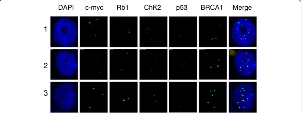
Table 2 The three subclones obtained from a cancer stem-cell preparation for a patient with low-differentiated ovarian adenocarcinoma
Clone c-myc Rb1 Chk2 P53 BRCA1
1 9 1 1 2 2
2 5 2 1 1 1
3 4 1 1 1 2
Note: the numbers in Clone column indicate the coding number of the subclones; the numbers in FISH probe columns (c-myc, Rb1, Chk2, p53 and BRCA1) indicate copy numbers of each gene within a single nucleus of the cancer clone.
DAPI c-myc Rb1 ChK2 p53 BRCA1 Merge
1
2
3
targeted therapy in cases of advanced metastatic melan-oma [79].
Progress in FISH methodology
FISH automation
Manual evaluation of large numbers of clinical FISH samples is no doubt a time-consuming, exhausting, and error-prone procedure. Moreover, the inter-observer vari-ability could lead to scoring inconsistency and even misdiagnosis. Thus, an automated system would greatly reduce such errors. Such an automated system is gener-ally comprised of the following parts: a light source, filter set, objectives, signal detector, motorized scanning stage, and a computer equipped with dedicated soft-ware in charge of automated tissue-area selection, signal evaluation and data calculation [80]. There were several reports of the successful automated evaluation of HER2 gene amplification using breast cancer speci-mens [81-83]. An automated method has also been applied to detecting balanced rearrangements, such as the BCR/ABL1 gene rearrangements in patients with CML [84]. Nevertheless, automated FISH is still in a premature stage with more standardization and large scale clinical trials pending.
QM-FISH
Previously, most commercial FISH detection kits con-tained one probe labeled with a single fluorochrome or two probes labeled with two distinct fluorochromes. These single or dual-color kits were used to detect a deletion or an amplification of a single locus-specific genomic fragment or a balanced chromosomal trans-location. With the rapid progress in disease gene discov-eries, there is a need to simultaneously detect multiple genes. Thus, a FISH method that employs multiple
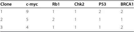
Table 3 The six subclones from the bone marrow sample taken from an ALL patient
Clone Fusion TEL AML1 PAX5 P16 IKZF1
1 1 1 2 1 2 2
2 1 1 2 2 2 1
3 1 1 2 2 2 0
4 1 1 2 1 1 1
5 1 1 3 2 2 2
6 1 1 3 2 2 0
Note: the numbers in Clone column indicate the coding number of the subclones; the numbers in FISH probe columns (Fusion, TEL, AML1, PAX5, p16 and IKZF1) indicate copy numbers of each gene (or gene fusion) within a single nucleus of the cancer clone.
1
2
3
4
5
6
probes, called quantitative multi-gene FISH (qmFISH)
has recently become popular. Abbott’s MultiVysion PB
multi-color probe kit, a five-color FISH kit that detects chromosomes 13, 16, 18, 21 and 22 was developed to assist in preimplantation diagnosis (PGD) by polar body analysis [85]. A four-color FISH assay, targeting chromo-somes 1, 2, 6, 9, 7, 17, the loci 3p24pter, and 3p13p14 has been used for the early diagnosis of renal carcinoma in biopsies of uncertain renal masses [86]. LAVysion FISH, a four-color FISH kit for simultaneously detecting chromosome 6 and the 5p15, 7p12 (EGFR gene), and 8q24 (MYC gene) loci was developed to assist in the differential diagnosis of ambiguous lung cancers [87]. In recent years, qmFISH has been used in genetic variega-tion and clonal evoluvariega-tion studies of both hematological and non-hematological cancers [88-92].
We have developed a state-of-the-art qmFISH system that can use as many as 10-20 fluorochromes, and the signals could only be analyzed by a computer-controlled detector (Zetterberg, A. et al, unpublished). This system is particularly useful for large-scale multi-gene clinical investigations of solid tumors or blood malignancies.
Currently, we are applying qmFISH to study the gen-etic architecture and clonal evolution in cases of ovarian cancer and leukemia. Ovarian cancer is a heterogeneous female malignancy characterized by various genomic aberrations. To define the molecular subgroups of ovar-ian cancer, we have chosen five genes, c-myc, Rb1, Chk2, p53 and BRCA1, which are known to be associated with
the pathogenesis of ovarian cancer. The ascites fluid samples were collected from ovarian cancer patients from Tianjin Medical University Cancer Institute and Hospital from January to December 2012, with hospital ethical review committee approval. The ovarian cancer cell sections were prepared by Cytospin procedure and fixed overnight in methanol. qmFISH was performed as previously described [93]. In each case, at least 200 nuclei were scored for CNAs (copy number alterations) of c-myc, Rb1, Chk2, p53 and BRCA1. The results showed that an ascites fluid cytospin sample from a rep-resentative case of progressive epithelial ovarian cancer contained at least three subclones with distinct molecu-lar profiles for the selected genes (Figure 1 and Table 1). Furthermore, a CD133 + ALDH + cancer stem cell [94,95]
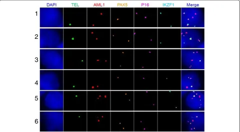
Table 4 The clonal components of bone marrow samples taken from an ALL patient upon the initial diagnosis and post chemotherapy
Clone Fusion TEL AML1 PAX5 P16 IKZF1
Initial diagnosis 1 1 2 2 2 2
1 1 3 2 2 2
1 1 3 2 1 2
Post chemotherapy 1 1 2 1 0 1
1 1 3 2 2 1
1 1 3 2 1 1
Note: the numbers in FISH probe columns (Fusion, TEL, AML1, PAX5, p16 and IKZF1) indicate copy numbers of each gene (or gene fusion) within a single nucleus of the cancer clone.
preparation isolated from a case of low-differentiated ovar-ian adenocarcinoma included three subclones with distinct qmFISH fingerprints (Figure 2 and Table 2). Thus, qmFISH analysis appears to be an excellent tool for molecular pro-filing of ovarian cancer at the single cell level.
Another of our projects is to apply qmFISH to mo-lecular profiling of acute lymphatic leukemia (ALL). It has been reported that t (12; 21) leads to fusion of an almost complete RUNX1(AML1) protein to part of the ETV6 (TEL) protein, which is found in 20-25% of the ALL patients. The ALL patients with the ETV6 (TEL)/ RUNX1(AML1) translocation generally have a better clinical outcome, but are more likely to experience a recurrence. The deletion of ETV6 (TEL) gene, which is associated with the ETV6 (TEL)/RUNX1(AML1) trans-location, is common in ALL and leads to LOH of
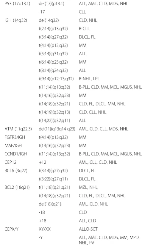
Table 5 FISH probes that are commonly used for clinical diagnosis of hematological diseases
Probes Cytogenetic anomaly
Associated disorders
BCR/ABL t(9;22)(q34;q11) CML, ALL, AML-M1, AML-M2, MPD
PML/RARα t(15;17)(q22;q21) AML-M3, CML Ph+
AML1/ETO t(8;21)(q22;q22) AML-M2, AML-M4, MDS
MLL (11q23) t(1;11)(p32;q23) ALL, AML
t(1;11)(q21;q23) AML-M4, AML-M5
t(2;11)(p21;q23) MDS
t(4;11)(q21;q23) ALL, AML
t(6;11)(q27;q23) AML-M4, AMl-M5
t(9;11)(p22;q23) ALL, AML-M5, MDS, t-MDS
t(10;11)(p13;q23) AML-M4, AMl-M5
t(11;17)(q23;q21) AML-M3, AML-M4, AMl-M5
t(11;19)(q23;p13) ALL, AML-M4, AML-M5, t-AMl
t(X;11)(q13;q23) T-ALL
del(11q23) AML, ALL, CLD, CLL, MDS, NHL
CBFB (16q22) t(16;16)(p13;q22) AML-M4Eo, MDS
inv(16)(p13q22) AML-M4Eo
del(16q22) AML, AML-M4Eo, NHL
EVI1 (3q26) t(3;3)(q21;q26) AML, MDS
inv(3)(q21q26) AML-M4, AML-M6, CML Ph+, MDS
t(3;21)(q26;q22) AML, CML Ph+, MDS
FGFR1/D8Z2 (8p11)
t(8;13)(p11;q12) MPD
t(8;16)(p11;p13) AML-M4, AML-M5
TEL/AML1 t(12;21)(p13;q22) ALL
TCF3/PBX1 t(1;19)(q23;p13) pre-B ALL
CKS1B (1q21)/ CDKN2C (1p32)
dup(1)(q21q32) ALL, CLD, NHL
del(1)(q21) NHL
del(1)(p32p36) CLD, NHL
MYC (8q24) t(2;8)(p12;q24) ALL-L3, BL, NHL
t(8;14)(q24;q32) ALL-L3, BL, MM, NHL
t(8;14)(q24;q11) T-ALL
t(8;22)(q24;q11) ALL-L3, BL
CEP8 +8 ALL, AML, CLD, MPD, MDS, PV
EGR1 (5q31)/ D5S721 (5p15.2)
-5 AML, MDS
del(5)(q13q33) AML, MDS, MPD, 5q- syn
D7S486 (7q31)/ CEP7 (7p11.1-7q11.1)
-7 AML, MDS, MPD
del(7)(q11) ALL, AML, MDS
del(7)(q22q34) AML, CLD, CMD, MDS, NHL
D20S108 (20q12)
del(20)(q11q13) AML, CMD, MDS, PV
-20 ALL
RB-1 (13q14) del(13)(q12-q22) AML, AMM, CLD, CLL, MM, MDS, NHL
del(13)(q12-q14) AML, AMM, CLD, MDS, NHL
Table 5 FISH probes that are commonly used for clinical diagnosis of hematological diseases(Continued)
P53 (17p13.1) del(17)(p13.1) ALL, AML, CLD, MDS, NHL
-17 CLL
IGH (14q32) del(14q32) CLD, NHL
t(2;14)(p13;q32) B-CLL
t(3;14)(q27;q32) DLCL, FL
t(4;14)(p13;q32) MM
t(5;14)(q31;q32) ALL
t(6;14)(p25;q32) MM
t(8;14)(q24;q32) ALL
t(9;14)(p12-13;q32) B-NHL, LPL
t(11;14)(q13;q32) B-PLL, CLD, MM, MCL, MGUS, NHL
t(14;16)(q32;q23) MM
t(14;18)(q32;q21) CLD, FL, DLCL, MM, NHL
t(14;19)(q32;q13) CLD, CLL, NHL
t(14;22)(q32;q11) ALL
ATM (11q22.3) del(11)(q13q14-q23) AML, CLD, CLL, MDS, NHL
FGFR3/IGH t(4;14)(p13;q32) MM
MAF/IGH t(14;16)(q32;q23) MM
CCND1/IGH t(11;14)(q13;q32) B-PLL, CLD, MM, MCL, MGUS, NHL
CEP12 +12 AML, CLL, CLD, NHL
BCL6 (3q27) t(3;14)(q27;q32) DLCL, FL
t(3;22)(q27;q11) DLCL, FL
BCL2 (18q21) t(11;18)(q21;q21) MZL, NHL
t(14;18)(q32;q21) CLD, FL, DLCL, MM, NHL
del(18)(q21) AML, CLD, NHL
-18 CLD
+18 ALL, CLD
CEPX/Y XY/XX ALLO-SCT
-Y ALL, AML, CLD, MDS, MM, MPD,
12p12-13. In addition to ETV6 (TEL) and RUNX1 (AML1), we selected PAX5, CDKN2A(P16) and IKZF, which are known to be associated with ALL pathogen-esis, as target genes for the molecular profiling [88,96]. The bone marrow (BM) samples were collected from the ETV6 (TEL)/RUNX1(AML1) positive ALL patients from Tianjin Blood Diseases Hospital from January to December 2012, with hospital ethical review committee approval. The BM mononuclear cells were fixed in me-thanol:acetic acid solution. qmFISH was performed as previously described [93]. In each case, at least 200 nu-clei were scored for the presence of the ETV6 (TEL)/ RUNX1(AML1) fusion gene in combination with CNAs of ETV6 (TEL), RUNX1(AML1), PAX5, CDKN2A(p16) and IKZF. The results showed that the bone marrow sample of a representative case of ALL contained at least 6 subclones with distinct combinations of molecular pat-terns of the five selected genes (Figure 3 and Table 3). The qmFISH studies have also been successfully used to compare the clonal components of bone marrow sam-ples taken from the same ALL patient upon the initial diagnosis and post chemotherapy, which demonstrated a clonal evolution phenomenon (Figure 4 and Table 4). Taken together, our results showed that qmFISH is a useful tool for analyzing the genetic architecture and clonal evolution of leukemia cells, which could provide important information for monitoring the disease process and appropriately selecting the therapy.
Concluding remarks
While high-resolution molecular profiling techniques such as aCGH, SNP array analysis and whole-genome sequencing play critical roles in identifying novel chromo-somal abnormalities, they are not practical for a routine application of clinical diagnosis due to various reasons. In
contrast, FISH continues to work as a cornerstone in genetic labs due to its specificity, simplicity and reliability. Given the theory that cancer may be derived from tumor stem cells [97], the genetic abnormalities detected by FISH are likely to represent the initial or crucial genetic lesions responsible for cancer at the stem-cell level. Currently, FISH is widely used for detecting specific genomic aberra-tions, providing important information for disease diagno-sis, risk stratification and prognosis (Tables 5 and 6). Furthermore, FISH is the gold standard for evaluating some key biomarkers, such as BCR/ABL1, HER2 and ALK rearrangements, and plays a critical role in guiding tar-geted therapies. In conclusion, FISH has evolved to be-come a vital diagnostic tool for personalized medicine.
Abbreviations
FISH:Fluorescence in situ hybridization; CC: Conventional cytogenetics; aCGH: Array-based comparative genomic hybridization; SNP-arrays: Single nucleotide polymorphism arrays; CNAs: Copy number alterations.
Competing interests
All authors declare that they have no conflicts of interests.
Authors’contributions
WM and KR wrote the article; LH, LZ, YH, XZ, HL and WM were involved in qmFISH research; AZ and TC reviewed the article. All authors read and approved the final manuscript.
Acknowledgement
This work was supported by research grants from the Ministry of Science and Technology of China (2010DFB30270, 2011ZX09102-010-04,
2013BAI01B09), National Natural Science Foundation (81370598), the Tianjin Science and Technology Commission (09ZCZDSF03800, 11JCZDJC27900) and Jiangsu Nantong Science and Technology Commission (AS2013007).
Author details 1
State Key Laboratory of Experimental Hematology, Institute of Hematology and Blood Diseases Hospital, Chinese Academy of Medical Sciences and Peking Union Medical College, Tianjin, China.2Department of Pathology, Institute of Hematology and Blood Diseases Hospital, Chinese Academy of Medical Sciences and Peking Union Medical College, Tianjin, China. 3Department of Pediatrics, Institute of Hematology and Blood Diseases
Hospital, Chinese Academy of Medical Sciences and Peking Union Medical College, Tianjin, China.4Tianjin Medical University Cancer Institute and Hospital, National Clinical Research Center of Cancer, Tianjin, China. 5Department of Oncology-Pathology and Karolinska Cancer Center,
Karolinska Institute, Stockholm, Sweden.6Center for Stem Cell Medicine, Chinese Academy of Medical Sciences and Peking Union Medical College, Nanjing Road 288, Tianjin 300020, P.R. China.
Received: 16 November 2013 Accepted: 30 January 2014 Published: 5 February 2014
References
1. Wertheim GB, Hexner E, Bagg A:Molecular-based classification of acute myeloid leukemia and its role in directing rational therapy: personalized medicine for profoundly promiscuous proliferations.Mol Diagn Ther2012, 16(6):357–369.
2. Landau DA, Wu CJ:Chronic lymphocytic leukemia: molecular heterogeneity revealed by high-throughput genomics.Genome Med
2013,5(5):47.
3. Dietel M, Johrens K, Laffert M, Hummel M, Blaker H, Muller BM, Lehmann A, Denkert C, Heppner FL, Koch A,et al:Predictive molecular pathology and its role in targeted cancer therapy: a review focussing on clinical relevance.Cancer Gene Ther2013,20(4):211–221.
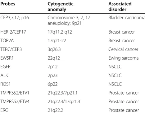
Table 6 FISH probes that are commonly used for clinical diagnosis of solid tumors
Probes Cytogenetic
anomaly
Associated disorder
CEP3,7,17; p16 Chromosome 3, 7, 17 aneuploidy; 9p21
Bladder carcinoma
HER-2/CEP17 17q11.2-q12 Breast cancer
TOP2A 17q21-22 Breast cancer
TERC/CEP3 3q26.3 Cervical cancer
EWSR1 22q12 Ewing sarcoma
EGFR 7p12 NSCLC
ALK 2p23 NSCLC
ROS1 6p22 NSCLC
TMPRSS2/ETV1 21q22.3/7p21.1 Prostate cancer
TMPRSS2/ETV4 21q22.3/17q21.3 Prostate cancer
4. Boyd C, Boyle DP:Molecular diagnosis on tissues and cells: how it affects training and will affect practice in the future.Cytopathology2012, 23(5):286–294.
5. Jiang H, Xue Y, Wang Q, Pan J, Wu Y, Zhang J, Bai S, Wang Q, He G, Sun A,
et al:The utility of fluorescence in situ hybridization analysis in diagnosing myelodysplastic syndromes is limited to cases with karyotype failure.Leuk Res2012,36(4):448–452.
6. Pathmanathan N, Bilous AM:HER2 testing in breast cancer: an overview of current techniques and recent developments.Pathology2012, 44(7):587–595.
7. Kim HR, Lim SM, Kim HJ, Hwang SK, Park JK, Shin E, Bae MK, Ou SH, Wang J, Jewell SS,et al:The frequency and impact of ROS1 rearrangement on clinical outcomes in never smokers with lung adenocarcinoma.
Ann Oncol2013,24(9):2364–2370.
8. Casaluce F, Sgambato A, Maione P, Rossi A, Ferrara C, Napolitano A, Palazzolo G, Ciardiello F, Gridelli C:ALK inhibitors: a new targeted therapy in the treatment of advanced NSCLC.Target Oncol2013,8(1):55–67. 9. Gandhi L, Janne PA:Crizotinib for ALK-rearranged non-small cell lung
cancer: a new targeted therapy for a new target.Clin Cancer Res2012, 18(14):3737–3742.
10. Rowley JD:Letter: A new consistent chromosomal abnormality in chronic myelogenous leukaemia identified by quinacrine fluorescence and Giemsa staining.Nature1973,243(5405):290–293.
11. Zhang Y, Rowley JD:Chronic myeloid leukemia: current perspectives.
Clin Lab Med2011,31(4):687–698. x.
12. Latagliata R, Avvisati G, Lo Coco F, Petti MC, Diverio D, Spadea A, Fazi P, Torromeo C, Breccia M, Malagnino F,et al:The role of all-trans-retinoic acid (ATRA) treatment in newly-diagnosed acute promyelocytic leukemia patients aged > 60 years.Ann Oncol1997,8(12):1273–1275.
13. Ablain J, de The H:Revisiting the differentiation paradigm in acute promyelocytic leukemia.Blood2011,117(22):5795–5802.
14. Costa D, Vidal A, Carrio A, Munoz C, Arias A, Gomez C, Berneaga D, Colomer D, Rozman M, Pratcorona M,et al:Refining the diagnosis and prognostic categorization of acute myeloid leukemia patients with an integrated use of cytogenetic and molecular studies.Acta Haematol2013,129(2):65–71. 15. Smith ML, Hills RK, Grimwade D:Independent prognostic variables in
acute myeloid leukaemia.Blood Rev2011,25(1):39–51. 16. Ritterbach J, Hiddemann W, Beck JD, Schrappe M, Janka-Schaub G,
Ludwig WD, Harbott J, Lampert F:Detection of hyperdiploid karyotypes (>50 chromosomes) in childhood acute lymphoblastic leukemia (ALL) using fluorescence in situ hybridization (FISH).Leukemia1998,12(3):427–433. 17. Lampert F, Harbott J, Borkhardt A:Cytogenetic aspects of childhood
leukemias.Klin Padiatr2013,225(Suppl 1):S30–S33.
18. Smoley SA, Brockman SR, Paternoster SF, Meyer RG, Dewald GW:A novel tricolor, dual-fusion fluorescence in situ hybridization method to detect BCR/ABL fusion in cells with t(9;22)(q34;q11.2) associated with deletion of DNA on the derivative chromosome 9 in chronic myelocytic leukemia.
Cancer Genet Cytogenet2004,148(1):1–6.
19. Dohner H, Stilgenbauer S, Benner A, Leupolt E, Krober A, Bullinger L, Dohner K, Bentz M, Lichter P:Genomic aberrations and survival in chronic lymphocytic leukemia.N Engl J Med2000,343(26):1910–1916.
20. Rodriguez-Vicente AE, Diaz MG, Hernandez-Rivas JM:Chronic lymphocytic leukemia: a clinical and molecular heterogenous disease.Cancer genetics
2013,206(3):49–62.
21. Rosenquist R, Cortese D, Bhoi S, Mansouri L, Gunnarsson R:Prognostic markers and their clinical applicability in chronic lymphocytic leukemia: where do we stand?Leuk Lymphoma2013,54(11):2351–2364.
22. Hallek M, Fischer K, Fingerle-Rowson G, Fink AM, Busch R, Mayer J, Hensel M, Hopfinger G, Hess G, von Grunhagen U,et al:Addition of rituximab to fludarabine and cyclophosphamide in patients with chronic lymphocytic leukaemia: a randomised, open-label, phase 3 trial.Lancet2010, 376(9747):1164–1174.
23. Nilsson T, Hoglund M, Lenhoff S, Rylander L, Turesson I, Westin J, Mitelman F, Johansson B:A pooled analysis of karyotypic patterns, breakpoints and imbalances in 783 cytogenetically abnormal multiple myelomas reveals frequently involved chromosome segments as well as significant age- and sex-related differences.Brit J Haematol2003,120(6):960–969. 24. Gutierrez NC, Garcia JL, Hernandez JM, Lumbreras E, Castellanos M, Rasillo A,
Mateo G, Hernandez JM, Perez S, Orfao A,et al:Prognostic and biologic significance of chromosomal imbalances assessed by comparative genomic hybridization in multiple myeloma.Blood2004,104(9):2661–2666.
25. Smadja NV, Bastard C, Brigaudeau C, Leroux D, Fruchart C, Groupe Francais de Cytogenetique H:Hypodiploidy is a major prognostic factor in multiple myeloma.Blood2001,98(7):2229–2238.
26. Sawyer JR:The prognostic significance of cytogenetics and molecular profiling in multiple myeloma.Cancer Genet2011,204(1):3–12. 27. Kuehl WM, Bergsagel PL:Multiple myeloma: evolving genetic events and
host interactions.Nat Rev Cancer2002,2(3):175–187.
28. Bergsagel PL, Kuehl WM:Chromosome translocations in multiple myeloma.Oncogene2001,20(40):5611–5622.
29. Avet-Loiseau H, Gerson F, Magrangeas F, Minvielle S, Harousseau JL, Bataille R, Intergroupe Francophone du M:Rearrangements of the c-myc oncogene are present in 15% of primary human multiple myeloma tumors.
Blood2001,98(10):3082–3086.
30. Tricot G, Barlogie B, Jagannath S, Bracy D, Mattox S, Vesole DH, Naucke S, Sawyer JR:Poor prognosis in multiple myeloma is associated only with partial or complete deletions of chromosome 13 or abnormalities involving 11q and not with other karyotype abnormalities.Blood1995, 86(11):4250–4256.
31. Fonseca R, Oken MM, Harrington D, Bailey RJ, Van Wier SA, Henderson KJ, Kay NE, Van Ness B, Greipp PR, Dewald GW:Deletions of chromosome 13 in multiple myeloma identified by interphase FISH usually denote large deletions of the q arm or monosomy.Leukemia2001,15(6):981–986. 32. Drach J, Ackermann J, Fritz E, Kromer E, Schuster R, Gisslinger H, DeSantis M,
Zojer N, Fiegl M, Roka S,et al:Presence of a p53 gene deletion in patients with multiple myeloma predicts for short survival after conventional-dose chemotherapy.Blood1998,92(3):802–809.
33. Fonseca R, Blood E, Rue M, Harrington D, Oken MM, Kyle RA, Dewald GW, Van Ness B, Van Wier SA, Henderson KJ,et al:Clinical and biologic implications of recurrent genomic aberrations in myeloma.Blood2003, 101(11):4569–4575.
34. Qazilbash MH, Saliba RM, Ahmed B, Parikh G, Mendoza F, Ashraf N, Hosing C, Flosser T, Weber DM, Wang M,et al:Deletion of the short arm of chromosome 1 (del 1p) is a strong predictor of poor outcome in myeloma patients undergoing an autotransplant.Biol Blood Marrow Transplant2007, 13(9):1066–1072.
35. Wu KL, Beverloo B, Lokhorst HM, Segeren CM, van der Holt B, Steijaert MM, Westveer PH, Poddighe PJ, Verhoef GE, Sonneveld P,et al:Abnormalities of chromosome 1p/q are highly associated with chromosome 13/13q deletions and are an adverse prognostic factor for the outcome of high-dose chemotherapy in patients with multiple myeloma.Brit J Haematol2007,136(4):615–623.
36. Shaughnessy JD Jr, Zhan F, Burington BE, Huang Y, Colla S, Hanamura I, Stewart JP, Kordsmeier B, Randolph C, Williams DR,et al:A validated gene expression model of high-risk multiple myeloma is defined by deregulated expression of genes mapping to chromosome 1.Blood
2007,109(6):2276–2284.
37. Kawankar N, Jijina F, Ghosh K, Vundinti BR:Cytogenetic and comparative genomic hybridization study of Indian myelodysplastic syndromes.
Cancer Epidemiol2011,35(4):e1–e5.
38. Voso MT, Fenu S, Latagliata R, Buccisano F, Piciocchi A, Aloe-Spiriti MA, Breccia M, Criscuolo M, Andriani A, Mancini S,et al:Revised International Prognostic Scoring System (IPSS) predicts survival and leukemic evolution of myelodysplastic syndromes significantly better than IPSS and WHO prognostic scoring system: validation by the Gruppo Romano Mielodisplasie Italian Regional Database.J Clin Oncol2013,31(21):2671–2677.
39. Adema V, Hernandez JM, Abaigar M, Lumbreras E, Such E, Calull A, Dominguez E, Arenillas L, Mallo M, Cervera J,et al:Application of FISH 7q in MDS patients without monosomy 7 or 7q deletion by conventional G-banding cytogenetics: does−7/7q- detection by FISH have prognostic value?Leuk Res2013,37(4):416–421.
40. Itzykson R, Kosmider O, Cluzeau T, Mansat-De Mas V, Dreyfus F, Beyne-Rauzy O, Quesnel B, Vey N, Gelsi-Boyer V, Raynaud S,et al:Impact of TET2 mutations on response rate to azacitidine in myelodysplastic syndromes and low blast count acute myeloid leukemias.Leukemia2011,25(7):1147–1152.
41. Otrock ZK, Tiu RV, Maciejewski JP, Sekeres MA:The need for additional genetic markers for myelodysplastic syndrome stratification: what does the future hold for prognostication?Expert Rev Hematol2013, 6(1):59–68.
43. Gainor JF, Varghese AM, Ou SH, Kabraji S, Awad MM, Katayama R, Pawlak A, Mino-Kenudson M, Yeap BY, Riely GJ,et al:ALK rearrangements are mutually exclusive with mutations in EGFR or KRAS: an analysis of 1,683 patients with non-small cell lung cancer.Clin Cancer Res2013,19(15):4273–4281. 44. Savic S, Bubendorf L:Role of fluorescence in situ hybridization in lung
cancer cytology.Acta Cytol2012,56(6):611–621.
45. Yoshida A, Kohno T, Tsuta K, Wakai S, Arai Y, Shimada Y, Asamura H, Furuta K, Shibata T, Tsuda H:ROS1-rearranged lung cancer: a clinicopathologic and molecular study of 15 surgical cases.Am J Surg Pathol2013,
37(4):554–562.
46. Rothschild SI, Gautschi O:Crizotinib in the treatment of non-small-cell lung cancer.Clin Lung Cancer2013,14(5):473–480.
47. Mosquera JM, Mehra R, Regan MM, Perner S, Genega EM, Bueti G, Shah RB, Gaston S, Tomlins SA, Wei JT,et al:Prevalence of TMPRSS2-ERG fusion prostate cancer among men undergoing prostate biopsy in the United States.Clin Cancer Res2009,15(14):4706–4711.
48. Attard G, Clark J, Ambroisine L, Fisher G, Kovacs G, Flohr P, Berney D, Foster CS, Fletcher A, Gerald WL,et al:Duplication of the fusion of TMPRSS2 to ERG sequences identifies fatal human prostate cancer.Oncogene2008, 27(3):253–263.
49. Demichelis F, Fall K, Perner S, Andren O, Schmidt F, Setlur SR, Hoshida Y, Mosquera JM, Pawitan Y, Lee C,et al:TMPRSS2:ERG gene fusion associated with lethal prostate cancer in a watchful waiting cohort.
Oncogene2007,26(31):4596–4599.
50. Gopalan A, Leversha MA, Satagopan JM, Zhou Q, Al-Ahmadie HA, Fine SW, Eastham JA, Scardino PT, Scher HI, Tickoo SK,et al:TMPRSS2-ERG gene fusion is not associated with outcome in patients treated by prostatectomy.
Cancer Res2009,69(4):1400–1406.
51. Saramaki OR, Harjula AE, Martikainen PM, Vessella RL, Tammela TL, Visakorpi T: TMPRSS2:ERG fusion identifies a subgroup of prostate cancers with a favorable prognosis.Clin Cancer Res2008,14(11):3395–3400.
52. van de Vijver M, van de Bersselaar R, Devilee P, Cornelisse C, Peterse J, Nusse R: Amplification of the neu (c-erbB-2) oncogene in human mammmary tumors is relatively frequent and is often accompanied by amplification of the linked c-erbA oncogene.Mol Cell Biol1987,7(5):2019–2023.
53. Slamon DJ, Godolphin W, Jones LA, Holt JA, Wong SG, Keith DE, Levin WJ, Stuart SG, Udove J, Ullrich A,et al:Studies of the HER-2/neu proto-oncogene in human breast and ovarian cancer.Science1989,244(4905):707–712. 54. Menard S, Fortis S, Castiglioni F, Agresti R, Balsari A:HER2 as a prognostic
factor in breast cancer.Oncology2001,61(Suppl 2):67–72.
55. Rouzier R, Perou CM, Symmans WF, Ibrahim N, Cristofanilli M, Anderson K, Hess KR, Stec J, Ayers M, Wagner P,et al:Breast cancer molecular subtypes respond differently to preoperative chemotherapy.Clin Cancer Res2005, 11(16):5678–5685.
56. Cleator S, Ashworth A:Molecular profiling of breast cancer: clinical implications.Brit J Cancer2004,90(6):1120–1124.
57. Perou CM, Sorlie T, Eisen MB, van de Rijn M, Jeffrey SS, Rees CA, Pollack JR, Ross DT, Johnsen H, Akslen LA,et al:Molecular portraits of human breast tumours.Nature2000,406(6797):747–752.
58. Sorlie T, Perou CM, Tibshirani R, Aas T, Geisler S, Johnsen H, Hastie T, Eisen MB, van de Rijn M, Jeffrey SS,et al:Gene expression patterns of breast carcinomas distinguish tumor subclasses with clinical implications.
Proc Natl Acad Sci USA2001,98(19):10869–10874.
59. Hicks J, Krasnitz A, Lakshmi B, Navin NE, Riggs M, Leibu E, Esposito D, Alexander J, Troge J, Grubor V,et al:Novel patterns of genome rearrangement and their association with survival in breast cancer.
Genome Res2006,16(12):1465–1479.
60. Chin K, DeVries S, Fridlyand J, Spellman PT, Roydasgupta R, Kuo WL, Lapuk A, Neve RM, Qian Z, Ryder T,et al:Genomic and transcriptional aberrations linked to breast cancer pathophysiologies.Cancer Cell2006, 10(6):529–541.
61. Fridlyand J, Snijders AM, Ylstra B, Li H, Olshen A, Segraves R, Dairkee S, Tokuyasu T, Ljung BM, Jain AN,et al:Breast tumor copy number aberration phenotypes and genomic instability.BMC Cancer2006,6:96. 62. Bergamaschi A, Kim YH, Wang P, Sorlie T, Hernandez-Boussard T, Lonning
PE, Tibshirani R, Borresen-Dale AL, Pollack JR:Distinct patterns of DNA copy number alteration are associated with different clinicopathological features and gene-expression subtypes of breast cancer.
Gene Chromosome Canc2006,45(11):1033–1040.
63. Letessier A, Sircoulomb F, Ginestier C, Cervera N, Monville F, Gelsi-Boyer V, Esterni B, Geneix J, Finetti P, Zemmour C,et al:Frequency, prognostic
impact, and subtype association of 8p12, 8q24, 11q13, 12p13, 17q12, and 20q13 amplifications in breast cancers.BMC cancer2006,6:245. 64. Creighton CJ, Kent Osborne C, van de Vijver MJ, Foekens JA, Klijn JG,
Horlings HM, Nuyten D, Wang Y, Zhang Y, Chamness GC,et al:Molecular profiles of progesterone receptor loss in human breast tumors.
Breast Cancer Res Treat2009,114(2):287–299.
65. Carracedo A, Salido M, Corominas JM, Rojo F, Ferreira BI, Suela J, Tusquets I, Corzo C, Segura M, Espinet B,et al:Are ER+PR+ and ER+PR- breast tumors genetically different? A CGH array study.Cancer Genet2012,205(4):138–146. 66. Ly M, Valent A, Diallo G, Penault-Lorca F, Dumke K, Marty V, Viehl P, Lazar V, Job B, Richon C,et al:Gene copy number variations in breast cancer of Sub-Saharan African women.Breast2013,22(3):295–300.
67. Ioannidis P, Mahaira L, Papadopoulou A, Teixeira MR, Heim S, Andersen JA, Evangelou E, Dafni U, Pandis N, Trangas T:8q24 Copy number gains and expression of the c-myc mRNA stabilizing protein CRD-BP in primary breast carcinomas.Int J Cancer2003,104(1):54–59.
68. Karlseder J, Zeillinger R, Schneeberger C, Czerwenka K, Speiser P, Kubista E, Birnbaum D, Gaudray P, Theillet C:Patterns of DNA amplification at band q13 of chromosome 11 in human breast cancer.Gene Chromosome Canc
1994,9(1):42–48.
69. Hu G, Chong RA, Yang Q, Wei Y, Blanco MA, Li F, Reiss M, Au JL, Haffty BG, Kang Y:MTDH activation by 8q22 genomic gain promotes
chemoresistance and metastasis of poor-prognosis breast cancer.
Cancer Cell2009,15(1):9–20.
70. Walker LC, McDonald M, Wells JE, Harris GC, Robinson BA, Morris CM: Dual-color fluorescence in situ hybridization reveals an association of chromosome 8q22 but not 8p21 imbalance with high grade invasive breast carcinoma.PloS One2013,8(7):e70790.
71. Mesquita B, Lopes P, Rodrigues A, Pereira D, Afonso M, Leal C, Henrique R, Lind GE, Jeronimo C, Lothe RA,et al:Frequent copy number gains at 1q21 and 1q32 are associated with overexpression of the ETS transcription factors ETV3 and ELF3 in breast cancer irrespective of molecular subtypes.Breast Cancer Res Treat2013,138(1):37–45.
72. Curtis C, Shah SP, Chin SF, Turashvili G, Rueda OM, Dunning MJ, Speed D, Lynch AG, Samarajiwa S, Yuan Y,et al:The genomic and transcriptomic architecture of 2,000 breast tumours reveals novel subgroups.Nature
2012,486(7403):346–352.
73. Shahbain H, Cooper C, Gerami P:Molecular diagnostics for ambiguous melanocytic tumors.Semin Cutan Med Surg2012,31(4):274–278. 74. Nijhawan RI, Votava HJ, Mariwalla K:Clinical application and limitations of
the fluorescence in situ hybridization (FISH) assay in the diagnosis and management of melanocytic lesions: a report of 3 cases.Cutis2012, 90(4):189–195.
75. Bastian BC, LeBoit PE, Hamm H, Brocker EB, Pinkel D:Chromosomal gains and losses in primary cutaneous melanomas detected by comparative genomic hybridization.Cancer Res1998,58(10):2170–2175.
76. Bastian BC, Olshen AB, LeBoit PE, Pinkel D:Classifying melanocytic tumors based on DNA copy number changes.Am J Pathol2003,163(5):1765–1770. 77. Horst BA, Fang Y, Silvers DN, Busam KJ:Chromosomal aberrations by
4-color fluorescence in situ hybridization not detected in Spitz nevi of older individuals.Arch Dermatol2012,148(10):1152–1156.
78. Bangash HK, Romegialli A, Dadras SS:What's new in prognostication of melanoma in the dermatopathology laboratory?Clin Dermatol2013, 31(3):317–323.
79. Julia F, Thomas L, Dalle S:New therapeutical strategies in the treatment of metastatic disease.Dermatol Ther2012,25(5):452–457.
80. Pajor G, Alpar D, Kajtar B, Melegh B, Somogyi L, Kneif M, Bollmann D, Pajor L, Sule N:Automated signal pattern evaluation of a bladder cancer specific multiprobe-fish assay applying a user-trainable workstation.
Microsc Res Tech2012,75(6):814–820.
81. Raimondo F, Gavrielides MA, Karayannopoulou G, Lyroudia K, Pitas I, Kostopoulos I:Automated evaluation of Her-2/neu status in breast tissue from fluorescent in situ hybridization images.IEEE Trans Image Process
2005,14(9):1288–1299.
82. Theodosiou Z, Kasampalidis IN, Karayannopoulou G, Kostopoulos I, Bobos M, Bevilacqua G, Aretini P, Starita A, Lyroudia K, Pitas I:Evaluation of FISH image analysis system on assessing HER2 amplification in breast carcinoma cases.Breast2008,17(1):80–84.
84. Kajtar B, Mehes G, Lorch T, Deak L, Kneifne M, Alpar D, Pajor L:Automated fluorescent in situ hybridization (FISH) analysis of t(9;22)(q34;q11) in interphase nuclei.Cytometry A2006,69(6):506–514.
85. Stumm M, Wegner RD, Bloechle M, Eckel H:Interphase M-FISH applications using commercial probes in prenatal and PGD diagnostics.Cytogenet Genome Res2006,114(3–4):296–301.
86. Chyhrai A, Sanjmyatav J, Gajda M, Reichelt O, Wunderlich H, Steiner T, Tanovic E, Junker K:Multi-colour FISH on preoperative renal tumour biopsies to confirm the diagnosis of uncertain renal masses.World J Urol
2010,28(3):269–274.
87. Schramm M, Wrobel C, Born I, Kazimirek M, Pomjanski N, William M, Kappes R, Gerharz CD, Biesterfeld S, Bocking A:Equivocal cytology in lung cancer diagnosis: improvement of diagnostic accuracy using adjuvant multicolor FISH, DNA-image cytometry, and quantitative promoter hypermethylation analysis.Cancer Cytopathol2011,119(3):177–192. 88. Anderson K, Lutz C, van Delft FW, Bateman CM, Guo Y, Colman SM,
Kempski H, Moorman AV, Titley I, Swansbury J,et al:Genetic variegation of clonal architecture and propagating cells in leukaemia.Nature2011, 469(7330):356–361.
89. Janssens A, Van Roy N, Poppe B, Noens L, Philippe J, Speleman F, Offner F: High-risk clonal evolution in chronic B-lymphocytic leukemia: single-center interphase fluorescence in situ hybridization study and review of the literature.Eur J Haematol2012,89(1):72–80.
90. Wawrzyniak E, Kotkowska A, Blonski JZ, Siemieniuk-Rys M, Ziolkowska E, Giannopoulos K, Robak T, Korycka-Wolowiec A:Clonal evolution in CLL patients as detected by FISH versus chromosome banding analysis, and its clinical significance.Eur J Haematol2013,92(2):91–101. 91. Wang YC, Hu LP, Lin D, Li CW, Yuan T, Jia YJ, Tian Z, Tang KJ, Wang M,
Wang JX:Detection of heterogeneity and evolution of subclones in t(8;21) AML by QM-FISH.Zhonghua Xue Ye Xue Za Zhi2013,34(10):844–850. 92. Vital AL, Tabernero MD, Crespo I, Rebelo O, Tao H, Gomes F, Lopes MC,
Orfao A:Intratumoral patterns of clonal evolution in gliomas.
Neurogenetics2010,11(2):227–239.
93. Zhang S, Shao Y, Hou G, Bai J, Yuan W, Hu L, Cheng T, Zetterberg A, Zhang J: QM-FISH analysis of the genes involved in the G1/S checkpoint signaling pathway in triple-negative breast cancer.Tumour Biol2013.
(Epub ahead of print).
94. Tomao F, Papa A, Rossi L, Strudel M, Vici P, Lo Russo G, Tomao S:Emerging role of cancer stem cells in the biology and treatment of ovarian cancer: basic knowledge and therapeutic possibilities for an innovative approach.J Exp Clin Cancer Res2013,32:48.
95. Li Z:CD133: a stem cell biomarker and beyond.Exp Hematol Oncol2013, 2(1):17.
96. Schwab CJ, Jones LR, Morrison H, Ryan SL, Yigittop H, Schouten JP, Harrison CJ:Evaluation of multiplex ligation-dependent probe amplification as a method for the detection of copy number abnormalities in B-cell precursor acute lymphoblastic leukemia.Gene Chromosome Canc2010, 49(12):1104–1113.
97. Cheng T:Cell cycle inhibitors in normal and tumor stem cells.Oncogene
2004,23(43):7256–7266.
doi:10.1186/2050-7771-2-3
Cite this article as:Huet al.:Fluorescence in situ hybridization (FISH): an increasingly demanded tool for biomarker research and personalized medicine.Biomarker Research20142:3.
Submit your next manuscript to BioMed Central and take full advantage of:
• Convenient online submission
• Thorough peer review
• No space constraints or color figure charges
• Immediate publication on acceptance
• Inclusion in PubMed, CAS, Scopus and Google Scholar
• Research which is freely available for redistribution