R E S E A R C H
Open Access
Hyperthermia induced HIF-1a expression of
lung cancer through AKT and ERK signaling
pathways
Jun Wan
1*and Wei Wu
2Abstract
Background:Hyperthermia is a promising treatment for human lung cancer, but recurrence of the primary lesion is common, as the residual tumor becomes adapted to heat treatment and growth is induced by hypoxia-triggered HIF-1a expression. Here, we explored the effects of hyperthermia on HIF-1a expression, proliferation, and lung cancer angiogenesis.
Methods:Human NSCLC NCI-H1650 and SCLC NCI-H446 cell lines were used to examine cell viability, apoptosis, and HIF-1a expression level under a gradient of thermal conditions (37, 42 and 47 °C for 40 min). The 47 °C heat-adapted NCI-H1650 and NCI-H446 sublines (also called NCI-H1650-b and NCI-H446-b cells) had enhanced viability and HIF-1a expression levels compared to the parental and 42 °C heat-adapted cells and were thus used for subsequent research. Concentration gradients of wortmannin and PD98095 were used to inhibit AKT and ERK expression, respectively in the NSCLC NCI-H1650-b and SCLC NCI-H446-b cell lines, and cell growth curves were drawn. Western blots were used to detect the expression of HIF-1a, extracellular signal-regulated kinase (ERK), protein kinase B (AKT), phospho-ERK, and phospho-AKT. We established a subcutaneous transplantation tumor model with wortmannin and PD98095 intervention. Immunohistochemistry was used to detect the expression of HIF-1a and the vascular specific marker CD34, and tumor growth curves were drawn.
Results:Following hyperthermia treatment, HIF-1a expression in 47 °C heat-adapted NSCLC and SCLC cell lines was regulated by the AKT pathway. However, HIF-1a expression was also regulated by the ERK pathway in NSCLCs, while SCLCs did not exhibit changes in ERK. These biological behaviors are governed by signaling pathway protein phosphorylation. Furthermore, inhibiting the AKT pathway can suppress the proliferation and angiogenesis potential of both 47 °C heat-adapted NSCLCs and SCLCs, but inhibiting the ERK pathway only affects SCLCs. Conclusion:Our study suggests that following hyperthermia, the proliferation and angiogenesis potential of residual NSCLCs and SCLCs is induced by HIF-1a. However, HIF-1a expression in NSCLCs is regulated by both the AKT and ERK signaling pathway, but HIF-1a expression in SCLCs is regulated only by the AKT signaling pathway. This study sheds light on the molecular regulatory mechanisms of lung cancer recurrence following hyperthermia treatment.
Keywords:Angiogenesis potential, Cd34, Extracellular regulated protein kinases (erk), Hyperthermia, Hypoxia-inducible factor-1 alpha (hif-1a), Non small cell lung cancer (nsclc), Protein kinase b (akt), Small cell lung cancer (sclc), Signaling pathway protein, Tumor proliferation
* Correspondence:junwandr@yahoo.com
1Department of Thoracic Surgery, The First Affiliated Hospital of Anhui Medical University, NO.218, Jixi Road, Hefei 230022, Anhui, People’s Republic of China
Full list of author information is available at the end of the article
Background
In recent years, hyperthermia has been recognized as a clinical treatment for certain cancers (e.g. lung cancer), which can be induced by therapeutic techniques, such as radiofrequency ablation (RFA). However, one of the major problems with hyperthermia is that it is difficult to achieve complete tumor destruction using this tech-nique, and recurrence is common, because the residual tumor is now adapted to heat treatment and growth is induced [1]. Accumulating evidence indicates that both hypoxia and hypoxia-driven angiogenesis are the conse-quence of hyperthermia, and both of these factors play important roles in tumor growth [2]. Under conditions of hypoxia, a signaling pathway involving a crucial oxy-gen response regulator, defined hypoxia-inducible factor (HIF), is turned on. HIF protein, especially HIF-1α and HIF-2α, are correlation with tumor development, metas-tasis and promote epithelial-mesenchymal transition [3]. In contrast with HIF-2α, which is expressed in certain cell types of vertebrate species, the expression of HIF-1α is observed in most metazoan species and involved in the regulation of epithelial-mesenchymal transition of tumor [4]. As a key transcriptional regulator, HIF-1α plays a central role in the adaptation of tumor cells to hypoxia and helps to regulate the expression of muitiple cytokines, such as vascular endothelial growth factor-A (VEGF-A), and promotes the proliferation [5] and angio-genesis potential [6] of small cell lung cancers (SCLCs). Earlier research also showed that HIF-1a is involved in apoptosis and the proliferation of non-small cell lung cancers (NSCLCs) [7, 8]. In our previous study, we found that local recurrences of SCLC following RFA treatment were driven by HIF-1a, while the thermal ef-fects of RFA can promote the growth of residual NSCLCs by up-regulating HIF-1a expression [9].
The expression and activity of HIF-1a is not only in-duced in response to limited oxygen availability, but it is also modulated through related signaling pathways [10]. Some studies have demonstrated that the expres-sion of HIF-1a is regulated by major signaling path-ways, including the extracellular signal-regulated kinase (ERK) pathway [11] and the protein kinase B (AKT) pathway [12]. Intensive studies of the ERK and AKT pathways have revealed these signaling pathways play their most important roles in the molecular sig-naling network that governs growth, proliferation, dif-ferentiation and survival in many, if not all, cell types [13, 14]. Therefore, to uncover the molecular mecha-nisms of lung cancer recurrence following hyperther-mia treatment, the present study explores the effects of hyperthermia on HIF-1a expression and cell prolif-eration and angiogenesis. We then went on to investi-gate the regulation and function of the Akt and ERK signaling pathways.
Methods
Chemicals and antibodies
Specific inhibitor wortmannin, PD98059, and Recombinant Human Heregulin were purchased from Sigma, St. Louis, MO, USA. TRIzol reagent was purchased from Invitrogen, Carlsbad, CA. RIPA lysis buffer was purchased from the Beyotime Institute of Biotechnology, China. The following primary antibodies: anti-CD34 (1:40 dilution), anti-HIF-1a (1:500 dilution), ERK1/2 (1:1000 dilution), and anti-AKT (1:1000 dilution) were purchased from Wuhan Boster Biological Engineering Technology Limited Company. Anti-phospho-AKT (1:1000 dilution) was purchased from Cell Signaling Technology, Beverly, MA, USA. and anti-phospho-ERK1/2 (1:800 dilution) was purchased from Santa Cruz, CA, USA.
Cell culture, heat treatment of cells, and establishment of sublines
Human NSCLC H1650 cells and SCLC NCI-H446 cells were maintained in RPMI-1640 medium (Sigma-Aldrich Co., St. Louis, MO, USA) supplemented with 10 % fetal bovine serum (FBS), 100 units/mL penicil-lin, and 100μg/ml kanamycin at 37 °C in a humidified at-mosphere containing 5 % CO2and 20 % O2. The medium
was routinely changed 2-3 days after seeding. Cells were detached with trypsin/EDTA (GibcoBRL, Paisley, UK) and were resuspended in a 1:1 solution of serum-free RPMI-1640 medium to a final concentration of approximately 5× 10⁵cells/10μl.
During the exponential phase, cells were exposed to hyperthermic stress in cell culture plates for 24, 48, or 72 h. The plates were sealed with parafilm and sub-merged in a water bath at the desired temperature for 10 min. The three desired temperatures were 37, 42, and 47 °C. The cells cultured at 42 and 47 °C generated 2 cell sublines each, named NCI-H1650-a and NCI-H1650-b; NCI-H446-a and NCI-H446-b respectively. After the hyperthermic treatment, fresh culture medium was added to each well, and the surviving cells were main-tained at 37 °C with an atmosphere containing 5 % CO2
and 1 % O2for 4 h. After determining cell viability and
HIF-1a expression levels, we chose one subline for the following experiment.
TUNEL staining for apoptosis assay
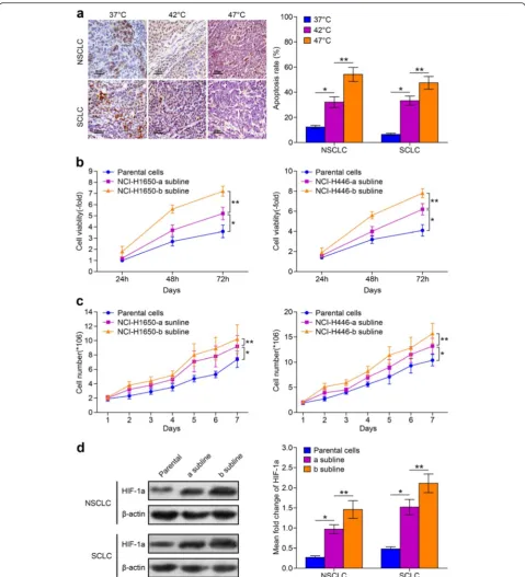
Fig. 1The viability of NCI-H1650 cells and NCI-H446 cells and their sublines adapted to hyperthermia treatment.aNCI-H1650 and NCI-H446 cells are cultured under 37, 42, and 47 °C. Apoptosis rate is measured using a Tunnel staining assay. The brown stained cells are apoptocic cells, and semi-quantitative analysis of apoptosis rates are measured. (*p< 0.05 37 °C vs 42 °C heat treatment group,**p< 0.05 47 °C vs 42 °C heat treatment group) (b) 2 sublines were established following incubation at 42 and 47 °C. The 24, 48, and 72 h viability was evaluated by MTT assay. Parental cells: cells cultured under 37 °C; NCI-H1650-a and NCI-H446-a subline: cells adapted to 42 °C heat treatment; NCI-H1650-b and NCI-H446-b subline: cells adapted to 47 °C heat treatment (*p< 0.05 NCI-H1650-a or NCI-H446-a subline vs parental cells,**p< 0.05 NCI-H1650-a subline vs NCI-H1650-b subline, NCI-H446-a subline vs NCI-H446-b subline) (c) Growth curve of parental cells and the cells subline a and b are drawn. Data are the representative results of three independent experiments (*p< 0.05 from day 2-7 parental cells vs cells subline-a,**p< 0.05 from day 2-7 cells subline-a vs cells subline-b).
tetrahydrochloride (DAB) solution for 8 min was used for counterstaining. Cells were examined using a Nikon microscope with Image-Pro Plus 6.x software (Diagnostic Instruments, USA) for image analysis.
MTT assay for lung cancer cells viability
Cells were cultured at a concentration of 1 × 10⁴ cells/ well in 48-well plates. The 3-(4,5-dimethylthiazol-2-yl)-2,5-diphenyl tetrazolium bromide (MTT) solution was added to each well at a final concentration of 0.5 mg/ml and incubated for 4 h. At the end of the incubation, for-mazan crystals, resulting from MTT reduction, were dis-solved by addition of 150 ml DMSO per well. The optical density was read at 570 nm, and the average values were determined from replicate wells.
Growth of xenografts in nude mice
Male congenital athymic BALB/c nude mice were ob-tained from the Experimental Animal Center of the Shang Hai Jiao Tong University School of Medicine. They were maintained under pathogen-free conditions in accordance with established institutional guidance and approved protocols. All experiments were carried out using 6-8-week-old mice weighting 16-22 g. Sublines of NCI-H1650 and NCI-H446 cells were cultured in vitro (1 × 10⁷), and a final concentration of approxi-mately 5 × 10⁵ cells/10 μl were suspended in PBS and subcutaneously injected into the flank area of mice. After tumors reached 3-5 mm in diameter, mice were injected with either vehicle (10 % DMSO/PBS), 4 mg/kg, or 5 mg/kg twice weekly. The tumor size was measured with calipers every 3 days, and tumor volume was calcu-lated according to the formula: volume = width2× length × 0.5. Tumors were removed and weighed 30 days after inoculation. All surgical procedures were per-formed under isoflurane inhalation anesthesia. Bupre-norfine was injected intramuscularly prior to surgery for perioperative analgesia.
Immunohistochemistry detection for HIF-1a and vascular specific markers
All tumor tissue sections were cut into 4 μM sections, deparaffinized, and endogenous peroxidases were inhib-ited with 0.3 % hydrogen peroxide in methanol for 30 min. Antigen retrieval was achieved using 0.05 % pro-tease XIV at 37 °C for 5 min. Sections were then incu-bated with a mouse anti-human HIF-1a or CD34 primary antibody overnight at 4oC. Next, the slides were incubated with biotin-conjugated rabbit anti-mouse secondary antibody at room temperature for 45 min. The sections were subsequently incubated with a streptavidin-biotin-peroxidase complex (Vectastain ABC kit, Vector Laboratories, Burlingame, CA, USA) at room temperature for 45 min. The reaction was visualized
using chromogen diaminobenzidine (DAB) for 10 s. Fi-nally, the slides were counterstained with hematoxylin and mounted. The slides were examined with a Nikon Eclipse Ti microscope under a 40X objective.
Western-blot analysis of HIF-1a and signaling protein
Cells and tissues were harvested and analyzed for the ex-pression of HIF-1a, ERK, p-ERK, AKT, and p-AKT. Briefly, total protein was extracted by disrupting cells in RIPA lysis buffer and separating on a polyacrylamide gel and transferring to PVDF membrane. The membranes were then blocked at room temperature for 1 h with 5 % non-fat milk in Tris buffered saline containing Tween 20 (TBST). The membrane was incubated with anti-HIF-1a, ERK1/2, AKT, phospho-AKT, and anti-phospho-ERK1/2 primary antibodies at 37oC for 2 h, and then with peroxidase-conjugated IgG at room temperature for 1 h. The membranes were subsequently incubated with goat anti-rabbit peroxidase-conjugated secondary antibodies, and immunoreactivity was de-tected by using an enhanced chemiluminescence kit, and captured on X-ray film. β-actin was used as an internal control.
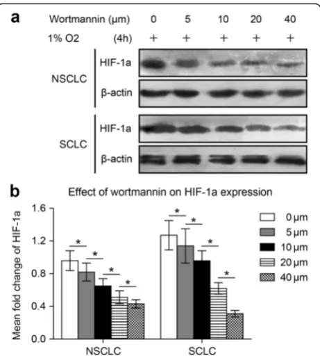
Fig. 2Wortmannin inhibits HIF-1a expression in NSCLC and SCLC cells following 47 °C heat treatment. NCI-H1650-b and NCI-H446-b sublines are cultured to 80-90 % confluence and exposed to hypoxia.
Statistical analysis
The SPSS 13.0 software (SPSS, USA) was applied to complete data processing. An independent-samples t-test was used to evaluate the differences in optical density (OD) values or tumor cell numbers between groups with various treatments. All data are represented as the mean ± SD for three independent experiments. Results were considered statistically significant when the p-value was less than 0.05.
Results
In vitro heat treatment can generate NCI-H1650 cell sublines with increased viability
To detect the potential effect of hyperthermia on the pro-liferative activity of lung cancer cells, first we monitored cell viability at 24, 48, and 72 h after incubation at 37, 42, or 47 °C for 10 min. We found that the apoptosis rate of NCI-H1650 and NCI-H446 cells after incubation at 47 °C was significantly higher compared to cells incubated at 37 °C or 42 °C (Fig. 1a). In addition, the apoptosis rate of cells incubated at 42 °C was higher than at 37 °C. The cells cultured at conventional culture temperature (37 °C) are called parental cells, and the cells cultured and adapted to 42 and 47 °C were named the H1650-a and
NCI-H446-a and NCI-H1650-b and NCI-H446-b sublines respectively. The cellular viability of parental cells and sublines all reach their highest viability at 72 h. However, the cell viability of sublines adapted to 47 °C are higher than the sublines adapted to 42 °C or the parental cells. The viability of sublines adapted to 42 °C are higher than parental cells (Fig. 1b). From the growth curve, we can see that the proliferation activity of sublines adapted to 47 °C are higher than the sublines adapted to 42 °C or the parental cells (Fig. 1c). Furthermore, the HIF-1a expres-sion level of sublines adapted to 47 °C is also higher than the subline adapted to 42 °C or the parental cells (Fig. 1d). Therefore, we conclude that thermal treatment can partially kill lung cells, while generating sublines with increased viability with higher HIF-1a expression levels. Therefore, we used the NCI-H1650 subline adapted to 47 °C for our subsequent experiments.
Specific inhibitor wortmannin inhibited HIF-1a expression, and the proliferation, and angiogenesis potential of NSCLCs and SCLCs following 47 °C heat treatment
We investigated the effect of Akt signaling on HIF-1a expression in both NSCLC NCI-H1650 cells and SCLC
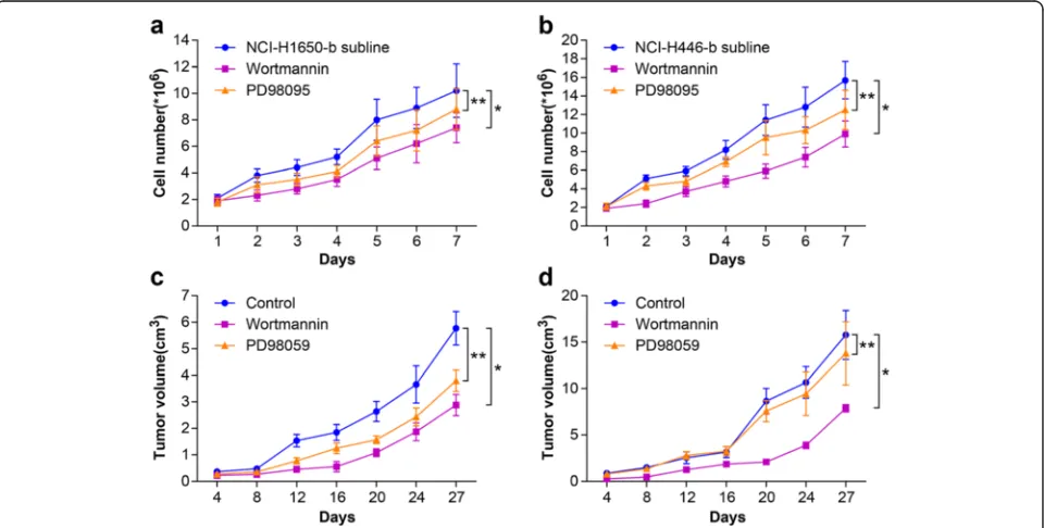
Fig. 3Effect of Wortmannin or PD98095 on the proliferation of NSCLC and SCLC following heat treatment. In vitro, the H1650-b and NCI-H446-b sublines (in logarithmic phase) are treated with wortmannin or PD98095 (40μM). We compared the growth curve of the cells in control group, wortmannin group, and PD98095 group.aWhen the NCI-H1650-b subline was treated with wortmannin or PD98095, the growth curve shifted to the right and the growth rate was slowed (*p< 0.05 wortmannin group vs control group,**p< 0.05 PD98095 group vs control group).
NCI-H446 cells adapted to 47 °C. Because HIF-1a ex-pression in the NCI-H1650-b and NCI-H446-b sublines was elevated, we cultured the cells under hypoxic con-ditions (1 % oxygen concentration). Wortmannin, a specific inhibitor of the Akt signaling pathway, reduced HIF-1a protein expression in a dose-dependent manner in both the NCI-H1650-b and NCI-H446-b cell sub-lines (Fig. 2a, b). From the cell growth curve, we found that the proliferation of the H1650-b and NCI-H446-b sublines was inhibited by wortmannin (Fig. 3a, b). To further evaluate the effect of AKT signaling on
HIF-1a expression and angiogenesis potential, we next examined the effects of wortmannin on NCI-H1650-b and NCI-H446-b subline xenografts. NCI-H1650-b and NCI-H446-b subline cells were injected into the flanks of athymic BALB/c nude mice to facilitate development of a xenograft. One week following injection of the tumor cells, wortmannin mixed with 5 % sodium bicar-bonate was intraperitoneally delivered daily for three weeks. We found that the growth of the tumor xeno-grafts was significantly inhibited by wortmannin (Fig. 3c, d), while immunohistochemical staining
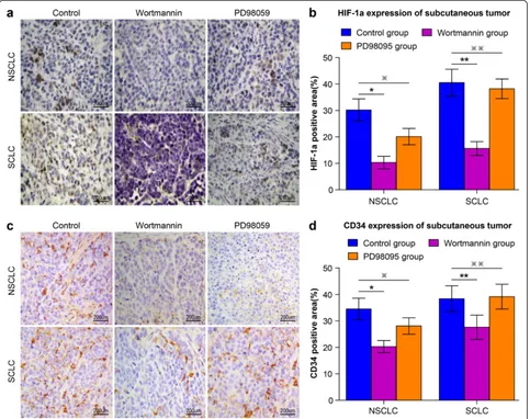
showed HIF-1a and CD34 expression was significantly decreased (Fig. 4a-d).
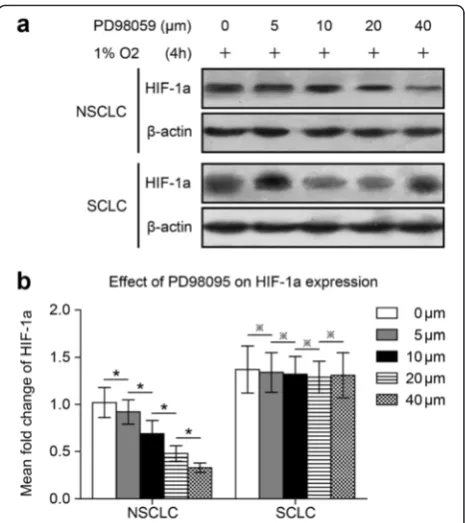
Specific inhibitor PD98095 inhibited HIF-1a expression, proliferation, and angiogenesis potential in NSCLCs but not SCLCs following 47 °C heat treatment
To determine if ERK signaling affected HIF-1a ex-pression in NSCLC NCI-H1650 and SCLC NCI-H446 cells adapted to 47 °C, we used the ERK specific in-hibitor, PD98095. PD98095 inhibited hypoxia-induced HIF-1a protein expression in a dose-dependent man-ner in NCI-H1650-b cells (Fig. 5a, b), while inhibiting cell proliferation (Fig. 3a). On the contrary, HIF-1a expression was not inhibited by PD98095 in NCI-H1650-b cells (Fig. 5a, b); proliferation of these cells was also unaffected (Fig. 3b). After the establishment of NCI-H1650-b and NCI-H446-b subline xenografts, we injected PD98095 mixed with 5 % sodium bicar-bonate daily for three weeks. We found that the growth of the NSCLC tumor xenografts was inhibited by PD98095 (Fig. 3c), while immunohistochemical staining showed that HIF-1a and CD34 expression was signifi-cantly decreased (Fig. 4a-d). However, PD98095 had no obvious effect on the growth of the SCLC tumor xeno-grafts (Fig. 3d), and HIF-1a and CD34 expression in this tumor tissue showed only marginal changes (Fig. 4a-d).
HIF-1a expression in the NSCLC cell subline adapted to 47 °C was induced by both AKT and ERK signaling
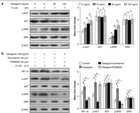
We next studied the signaling pathways regulating the proliferation and angiogenesis potential of NSCLC cells adapted to 47 °C. Previous reports confirmed that AKT and ERK signaling pathways are involved in regulating the important biological characteristics of tumors de-rived from multiple tissue sources. To characterize the roles of AKT and ERK in regulating HIF-1a expression, wortmannin and PD98059 were used again. NCI-H1650-b cells were cultured under hypoxic conditions, follow-ing treatment by heregulin, which induces HIF-1a ex-pression [16]. We found that AKT and ERK total expression levels did not change much with treatment, but the abundance of their phosphorylated forms, p-AKT and p-ERK, increased in a dose dependent manner (Fig. 6a). These results indicate that the phosphorylated forms of AKT and ERK play a major role in the regula-tion of HIF-1a expression. To confirm this finding, we pre-treated NCI-H1650-b cells with the heregulin and then treated with wortmannin or PD98095 and incu-bated under hypoxic conditions for 6 h. Our study showed that wortmannin abolishes heregulin-induced HIF-1a expression, and PD98059 also significantly atten-uates HIF-1a expression (Fig. 6b). Taken together, these results indicate that the inactivation of the AKT and
ERK signaling pathways is important for the inhibition of HIF-1a expression in NSCLCs following hyperthermic treatment.
HIF-1a expression of the SCLC cell subline adapted to 47 °C is induced by the AKT, but not the ERK signaling pathway
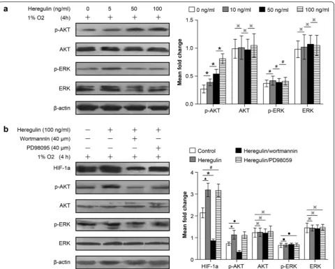
Because we found that the AKT and ERK signaling path-ways were both involved in regulating HIF-1a expression in NSCLCs following hyperthermic treatment, we inves-tigated the role of these pathways in HIF-1a regulation in SCLCs. As with NSCLCs, heregulin upregulated p-AKT expression in a dose-dependent manner, but had little effect on p-ERK expression (Fig. 7a). We pre-treated the NCI-H446-b cell subline with heregulin and then followed with wortmannin or PD98095, incubating under hypoxic conditions for 6 h. Our study showed that wortmannin also abolished heregulin induction of HIF-1a expression in the NCI-H446-b cell subline (Fig. 7b).
Fig. 5PD98095 inhibits HIF-1a expression in NSCLC cells but not SCLC cells following heat treatment. After cultured to 80-90 % confluence, NCI-H1650-b and NCI-H446-b cell sublines are exposed to hypoxia and harvested to determine HIF-1a expression levels.
Although PD98059 inhibits ERK, it had no significant ef-fect on HIF-1a expression in SCLCs (Fig. 7b). These in-dicate that: (1) AKT signaling pathways help to regulate HIF-1a expression in both NSCLCs and SCLCs follow-ing hyperthermic treatment, and (2) the change in p-ERK expression has no significant relationship with HIF-1a expression in SCLCs following hyperthermia. Therefore, HIF-1a expression in SCLCs following hyperthermia is not regulated through the ERK signal-ing pathway.
Discussion
Lung cancer is a heterogeneous, complex, and challen-ging disease to treat. Hyperthermia is an approach that takes advantage of the biological effects of heat to target tumors. Hyperthermia techniques have been the subject of extensive research with the overall goal of medicine development and equipment advancement [17]. Now, hyperthermia is regarded as an effective treatment for lung cancer, which may be as successful as surgery, radiotherapy, and chemotherapy. At present, clinical Fig. 6HIF-1a expression in NSCLCs following heat treatment is regulated through both the AKT and ERK signaling pathways. NCI-H1650-b sublines are seeded and pretreated with different concentrations of heregulin and then exposed to 1 % oxygen. Expression levels of AKT, ERK, phosphorylated AKT, and phosphorylated ERK were determined by western blot analysis.aAfter treatment with heregulin, AKT and ERK total protein expression levels showed no obvious change (*p> 0.05 between different groups), but p-AKT and p-ERK were significantly up-regulated (※p< 0.05 between different
hyperthermia treatment methods for lung cancers in-clude laser ablation, radiofrequency ablation, and mag-netic hyperthermia [18]. Among these, radiofrequency ablation is becoming an increasingly accepted treatment for primary lung cancers in patients who are not candi-dates for subsegmental resection or lobectomy [19]. The prognosis for tumor patients has been closely correlated with activation and expression level of HIF-1a which are regulated by many factors such as DEC2 (differentiated embryonic chondrocyte gene 2) [20]. Besides this, heat treatment can cause hypoxia in the local tissue and
increase HIF-1a expression levels, which can induce the over-proliferation of any residual tumors, leading to the recurrence of lung cancer [21]. Additionally, the angio-genesis potential increased by heat-induced hypoxia can also play a role in the rapid growth of residual tumor cells that have escaped heat ablation. This phenomenon is often referred to as a“malignant transformation”[22]. Hypoxia conditions present with, or induced by, heat treatment can be replicated in in vitro cell culture. The cell lines we used were already established and used in previous studies, so that further spontaneous Fig. 7HIF-1a expression in SCLCs following heat treatment was regulated through the AKT but not the ERK signaling pathway. The method used was described in Fig. 6; NCI-H446-b subline cells are harvested and western blot analysis for protein expression is applied.aThere were no obvious changes in AKT and ERK total protein expression levels (※p> 0.05 between different groups). p-AKT was gradually induced with increasing heregulin concentrations (*p< 0.05 between different groups), but p-ERK expression showed no significant change (#p> 0.05 between different groups).bHIF-1a expression was upregulated after treated by heregulin (▲p< 0.05 heregulin group vs control group), but was inhibited after co-treatment with wortmannin, but not PD98059 (*p< 0.05 heregulin group vs heregulin + wortmannin group,#p> 0.05 heregulin
transformation under normal culture conditions would be unlikely. Therefore, we concluded that the biological microenvironment associated with tumor growth chan-ged due to the exposure to heat stress. Heat-adapted sublines became more proliferative, which became the focus of our subsequent work to identify the molecular/ biological mechanisms involved in tumor recurrence.
In our previous study, HIF-1a was found overex-pressed in many human cancers and various cell types, and the levels of HIF-1a activity were correlated with tumorigenicity, angiogenesis, and metastasis [23]. Its tar-get gene, VEGF-A, is mainly regulated by HIF-1a at the transcriptional level, and VEGF-A also plays a critical role in tumor angiogenesis, growth, and metastasis. Targeting the HIF-1a/VEGF-A axis may be a promising strategy for combating tumor recurrences following hyperthermia treatment [24]. There has been a signifi-cant increase in the understanding of the importance of the signaling pathways that regulate the biological be-havior of both NSCLCs and SCLCs (e.g. proliferation and angiogenesis) in recent years [25, 26]. Inhibitors of HIF-1a represent a new tool for improved cancer therap-ies [27]. A number of chemical, protein, and nucleic acid inhibitors are included and classified based on their mechanisms of inhibitory action. Among these, inhibit-ing upsteam signalinhibit-ing to block HIF-1a expression has proven effective [28]. HIF-1α is situated at the conver-gence of multiple oncogenic and tumor suppressor path-ways, including the PI3K/AKT and MAPK/ERK pathways [29]. These signaling pathways are at the heart of a molecular signaling network that governs growth, proliferation, differentiation, and survival in many cell types [30]. They are dysregulated in various diseases, ranging from cancer to immunological, inflammatory, and degenerative syndromes, and thus represent an im-portant drug target [31, 32]. A previous study found that activating the AKT and ERK signaling pathways could enhance HIF-1a/VEGF expression in some malignant tu-mors [33]. From the study about the mechanical regula-tion of signaling pathway in lung cancer, some scholars find that inactivation of Akt signaling can inhibit the growth of human lung cancer cells through reducing SP1 and p65 protein expression [34]. ERK and PI3K/ AKT pathways can promote tumor cell growth and me-tastasis and a notable decrease of ERK and PI3K/AKT activation can be found in TRIM11 knocked down lung cancer cells [35]. These results are in congruence with our results that show inhibiting AKT and ERK expres-sion through specific inhibitors in NSCLs can down-regulate HIF-1a expression and inhibit angiogenesis. However, inhibition of ERK expression had no signifi-cant effect on HIF-1a expression in SCLCs, which was regulated by the AKT signaling pathways. We hypothe-sized that the prime cause for this observation was that
the regulatory mechanisms governing tumor properties are different, based on histological origin. Therefore, we intend to investigate these regulatory mechanisms fur-ther in the future.
Hyperthermia is a promising treatment for human lung cancer, but local recurrence is common, as growth of residual tumors can be induced by thermal ablation. Our study demonstrates that hyperthermia inherently changes the properties of cancer cells, facilitating the creation of different cancer sublines. These sublines ex-hibited enhanced viability and angiogenesis potential, and we determined that HIF-1a/VEGF-A plays a central role during this biological process. Furthermore, we in-vestigated the molecular pathways induced/affected by hyperthermia. We found that HIF-1a expression was in-duced in heat adapted cell lines and that in NSCLCs this involves both PI3K/AKT and ERK signaling pathways, while only the PI3K/AKT signaling pathway is affected in SCLCs. Altogether, we hope that through investigating the molecular mechanisms of lung cancer recurrence following hyperthermia, our work will supply additional evidence for the use of multidisciplinary synthetic ther-apies to treat lung cancer.
Conclusions
In this research we find that HIF-1a plays a critical role in the recurrence of lung cancer following hyperthermia treatment as the proliferation and angiogenesis potential of residual NSCLCs and SCLCs are induced by HIF-1a. However, HIF-1a expression in NSCLCs is regulated by both the AKT and ERK signaling pathway, but HIF-1a expression in SCLCs is regulated only by the AKT signaling pathway. Our study sheds light on the molecular regulatory mechanisms of lung cancer recurrence following hyper-thermia treatment.
Abbreviations
AKT, proteinkinase B; ERK, extracellular signal-regulated kinase; HIF-1a, hypoxia-inducible factor-1 alpha; NSCLC, non small cell lung cancer; SCLC, small cell lung cancer
Acknowledgements
We would like to thank the Research Center of the First Affiliated Hospital of Anhui Medical University and Laboratory Animal Center of Anhui Medical University for providing technical assistance. The authors would like to thank the Duoease Scientific Service Center for excellent language editing service and suggestions for figure revision.
Authors’contributions
JW and WW conceived and designed the study. JW and WW performed the experiments. WW analyzed the data. JW contributed reagents/materials/ analysis tools. JW wrote the paper. WW read and revised the manuscript, accepted the final version. All authors read and approved the final manuscript.
Competing interests
Author details
1Department of Thoracic Surgery, The First Affiliated Hospital of Anhui Medical University, NO.218, Jixi Road, Hefei 230022, Anhui, People’s Republic of China.2Department of Hematology, The First Affiliated Hospital of Anhui Medical University, Hefei 230022, China.
Received: 21 June 2016 Accepted: 19 July 2016
References
1. Wang Y, Li G, Li W, He X, Xu L. Radiofrequency ablation of advanced lung tumors: imaging features, local control, and follow-up protocol. Int J Clin Exp Med. 2015;8(10):18137–43.
2. Wan J, Wu W, Chen Y, Kang N, Zhang R. Insufficient radiofrequency ablation promotes the growth of non-small cell lung cancer cells through PI3K/Akt/ HIF-1alpha signals. Acta Biochim Biophys Sin (Shanghai). 2016;48(4):371–7. 3. Yang J, Zhang X, Zhang Y, Zhu D, Zhang L, Li Y, et al. HIF-2alpha promotes
epithelial-mesenchymal transition through regulating Twist2 binding to the promoter of E-cadherin in pancreatic cancer. J Exp Clin Cancer Res. 2016;35:26.
4. Zhang J, Zhu L, Fang J, Ge Z, Li X. LRG1 modulates epithelial-mesenchymal transition and angiogenesis in colorectal cancer via HIF-1alpha activation. J Exp Clin Cancer Res. 2016;35:29.
5. Wan J, Ma J, Mei J, Shan G. The effects of HIF-1alpha on gene expression profiles of NCI-H446 human small cell lung cancer cells. J Exp Clin Cancer Res. 2009;28:150.
6. Wan J, Chai H, Yu Z, Ge W, Kang N, Xia W, et al. HIF-1alpha effects on angiogenic potential in human small cell lung carcinoma. J Exp Clin Cancer Res. 2011;30:77.
7. Fan LF, Diao LM, Chen DJ, Liu MQ, Zhu LQ, Li HG, et al. Expression of HIF-1 alpha and its relationship to apoptosis and proliferation in lung cancer. Ai Zheng. 2002;21(3):254–8.
8. Swinson DE, O'Byrne KJ. Interactions between hypoxia and epidermal growth factor receptor in non-small-cell lung cancer. Clin Lung Cancer. 2006;7(4):250–6.
9. Wan J, Wu W, Zhang R. Local recurrence of small cell lung cancer following radiofrequency ablation is induced by HIF1alpha expression in the transition zone. Oncol Rep. 2016;35(3):1297–308.
10. Zhang QL, Cui BR, Li HY, Li P, Hong L, Liu LP, et al. MAPK and PI3K pathways regulate hypoxia-induced atrial natriuretic peptide secretion by controlling HIF-1 alpha expression in beating rabbit atria. Biochem Biophys Res Commun. 2013;438(3):507–12.
11. Agani F, Jiang BH. Oxygen-independent regulation of HIF-1: novel involvement of PI3K/AKT/mTOR pathway in cancer. Curr Cancer Drug Targets. 2013;13(3):245–51.
12. Belaiba RS, Bonello S, Zahringer C, Schmidt S, Hess J, Kietzmann T, et al. Hypoxia up-regulates hypoxia-inducible factor-1alpha transcription by involving phosphatidylinositol 3-kinase and nuclear factor kappaB in pulmonary artery smooth muscle cells. Mol Biol Cell. 2007;18(12):4691–7. 13. Dent P. ERK plays the baddie (again). Cancer Biol Ther. 2013;14(11):997–8. 14. Klement GL, Goukassian D, Hlatky L, Carrozza J, Morgan JP, Yan X. Cancer Therapy Targeting the HER2-PI3K Pathway: Potential Impact on the Heart. Front Pharmacol. 2012;3:113.
15. Ren A, Qiu Y, Cui H, Fu G. Inhibition of H3K9 methyltransferase G9a induces autophagy and apoptosis in oral squamous cell carcinoma. Biochem Biophys Res Commun. 2015;459(1):10–7.
16. Xu Q, Briggs J, Park S, Niu G, Kortylewski M, Zhang S, et al. Targeting Stat3 blocks both HIF-1 and VEGF expression induced by multiple oncogenic growth signaling pathways. Oncogene. 2005;24(36):5552–60. 17. Zhou J, Wang X, Du L, Zhao L, Lei F, Ouyang W, et al. Effect of
hyperthermia on the apoptosis and proliferation of CaSki cells. Mol Med Rep. 2011;4(1):187–91.
18. McTaggart RA, Dupuy DE. Thermal ablation of lung tumors. Tech Vasc Interv Radiol. 2007;10(2):102–13.
19. Hiraki T, Gobara H, Fujiwara H, Ishii H, Tomita K, Uka M, et al. Lung cancer ablation: complications. Semin Intervent Radiol. 2013;30(2):169–75. 20. Hu T, He N, Yang Y, Yin C, Sang N, Yang Q. DEC2 expression is positively
correlated with HIF-1 activation and the invasiveness of human osteosarcomas. J Exp Clin Cancer Res. 2015;34:22.
21. Ke S, Ding XM, Kong J, Gao J, Wang SH, Cheng Y, et al. Low temperature of radiofrequency ablation at the target sites can facilitate rapid progression of residual hepatic VX2 carcinoma. J Transl Med. 2010;8:73.
22. Obara K, Matsumoto N, Okamoto M, Kobayashi M, Ikeda H, Takahashi H, et al. Insufficient radiofrequency ablation therapy may induce further malignant transformation of hepatocellular carcinoma. Hepatol Int. 2008;2(1):116–23.
23. Yao H, Wang H, Zhang Z, Jiang BH, Luo J, Shi X. Sulforaphane inhibited expression of hypoxia-inducible factor-1alpha in human tongue squamous cancer cells and prostate cancer cells. Int J Cancer. 2008;123(6):1255–61. 24. Wan J, Che Y, Kang N, Wu W. SOCS3 blocks HIF-1alpha expression to inhibit
proliferation and angiogenesis of human small cell lung cancer by downregulating activation of Akt, but not STAT3. Mol Med Rep. 2015;12(1):83–92.
25. Jett JR, Carr LL. Targeted therapy for non-small cell lung cancer. Am J Respir Crit Care Med. 2013;188(8):907–12.
26. Inazu M, Yamada T, Kubota N, Yamanaka T. Functional expression of choline transporter-like protein 1 (CTL1) in small cell lung carcinoma cells: a target molecule for lung cancer therapy. Pharmacol Res. 2013;76:119–31. 27. Wan J, Wu W, Che Y, Kang N, Zhang R. Low dose photodynamic-therapy
induce immune escape of tumor cells in a HIF-1alpha dependent manner through PI3K/Akt pathway. Int Immunopharmacol. 2015;28(1):44–51. 28. Gayed BA, O'Malley KJ, Pilch J, Wang Z. Digoxin inhibits blood vessel
density and HIF-1a expression in castration-resistant C4-2 xenograft prostate tumors. Clin Transl Sci. 2012;5(1):39–42.
29. Luo HY, Wei W, Shi YX, Chen XQ, Li YH, Wang F, et al. Cetuximab enhances the effect of oxaliplatin on hypoxic gastric cancer cell lines. Oncol Rep. 2010;23(6):1735–45.
30. Liu B, Kuang A. Genetic alterations in MAPK and PI3K/Akt signaling pathways and the generation, progression, diagnosis and therapy of thyroid cancer. Sheng Wu Yi Xue Gong Cheng Xue Za Zhi. 2012;29(6):1221–5. 31. Heavey S, O'Byrne KJ, Gately K. Strategies for co-targeting the PI3K/AKT/
mTOR pathway in NSCLC. Cancer Treat Rev. 2014;40(3):445–56.
32. Ding M, Zhang E, He R, Wang X. Newly developed strategies for improving sensitivity to radiation by targeting signal pathways in cancer therapy. Cancer Sci. 2013;104(11):1401–10.
33. Liu LZ, Li C, Chen Q, Jing Y, Carpenter R, Jiang Y, et al. MiR-21 induced angiogenesis through AKT and ERK activation and HIF-1alpha expression. PLoS One. 2011;6(4):e19139.
34. Chen Y, Tang Q, Wu J, Zheng F, Yang L, Hann SS. Inactivation of PI3-K/Akt and reduction of SP1 and p65 expression increase the effect of solamargine on suppressing EP4 expression in human lung cancer cells. J Exp Clin Cancer Res. 2015;34:154.
35. Wang X, Shi W, Shi H, Lu S, Wang K, Sun C, et al. TRIM11 overexpression promotes proliferation, migration and invasion of lung cancer cells. J Exp Clin Cancer Res. 2016;35(1):100.
• We accept pre-submission inquiries
• Our selector tool helps you to find the most relevant journal
• We provide round the clock customer support
• Convenient online submission
• Thorough peer review
• Inclusion in PubMed and all major indexing services
• Maximum visibility for your research
Submit your manuscript at www.biomedcentral.com/submit