R E S E A R C H
Open Access
Involvement of dopamine D
2
receptor in the
diurnal changes of tuberoinfundibular
dopaminergic neuron activity and prolactin
secretion in female rats
Shu-Ling Liang
1*, Sheng-Chieh Hsu
2and Jenn-Tser Pan
3Abstract
Background:An endogenous dopaminergic (DA) tone acting on D3receptors has been shown to inhibit
tuberoinfundibular (TI) DA neuron activity and stimulate prolactin (PRL) surge in the afternoon of estrogen-primed ovariectomized (OVX+E2) rats. Whether D2receptor (D2R) is also involved in the regulation of TIDA and PRL rhythms was determined in this study.
Results:Intracerebroventricular (icv) injection of PHNO, a D2R agonist, in the morning inhibited TIDA and midbrain DA neurons’activities, and stimulated PRL secretion. The effects of PHNO were significantly reversed by co-administration of raclopride, a D2R antagonist. A single injection of raclopride at 1200 h significantly reversed the lowered TIDA neuron activity and the increased serum PRL level at 1500 h. Dopamine D2R mRNA expression in medial basal hypothalamus (MBH) exhibited a diurnal rhythm, i.e., low in the morning and high in the afternoon, which was opposite to that of TIDA neuron activity. The D2R rhythm was abolished in OVX+E2rats kept under constant lighting but not in OVX rats with regular lighting exposures. Pretreatment with an antisense oligodeoxynucleotides (AODN, 10μg/3μl/day, icv) against D2R mRNA for 2 days significantly reduced D2R mRNAs in central DA neurons, and reversed both lowered TIDA neuron activity and increased serum PRL level in the afternoon on day 3. A diurnal rhythm of D2R mRNA expression was also observed in midbrain DA neurons and the rhythm was significantly knocked down by the AODN pretreatment.
Conclusions:We conclude that a diurnal change of D2R mRNA expression in MBH may underlie the diurnal rhythms of TIDA neuron activity and PRL secretion in OVX+E2rats.
Keywords:Circadian rhythm, 3, 4-dihydroxyphenylacetic acid (DOPAC), Prolactin, Dorsal medial arcuate nucleus, Median eminence, Dopamine receptor
Background
Dopamine (DA) is a well-established neurohormone that is synthesized by tuberoinfundibular dopaminergic (TIDA) neurons in hypothalamic dorsal medial arcuate nucleus (dmARN) and released from the median eminence (ME). It is then delivered to anterior pituitary by hypothalamo-hypophysial portal vessels and acts as a prolactin (PRL)-inhibiting hormone [1,2].
Intracerebroventricular (icv) injection of DA inhibits basal TIDA neuron activity in estrogen-primed ovariec-tomized (OVX+E2) rats [3]. Single-unit recording of dmARN neurons in brain slices obtained from OVX+E2 rats reveals that DA inhibits the firing rates of 63-74% dmARN neurons recorded [4-7], and the inhibitory effect is mimicked by selective D2and D3receptor (D2R and D3R) agonists [5]. The percentage of DA-inhibited dmARN neurons is significantly reduced (to 39-44%) in brain slices obtained from OVX+E2rats pretreated with antisense oligodeoxynucleotides (AODN) against D2R or D3R mRNA for 2 days [6]. Combined treatment of both AODNs reduces the percentage even further (to 18%).
* Correspondence:[email protected]
1
Department of Physiology and Pharmacology, College of Medicine, Chang Gung University, Tao-Yuan 33302, Taiwan
Full list of author information is available at the end of the article
Moreover, the firing rates of the DA-inhibited dmARN neurons exhibit a diurnal change similar to the rhythm observed in TIDA neuron activity as determined by neurochemical methods [7]. All the above findings indi-cate that TIDA neurons possess D2-like autoreceptors that may mediate the feedback control of their own activities.
The diurnal rhythm of TIDA neurons, i.e., high in the morning and low in the afternoon, is essential for the estrogen-induced PRL afternoon surge [8,9]. We recently reported that DA acting on D3R inhibits basal TIDA neuron activity, and an endogenous DA tone acting on D3R may be involved in the diurnal changes of TIDA neuron activities and PRL secretion in OVX+E2 rats [10]. Whether D2R is involved was determined in this study.
It has been shown that both mouse striatal D3R mRNA level [11] and D2/D3R proteins [12] exhibit diur-nal rhythms, which may correlate with motor functions. There is no report, however, on D2R mRNA level in medial basal hypothalamus (MBH) where TIDA neurons reside. Thus, we determined the diurnal changes of MBH D2R mRNA expression in this study. The D2R mRNAs in midbrain DA neurons were also measured for comparison.
PHNO and raclopride, selective D2R agonist and an-tagonist respectively, and AODN against D2R mRNA were used in this study. TIDA neuron activity was deter-mined by measuring DA and its metabolite DOPAC in the ME, where TIDA neurons terminate. For compari-son, the activities of nigrostriatal (NS) and mesolimbic (ML) DA neurons were also determined. The amounts of D2R mRNA in MBH, substantia nigra (SN) and ven-tral tegmental area (VTA) were determined by quantita-tive reverse transcription-polymerase chain reaction (QRT-PCR). The results clearly show that D2R may play a significant role in regulating diurnal rhythms of TIDA neuron activity and PRL secretion.
Methods
Animal sources and treatments
Adult female Sprague–Dawley rats (6- to 8-week-old) weighing between 220 and 250 g were purchased from National Yang-Ming University Animal Center (Taipei, Taiwan, ROC) and BioLASCO Taiwan Co., Ltd (Taipei, Taiwan, ROC). All animals were housed in a light (lights on between 06:00 and 20:00 h)- and temperature (23 ± 1°C)-controlled room with free access to tap water and rat chow except otherwise notice. All rats used in the ex-periments were surgically OVX one week before they were implanted subcutaneously each with a capsule (silicone tubing, A-M systems, Everett, WA, USA; id, 1.57 mm; od, 3.18 mm; active length, 20 mm) containing 17β-estradiol (E2; 150 μg/ml corn oil; Sigma-Aldrich,
St. Louis, MO, USA) except otherwise notice. Plasma E2levels of implanted animals are equivalent to proes-trous levels as previously reported [13]. Each rat also received implantation of a single icv cannula (23-gauge stainless steel) in the right lateral cerebroventricle using a stereotaxic instrument (DKI 900; David Kopf Instru-ments, Tujunga, CA, USA) at the same time when the E2capsule was implanted. Ether or isoflurane were used as anesthetics for ovariectomy, and equithesin (2 ml/kg BW, ip) or sodium pentobarbital (50 mg/kg BW, ip) were used in stereotaxic surgery. All experiments were performed one week after the implantations.
For icv injections of specific D2agonist, antagonist and AODN, a 30-gauge needle connected to a microsyringe (10 μl) with PE-10 tubing was inserted into the pre-implanted cannula in each conscious, free moving rat. All chemicals and vehicles were slowly injected (3 μl/2 min) into the right lateral cerebroventricle and the animals were decapitated at specific times afterwards without anesthesia.
The handlings and surgical procedures of animals were in accordance with the protocols approved by the Insti-tutional Animal Care and Use committee of Chang Gung University.
Experimental designs and sample collections
In the first study, both time- and dose-dependent effects of PHNO were determined in the morning around 1000 h. Groups of rats were sacrificed at 15, 30, 60 or 120 min following the injection of PHNO (0.1 μg, icv). Rats in the control group received injections of artificial cerebrospinal fluid (aCSF) and were decapitated at spe-cific time points afterwards. They were pooled as one control group since no significant difference was ob-served. In the dose-dependent study, groups of rats re-ceived various doses of PHNO (0.001-0.1 μg, icv) and they were sacrificed at 30 min after the injection. One group of rats received combined injection of PHNO (0.1μg) and raclopride (0.1μg), a D2R antagonist.
In the second study, a group of rats received injections of raclopride (10μg/3μl, icv) around 1200 h on the ex-perimentation day and they were sacrificed around 1500 h on the same day. Rats in the control groups re-ceived vehicle injections and were sacrificed either in the morning or afternoon on the same day.
(OVX, LD). All the rats were sacrificed in the morning and afternoon on day 14.
In the fourth study, OVX+E2 rats were divided into three groups; each received daily icv injections of aCSF,
AODN or random AODN against D2R mRNA (10 μg/
3μl) for 2 days. On day 3, each group was further divided into two subgroups: one was sacrificed in the morning and the other in the afternoon.
After sacrifice, the rat’s brain was quickly removed from the skull and frozen on dry ice. Thick (600 μm) coronal brain sections were prepared with a cryostat and thaw mounted onto glass slides. ME, MBH, SN, VTA, dorsal lateral striatum (dlST) and nucleus accumbens (NA) were removed using a modified micropunch tech-nique [14]. Briefly, the tissues were punched using a stainless steel needle from one or two frozen slices that are 2.2-1.0 mm anterior to bregma for punching dlST and NA, 2.0-3.2 mm posterior to bregma for punching ME and MBH, and 4.8-6.0 mm posterior to bregma for punching VTA and SN [15].
For measurement of their monoamine contents, the punched brain tissues of ME, dlST and NA from each rat were individually stored in 40 μl of 0.15 M sodium phosphate buffer containing 0.65 mM sodium octanesul-phonate, 0.5 mM EDTA, and 12% methanol, with its pH adjusted to 2.6 using phosphoric acid, and kept at−20°C until assayed by high-performance liquid chromatog-raphy (HPLC) plus electrochemical detection. For quan-titative analysis of D2 mRNA level, the punched brain tissues of MBH, VTA and SN from each rat were placed individually in eppendorf vials and stored at−80°C until assayed by QRT-PCR. The trunk blood of each rat was individually collected right after decapitation, and the serum was obtained after centrifugation of the coagu-lated blood and stored at−20°C until assayed for its PRL content by radioimmunoassay (RIA).
QRT-PCR
The punched brain tissues from two to three animals of the same treatment group were pooled together for RNA extractions using the RNeasy lipid tissue kit (QIAGEN, Hilden, Germany). The cDNA was then synthesized from mRNA using the RevertAid™First Strand cDNA Synthesis kit (ThermoFisher Scientific, Rockford, IL, USA). Quanti-tative PCR reactions were carried out using the Maxima SYBR Green/ROX qPCR master mix (ThermoFisher Scientific, Rockford, IL, USA) in a PCR machine (7500 Fast Real-Time PCR system, Applied Biosystem, Carlsbad, CA, USA). Relative quantification of D2R mRNA was ob-tained via the comparative CT method, and the relative amounts of the targets were normalized to their own en-dogenous control GAPDH. The primers used for amplifi-cation were as follows: GAPDH (BC059110): (forward) 5′ -acagcaacagggtggtggac-3′and (reverse) 5′-tttgagggtgcagcga
actt-3′; D2R (X56065.1): 5′-gtcctctacagcgccttcac-3′and (reverse) 5′-atgaggtctggcctgcatag- 3′. PCR cycle condi-tions were as follows: 15 s at 95°C, 60 s at 60°C for 45 cycles. The assays were performed at least twice for all samples.
Resources of chemicals and preparations
PHNO (a gift from Merck, Sharp and Dohme, Whitehouse Station, NJ, USA) and raclopride (RBI, Natick, MA, USA) were dissolved in distilled water. The D2AODN was a 19-mer (5′-AGG-ACA-GGT-TCA-GTG-GAT-C-3′) comple-mentary to codons 2–8 (nucleotides 4–22) of the D2R mRNA. The random sequence for D2 AODN (5′ -AGA-ACG-GCA-CTT-AGT-GGG-T-3′), was used as control. All sequences were adopted from previous studies [6,16] and were synthesized by a local company (Watson Bio-technology Co. LTD. Taipei, Taiwan) or IDT (Integrated DNA Technologies, Inc. Coralville, IA, USA). The ODN were phosphorothioate-modified to increase the resistance from degradation by endogenous nucleases [17,18], and they were dissolved in aCSF for icv injections. The com-position of aCSF is as follows (in mM): NaCl 125.1, KCl 3.8, KH2PO41.2, MgSO41.3, CaCl22.4, NaHCO326, and dextrose 10.
Chemical assays and statistical analysis
Dopamine and its metabolite 3,4-dihydroxyphenylacetic acid (DOPAC) levels in the ME, dlST and NA were de-termined by HPLC with electrochemical detection as previously described [3,8,9,19]. Protein contents in punched brain tissues used for HPLC were measured by the Lowry method [20]. Activities of TIDA, NSDA and MLDA neurons were estimated using the index of DOPAC/DA ratio. Materials used in PRL RIA were kindly provided by Dr. A.F. Parlow of the National Hormone and Pituitary Programme of NIDDK, USA.
Statistical analyses were conducted using either two-way or one-way ANOVA for testing significant differences among time points and/or treatments. One-way ANOVA followed by the Student-Newman-Keuls’ multiple range test were performed for all groups. Differences were con-sidered significant at p < 0.05 or p < 0.01.
Results
Effects of PHNO, a D2receptor agonist, on basal TIDA,
NSDA and MLDA neuron activities and serum PRL levels in OVX+E2rats
In the time-dependent study, icv injection of 0.1 μg PHNO significantly decreased DOPAC/DA ratios in ME, dlST and NA (p < 0.05 for ME and NA; p < 0.01 for ST; Figures 1 and 2), and increased serum PRL levels (p < 0.05; Figure 1) at 30, but not 15, 60, or 120 min. In the dose–response study, injection of 0.1, but not 0.001 or
ratios in ME, dlST and NA (p < 0.05 for ME and NA; p < 0.01 for dlST; Figures 3 and 4) and increased serum PRL level (p < 0.05; Figure 3) at 30 min. Co-administration with raclopride (0.1 μg) significantly reversed the in-hibitory effects of PHNO on DOPAC/DA ratios in ME (p < 0.05; Figure 3), dlST and NA (p < 0.05; Figure 4), and the stimulatory effect of PHNO on serum PRL level (p < 0.01; Figure 3).
Involvement of D2R in diurnal changes of TIDA neuron
activity and PRL secretion in OVX+E2rats
The diurnal changes of TIDA neuron activity and serum PRL level were observed in aCSF-injected rats in which ME DOPAC/DA ratio in the afternoon was significantly lower than that in the morning (p < 0.05; Figure 5) and serum PRL level in the afternoon was significantly higher than that in the morning (p < 0.01; Figure 5). Injection of raclopride (10μg/3μl, icv) at 1200 h significantly reversed the lowered ME DOPAC/DA ratio (p < 0.05; Figure 5),
and prevented the serum PRL level at 1500 h (p < 0.01; Figure 5). Nevertheless, serum PRL level at 1500 h was still significantly higher than that in the morning (p < 0.05; Figure 5).
Light- but not E2-dependent diurnal expression of D2R
mRNA in MBH
The D2R mRNA ratio in MBH of OVX+E2rats exhibited a prominent diurnal change, i.e., low in the morning and high in the afternoon (p < 0.01; Figure 6). This rhythm persisted in OVX animals (p < 0.01; Figure 6) but was abolished in OVX+E2animals kept under constant light exposure for 7 days (p < 0.05; Figure 6).
Effects of AODN and random AODN against D2R mRNA
on expressions of D2R mRNA in MBH, SN and VTA, and on
TIDA neuron activities and serum PRL levels in both morning and afternoon of OVX+E2rats
The diurnal changes in the expression of MBH D2R mRNA (higher in the afternoon), TIDA neuron activity (lower in the afternoon using ME DOPAC/DA as the Figure 1Effects of a D2receptor agonist, PHNO (0.1μg/3μl, icv),
on ME DOPAC/DA ratio (upper) and serum PRL (lower) level in OVX+E2rats in the morning.Data are expressed as mean ± SEM (n = 5–7 rats). *, p < 0.05 compared with respective control groups.
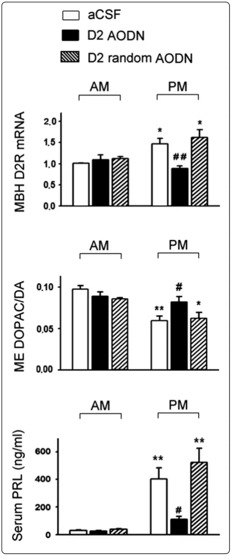
index) and serum PRL levels (higher in the afternoon) were confirmed in aCSF-treated control rats (Figure 7). Pretreatment with AODN against D2R mRNA signifi-cantly lowered MBH D2R mRNA and serum PRL level (p < 0.01; Figure 7), and increased ME DOPAC/DA ratio (p < 0.05; Figure 7) in the afternoon, while it had no sig-nificant effects in the morning. Pretreatment with ran-dom AODN against D2R mRNA had no significant effect on any of the three variables compared with those in aCSF-treated control (Figure 7).
D2R mRNAs in NS and VTA also exhibited a diurnal rhythm with their afternoon levels significantly lower than those in the morning (p < 0.01; Table 1). Pretreat-ment with AODN significantly lowered morning levels of D2R mRNA in NS and VTA (p < 0.05 and 0.01 respect-ively; Table 1) plus afternoon level in VTA (p < 0.01; Table 1), and disrupted the diurnal rhythm in both re-gions. Pretreatment with the random AODN had no sig-nificant effects on D2R mRNA ratios in NS and VTA (Table 1).
Discussion
We recently reported that an endogenous DA tone act-ing on DA D3Rs may be involved in diurnal changes of TIDA neuron activity and PRL secretion in OVX+E2rats [10]. We further demonstrated in this study that D2Rs were also involved. Moreover, we found that the expres-sion of D2R mRNA in the MBH exhibited a diurnal change that persisted in OVX animals but was abolished in OVX+E2 animals kept under constant lighting expos-ure, so was the response of TIDA neuron activity. To our knowledge, this is the first report showing that a di-urnal change in the expression of D2R mRNA exists in MBH neurons.
PHNO has been characterized as a highly specific D2 receptor agonist which binds primarily to the high-affinity state of D2Rs rather than D3Rs in the striatum [21] and exhibits a 67-fold selectivity for D2Rs versus D1Rs [22,23]. The findings that PHNO inhibited the ac-tivities of all three central DA neurons tested and its in-hibitory effects were blocked by co-administration of Figure 3Effects of PHNO (0.001, 0.01, 0.1μg/3μl/rat) and
co-administration of PHNO (0.1μg) with a D2receptor antagonist,
raclopride (0.1μg), on ME DOPAC/DA ratio (upper) and serum PRL (lower) level in OVX+E2rats in the morning.Data are expressed as mean ± SEM (n = 5–7 rats). *, p < 0.05, compared with respective control groups. #, p < 0.05; ##, p < 0.01 compared with the PHNO (0.1μg)-injected group.
Figure 4Effects of PHNO (0.001, 0.01, 0.1μg/3μl/rat) and co-administration of PHNO (0.1μg) with raclopride (0.1μg) on dlST (upper) and NA (lower) DOPAC/DA ratios in OVX+E2rats
raclopride further strengthen the notion that D2 autore-ceptors are involved in the control of central DA neu-rons. Raclopride not only blocked the effect of PHNO on TIDA neuron activity, it further lowered serum PRL to below control levels, indicating that not only D2Rs but other D2R-like receptors may be involved. Due to technical limitations, the time-dependent changes in TIDA neuron activity can only be determined using dif-ferent groups of rats sacrificed at difdif-ferent time points, but not in individual rats. It is possible that the effect of PHNO was maximal at 30 min and the effects at earlier and later time points may be obscured in group averages.
We used OVX rats with estrogen replacement because the diurnal changes of TIDA neuron activity and after-noon PRL surge are gender specific and induced by
estrogen [8]. We have repeatedly shown that the diurnal change in TIDA neuron activity is prerequisite for the estrogen-induced PRL afternoon surge [8,9,24]. Disrupt-ing the TIDA rhythm invariably blunts the PRL surge. The previous [10] and present studies using D3R and D2R antagonists to disrupt the TIDA rhythm further confirm the notion. It appears that an endogenous DA tone acting on D2-like receptors during a critical period of time (from 1200 to 1400 h) is responsible for the low-ered TIDA neuron activity in the afternoon.
Although raclopride treatment completely reversed the afternoon decrease of TIDA neuron activity and signifi-cantly blunted the PRL surge, it did not block the surge completely, i.e., serum PRL level in the afternoon was still higher than that in the morning. This indicates that factor(s) other than withdrawal of DA inhibition Figure 5Effects of a single injection of raclopride (10μg/3μl,
icv) at 1200 h on ME DOPAC/DA ratio (upper) and serum PRL (lower) level at 1500 h in OVX+E2rats.Data are expressed as mean ± SEM (n = 5–7 rats). *, p < 0.05; ** p < 0.01, compared with the control groups in the morning; #, p < 0.05; ##, p < 0.01 compared with respective control groups in the afternoon.
Figure 6Morning and afternoon D2R mRNA ratios in MBH and
their responses to constant light and E2exposures (A) and a
stimulates the PRL secretion. It is well-established that de-creased inhibition by DA (PRL-inhibiting hormone) plus increased stimulation by a putative PRL-releasing hor-mone are required for a full-blown PRL surge [25-27]. The present finding further substantiates the notion.
That the mRNA of D2Rs in MBH exhibited a diurnal rhythm and was disrupted by constant light exposure are novel findings. It appears that when TIDA neuron activity was high, the expression of D2R mRNA was low, and vice versa. Moreover, the findings that both D2R mRNA and TIDA diurnal rhythms are sensitive to con-stant light exposure but persist in OVX rats are consist-ent with the criteria of true circadian rhythms [8,28]. Pretreatment with an AODN against D2receptor mRNA not only reversed the increased expression of D2 recep-tor mRNA in the afternoon, but also prevented the diur-nal changes of TIDA neuron activity and PRL secretion. This finding indicates that the diurnal change in the ex-pression of D2R mRNA may underlie the changes of TIDA neuron activity and in turn the PRL surge, al-though the exact mechanism needs further work.
This study raised another interesting issue on the ori-gin of dopamine acting on TIDA neurons. Dopamine may arise from recurrent collaterals of TIDA neurons or
from other hypothalamic DA neurons including
periventricular-hypophysial (PH) and tuberohypophysial (TH) DA neurons [29]. It has been shown that rhythm-related genes are present not only in suprachiasmatic nucleus (SCN) and ARN [30], but also in hypothalamic DA neurons, including TIDA, PHDA and THDA neurons [31], indicating that these neurons may generate their own
Table 1 Dopamine D2receptor mRNA ratios in SN and VTA of OVX+E2rats as determined by QRT-PCR
aCSF D2AODN D2random AODN
SN AM 1.00 ± 0.01 0.76 ± 0.06# 1.33 ± 0.13
PM 0.78 ± 0.02** 0.86 ± 0.03 0.99 ± 0.05* VTA AM 1.02 ± 0.02 0.42 ± 0.02## 1.38 ± 0.19
PM 0.77 ± 0.01** 0.46 ± 0.06# 0.53 ± 0.08**
Data are expressed as mean ± SEM (n = 8-12 rats in each group). *, p < 0.05; **, p < 0.01, compared with respective control groups in the morning; #, p < 0.05; ##, p < 0.01 compared with respective aCSF-treated groups at the same time point. Groups of OVX+E2rats received icv injections of aCSF, antisense
oligodeoxynucleotides (AODN, 10μg/3μl), or random AODN against D2
receptor mRNA for 2 days were sacrificed either in the morning or afternoon. Figure 7Effects of antisense oligodeoxynucleotides (AODN) or random AODN (10μg/3μl, icv) pretreatment against D2R
mRNA for 2 days on MBH D2R mRNA ratios (upper; arbitrary unit),
rhythms. Single-unit recording of DA-responsive dmARN neuronsin vitroalso reveals a diurnal rhythm of their fir-ing rates [7]. On the other hand, it is well-established that SCN is the master biological clock that coordinates most circadian rhythms observed in mammals and bilateral le-sions of the SCN eliminate both TIDA diurnal rhythm and the estrogen-induced PRL surge [9], indicating an entraining signal originated from SCN can affect directly or indirectly on TIDA neurons.
Circadian changes of midbrain DA neurons’ activities in rats have been reported with their levels higher during dark hours when the animals exhibit nocturnal motor activities [28,32]. Recent studies have shown that striatal D2/D3R proteins and D3R mRNA levels in mouse also exhibit diurnal rhythms [11,12]. Our findings that D2R mRNAs in SN and VTA exhibited a morning high, after-noon low rhythm are in agreement with the changes of D3R mRNA levels in mouse striatum [12]. That the D2R mRNAs in SN and VTA were significantly lowered by pretreatment of AODN against D2R mRNA further con-firms the knockdown effects of AODN.
Conclusions
The present study demonstrates that D2Rs are involved in the inhibitory actions of exogenous and endogenous dopamine on TIDA neuron activity. Moreover, a diurnal change of D2R mRNA expression in the MBH may be involved in the diurnal rhythms of TIDA neuron activity and PRL secretion in OVX+E2rats.
Competing interests
The authors declare that they have no competing interests.
Authors’contributions
SLL designed and carried out all studies except QRT-PCR, and analyzed, plotted all the data and drafted the manuscript. SCH carried out QRT-PCR study and participated in its design. JTP conceived of the study, participated in its design and coordination, and revised the manuscript. All authors read and approved the final manuscript.
Acknowledgments
We thank Merck, Sharp and Dohme for providing PHNO. This work was supported in part by grants CMRPD180253 (to SCH), CMRPD180263 and CMRPD1C0181 (to SLL) from Chang Gung Medical Research and Development Council of Taiwan.
Author details
1Department of Physiology and Pharmacology, College of Medicine, Chang
Gung University, Tao-Yuan 33302, Taiwan.2Department of Biomedical Sciences, College of Medicine, Chang Gung University, Tao-Yuan 33302, Taiwan.3Department of Biological Sciences, Oakland University, Rochester, MI 48309, USA.
Received: 11 February 2014 Accepted: 29 April 2014 Published: 3 May 2014
References
1. Ben-Jonathan N:Dopamine: a prolactin-inhibiting hormone.Endocr Rev 1985,6:564–589.
2. Pan JT:Neuroendocrine functions of dopamine. InCNS Neurotransmitters and Neuromodulators: Dopamine.Edited by Stone TW. Boca Raton: CRC Press; 1996:213–232.
3. Lin JY, Yen SH, Shieh KR, Liang SL, Pan JT:Dopamine and 7-OH-DPAT may act on D(3) receptors to inhibit tuberoinfundibular dopaminergic neu-rons.Brain Res Bull2000,52:567–572.
4. Yen SH, Lai CJ, Lin JY, Pan JT:Inhibitory effect of dopamine on dorsomedial arcuate neurons in rat brain slices: potentiation by coadministration of cocaine.Brain Res Bull1997,42:347–351.
5. Liang SL, Pan JT:Potent inhibitory effect of selective D2 and D3 agonists on dopamine-responsive dorsomedial arcuate neurons in brain slices of estrogen-primed rats.Life Sci2001,69:2653–2662.
6. Liang SL, Pan JT:Pretreatment with antisense oligodeoxynucleotide against D(2) or D(3) receptor mRNA diminished dopamine's inhibitory effect on dorsomedial arcuate neurons in brain slices of estrogen-treated ovariectomized rats.Brain Res2002,926:156–164.
7. Liang SL, Pan JT:The spontaneous firing rates of dopamine-inhibited dorsomedial arcuate neurons exhibit a diurnal rhythm in brain slices obtained from ovariectomized plus estrogen-treated rats.Brain Res Bull 2011,85:189–193.
8. Shieh KR, Pan JT:An endogenous cholinergic rhythm may be involved in the circadian changes of tuberoinfundibular dopaminergic neuron activity in ovariectomized rats treated with or without estrogen. Endocrinology1995,136:2383–2388.
9. Mai LM, Shieh KR, Pan JT:Circadian changes of serum prolactin levels and tuberoinfundibular dopaminergic neuron activities in ovariectomized rats treated with or without estrogen: the role of the suprachiasmatic nuclei.Neuroendocrinology1994,60:520–526.
10. Liang SL, Pan JT:An endogenous dopaminergic tone acting on dopamine D3 receptors may be involved in diurnal changes of tuberoinfundibular dopaminergic neuron activity and prolactin secretion in estrogen-primed ovariectomized rats.Brain Res Bull2012,87:334–339. 11. Akhisaroglu M, Kurtuncu M, Manev H, Uz T:Diurnal rhythms in
quinpirole-induced locomotor behaviors and striatal D2/D3 receptor levels in mice. Pharmacol Biochem Behav2005,80:371–377.
12. Ikeda E, Matsunaga N, Kakimoto K, Hamamura K, Hayashi A, Koyanagi S, Ohdo S:Molecular mechanism regulating 24-hour rhythm of dopamine D3 receptor expression in mouse ventral striatum.Mol Pharmacol2013, 83:959–967.
13. Goodman RL:A quantitative analysis of the physiological role of estradiol and progesterone in the control of tonic and surge secretion of luteinizing hormone in the rat.Endocrinology1978,102:142–150. 14. Palkovits M:Isolated removal of hypothalamic or other brain nuclei of
the rat.Brain Res1973,59:449–450.
15. Paxinos G, Watson C:The Rat Brain in Stereotaxic Coordinates, compact. sixthth edition. New York: Academic Press; 2009.
16. Tepper JM, Sun BC, Martin LP, Creese I:Functional roles of dopamine D2 and D3 autoreceptors on nigrostriatal neurons analyzed by antisense knockdown in vivo.J Neurosci1997,17:2519–2530.
17. Szklarczyk A, Kaczmarek L:Antisense oligodeoxyribonucleotides: stability and distribution after intracerebral injection into rat brain.J Neurosci Methods1995,60:181–187.
18. Agrawal S, Temsamani J, Tang JY:Pharmacokinetics, biodistribution, and stability of oligodeoxynucleotide phosphorothioates in mice.Proc Natl Acad Sci U S A1991,88:7595–7599.
19. Liang SL, Pan JT:An endogenous serotonergic rhythm acting on 5-HT(2A) receptors may be involved in the diurnal changes in tuberoinfundibular dopaminergic neuronal activity and prolactin secretion in female rats. Neuroendocrinology2000,72:11–19.
20. Lowry OH, Rosebrough NJ, Farr AL, Randall RJ:Protein measurement with the Folin phenol reagent.J Biol Chem1951,193:265–275.
21. Kiss B, Horti F, Bobok A:In vitro and in vivo comparison of [(3)H](+)-PHNO and [(3)H]raclopride binding to rat striatum and lobes 9 and 10 of the cerebellum: a method to distinguish dopamine D(3) from D(2) receptor sites.Synapse2011,65:467–478.
22. Seeman P, Ulpian C, Larsen RD, Anderson PS:Dopamine receptors labelled by PHNO.Synapse1993,14:254–262.
23. Nobrega JN, Seeman P:Dopamine D2 receptors mapped in rat brain with [3H](+)PHNO.Synapse1994,17:167–172.
24. Mai LM, Pan JT:Bombesin acts in the suprachiasmatic nucleus to affect circadian changes in tuberoinfundibular dopaminergic neuron activity and prolactin secretion.Endocrinology1995,136:4163–4167.
prolactin release in ovariectomized rats treated with estradiol and/or progesterone.Endocrinology1986,119:1996–2003.
26. Pan JT, Gala RR:The pharmacologic induction of prolactin release in the medial preoptic/suprachiasmatic nuclei-lesioned, estrogen-treated ovariectomized rat.Life Sci1988,43:1929–1934.
27. Yuan ZF, Pan JT:Involvement of angiotensin II, TRH and prolactin-releasing peptide in the estrogen-induced afternoon prolactin surge in female rats: studies using antisense technology.Life Sci2002,71:899–910. 28. Shieh KR, Chu YS, Pan JT:Circadian change of dopaminergic neuron
activity: effects of constant light and melatonin.Neuroreport1997, 8:2283–2287.
29. Moore KE, Lookingland KJ:Dopaminergic neuronal systems in the hypothalamus. InPsychopharmacology.Edited by Bloom KE, Kuper DJ. New York: Raven Press; 1995:245–256.
30. Shieh KR:Distribution of the rhythm-related genes rPERIOD1, rPERIOD2, and rCLOCK, in the rat brain.Neuroscience2003,118:831–843. 31. Sellix MT, Egli M, Poletini MO, McKee DT, Bosworth MD, Fitch CA, Freeman
ME:Anatomical and functional characterization of clock gene expression in neuroendocrine dopaminergic neurons.Am J Physiol Regul Integr Comp Physiol2006,290:R1309–R1323.
32. Paulson PE, Robinson TE:Relationship between circadian changes in spontaneous motor activity and dorsal versus ventral striatal dopamine neurotransmission assessed with on-line microdialysis.Behav Neurosci 1994,108:624–635.
doi:10.1186/1423-0127-21-37
Cite this article as:Lianget al.:Involvement of dopamine D2receptor in
the diurnal changes of tuberoinfundibular dopaminergic neuron activity and prolactin secretion in female rats.Journal of Biomedical Science
201421:37.
Submit your next manuscript to BioMed Central and take full advantage of:
• Convenient online submission
• Thorough peer review
• No space constraints or color figure charges
• Immediate publication on acceptance
• Inclusion in PubMed, CAS, Scopus and Google Scholar
• Research which is freely available for redistribution


![Figure 6 Morning and afternoon D2R mRNA ratios in MBH andtheir responses to constant light and E2 exposures (A) and aschematic drawing of the coronal section of the rat brain (B)showing the punched area of MBH [grey circles, [15]]](https://thumb-us.123doks.com/thumbv2/123dok_us/9028267.1895631/6.595.57.292.89.471/morning-afternoon-andtheir-responses-constant-exposures-aschematic-circles.webp)