R E S E A R C H
Open Access
Increased gait variability during
robot-assisted walking is accompanied by
increased sensorimotor brain activity in
healthy people
Alisa Berger
1*, Fabian Horst
2, Fabian Steinberg
1,3, Fabian Thomas
1, Claudia Müller-Eising
4,
Wolfgang I. Schöllhorn
2and Michael Doppelmayr
1,5Abstract
Background:Gait disorders are major symptoms of neurological diseases affecting the quality of life. Interventions that restore walking and allow patients to maintain safe and independent mobility are essential. Robot-assisted gait training (RAGT) proved to be a promising treatment for restoring and improving the ability to walk. Due to
heterogenuous study designs and fragmentary knowlegde about the neural correlates associated with RAGT and the relation to motor recovery, guidelines for an individually optimized therapy can hardly be derived. To optimize robotic rehabilitation, it is crucial to understand how robotic assistance affect locomotor control and its underlying brain activity. Thus, this study aimed to investigate the effects of robotic assistance (RA) during treadmill walking (TW) on cortical activity and the relationship between RA-related changes of cortical activity and biomechanical gait characteristics.
Methods:Twelve healthy, right-handed volunteers (9 females; M = 25 ± 4 years) performed unassisted walking (UAW) and robot-assisted walking (RAW) trials on a treadmill, at 2.8 km/h, in a randomized, within-subject design. Ground reaction forces (GRFs) provided information regarding the individual gait patterns, while brain activity was examined by measuring cerebral hemodynamic changes in brain regions associated with the cortical locomotor network, including the sensorimotor cortex (SMC), premotor cortex (PMC) and supplementary motor area (SMA), using functional near-infrared spectroscopy (fNIRS).
Results:A statistically significant increase in brain activity was observed in the SMC compared with the PMC and SMA (p< 0.05), and a classical double bump in the vertical GRF was observed during both UAW and RAW throughout the stance phase. However, intraindividual gait variability increased significantly with RA and was correlated with increased brain activity in the SMC (p= 0.05;r= 0.57).
(Continued on next page)
© The Author(s). 2019Open AccessThis article is distributed under the terms of the Creative Commons Attribution 4.0 International License (http://creativecommons.org/licenses/by/4.0/), which permits unrestricted use, distribution, and reproduction in any medium, provided you give appropriate credit to the original author(s) and the source, provide a link to the Creative Commons license, and indicate if changes were made. The Creative Commons Public Domain Dedication waiver (http://creativecommons.org/publicdomain/zero/1.0/) applies to the data made available in this article, unless otherwise stated.
* Correspondence:[email protected]
1Department of Sport Psychology, Institute of Sport Science, Johannes Gutenberg-University Mainz, Albert Schweitzer Straße 22, 55128 Mainz, Germany
(Continued from previous page)
Conclusions:On the one hand, robotic guidance could generate sensory feedback that promotes active
participation, leading to increased gait variability and somatosensory brain activity. On the other hand, changes in brain activity and biomechanical gait characteristics may also be due to the sensory feedback of the robot, which disrupts the cortical network of automated walking in healthy individuals. More comprehensive neurophysiological studies both in laboratory and in clinical settings are necessary to investigate the entire brain network associated with RAW.
Keywords:Walking, Gait variability, GRF, Brain activity, Neuroimaging, Functional near-infrared spectroscopy, fNIRS, Robotic rehabilitation, RAGT, Neurorehabilitation
Background
Safe and independent locomotion represents a funda-mental motor function for humans that is essential for self-contained living and good quality of life [1–5]. Loco-motion requires the ability to coordinate a number of different muscles acting on different joints [6–8], which are guided by cortical and subcortical brain structures within the locomotor network [9]. Structural and func-tional changes within the locomotor network are often accompanied by gait and balance impairments which are frequently considered to be the most significant con-cerns in individuals suffering from brain injuries or neurological diseases [5,10,11]. Reduced walking speeds and step lengths [12] as well as non-optimal amount of gait variability [13–15] are common symptoms associ-ated with gait impairments that increase the risk of fall-ing [16].
In addition to manual-assisted therapy, robotic neu-rorehabilitation has often been applied in recent years [17, 18] because it provides early, intensive, task-specific and multi-sensory training which is thought to be effective for balance and gait recovery [17, 19,
20]. Depending on the severity of the disease,
move-ments can be completely guided or assisted, tailored to individual needs [17], using either stationary ro-botic systems or wearable powered exoskeletons.
Previous studies investigated the effectiveness of robot-assisted gait training (RAGT) in patients suffering from stroke [21, 22], multiple sclerosis [23–26], Parkin-son’s disease [27, 28], traumatic brain injury [29] or spinal cord injury [30–32]. Positive effects of RAGT on walking speed [33,34], leg muscle force [23] step length, and gait symmetry [29, 35] were reported. However, the results of different studies are difficult to summarize due to the lack of consistency in protocols and settings of robotic-assisted treatments (e.g., amount and frequency of training sessions, amount and type of provided ro-botic support) as well as fragmentary knowledge of the effects on functional brain reorganization, motor recov-ery and their relation [36,37]. Therefore, it is currently a huge challenge to draw guidelines for robotic
rehabilita-tion protocols [22, 36–38]. To design prologned
personalized training protocols in robotic rehabilitation to maximize individual treatment effects [37], it is cru-cial to increase the understanding of changes in loco-motor patterns [39] and brain signals [40] underlying RAGT and how they are related [36,41].
A series of studies investigated the effects of robotic assistance (RA) on biomechanical gait patterns in healthy people [39, 42–44]. On one side, altered gait patterns were reported during robot-assisted walking (RAW) compared to unassisted walking (UAW), in particular, substantially higher muscle activity in the quadriceps, gluteus and adductor longus leg muscles and lower muscle activity in the gastrocnemius and tibialis anterior ankle muscles [39, 42] as well as
re-duced lower-body joint angles due to the little
medial-lateral hip movements [45–47]. On the other side, similar muscle activation patterns were observed
during RAW compared to UAW [44, 48, 49],
indicat-ing that robotic devices allow physiological muscle
ac-tivation patterns during gait [48]. However, it is
hypothesized that the ability to execute a physio-logical gait pattern depends on how the training
pa-rameters such as body weight support (BWS),
guidance force (GF) or kinematic restrictions in the robotic devices are set [44, 48, 50]. For example,
Aurich-Schuler et al. [48] reported that the
variability in relative knee and hip movements. Never-theless, Gizzi et al. [49] showed that the muscular ac-tivity during RAW was clearly more stereotyped and similar among individuals compared to UAW. They concluded that RAW provides a therapeutic approach to restore and improve walking that is more repeat-able and standardized than approaches based on exer-cising during UAW [49].
In addition to biomechanical gait changes, insights into brain activity and intervention-related changes in brain activity that relate to gait responses, will contribute to the optimization of therapy interventions [41, 51]. Whereas the application of functional magnetic reson-ance imaging (fMRI), considered as gold standard for the assessment of activity in cortical and subcortical structures, is restricted due to the vulnerability for movement artifacts and the range of motion in the scan-ner [52], functional near infrared spectroscopy (fNIRS) is affordable and easily implementable in a portable sys-tem, less susceptible to motion artifacts, thus facilitation a wider range of application with special cohorts (e.g., children, patients) and in everyday environments (e.g., during a therapeutic session of RAW or UAW) [53,54]. Although with lower resolution compared to fMRI [55], fNIRS also relies on the principle of neurovascular coup-ling and allows the indirect evaluation of cortical activa-tion [56, 57] based on hemodynamic changes which are analogous to the blood-oxygenation-level-dependent re-sponses measured by fMRI [56]. Despite limited depth sensitivity, which restricts the measurement of brain ac-tivity to cortical layers, it is a promising tool to investi-gate the contribution of cortical areas to the neuromotor control of gross motor skills, such as walking [53]. Regarding the cortical correlates of walking, numerous
studies identified either increaesed oxygenated
hemoglobin (Hboxy) concentration changes in the sen-sorimotor cortex (SMC) by using fNIRS [53, 57–59] or suppressed alpha and beta power in sensorimotor areas by using electroencephalography (EEG) [60–62] demon-strating that motor cortex and corticospinal tract con-tribute directly to the muscle activity of locomotion [63]. However, brain activity during RAW [36,61,64–68], es-pecially in patients [69,70] or by using fNIRS [68,69], is rarely studied [71].
Analyzing the effects of RA on brain activity in healthy volunteers, Knaepen et al. [36] reported significantly suppressed alpha and beta rhythms in the right sensory cortex during UAW compared to RAW with 100% GF and 0% BWS. Thus, significantly larger involvement of the SMC during UAW compared to RAW were con-cluded [36]. In contrast, increases of Hboxy were ob-served in motor areas during RAW compared UAW, leading to the conclusion that RA facilitated increased cortical activation within locomotor control systems
[68]. Furthermore, Simis et al. [69] demonstrated the feasibility of fNIRS to evaluate the real-time activation of the primary motor cortex (M1) in both hemispheres during RAW in patients suffering from spinal cord in-jury. Two out of three patients exhibited enhanced M1 activation during RAW compared with standing which indicate the enhanced involvement of motor cortical areas in walking with RA [69].
To summarize, previous studies mostly focused the effects of RA on either gait characteristics or brain activity. Combined measurements investigating the ef-fects of RA on both biomechanical and hemodynamic patterns might help for a better understanding of the neurophysiological mechanisms underlying gait and gait disorders as well as the effectiveness of robotic rehabilitation on motor recovery [37, 71]. Up to now, no consensus exists regarding how robotic devices should be designed, controlled or adjusted (i.e., device settings, such as the level of support) for synergistic interactions with the human body to achieve optimal neurorehabilitation [37, 72]. Therefore, further re-search concerning behavioral and neurophysiological mechanisms underlying RAW as well as the modula-tory effect of RAGT on neuroplasticy and gait recov-ery are required giving the fact that such knowledge is of clinical relevance for the development of gait re-habilitation strategies.
Consequently, the central purpose of this study was
to investigate both gait characteristics and
hemodynamic activity during RAW to identify RAW-related changes in brain activity and their relationship to gait responses. Assuming that sensorimotor areas play a pivotal role within the cortical network of automatic gait [9, 53] and that RA affects gait and brain patterns in young, healthy volunteers [39, 42,
45, 68], we hypothesized that RA result in both
al-tered gait and brain activity patterns. Based on previ-ous studies, more stereotypical gait characteristics with less inter- and intraindividual variability are ex-pected during RAW due to 100% GF and the fixed
pelvis compared to UAW [45, 48], wheares brain
ac-tivity in SMC can be either decreased [36] or
in-creased [68].
Methods
Participants
Twelve healthy subjects (9 female, 3 male; aged 25 ± 4 years), without any gait pathologies and free of extremity injuries, were recruited to participate in this study. All participants were right-handed, according to the Edin-burg handedness-scale [73], without any neurological or psychological disorders and with normal or corrected-to-normal vision. All participants were requested to disclose pre-existing neurological and psychological con-ditions, medical concon-ditions, drug intake, and alcohol or caffeine intake during the preceding week.
Experimental equipment
The Lokomat (Hocoma AG, Volketswil, Switzerland) is a robotic gait-orthosis, consisting of a motorized treadmill and a BWS system. Two robotic actuators can guide the knee and hip joints of participants to match pre-programmed gait patterns, which were derived from average joint trajectories of healthy walkers, using a GF ranging from 0 to 100% [74,75] (Fig.1a). Kinematic tra-jectories can be adjusted to each individual’s size and
step preferences [45]. The BWS was adjusted to 30%
body weight for each participant, and the control mode was set to provide 100% guidance [64].
Functional activation of the human cerebral cortex was recorded using a near-infrared optical tomographic imaging device (NIRSport, NIRx, Germany; Wave-lengths: 760 nm, 850 nm; Sampling rate: 7.81 Hz). The methodology and the underlying physiology are ex-plained in detail elsewhere [76]. A total of 16 optodes (8 emittors, 8 detectors) were placed with an interoptode distance of 3 cm [53, 54] above the motor cortex, based
on the landmarks from the international 10–5 EEG sys-tem [77], resulting in 24 channels (source-detector pairs) of measurement (Fig. 1b). The spatial reolution was up to 1 cm. Head dimensions were individually measured and corresponding cap sizes assigned. Channel positions covered identical regions of both hemispheres including
the SMC (Brodmann Area [BA] 1–4), and the
supple-mentary motor area/premotor cortex (SMA/PMC; BA6) (Fig.1c).
Participants were equipped with standardized running shoes (Saucony Ride 9, Saucony, USA). Pressure insoles (Pedar mobile system, Novel GmbH, Germany) were inserted into the shoes for the synchronized measure-ment of plantar foot pressure, at a frequency of 100 Hz. Each insole consists of 99 capacitive sensors and covers the entire plantar area. The data recording process was managed by the software Novel Pedar-X Recorder 25.6.3 (Novel GmbH, Germany), and the vertical ground reac-tion force (GRF) was estimated for the analysis of kinetic and temporal gait variables.
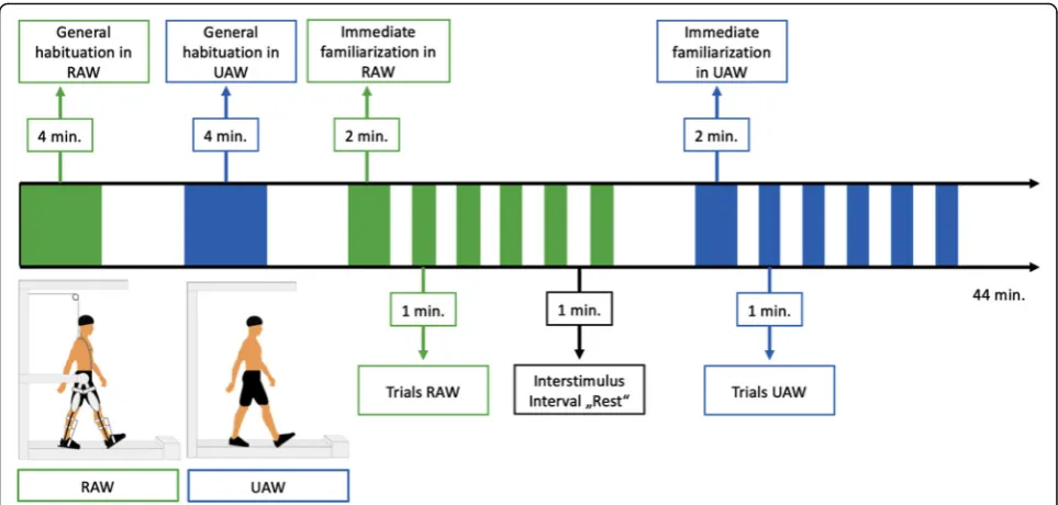
Experimental design
Participants performed two blocks, (1) UAW and (2) RAW, in a randomized order. Each block consisted of five walking trials (60 s) and intertrail standing intervals of 60 s (s) [41, 53, 68, 78] (Fig. 2). While walking, the participants were instructed to actively follow the ortho-sis’s guidance while watching a neutral symbol (black cross) on a screen at eye level to ensure the most natural walking possible in an upright posture. During standing (rest), participants were instructed to stand with their feet shoulder-width apart while watching the same black
Fig. 1Montage and Setup.aParticipant during robot-assisted walking (RAW), with functional near-infrared spectroscopy (fNIRS) montage.bfNIRS
montage; S = Sources; D = DetectorscClassification of regions of interest (ROI): supplementary motor area/premotor cortex (SMA/PMC) and
cross. Furthermore, the participants were requested to avoid head movements and talking during the entire ex-periment, to reduce motion and physiological artifacts [78]. Prior to the experiment, individual adjustments of the Lokomat were undertaken, according to common practices in clinical therapy. The safety procedures of the rehabilitation center required that all subjects wore straps around the front foot to assist with ankle dorsi-flexion. To familiarize themselves with the robotic device and treadmill walking (TW), participants walked with and without the Lokomat for 4 min before the experi-ment started.
Data processing and analysis
fNIRS raw data were preprocessed and analyzed using the time series analysis routine available in the MATLAB-based NIRSlab analysis package (v2017.05,
Nirx Medical Technologies, Glen Head, NY, [“
Biomed-ical Optics”]) [79] following current recommendations when possible [53, 78]. In each channel of individual participant, fNIRS signal was visually inspected with re-spect to transient spikes and abrupt discontinuities which represent two most common forms of movement artifacts in fNIRS data. First, sections containing discon-tinuities (or“jumps”) as well as long term drifts were de-tected and corrected (standard deviation threshold = 5) [79]. Second, spikes were smoothed by a procedure that replaces contaminated data with the nearest signal [79]. Third, a band-pass filter (0.01 to 0.2 Hz) was applied to attenuate slow drifts and high frequency noises to re-duce unknown global trend due to breathing, respiratory or cardiac rhythms, vasomotion, or other movement
artifacts [59]. Then, time series of hemodynamic states of Hboxy and deoxygenated hemoglobin (Hbdeoxy) were computed using the the modified Beer-Lambert law [80, 81]. Following parameters were specified: wavelengths (WL1 = 760 nm; WL2 = 850 nm), differential pathlength factors (7.25 for WL1; 6.38 for WL2), interoptode dis-tances (3 cm), background tissue values (totHb: 75 uM; MVO2Sat: 70%).
Preprocessed Hboxy concentration changes (ΔHboxy)
were exported and processed as follows: 50 s per walking trial were used to analyze the hemodynamic responses during (1) UAW and (2) RAW due to the time needed for the acceleration and deceleration of the treadmill. The averaged baseline concentration values of rest be-fore each walking trial were subtracted from the task-evoked concentration measurements, to account for time-dependent changes in cerebral oxygenation [78].
ΔHboxy were calculated for regions of interest (ROI)
(see Fig. 1c) during both UAW and RAW and used as a
marker for the regional cortical activation, since it is more sensitive to locomotion-related activities than Hbdeoxy [82] and represents an accurate indicator of hemodynamic activity [83].
with the acceleration and deceleration phases of the treadmill. The swing and stance phase times were measured. The stance phase was also subdivided into initial limb, single-limb and terminal double-limb support times. Furthermore, the number of steps and the cadence was calculated. Kinetic variables were analyzed during the stance phase of walking. The GRF values were normalized against body mass and were time-normalized against 101 data points corre-sponding with the stance phase of walking. Gait vari-ability was estimated for time-continuous GRF during the stance phase, using the coefficient of variation (CV) [84]. According to Eq. (1), the intraindividual
CV was calculated based on the mean (GRFs;b;i) and
standard deviation (σs, b, i) of the normalized GRF at the i-th interval of a concanated vector of the right and left leg stance phases. The intraindividual CV
was calculated for each subject s and both blocks b
(RAW and UAW).
IntraindividualCVðs;bÞ ¼
ffiffiffiffiffiffiffiffiffiffiffiffiffiffiffiffiffiffiffiffiffiffiffiffiffiffiffiffiffiffiffiffiffiffi
1 202
X202
i¼1σs;b;i 2
r
1 202
X202
i¼1jGR Fs;b;ij
100½% ð1Þ
Similarly, interindividual variability was estimated across the subject’s mean GRF, calculated across the time-continuous GRF from all stance phases from one subject. According to Eq. (2), the interindividual CV
was calculated based on the mean (GRFs;b;i) and
standard deviation (σs;b;i) of the normalized subject’s
mean GRF at the i-th interval of the concanated
vec-tor of the right and left leg stance phases.
Interindi-vidual CV was calculated for both blocks b (RAW
and UAW).
InterindividualCVðbÞ ¼
ffiffiffiffiffiffiffiffiffiffiffiffiffiffiffiffiffiffiffiffiffiffiffiffiffiffiffiffiffiffiffiffiffiffi
1 202
X202
i¼1σs;b;i 2
r
1 202
X202
i¼1jGR Fs;b;ij
100½% ð2Þ
The absolute magnitude of the symmetry index, ac-cording to Herzog et al. [85], was adapted for i time-intervals of time-continuous GRF. The symmetry index (SI) is a method of assessing the differences be-tween the variables associated with both lower limbs during walking. According to Eq. (3), the SI was cal-culated based on the absolute difference of the mean normalized GRF (GRF righti and GRF lefti) at the i-th
interval for each subject s and both blocks b (RAW
and UAW). An SI value of 0% indicates full sym-metry, while an SI value > 0% indicates the degree of asymmetry [85].
SIðs;bÞ ¼ 1
101ð
X101
i¼1
jGR Fright s;b;i−GR Fle f t s;b;ij
1
2jGR Fright s;b;iþGR Fle f t s;b;ij
100Þ½% ð3Þ
Based on the time-continuous vertical GRF waveforms, three time-discrete variables were derived within the stance phase: the magnitude of the first peak (weight ac-ceptance), the valley (mid-stance) and the magnitude of the second peak (push-off), as well as their temporal ap-pearances during the stance phase.
The statistical analysis was conducted using SPSS 23 (IBM, Armonk, New York, USA). Normal distribution was examined for both hemodynamic and kinetic/tem-poral variables using the Shapiro-Wilk test (p≥0.05). Averaged Hboxy values were computed for each subject and ROI (SMA/PMC, SMC) during both UAW and
RAW [53, 78] and were normalized (normHboxy) by
dividing them by the corresponding signal amplitude for the whole experiment [41, 59]. A two-way analysis of variance (ANOVA), with the factors condition (UAW or RAW) and ROI (SMA/PMC, SMC), was used to analyze differences in cortical hemodynamic patterns. In cases of significant main effects, Bonferroni-adjusted post hoc analyses provided statistical information regarding the differences among the ROIs by condition. Temporal and kinetic gait variables were statistically tested for differ-ences between the experimental conditions (UAW and RAW) using paired t-tests. The overall level of signifi-cance was set to p ≤ 0.05. Mauchly’s test was used to check for any violations of sphericity. If a violation of sphericity was detected (p< 0.05) and a Greenhouse-Geisser epsilon ε> 0.75 existed, the Huynh-Feldt cor-rected p-values were reported. Otherwise (epsilon ε< 0.75), a Greenhouse-Geisser correction was applied. Ef-fect sizes were given in partial eta-squared (ƞp2) or interpreted, according to Cohen. The association be-tween cortical activation and gait characteristics was ex-plored using Pearson’s correlation coefficient.
Results
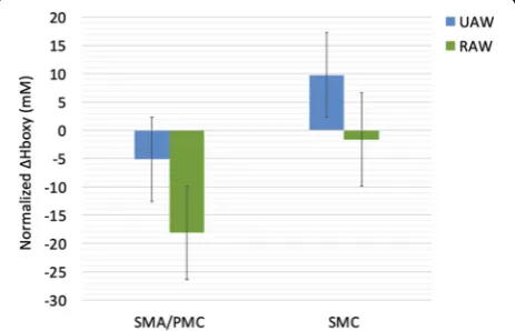
Cortical activity (Hboxy)
The effect of RAW on ΔHboxy in locomotor cortical
areas was analyzed using a two-way repeated measure-ments ANOVA with the factors ROI (SMA/PMC, SMC)
and CONDITION (UAW, RAW). ΔHboxy served as
dependent variable. A significant main effect for ROI [F(1,11) = 11.610,p= 0.006, ƞp2= 0.513] was found indi-cating significant greater ΔHboxy values in the 7
chan-nels (1–3,13–16) covering regions of the SMA/PMC
ROI x CONDITION [F(1,11) = 0.092, p= 0.767, ƞp2= 0.008] were significant (Fig.3).
Gait characteristics
Descriptive analyses of the mean vertical GRFs show a
“classical” double bump (M-Shape) during the stance
phase [84] for both UAW and RAW (Fig. 4). However,
various differences in the gait characteristics were ob-served between the two conditions. First, the mean verti-cal GRFs were lower during RAW than during UAW. Second, the relative appearance of the peak values oc-curs earlier for the first peak and later for the second peak during RAW compared with UAW. Third, the ver-tical GRFs had higher standard deviations during RAW than during UAW. Statistical analyses of the time-discrete kinetic gait variables confirmed significantly lower GRFs and earlier and later appearances for the first and second vertical GRF peaks, respectively, during
RAW than during UAW (Table1).
Fourth, significantly increased inter- and intraindivi-dual variability and asymmetry between the time-continuous GRFs of the right and left feet (SI values) and significantly longer stance and swing phases emerge during RAW compared with UAW, despite the guidance of the robotic device and the same treadmill velocity
(Table 2). Accordingly, lower numbers of steps and
lower cadence values were observed during RAW than during UAW.
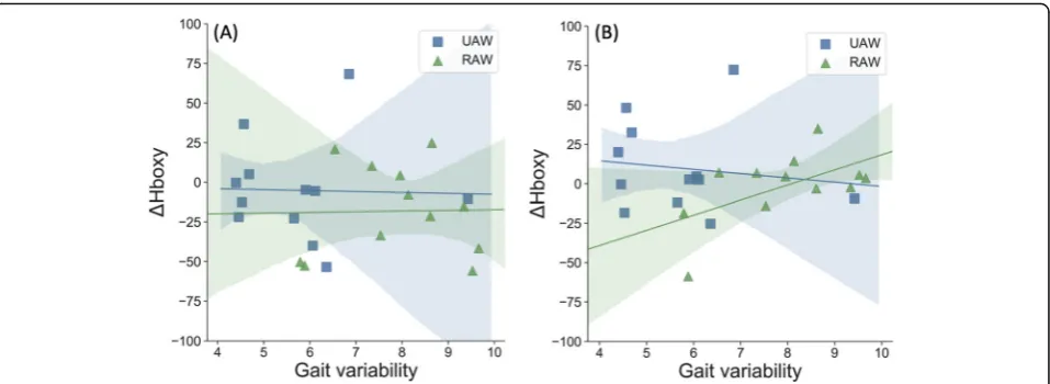
Association between changes in cortical activity and gait characteristics
Correlation analyses showed that changes in gait charac-teristics due to RA were also associated with changes in cortical activity. During RAW, a positive association be-tween gait variability and Hboxy was observed only in the SMC (p= 0.052, r= 0.570). No further correlations
were found during UAW or for other brain regions
(SMA/PMC p= 0.951, r= 0.020). Thus, increased gait
variability during RAW was associated with increased brain activity in the SMC (Fig.5b).
Discussion
In this study, the effects of RA on cortical activity during TW and the relationship to changes in gait characteris-tics were investigated. We identified a classical double bump in the GRF, throughout the stance phase during both UAW and RAW, which was accompanied by sig-nificantly increased brain activity in the SMC compared to premotor/supplementary motor areas. However, indi-vidual analyses showed significantly higher inter- and intraindividual gait variability due to RA that correlated
with increased hemodynamic activity in the SMC (p=
0.052;r= 0.570).
In both conditions, shape characteristics of the mean GRF curves during the stance phase were ob-served. This in not in line with the results of Neckel et al. [46] who did not report a classical double bump during the stance phase during RAW, which could be due to the age differences of our samples. Further-more, significantly altered kinematic patterns (lower GRF values and earlier and later appearances for the first and second vertical GRF peak values, respect-ively) as well as large inter- and intraindividual gait variability were observed during RAW compared to UAW. Results of the kinematic patterns are consist-ent with other biomechanical studies showing altered muscle activity [39, 42] or kinematic patterns [45–47] due to RA. The results of greater inter- and intraindi-vidual gait variability during RAW do not agree with the more stereotypical and similar patterns of Gizzi
et al. [49], nor with the assumption that the user
lacks the ability to vary and adapt gait patterns dur-ing RAW [45, 48, 50].
Fig. 3Normalized oxygenated hemoglobin (Hboxy; mean ± SME) for unassisted-walking (UAW) and robot-assisted walking (RAW). SMA/ PMC, supplementary motor area/premotor cortex; SMC,
sensorimotor cortex; SME = standard mean error
Fig. 4Normalized vertical ground reaction force (GRF; mean ± SD) during the stance phase of unassisted walking (UAW) and
robot-assisted walking (RAW). In Additional file1, normalized vertical GRF
Regarding brain activity during UAW, Hboxy concen-tration changes were significantly increased in sensori-motor areaes compared to areas of the SMA/PMC which is in line with other neurophysiological studies that showed increased Hboxy concentrations during walking [57,58]. This is further confirmed by EEG stud-ies reporting suppressed alpha and beta oscillations within the SMC [60–62] during active walking. This also demonstrates that the SMC and the corticospinal tract contribute directly to muscle activity in locomotion [9,
53, 63] representing a general marker of an active
movemet-related neuronal state [61].
Analyzing the effects of RA on cortical patterns, sig-nificantly increased Hboxy concentration changes were also observed in SMC compared to frontal areas. Whereas Kim et al. [68] observed more global network activation during RAW compared to UAW, Knaepen et al. [36] reported significantly suppressed alpha and beta power during UAW compared to RAW with the conclusion that walking with 100% GF leads to less ac-tive participation and little activation of the SMC, which should be avoided during RAGT.
However, during RAW, we observed a positive
correl-ation between ΔHboxy concentrations in the SMC and
intraindividual gait variability. Thus, individuals with lar-ger gait variability showed higher sensorimotor brain
activity, which is similar to the results reported of Vitorio et al. [41]. In this study, positive correlations
be-tween gait variability and ΔHboxy in the PMC and M1
were found in young healthy adults when walking with rhythmic auditory cueing [41]. The following two pos-sible explanations are suggested.
On one side, robotic guidance might induce additional and new sensory feedback that promotes active partici-pation, resulting in high gait variability and increased brain activity. This possibility is supported by previous observations that muscles exhibited marked and struc-turally phased activity, even under full guidance condi-tions [39, 42, 86–88]. Van Kammen et al. [88] found muscle activity in the vastus lateralis, suggesting that the leg muscles are still activated during RAW as opposed to the muscles related to stability and propulsion, in which activity is reduced under guidance conditions. This finding is remarkable because, in this state, the exo-skeleton is responsible for walking control, and theoret-ically, no voluntary activity from the performer is required [87, 89]. However, the instructions used in the present study (i.e.,‘actively move along with the device’) may have affected activity, as previous studies have shown that encouraging active involvement increases muscle activity [86, 87] as well as brain activity signifi-cantly during RAW [64]. More specifically, Wagner et al.
Table 1Comparison of vertical ground reaction force variables (GRF; mean ± SD) during the stance phase of unassisted walking
(UAW) and robot-assisted walking (RAW), SD = standard deviation
Kinetic Gait Variable UAW RAW t df p r
First Peak (weight acceptance) [N/kg] 8.2 ± 0.5 6.2 ± 1.0 6.807 11 0.000 0.899
First Peak (weight acceptance) [% Stance Phase] 31.4 ± 2.6 24.8 ± 3.2 7.366 11 0.000 0.912
Valley (mid-stance) [N/kg] 7.2 ± 0.4 4.2 ± 0.6 23.963 11 0.000 0.991
Valley (mid-stance) [% Stance Phase] 48.9 ± 2.9 49.6 ± 6.3 −0.277 11 0.787 0.083
Second Peak (weight acceptance) [N/kg] 8.6 ± 0.4 6.3 ± 0.7 12.898 11 0.000 0.968
Second Peak (weight acceptance) [% Stance Phase] 75.8 ± 3.0 82.4 ± 2.3 −7.789 11 0.000 0.920
Intraindividual Variability [%] 5.8 ± 1.4 7.9 ± 1.3 −4.427 11 0.001 0.800
Interindividual Variability [%] 7.0 15.7 – – – –
Symmetry Index [%] 4.3 ± 2.0 7.7 ± 2.7 −2.871 11 0.015 0.654
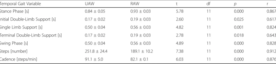
Table 2Comparison of temporal gait variables (mean ± SD) during unassisted walking (UAW) and robot-assisted walking (RAW)
Temporal Gait Variable UAW RAW t df p r
Stance Phase [s] 0.84 ± 0.05 0.93 ± 0.03 5.78 11 0.000 0.867
Initial Double-Limb Support [s] 0.17 ± 0.02 0.19 ± 0.03 2.60 11 0.025 0.617
Single Limb Support [s] 0.50 ± 0.04 0.56 ± 0.03 4.82 11 0.001 0.824
Terminal Double-Limb Support [s] 0.17 ± 0.02 0.19 ± 0.03 2.78 11 0.018 0.643
Swing Phase [s] 0.50 ± 0.04 0.56 ± 0.03 4.89 11 0.000 0.828
Steps [number] 251.8 ± 24.4 189.1 ± 10.2 7.38 11 0.000 0.912
[64] showed significantly suppressed alpha and beta power during active compared to passive RAW. Dobkin (1994) also showed that passive stepping can lead to task-specific sensory information that induces and mod-ulates step-like electromyography activity [90]. Thus, high guidance might also promote active contribution. Particularly in patients who are not able to walk un-assisted, successful stepping induces task-specific sen-sory information that may trigger plastic changes in the central nervous system [88, 91]. Since active participa-tion and the producparticipa-tion of variable movement patterns are prerequisites for activity-dependent neuroplasticity
[7, 20,89, 92–94], it is important to determine whether
the activation of the SMC can be triggered by changes in the levels of GF, BWS and kinematic freedom in order to specifically provoke gait variability due to active par-ticipation of the patient [45, 48,50]. High gait variability may indicate that people use multiple combinations of gait variables to walk more effectively [45,95], resulting in better and faster improvements during robotic rehabilitation.
On other side, the sensory feedback from robot guidance could also disturb the brain network underlying automatic walking, leading to increased gait variability and sensori-motor activity. According to Vitorio et al. [41], the require-ment to adapt to external stimuli leads to disturbances in automatic walking in young healthy people, resulting in higher gait variability and higher cortical costs. As previous study have shown, the ability to execute a physiological gait pattern depends on how the training parameters such as BWS, GF or kinematic freedom in the robotic devices are set. During RAW with fixed pelvis, significantly altered muscle activity [39, 42, 45] and kinematic patterns [48,50] were found. In addition to GF, BWS and kinematic freedom, the presence of foot support may also contribute to altered
patterns. The safety procedures of the therapy institution re-quired that all subjects wear straps around the front foot to assist with ankle dorsiflexion, which is known to reduce ac-tivity in the ankle dorsiflexors [39,42].
In summary, increased gait variability and sensori-motor activity during RAW could be the result of ac-tive participation or disrupted automatic locomotor control. However, the generalization of these results to other populations is not intended or recom-mended. Healthy elderly individuals [41] and patients with stroke [22], multiple sclerosis [23, 25, 26], Par-kinson’s disease [27, 28], brain injuries [29] or spinal cord injuries [30, 31] who suffer from gait and bal-ance disorders react differently to robotic support than healthy young people, which may lead to differ-ent gait and brain activation patterns [44]. In addition to high inter- and intraindividual variability within one sample, the heterogeneity of methodological pro-cedures between studies appears to pose another chal-lenge [71].
Therefore, one future goal should be to understand the mechanisms underlying RAGT and which parame-ters determine the effectiveness of a single treatment in the heterogenuous population of patients suffering from neurological diseases [37]. For this purpose, ob-jective biomarkers for motor recovery and neuroplas-tic changes have to be identified [37]. Then, specific training protocols and further interventions, such as
augmented feedback with virtual reality,
brain-machine interface or non-invasive brain stimulation, can be developed to deliver sustainable therapies for individualized rehabilitation that optimizes the out-come and efficacy of gait recovery, which together can foster independent living and improve the quality of life for neurological patients [37, 71].
Fig. 5Correlations between relative oxygenated hemoglobin (Hboxy) and gait variability calculated by intraindividual coefficient of variation (CV)
during unassisted-walking (UAW) and robot-assisted walking (RAW).aSMA/PMC, supplementary motor area/premotor cortex;bSMC,
Methodological limitations
Two methodological limitations that emerged using the present approach should be mentioned. First, the ability to walk is guided by an optimal interaction between cor-tical and subcorcor-tical brain structures within the
loco-motor network [53]. Using our NIRSport system, we
were only able to report brain activity patterns in motor cortical areas and were unable to monitor the activities of subcortical areas or other cortical involvements. Vari-ous studies have reported that patients with gait disor-ders recruit additional cortical regions to manage the demands of UAW and RAW, due to structural and/or functional changes in the brain. Measuring the entire cortical network underlying locomotion may be neces-sary to investigate neuronal compensations and cognitive resources used for neuroplastic processes during gait re-habilitation. Therefore, we must be careful when dis-cussing brain activity associated with other regions involved in locomotor control [9].
Secondly, we must take into account the small sample size of our healthy volunteers and their young age (mean: 25 ± 4 years), which also had no gait pathologies. Thus, RA guidance of gait movement might have differ-ent effects in elderly subjects or patidiffer-ents who are not able to walk without restrictions [96]. Therefore, the findings from our study are difficult to apply to other age or patient groups, as neurological patients often suf-fer from movement disorders and therefore use difsuf-ferent control strategies during RAW. Although the available results provide relevant insights into the mobile applica-tions of neurophysiological measurements during RAW, with approaches for further therapeutic interventions during robotic rehabilitation, the effects of RAW must also be investigated in other groups and in patients with gait disorders in the future.
Conclusions
The purpose of the present study was to investigate brain activity during UAW and RAW and how this ac-tivity was associated with gait characteristics. The results confirmed the involvement of the SMC during TW and significantly increased gait variability due to RA, which correlated positively with brain activity. Furthermore, this study highlights the interaction between cortical ac-tivity and gait variability, stressing the need to use holis-tic, multisystem approaches when investigating TW in elderly individuals or patients suffering from gait disor-ders. Assessing the effects of RA on brain activity and gait characteristics is essential to develop a better under-standing of how robotic devices affect human locomo-tion. This knowledge is essential for interventional studies examining the rehabilitation of motor disorders. Basic research regarding robotic rehabilitation is neces-sary to gain a deeper understanding of the brain and gait
patterns associated with RAW, which is essential for fur-ther investigations of gait recovery and neuroplastic changes. In addition, clinical longitudinal studies are re-quired to identify individual gait improvements and to identify the underlying neurophysiological changes to develop therapies with respect to interindividual differ-ences. RAGT devices should be designed to provide an amount of force that adapts to the patient’s capacity, to achieve an optimal balance between forced motor activ-ity and the promotion of the patient’s voluntary activity
[36, 92–94]. Further combined studies are necessary to
determine the relationship between brain activity and functional motor improvements and to evaluate the ef-fects of therapeutic interventions. Neurophysiological in-vestigations can contribute to the development of robotic rehabilitation and to individual, closed-loop treatments for future neurorehabilitation therapies.
Supplementary information
Supplementary informationaccompanies this paper athttps://doi.org/10. 1186/s12984-019-0636-3.
Additional file 1: Figure S1.Normalized vertical ground reaction force (GRF; mean) during the stance phase of unassisted walking (UAW) for
each individual participant.Figure S2.Normalized vertical ground
reaction force (GRF; mean) during the stance phase of robot-assisted walking (RAW) for each individual participant.
Abbreviations
ANOVA:Analysis of variance; BA: Brodmann area; BWS: Body weight support; EEG: Electroencephalography; fNIRS: Functional nearinfrared spectroscopy; GF: Guidance force; GRF: Ground reaction forces; Hbdeoxy: Deoxygenated hemoglobin; Hboxy: Oxygenated hemoglobin; M1: Primary motor cortex; RA: Robotic assistance; RAGT: Robot assisted gait training; RAW: Robot assisted walking; ROI: Regions of interest; SD: Standard deviation; SEM: Standard mean error; SI: Symmetry index; SMA: Supplementary motor area; SMC: Sensorimotor cortex; TW: Treadmill walking; UAW: Unassisted
walking;ΔHboxy: Relative changes of oxygenated hemoglobin
Acknowledgments
We thank Alina Hammer and Svenja Klink for assisting with the data collection, and Elmo Neuberger and Alexander Stahl for preparing the graphics included in this manuscript.
Authors’contribution
AB: Research project conception and execution, data acquisition, statistical analysis, interpretation and the manuscript writing. AB acts as the corresponding author. FH: Research project conception and execution, data acquisition, statistical analysis, and interpretation. CM: Research project conception. FS, FT, WS, and MD: Research project conception, manuscript review, and critique. All authors read and approved the final version of the manuscript.
Funding
Not applicable.
Availability of data and materials
The datasets used and analysed during the current study are available from the corresponding author on reasonable request.
Ethics approval and consent to participate
approved by the ethical committee of the Medical Association Hessen in Frankfurt (Germany). Participants were informed of all relevant issues regarding the study and provided their written informed consent prior to the initiation of the experiment.
Consent for publication
Consent from all authors has been acquired prior to submission of this article.
Competing interests
The authors declare that they have no competing interests.
Author details 1
Department of Sport Psychology, Institute of Sport Science, Johannes Gutenberg-University Mainz, Albert Schweitzer Straße 22, 55128 Mainz, Germany.2Department of Training and Movement Science, Institute of Sport Science, Johannes Gutenberg-University Mainz, Mainz, Germany.3School of Kinesiology, Louisiana State University, Baton Rouge, USA.4Center of Neurorehabilitation neuroneum, Bad Homburg, Germany.5Centre for Cognitive Neuroscience, Paris Lodron University of Salzburg, Salzburg, Austria.
Received: 20 August 2019 Accepted: 13 December 2019
References
1. Verghese J, LeValley A, Hall CB, Katz MJ, Ambrose AF, Lipton RB.
Epidemiology of gait disorders in community-residing older adults. J
Am Geriatr Soc. 2006;54:255–61.https://doi.org/10.1111/j.1532-5415.2005.
00580.x.
2. Forte R, Boreham CAG, de Vito G, Pesce C. Health and quality of life
perception in older adults: the joint role of cognitive efficiency and functional mobility. Int J Environ Res Public Health. 2015;12:11328–44.
https://doi.org/10.3390/ijerph120911328.
3. Fagerström C, Borglin G. Mobility, functional ability and health-related
quality of life among people of 60 years or older. Aging Clin Exp Res. 2010; 22:387–94.
4. Hirsch CH, Buzková P, Robbins JA, Patel KV, Newman AB. Predicting
late-life disability and death by the rate of decline in physical
performance measures. Age Ageing. 2012;41:155–61.https://doi.org/10.
1093/ageing/afr151.
5. Soh S-E, Morris ME, McGinley JL. Determinants of health-related quality of
life in Parkinson’s disease: a systematic review. Parkinsonism Relat Disord. 2011;17:1–9.https://doi.org/10.1016/j.parkreldis.2010.08.012.
6. Nielsen JB. How we walk: central control of muscle activity during human
walking. Neuroscientist. 2003;9:195–204.https://doi.org/10.1177/ 1073858403009003012.
7. Bernstein N. The co-ordination and regulation of movements. 1st ed.
Oxford: Pergamon Press; 1967.
8. Hatze H. Motion variability--its definition, quantification, and origin. J Mot
Behav. 1986;18:5–16.
9. La Fougère C, Zwergal A, Rominger A, Förster S, Fesl G, Dieterich M,
et al. Real versus imagined locomotion: a 18F-FDG PET-fMRI
comparison. Neuroimage. 2010;50:1589–98.https://doi.org/10.1016/j.
neuroimage.2009.12.060.
10. Ellis T, Cavanaugh JT, Earhart GM, Ford MP, Foreman KB, Dibble LE. Which
measures of physical function and motor impairment best predict quality of life in Parkinson's disease? Parkinsonism Relat Disord. 2011;17:693–7.https:// doi.org/10.1016/j.parkreldis.2011.07.004.
11. Schmid A, Duncan PW, Studenski S, Lai SM, Richards L, Perera S, Wu SS.
Improvements in speed-based gait classifications are meaningful. Stroke.
2007;38:2096–100.https://doi.org/10.1161/STROKEAHA.106.475921.
12. von Schroeder HP, Coutts RD, Lyden PD, Billings E, Nickel VL. Gait
parameters following stroke: a practical assessment. J Rehabil Res Dev. 1995; 32:25–31.
13. Stergiou N, Harbourne R, Cavanaugh J. Optimal movement variability: a
new theoretical perspective for neurologic physical therapy. J Neurol Phys
Ther. 2006;30:120–9.https://doi.org/10.1097/01.npt.0000281949.48193.d9.
14. Hausdorff JM. Gait dynamics, fractals and falls: finding meaning in the
stride-to-stride fluctuations of human walking. Hum Mov Sci. 2007;26:555–
89.https://doi.org/10.1016/j.humov.2007.05.003.
15. Chen G, Patten C, Kothari DH, Zajac FE. Gait differences between individuals
with post-stroke hemiparesis and non-disabled controls at matched speeds. Gait Posture. 2005;22:51–6.https://doi.org/10.1016/j.gaitpost.2004.06.009.
16. Titianova EB, Tarkka IM. Asymmetry in walking performance and postural
sway in patients with chronic unilateral cerebral infarction. J Rehabil Res Dev. 1995;32:236–44.
17. Turner DL, Ramos-Murguialday A, Birbaumer N, Hoffmann U, Luft A.
Neurophysiology of robot-mediated training and therapy: a perspective for future use in clinical populations. Front Neurol. 2013;4:184.https://doi.org/ 10.3389/fneur.2013.00184.
18. Calabrò RS, Cacciola A, Bertè F, Manuli A, Leo A, Bramanti A, et al. Robotic
gait rehabilitation and substitution devices in neurological disorders: where
are we now? Neurol Sci. 2016;37:503–14.
https://doi.org/10.1007/s10072-016-2474-4.
19. Galen SS, Clarke CJ, Allan DB, Conway BA. A portable gait assessment tool
to record temporal gait parameters in SCI. Med Eng Phys. 2011;33:626–32.
https://doi.org/10.1016/j.medengphy.2011.01.003.
20. Schmidt RA, Lee TD. Motor control and learning: A behavioral emphasis. 5th
ed. Champaign: Human Kinetics; 2011.
21. Bruni MF, Melegari C, de Cola MC, Bramanti A, Bramanti P, Calabrò RS. What
does best evidence tell us about robotic gait rehabilitation in stroke
patients: a systematic review and meta-analysis. J Clin Neurosci. 2018;48:11–
7.https://doi.org/10.1016/j.jocn.2017.10.048.
22. Mehrholz J, Thomas S, Werner C, Kugler J, Pohl M, Elsner B.
Electromechanical-assisted training for walking after stroke. Cochrane
Database Syst Rev. 2017;5:CD006185.https://doi.org/10.1002/14651858.
CD006185.pub4.
23. Beer S, Aschbacher B, Manoglou D, Gamper E, Kool J, Kesselring J.
Robot-assisted gait training in multiple sclerosis: a pilot randomized trial. Mult
Scler. 2008;14:231–6.https://doi.org/10.1177/1352458507082358.
24. Lo AC, Triche EW. Improving gait in multiple sclerosis using robot-assisted,
body weight supported treadmill training. Neurorehabil Neural Repair. 2008;
22:661–71.https://doi.org/10.1177/1545968308318473.
25. Schwartz I, Sajin A, Moreh E, Fisher I, Neeb M, Forest A, et al. Robot-assisted
gait training in multiple sclerosis patients: a randomized trial. Mult Scler.
2012;18:881–90.https://doi.org/10.1177/1352458511431075.
26. Straudi S, Fanciullacci C, Martinuzzi C, Pavarelli C, Rossi B, Chisari C, Basaglia N. The effects of robot-assisted gait training in progressive multiple sclerosis: a randomized controlled trial. Mult Scler. 2016;22:373–84.https:// doi.org/10.1177/1352458515620933.
27. Lo AC, Chang VC, Gianfrancesco MA, Friedman JH, Patterson TS, Benedicto
DF. Reduction of freezing of gait in Parkinson's disease by repetitive robot-assisted treadmill training: a pilot study. J Neuroeng Rehabil. 2010;7:51.
https://doi.org/10.1186/1743-0003-7-51.
28. Picelli A, Melotti C, Origano F, Waldner A, Fiaschi A, Santilli V, Smania N.
Robot-assisted gait training in patients with Parkinson disease: a
randomized controlled trial. Neurorehabil Neural Repair. 2012;26:353–61.
https://doi.org/10.1177/1545968311424417.
29. Esquenazi A, Lee S, Packel AT, Braitman L. A randomized comparative study
of manually assisted versus robotic-assisted body weight supported
treadmill training in persons with a traumatic brain injury. PM R. 2013;5:280–
90.https://doi.org/10.1016/j.pmrj.2012.10.009.
30. Nam KY, Kim HJ, Kwon BS, Park J-W, Lee HJ, Yoo A. Robot-assisted gait
training (Lokomat) improves walking function and activity in people with spinal cord injury: a systematic review. J Neuroeng Rehabil. 2017;14:24.
https://doi.org/10.1186/s12984-017-0232-3.
31. Schwartz I, Sajina A, Neeb M, Fisher I, Katz-Luerer M, Meiner Z. Locomotor
training using a robotic device in patients with subacute spinal cord injury. Spinal Cord. 2011;49:1062–7.https://doi.org/10.1038/sc.2011.59.
32. Wirz M, Zemon DH, Rupp R, Scheel A, Colombo G, Dietz V, Hornby TG.
Effectiveness of automated locomotor training in patients with chronic incomplete spinal cord injury: a multicenter trial. Arch Phys Med Rehabil. 2005;86:672–80.https://doi.org/10.1016/j.apmr.2004.08.004.
33. Benito-Penalva J, Edwards DJ, Opisso E, Cortes M, Lopez-Blazquez R, Murillo
N, et al. Gait training in human spinal cord injury using electromechanical systems: effect of device type and patient characteristics. Arch Phys Med Rehabil. 2012;93:404–12.https://doi.org/10.1016/j.apmr.2011.08.028.
34. Uçar DE, Paker N, BuğdaycıD. Lokomat: a therapeutic chance for patients
with chronic hemiplegia. NeuroRehabil. 2014;34:447–53.https://doi.org/10.
35. Husemann B, Müller F, Krewer C, Heller S, Koenig E. Effects of locomotion training with assistance of a robot-driven gait orthosis in hemiparetic patients after stroke: a randomized controlled pilot study. Stroke. 2007;38: 349–54.https://doi.org/10.1161/01.STR.0000254607.48765.cb.
36. Knaepen K, Mierau A, Swinnen E, Fernandez Tellez H, Michielsen M,
Kerckhofs E, et al. Human-robot interaction: does robotic guidance force affect gait-related brain dynamics during robot-assisted treadmill walking?
PLoS One. 2015;10:e0140626.https://doi.org/10.1371/journal.pone.0140626.
37. Coscia M, Wessel MJ, Chaudary U, Millán JDR, Micera S, Guggisberg A, et al. Neurotechnology-aided interventions for upper limb motor rehabilitation in severe chronic stroke. Brain. 2019;142:2182–97.https://doi.org/10.1093/brain/awz181.
38. Mehrholz J, Pohl M, Platz T, Kugler J, Elsner B. Electromechanical and
robot-assisted arm training for improving activities of daily living, arm function, and arm muscle strength after stroke. Cochrane Database Syst Rev. 2018;9:
CD006876.https://doi.org/10.1002/14651858.CD006876.pub5.
39. Moreno JC, Barroso F, Farina D, Gizzi L, Santos C, Molinari M, Pons JL. Effects
of robotic guidance on the coordination of locomotion. J Neuroeng
Rehabil. 2013;10:79.https://doi.org/10.1186/1743-0003-10-79.
40. Youssofzadeh V, Zanotto D, Stegall P, Naeem M, Wong-Lin K, Agrawal SK,
Prasad G. Directed neural connectivity changes in robot-assisted gait training: a partial granger causality analysis. Conf Proc IEEE Eng Med Biol
Soc. 2014;2014:6361–4.https://doi.org/10.1109/EMBC.2014.6945083.
41. Vitorio R, Stuart S, Gobbi LTB, Rochester L, Alcock L, Pantall A. Reduced gait
variability and enhanced brain activity in older adults with auditory cues: a functional near-infrared spectroscopy study. Neurorehabil Neural Repair.
2018;32:976–87.https://doi.org/10.1177/1545968318805159.
42. Hidler JM, Wall AE. Alterations in muscle activation patterns during
robotic-assisted walking. Clin Biomech (Bristol, Avon). 2005;20:184–93.https://doi. org/10.1016/j.clinbiomech.2004.09.016.
43. Hidler J, Nichols D, Pelliccio M, Brady K, Campbell DD, Kahn JH, Hornby TG.
Multicenter randomized clinical trial evaluating the effectiveness of the
Lokomat in subacute stroke. Neurorehabil Neural Repair. 2009;23:5–13.
https://doi.org/10.1177/1545968308326632.
44. van Kammen K, Boonstra AM, van der Woude LHV, Reinders-Messelink HA,
den Otter R. The combined effects of guidance force, bodyweight support and gait speed on muscle activity during able-bodied walking in the Lokomat. Clin Biomech (Bristol, Avon). 2016;36:65–73.https://doi.org/10. 1016/j.clinbiomech.2016.04.013.
45. Hidler J, Wisman W, Neckel N. Kinematic trajectories while walking within
the Lokomat robotic gait-orthosis. Clin Biomech (Bristol, Avon). 2008;23: 1251–9.https://doi.org/10.1016/j.clinbiomech.2008.08.004.
46. Neckel ND, Blonien N, Nichols D, Hidler J. Abnormal joint torque patterns
exhibited by chronic stroke subjects while walking with a prescribed
physiological gait pattern. J Neuroeng Rehabil. 2008;5:19.https://doi.org/10.
1186/1743-0003-5-19.
47. Neckel N, Wisman W, Hidler J. Limb alignment and kinematics inside a
Lokomat robotic orthosis. Conf Proc IEEE Eng Med Biol Soc. 2006;1:2698–
701.https://doi.org/10.1109/IEMBS.2006.259970.
48. Aurich-Schuler T, Gut A, Labruyère R. The FreeD module for the Lokomat
facilitates a physiological movement pattern in healthy people - a proof of
concept study. J Neuroeng Rehabil. 2019;16:26.https://doi.org/10.1186/
s12984-019-0496-x.
49. Gizzi L, Nielsen JF, Felici F, Moreno JC, Pons JL, Farina D. Motor modules in
robot-aided walking. J Neuroeng Rehabil. 2012;9:76.https://doi.org/10.1186/
1743-0003-9-76.
50. Aurich-Schuler T, Labruyère R. An increase in kinematic freedom in the
Lokomat is related to the ability to elicit a physiological muscle activity pattern: a secondary data analysis investigating differences between
guidance force, path control, and FreeD. Front Robot AI. 2019;6:387.https://
doi.org/10.3389/frobt.2019.00109.
51. Chen I-H, Yang Y-R, Lu C-F, Wang R-Y. Novel gait training alters functional
brain connectivity during walking in chronic stroke patients: a randomized controlled pilot trial. J Neuroeng Rehabil. 2019;16:33.https://doi.org/10. 1186/s12984-019-0503-2.
52. Cutini S, Brigadoi S. Unleashing the future potential of functional
near-infrared spectroscopy in brain sciences. J Neurosci Methods. 2014;232:152–6.
https://doi.org/10.1016/j.jneumeth.2014.05.024.
53. Herold F, Wiegel P, Scholkmann F, Thiers A, Hamacher D, Schega L.
Functional near-infrared spectroscopy in movement science: a systematic review on cortical activity in postural and walking tasks. Neurophotonics.
2017;4:41403.https://doi.org/10.1117/1.NPh.4.4.041403.
54. Quaresima V, Ferrari M. Functional near-infrared spectroscopy (fNIRS) for
assessing cerebral cortex function during human behavior in natural/social
situations: a concise review. Organ Res Methods. 2019;22:46–68.https://doi.
org/10.1177/1094428116658959.
55. Koch SP, Koendgen S, Bourayou R, Steinbrink J, Obrig H. Individual
alpha-frequency correlates with amplitude of visual evoked potential and
hemodynamic response. Neuroimage. 2008;41:233–42.https://doi.org/10.
1016/j.neuroimage.2008.02.018.
56. Ferrari M, Mottola L, Quaresima V. Principles, techniques, and limitations of
near infrared spectroscopy. Can J Appl Physiol. 2004;29:463–87.
57. Miyai I, Tanabe HC, Sase I, Eda H, Oda I, Konishi I, et al. Cortical mapping of
gait in humans: a near-infrared spectroscopic topography study.
Neuroimage. 2001;14:1186–92.https://doi.org/10.1006/nimg.2001.0905.
58. Hamacher D, Herold F, Wiegel P, Hamacher D, Schega L. Brain activity
during walking: a systematic review. Neurosci Biobehav Rev. 2015;57:310–27.
https://doi.org/10.1016/j.neubiorev.2015.08.002.
59. Koenraadt KLM, Roelofsen EGJ, Duysens J, Keijsers NLW. Cortical control of
normal gait and precision stepping: an fNIRS study. Neuroimage. 2014;85(Pt 1):415–22.https://doi.org/10.1016/j.neuroimage.2013.04.070.
60. Severens M, Nienhuis B, Desain P, Duysens J. Feasibility of measuring event
related desynchronization with electroencephalography during walking.
Conf Proc IEEE Eng Med Biol Soc. 2012;2012:2764–7.https://doi.org/10.1109/
EMBC.2012.6346537.
61. Seeber M, Scherer R, Wagner J, Solis-Escalante T, Müller-Putz GR. EEG beta
suppression and low gamma modulation are different elements of human
upright walking. Front Hum Neurosci. 2014;8:485.https://doi.org/10.3389/
fnhum.2014.00485.
62. Bulea TC, Kim J, Damiano DL, Stanley CJ, Park H-S. Prefrontal, posterior
parietal and sensorimotor network activity underlying speed control during
walking. Front Hum Neurosci. 2015;9:247.https://doi.org/10.3389/fnhum.
2015.00247.
63. Petersen TH, Willerslev-Olsen M, Conway BA, Nielsen JB. The motor cortex
drives the muscles during walking in human subjects. J Physiol Lond. 2012; 590:2443–52.https://doi.org/10.1113/jphysiol.2012.227397.
64. Wagner J, Solis-Escalante T, Grieshofer P, Neuper C, Müller-Putz G, Scherer R.
Level of participation in robotic-assisted treadmill walking modulates midline sensorimotor EEG rhythms in able-bodied subjects. Neuroimage.
2012;63:1203–11.https://doi.org/10.1016/j.neuroimage.2012.08.019.
65. Wagner J, Solis-Escalante T, Scherer R, Neuper C, Müller-Putz G. It’s how you
get there: walking down a virtual alley activates premotor and parietal
areas. Front Hum Neurosci. 2014;8:93.https://doi.org/10.3389/fnhum.2014.
00093.
66. Wagner J, Solis-Escalante T, Neuper C, Scherer R, Müller-Putz G. Robot
assisted walking affects the synchrony between premotor and
somatosensory areas. Biomed Tech (Berl). 2013.
https://doi.org/10.1515/bmt-2013-4434.
67. Seeber M, Scherer R, Wagner J, Solis-Escalante T, Müller-Putz GR. High and
low gamma EEG oscillations in central sensorimotor areas are conversely
modulated during the human gait cycle. Neuroimage. 2015;112:318–26.
https://doi.org/10.1016/j.neuroimage.2015.03.045.
68. Kim HY, Yang SP, Park GL, Kim EJ, You JSH. Best facilitated cortical activation
during different stepping, treadmill, and robot-assisted walking training paradigms and speeds: a functional near-infrared spectroscopy
neuroimaging study. NeuroRehabilitation. 2016;38:171–8.https://doi.org/10.
3233/NRE-161307.
69. Simis M, Santos K, Sato J, Fregni F, Battistella L. T107. Using Functional Near
Infrared Spectroscopy (fNIRS) to assess brain activity of spinal cord injury patient, during robot-assisted gait. Clin Neurophysiol. 2018;129:e43–4.
https://doi.org/10.1016/j.clinph.2018.04.108.
70. Calabrò RS, Naro A, Russo M, Bramanti P, Carioti L, Balletta T, et al. Shaping
neuroplasticity by using powered exoskeletons in patients with stroke: a
randomized clinical trial. J Neuroeng Rehabil. 2018;15:35.https://doi.org/10.
1186/s12984-018-0377-8.
71. Berger A, Horst F, Müller S, Steinberg F, Doppelmayr M. Current state and
future prospects of EEG and fNIRS in robot-assisted gait rehabilitation: a
brief review. Front Hum Neurosci. 2019;13:172.https://doi.org/10.3389/
fnhum.2019.00172.
72. Tucker MR, Olivier J, Pagel A, Bleuler H, Bouri M, Lambercy O, et al. Control
strategies for active lower extremity prosthetics and orthotics: a review. J
73. Oldfield RC. The assessment and analysis of handedness: the Edinburgh
inventory. Neuropsychologia. 1971;9:97–113.
https://doi.org/10.1016/0028-3932(71)90067-4.
74. Colombo G, Joerg M, Schreier R, Dietz V. Treadmill training of paraplegic
patients using a robotic orthosis. J Rehabil Res Dev. 2000;37:693–700.
75. Duschau-Wicke A, Caprez A, Riener R. Patient-cooperative control increases
active participation of individuals with SCI during robot-aided gait training.
J Neuroeng Rehabil. 2010;7:43.https://doi.org/10.1186/1743-0003-7-43.
76. Obrig H, Villringer A. Beyond the visible--imaging the human brain with
light. J Cereb Blood Flow Metab. 2003;23:1–18.https://doi.org/10.1097/01.
WCB.0000043472.45775.29.
77. Jurcak V, Tsuzuki D, Dan I. 10/20, 10/10, and 10/5 systems revisited: their
validity as relative head-surface-based positioning systems. Neuroimage.
2007;34:1600–11.https://doi.org/10.1016/j.neuroimage.2006.09.024.
78. Vitorio R, Stuart S, Rochester L, Alcock L, Pantall A. fNIRS response
during walking - Artefact or cortical activity? A systematic review.
Neurosci Biobehav Rev. 2017;83:160–72.https://doi.org/10.1016/j.
neubiorev.2017.10.002.
79. Xu Y, Graber HL, Barbour RL. nirsLAB: A Computing Environment for fNIRS
Neuroimaging Data Analysis. Miami: Optical Society of America; 2014. p. 1.
https://doi.org/10.1364/BIOMED.2014.BM3A.1.
80. Sassaroli A, Fantini S. Comment on the modified Beer-Lambert law for
scattering media. Phys Med Biol. 2004;49:N255–7.https://doi.org/10.1088/
0031-9155/49/14/N07.
81. Cope M, Delpy DT, Reynolds EO, Wray S, Wyatt J, van der Zee P. Methods of
quantitating cerebral near infrared spectroscopy data. Adv Exp Med Biol.
1988;222:183–9.https://doi.org/10.1007/978-1-4615-9510-6_21.
82. Suzuki M, Miyai I, Ono T, Oda I, Konishi I, Kochiyama T, Kubota K. Prefrontal
and premotor cortices are involved in adapting walking and running speed
on the treadmill: an optical imaging study. Neuroimage. 2004;23:1020–6.
https://doi.org/10.1016/j.neuroimage.2004.07.002.
83. Strangman G, Culver JP, Thompson JH, Boas DA. A quantitative comparison
of simultaneous BOLD fMRI and NIRS recordings during functional brain
activation. Neuroimage. 2002;17:719–31.
84. Winter DA. Kinematic and kinetic patterns in human gait: variability and
compensating effects. Hum Mov Sci. 1984;3:51–76.https://doi.org/10.1016/
0167-9457(84)90005-8.
85. Herzog W, Nigg BM, Read LJ, Olsson E. Asymmetries in ground reaction
force patterns in normal human gait. Med Sci Sports Exerc. 1989;21:110–4.
86. Aurich Schuler T, Müller R, van Hedel HJA. Leg surface
electromyography patterns in children with neuro-orthopedic disorders walking on a treadmill unassisted and assisted by a robot with and
without encouragement. J Neuroeng Rehabil. 2013;10:78.https://doi.org/
10.1186/1743-0003-10-78.
87. Israel JF, Campbell DD, Kahn JH, Hornby TG. Metabolic costs and muscle
activity patterns during robotic- and therapist-assisted treadmill walking in
individuals with incomplete spinal cord injury. Phys Ther. 2006;86:1466–78.
https://doi.org/10.2522/ptj.20050266.
88. van Kammen K, Boonstra AM, van der Woude LHV, Reinders-Messelink HA,
den Otter R. Differences in muscle activity and temporal step parameters between Lokomat guided walking and treadmill walking in post-stroke hemiparetic patients and healthy walkers. J Neuroeng Rehabil. 2017;14:32.
https://doi.org/10.1186/s12984-017-0244-z.
89. Duschau-Wicke A, von Zitzewitz J, Caprez A, Lunenburger L, Riener R. Path
control: a method for patient-cooperative robot-aided gait rehabilitation.
IEEE Trans Neural Syst Rehabil Eng. 2010;18:38–48.https://doi.org/10.1109/
TNSRE.2009.2033061.
90. Dobkin BH, Harkema S, Requejo P, Edgerton VR. Modulation of
locomotor-like EMG activity in subjects with complete and incomplete spinal cord injury. Neurorehabil Neural Repair. 1995;9:183–90.
91. Riener R, Lünenburger L, Jezernik S, Anderschitz M, Colombo G, Dietz V.
Patient-cooperative strategies for robot-aided treadmill training: first
experimental results. IEEE Trans Neural Syst Rehabil Eng. 2005;13:380–94.
https://doi.org/10.1109/TNSRE.2005.848628.
92. Riener R, Lünenburger L, Maier I, Colombo G, Dietz V. Locomotor training in
subjects with Sensori-motor deficits: an overview of the robotic gait
Orthosis Lokomat. J Healthc Eng. 2010;1:197–216.https://doi.org/10.1260/
2040-2295.1.2.197.
93. Lewek MD, Cruz TH, Moore JL, Roth HR, Dhaher YY, Hornby TG. Allowing
intralimb kinematic variability during locomotor training poststroke
improves kinematic consistency: a subgroup analysis from a randomized clinical trial. Phys Ther. 2009;89:829–39.https://doi.org/10.2522/ptj.20080180.
94. Krishnan C, Kotsapouikis D, Dhaher YY, Rymer WZ. Reducing robotic
guidance during robot-assisted gait training improves gait function: a case
report on a stroke survivor. Arch Phys Med Rehabil. 2013;94:1202–6.https://
doi.org/10.1016/j.apmr.2012.11.016.
95. Bohnsack-McLagan NK, Cusumano JP, Dingwell JB. Adaptability of
stride-to-stride control of stepping movements in human walking. J Biomech. 2016; 49:229–37.https://doi.org/10.1016/j.jbiomech.2015.12.010.
96. Yang JK, Ahn NE, Kim DH, Kim DY. Plantar pressure distribution during
robotic-assisted gait in post-stroke hemiplegic patients. Ann Rehabil Med. 2014;38:145–52.https://doi.org/10.5535/arm.2014.38.2.145.
Publisher’s Note