R E S E A R C H A R T I C L E
Open Access
Notch signaling during development requires the
function of
awd, the
Drosophila
homolog of
human metastasis suppressor gene
Nm23
Marilena Ignesti
1, Marilena Barraco
1,5, Gouthami Nallamothu
2, Julie A Woolworth
3, Serena Duchi
1,6,
Giuseppe Gargiulo
1, Valeria Cavaliere
1*and Tien Hsu
2,4*Abstract
Background:TheDrosophila abnormal wing discs(awd) belongs to a highly conserved family of genes implicated in metastasis suppression, metabolic homeostasis and epithelial morphogenesis. The cellular function of the mammalian members of this family, the Nm23 proteins, has not yet been clearly defined. Previousawdgenetic analyses unraveled its endocytic role that is required for proper internalization of receptors controlling different signaling pathways. In this study, we analyzed the role of Awd in controlling Notch signaling during development.
Results:To study theawdgene function we used genetic mosaic approaches to obtain cells homozygous for a loss of function allele. Inawdmutant follicle cells and wing disc cells, Notch accumulates in enlarged early endosomes, resulting in defective Notch signaling. Our results demonstrate thatawdfunction is required before γ-secretase mediated cleavage since over-expression of the constitutively active form of the Notch receptor inawd mutant follicle cells allows rescue of the signaling. By using markers of different endosomal compartments we show that Notch receptor accumulates in early endosomes inawdmutant follicle cells. A trafficking assay in living wing discs also shows that Notch accumulates in early endosomes. Importantly, constitutively active Rab5 cannot rescue theawdphenotype, suggesting thatawdis required for Rab5 function in early endosome maturation.
Conclusions:In this report we demonstrate thatawdis essential for Notch signaling via its endocytic role. In addition, we identify the endocytic step at which Awd function is required for Notch signaling and we obtain evidence indicating that Awd is necessary for Rab5 function. These findings provide new insights into the developmental and pathophysiological function of this important gene family.
Keywords:Awd, Notch signaling, Endocytosis
Background
The Drosophila awd (abnormal wing discs) gene was
identified in a genetic screen for genes involved in imaginal disc development [1,2]. It encodes theDrosophilahomolog
of human metastasis suppressor gene Nm23 [3,4]. The
Nm23gene family (also termed NME) consists of ten
re-lated genes in mammals [5] with theNME1andNME2
iso-forms most implicated in tumor progression and sharing about 78% of amino acid identity with the Awd protein.
DuringDrosophiladevelopment,awdis critical for epithe-lial morphogenesis [6] and has been linked to AMP
kinase-regulated energy-sensing [7]. Human and murine Nm23
has been shown in cancer cell xenografts to inhibit metasta-sis, but not primary tumor growth [8]. On the other hand, in other cancer cohorts, particularly those of ovarian can-cers, up-regulated Nm23 levels have been correlated with poor prognosis [9,10], suggesting an oncogenic function. These discrepancies have so far been difficult to reconcile because the exact cellular function of Nm23 has remained unclear, although several molecular activities have been assigned to the Nm23 family of proteins. Nm23 belongs to a classic nucleoside diphosphate kinase (NDPK) family that
generates nucleoside triphosphates using adenosine
* Correspondence:valeria.cavaliere@unibo.it;tienh@bu.edu
1
Dipartimento di Farmacia e Biotecnologie, Alma Mater Studiorum Università di Bologna, Via Selmi, 3, Bologna 40126, Italy
2
Department of Medicine, Boston University School of Medicine, Boston, Massachusetts 02118, USA
Full list of author information is available at the end of the article
triphosphates (ATP) as a phosphate source [11], but other activities, such as histidine-dependent protein kinase [12-14], nuclease [15-18] and lipid bilayer-binding [19,20], have also been documented. Interestingly, in Drosophila,
awdhas been shown to interact genetically withdynaminto promote endocytosis [6,21], although it is not yet clear which endocytic process is regulated by awd. In neurons,
awd has been shown to promote Dynamin-mediated
neurotransmitter uptake at the neuromuscular junction [22]. Proper tracheal branching morphogenesis requires
awdfunction to regulate internalization and signaling of the fibroblast growth factor receptor (FGFR) encoded by the
breathless gene [23]. During oogenesis awd is
down-regulated in border cells to allow for accumulation of and chemotactic signaling from the platelet-derived growth fac-tor/vascular endothelial growth factor (PDGF/VEGF) recep-tor (PVR) [24]. Awd also regulates Domeless signaling via modulating endocytosis [24]. Moreover, loss ofawdfunction in the follicular epithelium causes mislocalization of β -catenin and DE-cadherin, resulting in over-accumulation of these adherens junction components and disruption of epi-thelial integrity [25]. During our analyses ofawdfunction in the follicular epithelium, we also noted a proliferation ab-normality in awd mutant cells that is reminiscent of the Notch signaling defect. This observation prompted us to re-visit the original‘abnormal wing discs’phenotype, which led to the discovery of the classic‘notched wing’phenotype in flies carrying mosaicawdmutant clones. Notch pathway is a highly conserved cell-cell communication pathway and functions to regulate many different cellular processes dur-ing embryonic development and in adulthood [26]. Canon-ical Notch signaling requires binding of membrane-bound Notch receptor to membrane-bound ligand Delta/Serrate/ Lag2 (DSL) on the juxtaposed cells. The interaction triggers proteolytic cleavage in the extracellular juxtamembrane re-gion of Notch (S2 cleavage), separating the ligand-bound extracellular domain and the membrane-bound NEXT (Notch EXternal Truncation) [27]. NEXT is then subjected to intra-membrane proteolysis byγ-secretase (S3 cleavage). The proteolysis releases the intracellular domain of Notch (NICD), which translocates into the nucleus and regulates transcription of target genes by association with transcrip-tional cofactors of the CBF1-Su(H)-Lag1 (CSL) family [26,28-30]. More recently, it has been shown that in some cell types, Notch entry into the endocytic pathway is critical for proper Notch activation and signaling [31-34]. Since Notch signaling may function either as a tumor suppressor or as an oncogene, depending on the tissue context [35],
the functional relationship betweenNm23/awdand Notch
may provide important insights into the seemingly contra-dictory roles of Nm23 in tumor progression. In addition, elucidating the Notch signaling defect inawdmutant cells should also shed light on the awdaction in the endocytic pathway.
In the present study, we show thatawdfunction is re-quired for proper Notch signaling in follicle cells and imaginal disc cells. Genetic studies reveal that inawd mu-tants, Notch is blocked from entry into late endosomes and accumulates in abnormal, Avalanche (Avl)-positive vesicles, precluding signal activation.
Results
Notch signaling requiresawdfunction in follicle cells and imaginal disc cells
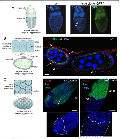
TheDrosophilaegg chamber consists of a 16-germ cell syn-cytium enveloped by a monolayer of follicular epithelium [36]. The process of proliferation and differentiation of the follicle cells is complex and under stringent control [37-39]. One critical event is the cessation of mitosis in mid-oogenesis. The proliferation of follicle cells occurs before stage 7 (up to 30 hours after the egg chamber buds off from the germarium at stage 2; total egg chamber development time from stage 2 to stage 14 is approximately 70 hours). Notch signaling that regulates cell proliferation in the follicle cells is activated at stage 6, which results in down-regulation ofcut andcyclin B, among other Notch target genes, and cessation of mitosis [40-43]. From stage 7 to 10A (approxi-mately 3 hours after stage 6) the follicle cell chromosomes continue to duplicate three times to generate polyploidity (endocycles). Disruption of Notch signaling causes extension of the proliferative program beyond stage 6 and follicle cells go through additional cell divisions without cell growth, resulting in increased cell number but reduced cell size.
We have previously shown that awd is involved in
regulating epithelial integrity of the follicle cells via its endocytic activity [25]. During the course of examining follicular function of awd, we also noticed that at later stages (after stage 8) the awdmutant clones often con-tain more numerous but smaller cells, suggesting faulty Notch signaling (Figure 1A). Since awd null alleles are lethal, the phenotypes in follicle cells, an adult tissue, are generated by mitotic recombination using the FLP/FRT system [44]. In this report, we employed different gen-etic methods that allow for induced mitotic recombin-ation using temporal or tissue-specific expression of the recombinase FLP [45] or allow for co-expression of
other transgenes in the awd mutant clones using the
mosaic analysis with a repressible cell marker (MARCM) system [46]. While specific genetic strategies will be pointed out when appropriate, it is worth noting that the
Notch phenotypes generated are consistent regardless of
the FLP/FRT variations.
Immunostaining with the mitotic marker
phosphory-lated histone H3 (p-H3) shows that awd mutant cells
A
C
st. 8
p-H3
Awd
DNA
awd
clone
st. 6
st. 8
p-H3
DE-cad
DNA
wt
wt
awd
clone (GFP
-)
DNA
DNA
GFP
st. 8
p-H3
Awd
DNA
awd
clone
B
only a few cells are stained at any given time. In awd
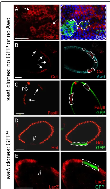
mutant cells p-H3 staining is detectable after stage 6 (Figure 1C). Again, theseawdmutant cells have smaller nu-clei (insets in Figure 1C). Consistent with increased prolif-eration in awd mutant follicle cells, prolonged expression of the mitotic marker cyclin B was also detected in these mosaic ovaries (GFP-negative cells are mutants in Figure 2A). Note that while cyclin B is absent inawd+cells, inawdmutant cells, cyclin B is not uniformly expressed at high levels. This is likely because the cell cycle is not syn-chronized in all follicle cells. In addition, the known Notch down-regulation target cut[40] is over-expressed in Awd-negative cells (Figure 2B). Compromised Notch signaling also results in expression of immature cell-fate markers in follicle cells beyond stage 6. In wild-type egg chambers Fasciclin III (FasIII) is expressed in all follicle cells up to stage 3 of oogenesis and then becomes restricted to the polar follicle cells (PC in Figure 2C) at the anterior and pos-terior poles of the follicular epithelium. Reduction of Notch activity arrests follicle cells in an undifferentiated state and up-regulates FasIII expression [42]. Follicle cell clones mu-tant forawdshow strong expression of FasIII after stage 6, indicating that they are defective in terminal differentiation (awd mutant cells lacking GFP expression in Figure 2C). Down-regulation ofcut in wild-type follicle cells is medi-ated by Hindsight (Hnt), an up-regulation target of Notch [40,47]. To examine loss of Notch target gene expression, we used the MARCM method of clonal analysis, which results in GFP-expression in mutant cells, so as to ensure that lack of gene expression is not the result of cell death (Figure 2D,E). In contrast to wild-type follicle cells, the
MARCM clone of awd mutant cells (GFP-positive) does
not express Hnt after stage 6 (Figure 2D). To further
con-firm that Notch signaling is attenuated in awd mutant
follicle cells, the expression ofGbeSu(H)m8-lacZ
transcrip-tional reporter for Notch activity [48] was examined. In MARCMawdclones,β-galactosidase staining is absent or strongly reduced (GFP-positive cells in Figure 2E).
The Notch signaling defect in awd mutant cells
sug-gested a potential mechanism for the original defining
phenotype ofawd- abnormal wing discs, because during
development Notch specifies the dorsal-ventral margin of the wing discs (which becomes the wing peripheral margin in the adult) and the vein-intervein boundary,
and is important for disc cell proliferation. Loss ofNotch
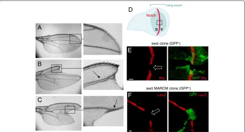
function causes wing margin defects and widening of wing veins [26]. As shown in Figure 3, 72% (18/25) of
adult mosaic flies show typical Notch phenotypes in
wings with‘notched’wing margins and wing vein thick-ening (Figure 3A-C). In wild type wing discs, activation of the Notch pathway at the dorsal-ventral boundary (Figure 3D) leads to the expression of target gene prod-ucts, such as the signaling molecule Wingless (Wg) [49]. Loss ofawdfunction abolished the Wg staining in third instar wing disc clones at the dorsal-ventral boundary (GFP-negative cells in Figure 3E). To further verify the
Notch signaling defect, we examined GbeSu(H)m8-lacZ
reporter expression using a different mosaic fly gener-ated by the MARCM system. Similar to our results in follicular epithelium, β-galactosidase expression in awd
mutant clones (GFP-positive cells) in the dorsal-ventral boundary is lost (Figure 3F).
awdfunction is required for signaling after the S2 cleavage of Notch
In the egg chamber, Notch functions in the follicle cells while the ligand Delta is expressed in the abutting germ-line cells [42]. Since the awdj2A4 clones were induced specifically in follicle cells, the defective Notch signaling in mutant follicle cells is not likely to be the result of a defect in Delta expression or endocytosis in the abutting
germline cells. Also importantly, inDeltamutant NICD
antibody-detected Notch accumulates on the follicle cell surface, which is consistent with the notion that ligand binding precedes intracellular trafficking and proteolytic processing of Notch [42].
To define the step where Notch signaling is stalled in
awd mutant follicle cells we over-expressed NICD or
NEXT inawdmutant follicle cells by using the MARCM
system. NICD is the cytoplasmic domain of Notch that functions as a cytoplasmic,γ-secretase-independent consti-tutively active Notch, while NEXT is the truncation gener-ated after the S2 cleavage devoid of the ligand-binding domain, S2 cleavage site, and the negative-regulatory region
(NRR) [50,51]. NEXT is a membrane-bound, γ
-secretase-dependent, constitutively active form of Notch that can function without ligand but still requires intracellular pro-teolytic processing and trafficking [52]. To assess rescue of
(See figure on previous page.)
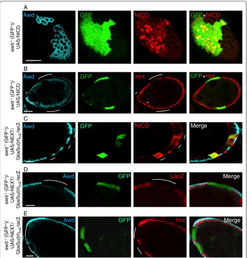
Notch signaling we analyzed the Hnt expression (no ex-pression in awd mutant). In stage 7–8 awd clones
over-expressing NICD (from the UAS-NICD transgene [53])
(GFP-positive cells in Figure 4A), 60.5% of mutant cells ex-press Hnt (199 out of 329 cells) (GFP-positive cells in
Figure 4B), representing a significant rescue of the lack of Hnt expression phenotype. This is also consistent with the observation that the over-expressed NICD is localized in the nuclei in a significant number of awd mutant follicle cells (Figure 4A). Furthermore follicle cells flp-out clones ex-pressing the same NICD transgene also show enhanced Hnt expression at stage 7–8 [see Additional file 1: Figure S1A], as well as enhanced the size of nuclei at stage 10B (not
shown) [54]. In contrast, expression of the UAS-NEXT
transgene [55] in the awd clone (GFP-positive cells in
Figure 4C) did not rescue Notch signaling as assessed by loss ofGbeSu(H)m8-lacZexpression (GFP-positive cells in
Figure 4D) and loss of Hnt expression (GFP-positive cells in Figure 4E). The same transgene is able to upregulate the Hnt expression in flp-out clones of follicle cells [see Additional file 1: Figure S1B]. Note that the over-expressed NEXT accumulates in the intracellular vesicles (Figure 4C), consistent with the notion that internalization of surface
Notch can occur inawd mutant cells but the subsequent
vesicle trafficking is defective.
It has recently been shown that transmission of Notch signal requires proper intracellular trafficking, at least in
Drosophila follicle cells and imaginal discs [32-34,55]. Therefore, our observed Notch processing and signaling defects may result from either defective proteolytic cleavage of Notch to release intracellular domain by γ -secretase or defective endocytic transport of Notch. We favor the latter mechanism since Awd has been shown to promote endocytosis of surface receptors in multiple tissues [21-25]. In addition, neither the expression level nor the punctate expression pattern of Presenilin [56-58], the catalytic component of theγ-secretase complex, are altered inawdmutant follicle cells [see Additional file 2: Figure S2]. To test the notion that the Notch signaling deficiency in
awdmutant cells is the result of defective endocytosis, we next examined the localization of Notch receptor inawd
follicle cell clones.
Notch accumulates in endocytic vesicles inawd mutant cells
While inawd+cells Notch is present in low abundance
in small punctates, Notch accumulates in large
vesicle-like aggregates near the apical surface in awd mutant
clones in follicular epithelium (Figure 5A,B) and in wing discs (Figure 5C). Such Notch accumulation phenotype
in awd mutant resembles that of mutants in avalanche
(avl; which encodes Syntaxin) and rab5 [34,55,59], two gene functions required for maturation of early endo-somes [59], but is different from the phenotype in dyna-min mutant (shits), in which Notch accumulates on the cell surface and in very large aggregates on apical and basal sides of the follicle cells (Figure 5D) as noted pre-viously [60]. This pattern is likely because of the failure to deliver Notch to apical membrane via
Dynamin-FasIII
GFP
cyclin B GFP
DNA
A
Hnt
GFP
Cut
Awd
cyclin B
FasIII Cut
Hnt
D
B
E
LacZ
LacZ GFP
awd
clones: no
GFP
or
no
A
w
d
awd
clones: GFP
+
PC
C
Figure 2Altered expression of Notch signaling target genes in
awdclones. (A and C)Stage 7–8 egg chambers were dissected fromhs-flp; +/+; Ubi-GFP, FRT82B/FRT82B, awdj2A4females, and theawd mutant clones were identified by lack of GFP staining (green). (B, D-E)Stage 7 egg chambers were dissected fromhs-flp/GbeSu(H)
m8-lacZ; act-Gal4, UAS-GFP/+; FRT82B, act-Gal80/FRT82B, awdj2A4
females. In(A-C)theawdmutant clones were identified by lack of Awd or GFP staining (green), while in(D-E)theawdmutant clones are GFP positive. In all panels,awdmutant clones are marked with dashed lines.(A)Cyclin B (red),(B)Cut (red) and(C)Fasciclin III (FasIII; in red) are normally negatively regulated by Notch signaling, but up-regulated inawdmutant clones (lack of GFP or Awd; arrows). (D)Hindsight (Hnt; in red) is normally induced by Notch signaling, and is down-regulated inawdclones (GFP-positive; empty arrowhead).(E)ThelacZreporter gene expression driven by Notch-activatedSu(H)promoter [GbeSu(H)m8-lacZ] is lost inawd
mediated transcytosis [61] as well as to internalize Notch
for signaling [55]. Since awd mutant cells do not show
these very large aggregates throughout the cells, it is un-likely thatawdfunction completely overlaps with that of
dynamin. Notch localization can also be influenced by
the integrity of the adherens junction [61]. Since we have
shown previously that the awd mutant can affect the
membrane localization of E-cadherin andβ-catenin [25], we also determined that Notch localization defect not only occurred inawdmutant pile-up epithelial cells [see
Additional file 3: Figure S3] but also occurred in awd
mutant follicle cells that show normal epithelial polarity, indicated by normal E-cadherin localization (Figure 5E).
awdmutant clones exhibiting normal epithelial integrity are most often observed in clones of small size (<10 cells; unpublished observation). We showed that small
awd mutant clones indeed lacked Hnt expression [see
Additional file 4: Figure S4]. We also showed that the epithelial polarity ofawdmutant cells in wing disc is un-affected as shown by normal E-cadherin localization
(Figure 5F,G in which GFP+ cells are awd mutants).
Since Notch processing in the follicle cells has been shown to occur during transition from mature early
endosomes to late endosomes [55,62], we suspected that
the endocytosis defect in awdmutant cells might be in
the step prior to the formation of late endosomes. To verify this notion, we first examined Notch
localization in the endocytic pathway in awd mutant
cells. In awd+ cells, NICD is in small punctates with
partial co-localization with Avl, a component of the early endosome (Figure 6A, upper panels), consistent
with previous observations [34,55]. In awd mutant
cells, the level of Notch-Avl colocalization increased by 2 fold (Figure 6A, bottom panels; statistical analysis reported in Additional file 5: Figure S5A,A’).
In order to determine whether these Avl-positive, Notch-containing vesicles are immature early endo-somes that cannot form multivesicular bodies (MVBs),
we examined the awd mutant vesicles in relation to
hepatocyte growth factor-regulated tyrosine kinase substrate (Hrs), which is involved in the maturation of early endosomes by promoting ubiquitinated cargo sorting [63]. It marks the mature early endosomes and MVBs. We observed similar, low-level co-localization
of Notch and Hrs in both awd+ and awd mutant
cells [see Additional file 5: Figure S5B,B’for statistical
A
B
C
D
Wg GFPWg
E
awdclone (GFP-)
LacZ
F
GFPLacZawdMARCM clone (GFP+) Notch
wing pouch
D V
Figure 3Notch signaling defect in adult wings and larval wing discs.Compared toywflies, representing wild-type(A), wings from flies of the genotypeen2.4-Gal4e22c, UAS-flp/+; FRT82B/FRT82B, awdj2A4(B-C)show typicalNotchphenotypes: enlarged wing veins (arrows) and loss of wing
margins (‘notched’wing blades).(D)Drawing of a third instar wing disc in apical view showing the dorsal-ventral (D, V) compartment border (red line) specified by the Notch activity. The wing disc pouch is the central fold of the disc (green) and will generate the wing blade. The black box approximately indicates the areas shown in E and F.(E)The discs were dissected fromhs-flp; +/+; FRT82B, Ubi-GFP/FRT82B, awdj2A4third instar larvae. wingless(wg) is a downstream activation target ofnotch. Wg protein expression is lost in theawdclone (loss of GFP; empty block arrow) overlapping the midline (dorsal-ventral boundary, where Notch specifieswgexpression).(F)The discs were dissected fromhs-flp/GbeSu(H)m8-lacZ; act-Gal4, UAS-GFP/+;
FRT82B, act-Gal80/FRT82B, awdj2A4third instar larvae.GbeSu(H)
m8-lacZexpression (red) is also lost inawdMARCM clones (expressing GFP; empty block arrow).
Awd
GFP
GFP
+
Hnt
B
A
NICD
Awd
GFP
Merge
C
awd
-/-(GFP
+)/
UAS
-NEXT/
Gbe
S
u
(H)
m8
-l
acZ
Awd
GFP
NICD
GFP
+
NICD
Hnt
awd
-/-(GFP
+)/
UAS
-NE
X
T/
Gbe
S
u(H)
m8
-l
acZ
Awd
GFP
LacZ
Merge
D
Hnt
Awd
GFP
Merge
E
awd
-/-(GFP
+)/
UAS
-NEXT/
Gbe
S
u
(H)
m8
-l
acZ
awd
-/
-(GFP +)/
UAS
-NICD
awd
-/-(GFP
+)/
UAS
-NICD
Figure 4Notch signaling defect inawdmutant cells is rescued by exogenous NICD.Stage 7–8 egg chambers were dissected from females of the genotypehs-flp/GbeSu(H)m8-lacZ; act-Gal4, UAS-GFP/UAS-NICD; FRT82B, act-Gal80/FRT82B, awdj2A4(A-B)orhs-flp/GbeSu(H)m8-lacZ; act-Gal4,
UAS-GFP/UAS-NEXT; FRT82B, act-Gal80/FRT82B, awdj2A4(C-E).(A, C)As controls, NICD and NEXT expression is verified.(B)A stage 7 egg chamber
with MARCM clones ofawd(Awd-negative and GFP+, marked by lines) simultaneously expressing NICD. The expression of endogenous Hnt (red)
is restored in a majority of the mutant cells. The Awd staining is in cyan. Note that the diffused Awd staining in regions abutting the apical side of the follicle cells is within the germline cells, occasionally observed in abnormal egg chambers.(D-E)Exogenously expressed NEXT cannot rescue theawdmutant phenotype.awdMARCM mutant clones lacking Awd staining (cyan) marked by the GFP expression and indicated by lines show loss ofGbeSu(H)m8-lacZreporter gene expression (red inD) as well as loss of Hindsight (Hnt) expression (red inE). Bars in(A-B)are 20μm.
µ µ
µ
µ µ
µ
µ
A
NICD NICD+Awd+DNAB
NICD NICD+AwdNICD Awd+NICD
C
D
5 m
GFP DE-Cad NICD DE-Cad+NICD
E
20 m 20 m
shi
tsNICD
10 m
10 m
10 m
NICD
shi
tsGFP+Wg+DE-Cad Wg+DE-Cad DE-Cad DE-Cad
GFP+DE-Cad
F
10 m
G
analysis]. Lack of significant Notch-Hrs co-localization even in awd+ cells is consistent with the finding that
normal Notch signaling is not affected in hrs mutants
[55]. Some co-localization of Hrs and Notch in awd
mutant cells is also consistent with the observation that a minor Rab5-independent route exists for Notch sorting [55]. On the other hand, this Notch accumula-tion pattern is very different from that of thephyllopod
mutation which blocks Notch entry into late endo-somes but not entry into mature early endoendo-somes, resulting in increased Notch signaling and significant co-localization of NICD and Hrs [64]. This suggests
that early endosome maturation is defective in awd
mutant cells.
Since awd can also act on the internalization of sur-face receptor [21], we examined whether constitutive
in-ternalization of full-length Notch is affected in awd
mutant cells. This was detected by using an antibody against the NECD. As shown in Figure 6B, NECD anti-body indeed detected increased accumulation of
full-length Notch in awd mutant cells. Therefore, Awd can
act on both internalization of surface Notch and intra-cellular trafficking of signaling Notch.
Notch does not traffic to late endosomes inawd mutant cells
It has been shown that Notch signaling can also be en-hanced by blocking MVB formation with mutations in
the endosomal sorting complex required for transport
(ESCRT) genestsg101,vps25andvps20, or by promoting early endosome maturation with over-expression of
con-stitutively active Rab5 [55]. Since the awd mutant is
defective in Notch signaling, it is unlikely that the Notch-containing vesicles inawdmutant cells have passed into late endosomes. This notion is supported by the lack of significant co-localization of Notch-containing vesicles
in MARCM awd mutant clones with Rab7, the
late-endosomal marker (GFP-positive cells in Figure 6C; statis-tical analysis reported in Additional file 6: Figure S6). As well, transition from early endosomes to late endosomes is
accompanied by acidification of the luminal contents, which can be detected by Lysotracker staining. Consistent with the notion that Notch-containing vesicles inawd mu-tant cells cannot enter MVB and late endosomes, we ob-served no difference in Lysotracker-positive vesicles inawd+
andawd mutant cells [see Additional file 7: Figure S7]. In
addition, the Notch-containing vesicles in MARCM awd
mutant clones are not Rab11-positive recycling endosomes, either (GFP-positive cells in Figure 6D; see Additional file 8: Figure S8 for statistical analysis).
We next sought to follow the time course of Notch localization in live cells. Wing discs are an ideal and standardized system for this purpose since they can be
cultured ex vivo for a prolonged period of time. Note
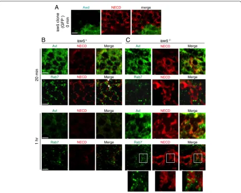
that in this establishedex vivo system, internalization of Notch is detected by binding to NECD antibody, without binding to spatially-expressed ligands. Therefore, the system strictly measures the kinetics of vesicular trans-port, not the endogenous signaling process. We first established that at the steady state (time 0), Notch accumulated on the awd−/−cell surface (Figure 7A). In wild-type cells, internalized Notch follows a typical time course: at 20 minutes after initiation of endocytosis, Notch is mostly in Avl-positive early endosomes while some has passed into Rab7-positive late endosomes (Figure 7B). At one hour after endocytosis, the Notch signal is barely detectable, consistent with the degrad-ation time course. Also, in wild-type cells, Avl staining is much more pronounced at 20 minutes than at one hour. This is likely because in this label-and-chase experiment, a large number of Avl-positive vesicles were formed syn-chronously after initiation of endocytosis. Concentrated Avl was then lost (therefore detected at a lesser extent by immunofluorescence) after early endosomes matured and were incorporated into late endosomes.
Inawdmutant, on the other hand, accumulated Notch
is mostly on cell surface or in Avl-positive early somes at 20 minutes and remains in these early endo-somes even one hour after internalization (Figure 7C). The Notch signal shows no localization to the late
(See figure on previous page.)
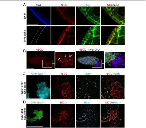
Figure 5Defective intracellular distribution of Notch inawdmutant cells. (A-B)Stage 8 egg chambers were dissected fromhs-flp; +/+; Ubi-GFP, FRT82B/FRT82B, awdj2A4females, and stained for NICD (red), Awd (green) and DNA (blue). Notch over-accumulates in vesicles near the cell periphery (insets in(A)and arrowheads in(B)).(C)The wing disc was dissected fromhs-flp; +/+; Ubi-GFP, FRT82B/FRT82B, awdj2A4third instar larva and stained for NICD (red).awdclones were identified by lack of Awd staining (pseudo-colored in green). Notch inawdmutant clones accumu-lates in large vesicles.(D)Surface and cross-section views ofshitsstage 7 egg chambers from females incubated at 29°C and stained for NICD. Very large aggregates are seen on the surface and throughout the cells.(E)A stage 7 egg chamber from ahs-flp/GbeSu(H)m8-lacZ; act-Gal4,
endosomes (Figure 7C). Note that some of the Rab7-positive vesicles shown in Figure 7C are very close to or surrounded by the Notch signal but are not overlapping (Figure 7C insets).
awdis required for Rab5 function
To further test the role of awdin early endosome matur-ation, we next tested how expression of constitutively active
Rab5 (Rab5CA) might affect Notch localization in awd
mutant. As mentioned above, Rab5CAhas been shown to
increase Notch signaling [55], presumably because the
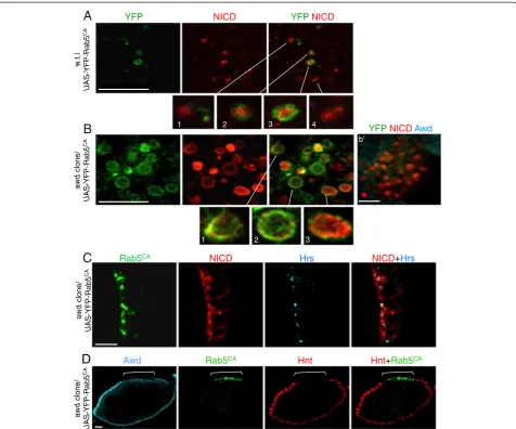
endocytic process is pushed through early endosomes. In awd+ cells, NICD is found in both Rab5CA-positive (Figure 8A, insets 2 and 3) and -negative (Figure 8A, insets 1 and 4) vesicles, and, importantly, the detectable NICD is almost exclusively in the lumen of these vesicles. The likely interpretation is that Rab5CA pushes endocytosis through early endosomal stages and Notch is processed. Processed endogenous NICD becomes diffused in the cytosol and nuclei, and undetectable by immunohistochemistry (IHC) in our assay system. Remaining, predominantly luminal, NICD is an unprocessed subpopulation that is internalized
NICD Avl
Awd NICD+Avl
awd
+
awd
clone
A
NICD Rab7 NICD+Rab7
GFP (awd-/-)
aw
d
+and
aw
d
clone
C
NICD Rab11 NICD+Rab11
GFP (awd-/-)
awd
+and
awd
clone
D
B
NECD NECD+Awd+DNAFigure 6Notch accumulates in early endosomal compartments inawdmutant cells. (A)Stage 8 egg chambers were dissected fromyw (wild-type; upper panel) oryw; en2.4-Gal4e22c, UAS-flp/+; FRT82B/FRT82B, awdj2A4(awdclone, lower panel) females. In wild-type, Notch shows low
level punctates that are partially co-localized with Avl. In theawdclone, most, if not all, large Notch-positive vesicles are also Avl-positive. Dashed line marks theawdclone.(B)Surface view of stage 7 egg chamber dissected from ayw; en2.4-Gal4e22c, UAS-flp/+; FRT82B/FRT82B, awdj2A4female
and stained for Notch extracellular domain peptide (NECD), Awd and DNA. There is accumulation of NECD on the surface ofawdmutant clones (empty arrowheads).(C-D)Stage 8 egg chambers were dissected fromhs-flp/GbeSu(H)m8-lacZ; act-Gal4, UAS-GFP/+; FRT82B, act-Gal80/FRT82B,
awdj2A4females.awdmutant clones were identified as GFP-expressing cells. Inawdclones, over-accumulated Notch does not co-localize with
in the MVBs or late endosomes destined for degradation [31,62]. Strikingly, in awd mutant clones, NICD is found exclusively in the Rab5CA-positive vesicles (Figure 8B). Most importantly, inawdmutant cells, much of the NICD signal is mostly present on the surface of these enlarged vesicles (Figure 8B insets 1–3). The result indicates that al-though cell surface-bound Notch can be internalized in
awdmutant cells in the presence of Rab5CA, it is not proc-essed and cannot enter late endosomes. In addition, inawd
mutant follicle cells 87.1% of Notch vesicles co-localize
with Rab5CA and 31.45% co-localize with Hrs (n = 124)
(Figure 8C; see Additional file 9: Figure S9 for co-localization analysis). Co-co-localization of NICD and Hrs inawdmutant cells increases by the over-expression of
Rab5CA [see Additional file 9: Figure S9B compared to
Additional file 5: Figure S5B’]. This suggests that Rab5CA partially stimulates vesicles to progress through the
endo-cytic pathway but awd function is necessary for
Rab5-mediated early endosome maturation. This notion is
supported by the increased number of Rab5CA-positive
vesicles in awd mutant clones (Figure 8B), indicating a block in vesicle trafficking downstream of Rab5 function. This interpretation is confirmed since Rab5CAcannot res-cue Notch signaling inawdmutant cells (Figure 8D).
Taken together, these results suggest that during Notch signalingawdfunction is downstream of or is re-quired for Rab5 function in promoting maturation of early endosomes.
20 min
1 hr
Avl NECD Merge
Rab7 NECD Merge
Avl NECD Merge
Rab7 NECD Merge
awd
-/-Avl NECD Merge
Rab7 NECD Merge
Avl NECD Merge
Rab7 NECD Merge
awd+
B
C
Awd NECD merge
A
awd
clone
(GFP
-)
0 min
Figure 7Endocytic defects inawdmutant cells.Notch trafficking assay was performed on wing discs from(A, C)yw; en2.4-Gal4e22c, UAS-flp/+;
Discussion
In this report we demonstrate a role ofawdin regulating Notch signaling via its endocytic function including sur-face internalization and vesicle trafficking. This conclu-sion is based on our results that show: (1) multiple Notch target genes are mis-expressed in follicle cells and wing discs; (2) Notch accumulates in enlarged early
endosomes; and (3) awd function is required for the
Rab5 activity in early endosome maturation. Our results also indicate that during vesicles trafficking, the Awd action is downstream of the S2 cleavage, since over-expressed of NEXT accumulated intracellularly and
could not rescue the awd defect. The same NEXT
over-expression strategy could rescue theshi/dynamin
defect [57,65], strongly supporting the notion that the Awd action on Notch signaling is post-membrane
awd
clone/
UAS
-YFP-Rab5
CA
w.t./
UAS
-YFP-Rab5
CA
1 2 3 4
1 2 3
A
B
YFPNICDAwdb’
YFP NICD YFPNICD
C
Awd Rab5CA Hnt Hnt+Rab5CA
Rab5CA NICD Hrs NICD+Hrs
D
awd
cl
one/
UAS
-YFP-Rab5
CA
awd
c
lone/
UAS
-YFP-Rab5
CA
Figure 8Awd is required for Rab5 function.YFP-tagged constitutively active Rab5 mutant Q88L (Rab5CA) were expressed in(A)wild-type or
(B)awdmutant clones, using the genetic combinationsen2.4-Gal4e22c, UAS-flp/UAS-YFP-RAB5Q88L; +/FRT82Boren2.4-Gal4e22c,
UAS-flp/UAS-YFP-RAB5Q88L; awdj2A4, FRT82B/FRT82B,respectively.Egg chambers were processed for staining for NICD (red) and YFP (green) as indicated.awdmutants
were verified by lack of Awd staining (cyan inb’).(A)YFP-Rab5CAexpressed in wild-type follicle cells. In Rab5CA-expressing wild-type follicle cells,
NICD is reduced and is present in either Rab5-positive (insets 2 and 3) or Rab5-negative (likely late endosomes; insets 1 and 4). Note that NICD is in the lumen of these vesicles.(B)YFP-Rab5CAexpressed inawdmutant follicle cells. In Rab5CA-expressingawdmutant cells, abundant NICD is
present in enlarged vesicles that are mostly Rab5-positive. NICD is enriched on the surface of these vesicles (insets 1–3).(C)A stage 8 egg cham-ber fromhs-flp/GbeSu(H)m8-lacZ; UAS-YFP-Rab5CA/+; tub-Gal4, FRT82B, tub-Gal80/FRT82B, awdj2A4was stained for Hrs (cyan), YFP (green) and NICD
(red). There is only partial co-localization of accumulated Notch with Hrs.(D)A stage 8 egg chamber fromhs-flp/GbeSu(H)m8-lacZ; UAS-YFP-Rab5CA/+;
tub-Gal4, FRT82B, tub-Gal80/FRT82B, awdj2A4was stained for Awd (cyan), YFP (green) and Hnt (red). Expression of Rab5CAin Awd-negative cells (bracket)
invagination. Since over-expression of NICD could
res-cue the awd defect, the Awd action is likely upstream
or in parallel to the S3 cleavage event (γ-secretase
activity). Although a role of awd in promoting the
activity of γ-secretase cannot be completely ruled out,
we considered this possibility unlikely. First, awd is
a known endocytic factor demonstrated in multiple tissues including neurons, trachea, and follicle cells [22-25]. Second, neither the expression level nor the expression pattern of Presenilin, the catalytic subunit of γ-secretase, is altered inawd mutant cells. Third, if the defect is in γ-secretase function, it would be ex-pected that Notch should accumulate in Hrs-positive MVBs [60]. On the contrary, we did not observe such ectopic accumulation of Notch in Hrs-positive vesicles. Therefore, our results, in aggregate, suggest that the main action of Awd on Notch signaling is via its endocytic activity promoting the transition from early endosomes to late endosomes. However, potential de-fects downstream ofγ-secretase cleavage, such as
traf-ficking to nucleus, in awd mutant cannot be formally
ruled out.
One curious exception for theawdfunction in relation to Notch signaling is found in the border cells. As we have re-cently reported [24], during the migration of these cells, Awd expression is down-regulated. Re-expression of Awd can lead to reduction of surface receptors, such as PVR that is critical for directional movement, resulting in defective migration. Interestingly, Notch signaling is also important for border cell migration [66]. It, therefore, appears that Notch signaling in these specialized cells does not require Awd activity or is insensitive to Awd protein levels. To test this, we compared Notch expression in border cells with or without Awd re-expression. In wild-type border cells (no Awd), Notch is located on the cell surface as well as in the cell body, consistent with active signaling (data not shown). Forced re-expression of Awd in the border cells does not alter this pattern. This may be because Notch is already ac-tively internalized; increasing the Awd level cannot further enhance such activity. Indeed, endocytosis is intrinsically highly active in border cells [24,67]. Alternatively, the differ-ential dependence of Notch on Awd activity may be a func-tion of how Notch is activated, not how Awd funcfunc-tions differently in different cell types. For example, Dobenset al. [68] have shown that the Notch ligand Delta may be co-expressed with Notch in the same border cells. Recent reports have hinted that the requirement of endocytosis for Notch signaling may depend on the ligand-receptor rela-tionship (for example, ligand-dependent or -independent, trans- or cis-activation, and so on) [62]. We, therefore, con-sider that the apparent Awd-independent Notch signaling in border cells has more to do with the intrinsic Notch sig-naling mechanism in these cells, and less to do with the function of Awd.
Our results indicate that the Notch signaling defect in
awd mutant cells is the failure to deliver Notch past the Rab5-dependent early endosomal stage. On the other hand, the ESCRT complex mutants, which are defective in late endosome formation, promote Notch signaling [34,55]. Taken together, it appears that Notch activation occurs in the intermediate stage between early endosome formation and late endosome entry. Transition from early endosomes to late endosomes is accompanied by cargo sorting, intrave-sicular invagination and acidification of the luminal con-tents. Curiously, the matured early endosome and MVB marker hrs mutant has no effect on Notch signaling [55], which indicates that endosomal cargo sortingper seis not required for Notch signaling. We have also shown thatawd
mutant cells do not exhibit altered levels of Lysotracker staining and that endosomal Notch remains on the surface
of enlarged endosomes in awdmutants. The exact nature
of this transition state that favors Notch processing, there-fore, requires further analysis. The endocytic function of
awdhas traditionally been described as a‘GTP supplier’for Dynamin, based on genetic interaction data and logical ex-trapolation because of the GTP producing activity of Awd [22]. In this report, we demonstrate that, in relation to Notch signaling,awdfunctions downstream of, but not dir-ectly on, dynamin. It is instead critical for Rab5 activity. This is supported by the following evidence: 1) Notch in
awdmutant accumulates in Avl-containing vesicles. There-fore, theawddefect is post Dynamin-mediated cleavage of
membrane invagination. 2) Rab5CA can push Notch into
enlarged early endosomes but failed to rescue the awd
phenotype, thereby strengthening the notion thatawd de-fect is post Shi/Dynamin function. 3) The Notch accumula-tion pattern in shi mutant is different from that in awd
mutant. 4) Over-expression of NEXT could not rescueawd
defect. The same NEXT over-expression strategy could res-cue the shidefect, strongly supporting the notion that the Awd action concerning Notch signaling is post-membrane invagination [57,65]. It should be noted that we did observe surface accumulation of NECD antibody-detected Notch molecules, likely representing the full-length Notch not en-gaged in ligand binding and signaling. This indicates that Awd can affect constitutive internalization of full-length Notch.
The requirement of endocytosis in the signal-receiving cells for Notch activation has been amply demonstrated [69]. It has been shown that Notch signaling in follicle cells after stage 6 requires Delta [42]. Since in this report we show that Notch signaling cannot occur in the follicle
cell without awd function, we conclude that, at least
in follicle cells, endocytosis is a requisite process for ligand-dependent Notch signaling.
carcinogenesis. For example, V-ATPase is required for Notch signaling while mutations in ESCRT components, such as Tsg101, result in increased Notch signaling. V-ATPase has generally been considered an oncogene [70] because it is associated with acidification of tumor cells. ESCRT components, on the other hand, have been shown to suppress tumor formation because they down-regulate surface growth factor receptor signaling [71]. As such, attempts to design therapeutics based on these prevalent functions should take into account the effects on Notch signaling, since the relationship between Notch sig-naling and carcinogenesis is context-dependent [35,72,73].
Conclusions
Awd belongs to the Nm23 family of protein that is
evolu-tionarily conserved from Drosophila to mammals. Our
in vivoanalyses demonstrate that loss ofawdgene function blocks Notch signaling by altering the receptor processing after the S2 cleavage and causes Notch accumulation in early endosomes. Furthermore, we obtained evidence indi-cating that Awd is required for Rab5 function in early en-dosome formation.
Nm23has been an enigmatic gene function. It is a house-keeping gene involved in nucleotide synthesis and energy metabolism, and yet exhibiting specific developmental func-tions [6,24]. It was the first metastasis suppressor gene identified [4,74], yet exhibits oncogenic functions in some cancer cohorts [9,10]. We have previously shown that ei-ther loss-of-function or over-expression of awd can affect different aspects of epithelial morphogenesis. That is, loss-of-function awd results in over-accumulation of adherens junction components and piling up of the epithelium, while over-expression of awd results in reduced adherens junc-tions and disintegration of epithelial structure [25]. These findings provided some explanation of the biphasic function of Nm23 in tumorigenesis. In light of the studies presented here, an additional level of complexity should be considered since Notch signaling can exert different cellular functions in different tissues and at different times during patho-physiological alterations of the same tissues [35].
Methods
Drosophilastrains and genetics
Stocks were raised on standard cornmeal/yeast/agar medium at 25°C. The stock carrying the protein-null awd
allele,awdj2A4, has been described [22-25]. Theawdj2A4
al-lele combined with theFRTchromosomeFRT82Bhas been
described [24,25]. Cell clones mutant forawdj2A4were gen-erated through mitotic recombination using the FLP/FRT system [44], either with the hs-flp recombinase transgene or using the directed mosaic technique with the UAS-flp
transgene under control of the ubiquitous somatic cell driver en2.4-Gal4e22c [45]. To obtain over-expression of specific transgenes inawdj2A4mutant follicle cells we used
either the directed mosaics or the MARCM [46] tech-niques. The transgenic line carrying the constitutively active (CA) variant of the YFP-Rab5 fusion genes was obtained from the Bloomington Stock Center (Bloomington, IN, USA) [75]. TheUAS-NICDand theGbeSu(H)m8-lacZlines
were a kind gift from S. Bray of University of Cambridge (Cambridge, UK). TheUAS-NEXTline was a kind gift from M. Fortini of Thomas Jefferson University (Philadelphia, PA, USA). The genotypes of flies and larvae used for the analyses are described in Additional file 10; Supplementary experimental procedures.
Immunohistochemistry
Ovaries were dissected, fixed and stained as previously de-scribed [76] with the exception of ovaries from shi2/shi2
(shits) females that were fixed at 29°C. Whole late third in-star larvae were dissected into room temperature PBS (pH 7.5), and fixed for 20 minutes in 4% formaldehyde. After three washes in PBS, larval tissues were permeabilized for one hour in PBT (0.3% Triton X-100 in PBS) and then were blocked in 2% BSA in PBT for 10 minutes at room temperature. Overnight incubation at 4°C with primary anti-bodies in 2% BSA in PBT was followed by three washes in PBT and 10 minutes incubation in 2% BSA in PBT. Larval samples were then incubated with fluorescence-tagged sec-ondary antibodies for two hours at room temperature and after extensive washes in PBT the wing discs were dissected. Primary antibodies used are: chicken anti-Avl (1:500) [59], mouse monoclonal anti-NICD (1:1000; C17.9c6, Develop-mental Studies Hybridoma Bank (DSHB, Iowa City, Iowa, USA)), mouse monoclonal anti-NECD (1:50; C458.2H, DSHB), mouse monoclonal anti-Cut (1:15; 2B10, DSHB), mouse monoclonal anti-Hnt (1:30; 1G9, DSHB), mouse monoclonal anti-Cyclin B (1:100; F2F4, DSHB), rat monoclonal anti-DE-cadherin (1:100; DCAD2, DSHB)
and mouse monoclonal anti-β-gal (1:25; 40-1A, DSHB);
and protein A-purified rabbit anti-Awd (1:2000) [23], rabbit anti-phosphohistone H3 (1:200; 06–570, Upstate Biotechnology, Lake Placid, NY, USA), rabbit anti-C-Psn (1:200) [58], rabbit Rab7 (1:2000) and rabbit anti-Rab11 (1:8000) [77]. Secondary antibodies used are: Cy3-(1:100; Jackson Lab, West Grove, PA, USA), DyLight 649- (1:200; Jackson Lab), or FITC- (1:250; Invitrogen, Molecular Probes, Eugene, OR, USA) conjugated anti-mouse immunoglobulin G (IgG); and Cy3- (1:1000; Sigma, Saint Louis, Missouri, USA), DyLight 649- (1:500; Jackson Lab), or BODIPY- (1:2000; Molecular Probes) conjugated anti-rabbit IgG.
DNA staining was carried out by incubating egg cham-bers and wing discs for 10 minutes with
4',6-diamidino-2-phenylindole (DAPI; Sigma) at 0.5 μg/ml in PBS
two hours with To-Pro-3 at 1 μM in PBS on a rotating wheel followed by several washes with PBT. Stained egg chambers or wing discs were mounted in Fluoromount-G (Electron Microscopy Sciences, Hatfield, PA, USA) and were subsequently analyzed with conventional epifluores-cence on a Nikon Eclipse 90i microscope and with a TCS SL Leica confocal system. Digital images were processed and assembled using the Adobe Photoshop software. No biased image manipulations were applied.
Cuticle preparation of adult wings
Adult flies of the genotype en2.4-Gal4e22c, UAS-flp/+; FRT82B/FRT82B, awdj2A4 were collected. Wings were re-moved at the hinge, dehydrated in ethanol and mounted on microscope slides in lactic acid/ethanol (6:5). Wing images were captured by a Nikon Eclipse 90i microscope and acquired with a Nikon Digital Sight camera.
Notch endocytosis assay
The assay was adopted from a published report [78] with modifications. Third instar larval wing discs were dissected
in Schneider’s Drosophila medium (SDM) containing 1%
fetal calf serum. The discs were cultured for 15 minutes on ice in the presence of the mouse monoclonal anti-NECD antibody. Excess antibody was rinsed away and the discs were incubated with fresh media at room temperature. The discs were dissected at different times and detected with anti mouse IgG.
Co-localization and statistical analysis
Thresholds of confocal images were set in Adobe Photoshop to exclude background staining. Images were processed with the CDA plugin of ImageJ to obtain the Pearson’s coeffi-cient. Statistical comparison was performed by two-tailed paired Student’s t-test (GraphPad Prism 6 software).
Lysotracker staining
For Lysotracker ex vivo staining, females were dissected in SDM. Ovaries were collected, separated and incubated in
medium containing 5 μM Lysotracker (DND-99, Molecular
Probes) in soft agitation for five minutes at room temperature in the dark. Ovaries were then rapidly washed three times with fresh SDM, mounted and imaged immediately.
Additional files
Additional file 1: Figure S1.Notch signaling in wild type follicle cells is upregulated by either NICD or NEXT over-expression. Females of the genotypehs-flp,UAS-mCD8GFP/act>CD2>Gal4;+/UAS-NICD(A) orhs-flp, UAS-mCD8GFP/act>CD2>Gal4;+/UAS-NEXT(B) were dissected and the egg chambers were stained for Hnt (red). Over-expression of NICD at stage 7–8 in wild type follicle cells marked by GFP expression (green) enhances the level of Hnt expression in 51% of follicle cells (n = 100). Over-expression of NEXT at stage 7–8 in wild type follicle cells marked by
the expression of GFP (green) enhances the level of Hnt expression in 92.5% of follicle cells (n = 40). Bars are 15μm.
Additional file 2: Figure S2.Presenilin expression pattern is not altered inawdmutant follicle cells. Polyclonal rabbit antibody against a C-terminal peptide in the putative hydrophilic loop region of Psn (anti-C-Psn) has been described [58]. Stage 6 and 7 egg chambers containing MARCM clones ofawdmutant (marked by positive GFP expression) were stained for Psn (cyan) and NICD (red). Psn is ubiquitously expressed in intracel-lular punctates in both follicle cells and germ cells. No changes in either the expression level or the punctate pattern are observed inawdmutant cells. The egg chambers were dissected fromhs-flp/GbeSu(H)m8-lacZ; act-Gal4,
UAS-GFP/+; FRT82B, act-Gal80/FRT82B, awdJ2A4females. Bars are 5μm.
Additional file 3: Figure S3.Disrupted epithelial cells inawdmutant clone show abnormal Notch accumulation. Females of the genotype en2.4-Gal4e22c, UAS-flp/+; FRT82B/FRT82B, awdj2A4were dissected and the egg chambers were stained for DNA (DAPI), Awd, NICD, and Avl as indicated. Abnormal Notch accumulation in large vesicles is observed in pile-up mutant epithelial cells (arrows), which co-localize with the early endosomal marker Avl (see also Additional file 5: Figure S5A,A’). Bar is 20μm.
Additional file 4: Figure S4.Smallawdmutant clones exhibit loss of Hnt expression. Stage 7–8 egg chambers were dissected fromhs-flp/ GbeSu(H)m8-lacZ; act-Gal4, UAS-GFP/+; FRT82B, act-Gal80/FRT82B, awdJ2A4
females and stained for Hnt (red) and DNA (cyan). Quantitative analysis of Hnt expression was perfomed inawdclones (GFP-positive cells, green) containing a maximum of 5 cells. In these small clones 93% ofawd mutant cells lack Hnt expression (n = 42). Bar are 5μm.
Additional file 5: Figure S5.Analysis of Notch vesicle co-localization with Avl and Hrs. Inawdmutant cells, Notch accumulates in Avl-positive and Hrs-negative early endosomes. Stage 7–8 egg chambers were dis-sected fromhs-flp/GbeSu(H)m8-lacZ; act-Gal4, UAS-GFP/+; FRT82B, act-Gal80/
FRT82B, awdj2A4females and stained for NICD and Avl(A,A’)or NICD and
Hrs(B,B’). Co-localization was analyzed by using ImageJ. The Pearson's coefficient ranges from +1 = complete correlation to−1 = anti-correlation, with 0 = no correlation. The mean values (n = 4) of Pearson’s coefficients for NICD and Avl(A)and for NICD and Hrs(B)inawd+andawdmutant
cells were plotted together with standard deviations (error bars). Statis-tical significance was calculated using the two-tailed paired t-test (** = P <0.01; N.S. = No Significant).(A’)Co-localization image of NICD and Avl based on the analysis ofawdmutant cells and neighboringawd+cells
showed in Figure 6A.(B’)Co-localization image of NICD and Hrs based on the analysis ofawdmutant cells and neighboringawd+cells.
Additional file 6: Figure S6.Analysis of Notch vesicle co-localization with Rab7. Inawdmutant cells, Notch does not accumulate in Rab7-positive endosomes. Stage 7–8 egg chambers were dissected fromhs-flp/ GbeSu(H)m8-lacZ; act-Gal4, UAS-GFP/+; FRT82B, act-Gal80/FRT82B, awdj2A4
females and stained for NICD and Rab7. Co-localization was analyzed by using ImageJ. The mean values (n = 6) of Pearson’s coefficients for NICD and Rab7 inawd+andawdmutant cells were plotted together with
standard deviations (error bars)(A). Statistical significance was calculated using the two-tailed paired t-test (N.S. = Not Significant).(A’)Co-localization image of NICD and Rab7 based on the analysis ofawdmutant cells and neighboringawd+cells showed in Figure 6C.
Additional file 7: Figure S7.awd+andawdmutant cells show similar
Lysotracker staining patterns. The egg chambers were dissected from hs-flp/GbeSu(H)m8-lacZ; act-Gal4, UAS-GFP/+; FRT82B, act-Gal80/FRT82B,
awdJ2A4females and stained for Lysotracker. GFP expression identifies
awdmutant clones. There is no difference in acidified endosomal compartments betweenawd+andawdmutant cells. Bar is 5μm.
Additional file 8: Figure S8.Analysis of Notch vesicle co-localization with Rab11. Inawdmutant cells, Notch does not accumulate in Rab11-positive endosomes. Stage 7–8 egg chambers were dissected fromhs-flp/ GbeSu(H)m8-lacZ; act-Gal4, UAS-GFP/+; FRT82B, act-Gal80/FRT82B, awdj2A4
fe-males and stained for NICD and Rab11. Co-localization was analyzed by using ImageJ. The mean values (n = 4) of Pearson’s coefficients for NICD and Rab11 inawd+andawdmutant cells were plotted together with
Co-localization image of NICD and Rab11 based on the analysis ofawd mutant cells and neighboringawd+cells showed in Figure 6D.
Additional file 9: Figure S9.Analysis of Notch vesicle co-localization with Rab5CAand Hrs. Stage 7–8 egg chambers were dissected from
hs-flp/GbeSu(H)m8-lacZ; UAS-YFP-Rab5CA/+; tub-Gal4, FRT82B, tub-Gal80/FRT82B,
awdj2A4females and stained for NICD and Hrs. Quantitative analysis of
Notch vesicle co-localization with Rab5CAand Hrs was performed. Inawd
mutant cells 87.1% of Notch vesicles co-localizes with Rab5CAand 31.45%
co-localizes with Hrs (n = 124). Co-localization was analyzed also by using ImageJ. The mean values (n = 6) of Pearson’s coefficients for NICD and Rab5CA(A) and for NICD and Hrs (B) inawdmutant cells are reported
with standard deviations. (A) Co-localization image of NICD and Rab5CA
and (B) co-localization image of NICD and Hrs are based on the analysis ofawdmutant cells showed in Figure 8C.
Additional file 10:Supplementary experimental procedures. Abbreviations
Avl:Avalanche; Awd: abnormal wing discs; BSA: bovine serum albumin; CSL: CBF1-Su(H)-Lag1; DSL: Delta/Serrate/Lag2; ESCRT: endosomal sorting complex required for transport; FasIII: Fasciclin III; FGFR: fibroblast growth factor receptor;GbeSu(H)m8: Grainyhead transcription factor binding site,
Suppressor of Hairless binding sites,Enhancer of split m8 gene; GFP: green fluorescent protein; Hnt: Hindsight; Hrs: hepatocyte growth factor-regulated tyrosine kinase substrate; IgG: immunoglobulin G; MARCM: mosaic analysis with a repressible cell marker; MVBs: multivesicular bodies; NDPK: nucleoside diphosphate kinase; NECD: Notch extracellular domain; NEXT: Notch External Truncation; NICD: Notch intracellular domain; Nm23/NME: non metastatic cells; NRR: negative regulatory region; PBS: phosphate-buffered saline; PBT: Triton-100 in PBS; PDGF: platelet-derived growth factor; PVR: PDGF/VEGF receptor; Rab5CA: constitutively active Rab5; SDM: Schneider’sDrosophila
medium; Shi: Shibire; UAS: upstream activating sequence; VEGF: vascular endothelial growth factor; YFP: yellow fluorescent protein.
Competing interests
The authors declare that they have no competing interests.
Authors' contributions
MI, MB, GN, JW and SD performed the experiments and participated in the discussion and conception part of the experiments. VC, GG and TH participated in the discussion, conceived and designed the experiments. VC and TH wrote the manuscript. All authors read and approved the final manuscript.
Acknowledgements
This paper is dedicated to the memory of our wonderful colleague Dr. Franco Graziani, a devoted scientist and great motivating mentor. We thank S. Bray, M. Fortini, M. Blanco, T. Vaccari, D. Bilder, A. Nakamura and B. Lemaitre for generous gifts of fly stocks and antibodies. We also thank the Bloomington Stock Center for providing us with fly stocks and the Developmental Studies Hybridoma Bank at the University of Iowa for monoclonal antibodies. We are particularly grateful to Carlo Taddei for his encouragements and insightful discussions throughout the course of this work. We thank P. Romani for helpful suggestions on preparing the manuscript. We also thank Marco Privitera for his graphic work. The work was supported by a research grant from the Association for International Cancer Research (AICR grant ref. 11–0738) to V.C., a grant from University of Bologna (RFO 2009) to G.G. and V.C., and a grant from the National Institutes of Health (R01CA109860) to T.H.
Author details
1
Dipartimento di Farmacia e Biotecnologie, Alma Mater Studiorum Università di Bologna, Via Selmi, 3, Bologna 40126, Italy.2Department of Medicine,
Boston University School of Medicine, Boston, Massachusetts 02118, USA.
3Department of Pathology and Laboratory Medicine, Medical University of
South Carolina, Charleston, South Carolina 29425, USA.4Graduate Institute of Systems Biology and Bioinformatics, National Central University, Jhongli, Taiwan.5Present address: Institute of Hematology“L. e A. Seràgnoli”, University of Bologna, Bologna, Italy.6Present address: Bone Regeneration
Laboratory, Research Institute Codivilla-Putti, Rizzoli Orthopaedic Institute, Bologna, Italy.
Received: 5 August 2013 Accepted: 10 February 2014 Published: 14 February 2014
References
1. Dearolf CR, Hersperger E, Shearn A:Developmental consequences of awdb3, a cell-autonomous lethal mutation of Drosophila induced by hybrid dysgenesis.Dev Biol1988,129:159–168.
2. Dearolf CR, Tripoulas N, Biggs J, Shearn A:Molecular consequences of awdb3, a cell-autonomous lethal mutation of Drosophila induced by hybrid dysgenesis.Dev Biol1988,129:169–178.
3. Rosengard AM, Krutzsch HC, Shearn A, Biggs JR, Barker E, Margulies IM, King CR, Liotta LA, Steeg PS:Reduced Nm23/Awd protein in tumour metastasis and aberrant Drosophila development.Nature1989,342:177–180. 4. Steeg PS, Bevilacqua G, Kopper L, Thorgeirsson UP, Talmadge JE, Liotta LA,
Sobel ME:Evidence for a novel gene associated with low tumor metastatic potential.J Natl Cancer Inst1988,80:200–204. 5. Desvignes T, Pontarotti P, Fauvel C, Bobe J:Nme protein family
evolutionary history, a vertebrate perspective.BMC Evol Biol2009,9:256. 6. Hsu T:NME genes in epithelial morphogenesis.Naunyn Schmiedebergs
Arch Pharmacol2011,384:363–372.
7. Onyenwoke RU, Forsberg LJ, Liu L, Williams T, Alzate O, Brenman JE:AMPK directly inhibits NDPK through a phosphoserine switch to maintain cellular homeostasis.Mol Biol Cell2011,23:381–389.
8. Ouatas T, Salerno M, Palmieri D, Steeg PS:Basic and translational advances in cancer metastasis: Nm23.J Bioenerg Biomembr2003,35:73–79. 9. Harlozinska A, Bar JK, Gerber J:nm23 expression in tissue sections and
tumor effusion cells of ovarian neoplasms.Int J Cancer1996,69:415–419. 10. Mandai M, Konishi I, Koshiyama M, Mori T, Arao S, Tashiro H, Okamura H,
Nomura H, Hiai H, Fukumoto M:Expression of metastasis-related nm23-H1 and nm23-H2 genes in ovarian carcinomas: correlation with clinicopathology, EGFR, c-erbB-2, and c-erbB-3 genes, and sex steroid receptor expression. Cancer Res1825–1830,1994:54.
11. Steeg PS, Zollo M, Wieland T:A critical evaluation of biochemical activities reported for the nucleoside diphosphate kinase/Nm23/Awd family proteins: opportunities and missteps in understanding their biological functions.Naunyn Schmiedebergs Arch Pharmacol2011,384:331–339. 12. Wagner PD, Steeg PS, Vu ND:Two-component kinase-like activity of
nm23 correlates with its motility-suppressing activity.Proc Natl Acad Sci U S A1997,94:9000–9005.
13. Inoue H, Takahashi M, Oomori A, Sekiguchi M, Yoshioka T:A novel function for nucleoside diphosphate kinase in Drosophila.Biochem Biophys Res Commun1996,218:887–892.
14. Engel M, Veron M, Theisinger B, Lacombe ML, Seib T, Dooley S, Welter C:A novel serine/threonine-specific protein phosphotransferase activity of Nm23/nucleoside-diphosphate kinase.Eur J Biochem1995,234:200–207. 15. Ma D, Xing Z, Liu B, Pedigo NG, Zimmer SG, Bai Z, Postel EH, Kaetzel DM:
NM23-H1 and NM23-H2 repress transcriptional activities of nuclease-hypersensitive elements in the platelet-derived growth factor-A promoter.J Biol Chem2002,277:1560–1567.
16. Postel EH, Berberich SJ, Rooney JW, Kaetzel DM:Human NM23/nucleoside diphosphate kinase regulates gene expression through DNA binding to nuclease-hypersensitive transcriptional elements.J Bioenerg Biomembr 2000,32:277–284.
17. Fan Z, Beresford PJ, Oh DY, Zhang D, Lieberman J:Tumor suppressor NM23-H1 is a granzyme A-activated DNase during CTL-mediated apoptosis, and the nucleosome assembly protein SET is its inhibitor.Cell2003,112:659–672. 18. Zhang Q, McCorkle JR, Novak M, Yang M, Kaetzel DM:Metastasis
suppressor function of NM23-H1 requires its 3'-5' exonuclease activity. Int J Cancer2011,128:40–50.
19. Tokarska-Schlattner M, Boissan M, Munier A, Borot C, Mailleau C, Speer O, Schlattner U, Lacombe ML:The nucleoside diphosphate kinase D (NM23-H4) binds the inner mitochondrial membrane with high affinity to cardiolipin and couples nucleotide transfer with respiration.J Biol Chem2008, 283:26198–26207.
20. Epand RF, Schlattner U, Wallimann T, Lacombe ML, Epand RM:Novel lipid transfer property of two mitochondrial proteins that bridge the inner and outer membranes.Biophys J2007,92:126–137.
21. Nallamothu G, Dammai V, Hsu T:Developmental function of Nm23/awd: a mediator of endocytosis.Mol Cell Biochem2009,329:35–44. 22. Krishnan KS, Rikhy R, Rao S, Shivalkar M, Mosko M, Narayanan R, Etter P,