R E S E A R C H
Open Access
Spheroid formation of human thyroid cancer cells
under simulated microgravity: a possible role of
CTGF and CAV1
Elisabeth Warnke
1, Jessica Pietsch
1, Markus Wehland
1, Johann Bauer
2, Manfred Infanger
1, Mark Görög
3,
Ruth Hemmersbach
3, Markus Braun
4, Xiao Ma
5, Jayashree Sahana
5and Daniela Grimm
1,5*Abstract
Background:Multicellular tumor spheroids (MCTS) formed scaffold-free under microgravity are of high interest for research and medicine. Their formation mechanism can be studied in space in real microgravity or on Earth using ground-based facilities (GBF), which simulate microgravity. On Earth, these experiments are more cost-efficient and easily performable. However, each GBF might exert device-specific and altered superimposingly gravity-dependent effects on the cells.
Results:FTC-133 human thyroid cancer cells were cultivated on a 2D clinostat (CN) and a random positioning machine (RPM) and compared with corresponding 1gcontrol cells. Harvested cell samples were investigated by microscopy, quantitative realtime-PCR and Multi-Analyte Profiling. Spheroid formation and growth occurred during 72 h of cultivation on both devices. Cytokine secretion and gene activation patterns frequently altered in different ways, when the cells were cultured either on the RPM or the CN. A decreased expression ofCAV1andCTGFin MCTS compared to adherent cells was observed after cultivation on both machines.
Conclusion:The development of MCTS proceeds similarly on the RPM and the CN resembling the situation observed under real microgravity conditions, while no MCTS formation was observed at 1gunder identical experimental conditions. Simultaneously, changes in the regulation ofCTGFandCAV1appeared in a comparable manner on both machines.A relationship between these molecules and MCTS formation is discussed.
Keywords:Adherent growth, Three-dimensional growth, Cytokine, Microgravity, MCTS, RPM, 2D clinostat
Background
During a recent flight on board the Shenzhou-8 space-craft human follicular thyroid cancer cells (FTC-133) were exposed to real microgravity for 10 days [1,2]. The returned samples revealed that scaffold-free formation of multicellular tumor cell spheroids (MCTS) occurred while the cells had been exposed to real microgravity. The spheroids obtained after landing of the space ship re-turn capsule showed a similar shape but larger diameters (5–10 mm) than those usually induced on Earth with the help of a random positioning machine (RPM) [1-4]. In
contrast, 1gcontrols kept under static conditions remained adherent and formed no spheroids. From these results we concluded that microgravity could be a major cause of tran-sition from 2- to 3-dimensional cellular growth. However, involved molecules and signaling pathways responsible for this change of growth behavior remained unknown [5].
In order to understand and explain the effects of altered gravity on spheroid formation, we complemented our studies using two different ground-based facilities in order
to simulate microgravity conditions – the 2D clinostat
(CN) and the RPM. Both devices are cost-efficient and en-able a sufficient number of experiments, which is rarely achieved under real microgravity conditions [6]. Each of these ground-based approaches prevents cell sedimenta-* Correspondence:[email protected]
1
Clinic for Plastic, Aesthetic and Hand Surgery, Otto-von-Guericke-University Magdeburg, Magdeburg, Germany
5
Institute of Biomedicine, Pharmacology, Aarhus University, Wilhelm Meyers Allé 4, DK-8000 Aarhus C, Denmark
Full list of author information is available at the end of the article
tion, however, in a device-specific manner. On the clino-stat, sedimentation is prevented by a fast and constant ro-tation of the samples around one horizontal axis, assuming that the sample does no longer perceive the gravity stimu-lus [7]. In contrast, the RPM consists of two independently rotating frames enabling a 3D rotation with random speed and random direction of the samples aiming to alter the in-fluence of the gravity vector [8,9].
Considering the construction of both machines, we con-cluded that a permanent change of the direction of the gravity vector and thus prevention of sedimentation is a common capacity of both machines, while their particular modes of operations are rather different. Therefore, we aimed to analyze whether biological processes triggered by altered gravity may show identical or different results after exposure on these two kinds of devices.
In order to prove ground-based microgravity simula-tion approaches, we investigated human follicular thy-roid cancer cells (FTC-133) cultivated either on the CN or the RPM in a parallel manner focusing on the formation of spheroids as well as on alterations of gene expression and protein secretion. We learned that spheroids are formed on both devices and concluded that caveolin-1 (CAV1) and connective tissue growth factor (CTGF) could be directly involved in the initi-ation of 3D cell growth.
Results
Spheroid formation on the RPM and the CN
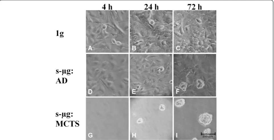
Subconfluent monolayers of human follicular thyroid car-cinoma cells (FTC-133) were cultivated either on the RPM or on the CN and in parallel to the 1gcontrols located in the same incubator, respectively. On both devices spher-oid formation progressed like shown in Figure 1 for
cells harvested from the CN. While under static 1g
conditions (1g controls), the cells remained adherent
(Figure 1 A-C), two cell populations developed within the culture flasks mounted on each of the two machines, respectively (Figure 1 D-I). Of these populations, one con-tinued to grow adherently (AD cells) (Figure 1 D-F), the other one detached from the bottom of the culture flask and assembled to MCTS (Figure 1 H-I). The separation of the two cell populations is delayed, as 4 h after exposure to the devices only adherent growth was observed in each sample (Figure 1 A, D, G). After approximately 24 h early spheroids became visible on each of both the devices in addition to the adherently growing cells (Figure 1 E, H). During the subsequent 48 h, spheroids became more nu-merous and larger, while adherently growing cells were still present (Figure 1 F, I). Spheroid size can be assumed to be around 100μm on the clinostat, as shown in Figure 1, but also reaching up to 1 mm on the RPM as previously pub-lished by Pietschet al.[4].
Differential gene expression in FTC-133 cells after exposure to CN and RPM
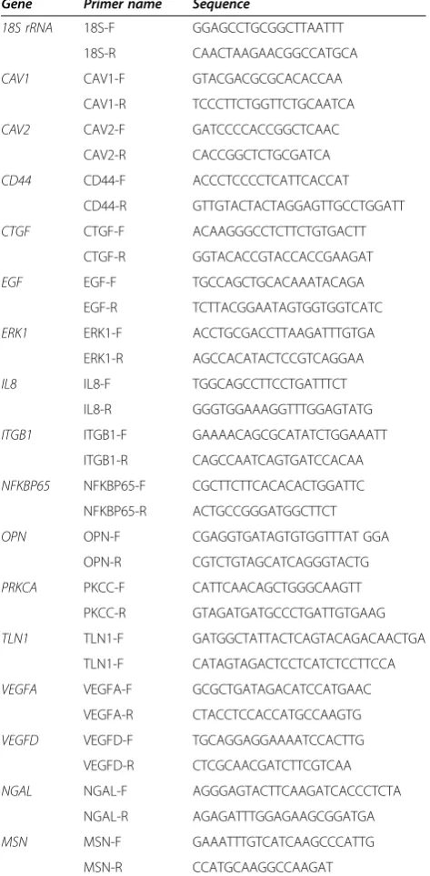
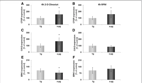
After spheroid formation was observed on the RPM and the CN, we were interested to see whether similar alter-ations of mRNA expression occurred on both machines. Therefore, qPCR on several types of mRNA was per-formed, which had been recognized in former experiments on FTC-133 cells exposed to the RPM for 24 h to be im-portant for MCTS formation [10]. The selected genes be-long to several biological categories including cytoskeletal proteins, and factors of growth, apoptosis, angiogenesis and signal transduction (Table 1).
As shown in Figure 1, after a 4 h-incubation on the RPM and on the CN the whole FTC-133 population grew adher-ently like in 1g-control samples. Nevertheless, some mRNA changes were already found in cells harvested from either machine as compared to control cells. An up-regulation in theCTGFgene expression was observed in cell cultures on
both devices. However, an up-regulation of CAV2 and a
down-regulation of ERK1 gene expression were only
evi-dent after culturing on the CN (Figure 2).
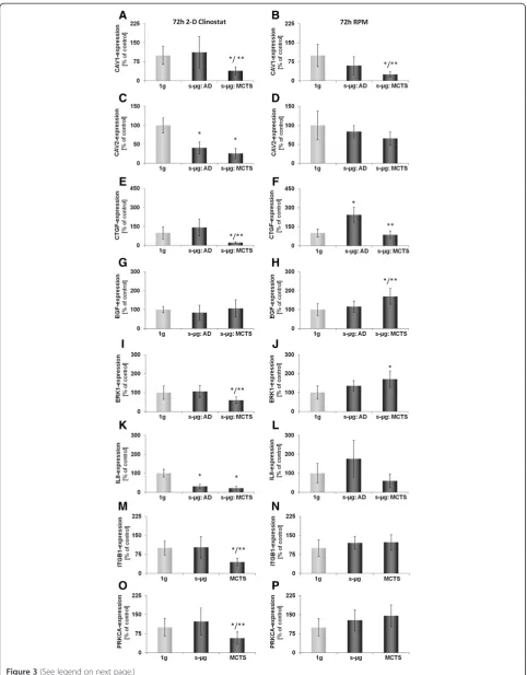
After 72 h of exposure to the CN or the RPM, cells had parted into adherent cells (AD) and cells forming multicel-lular tumor spheroids (MCTS), which floated in the super-natant (Figure 1 I). At this time, two fractions were harvested from the CN and the RPM, respectively. Subquently, it was investigated how the expression of the se-lected genes (Table 1) had been regulated within the 4 cell samples indicated in Figure 3 as compared to the corre-sponding static ground controls (1 g). Interestingly, the
regulation of CTGF and CAV1 split on both machines
equally. In adherent cells, CTGF remained up-regulated
and CAV1unchanged, while in MCTS the expression of
these two genes was decreased (Figure 3 E-F). Other genes have also changed their expression behavior, but differ-ently, when incubated on the two machines. Exposure of FTC-133 cells to the RPM led to an up-regulation ofERK1 andEGFgene-expression in MCTS, but did not affect one of the selected genes in AD cells. In contrast, exposure of the same type of cells to CN triggered down-regulation of ERK1,ITGB1,andPRKCAgene-expression in MCTS only (Figure 3 I, M, O), but decreased expression ofCAV2and IL8in both, AD and MCTS cells (Figure 3 C, K).
The translation from mRNA to protein is a complex process, and results obtained on mRNA level do not ne-cessarily reflect the situation on the protein level. There-fore, investigations of CN- and RPM- related effects on cytokine release were performed additionally using Multi-Analyte Profiling Technology (MAP).
Cytokine release of FTC-133 cells after 72 h exposure to RPM and CN
estimate the secretion activities of the cells (Table 2). As compared to the relevant 1gground controls, significantly higher amounts of GM-CSF, IL-6, IL-8, BDNF, Eotaxin-1, ICAM1, IL-1α, IL-1β, 1Ra, 12p40, 15, 17, IL-23, MMP-3 and SCF were detected in the supernatants of FTC-133 cells cultured on the RPM for 72 h. In contrast, significantly lower quantities of GM-CSF, IL-6, IL-8, MIP-1α, MIP-1βand BDNF were detected in cell
superna-tants of CN samples than in 1g control supernatants
(Table 2) after 72 h cell culturing. After cultivation on both machines, however, a tendency of the cells to in-crease eotaxin-1 and to dein-crease VEGF secretion was ob-served in a comparable manner (Table 2).
Discussion
Spheroid formation occurs on both machines
The main result of this study is that FTC-133 cells form spheroids on the CN as well as on the RPM like they did during the Shenzhou-8 spaceflight in real microgravity [1,2]. During the MCTS formation process, the cells were separated into two populations. One continued adherent cell growth, while the other one detached and formed MCTS. Cultivation on the CN and the RPM resulted in spheroid formation like it was observed in real micrograv-ity. Hence, both machines equally influence the cells in this respect. However, different device-specific alterations of gene-expression and cytokine secretion were found after 4 and 72 h cultivation, indicating device-specific characteris-tics. Common effects could be due to the prevention of
sedimentation. Nevertheless, data from real microgravity are needed to clarify the situation and to control for any pos-sibly confounding variables in the GBF and to choose the appropriate microgravity approach. A review on the suitabil-ity of diverse ground-based facilities was given by Herranz et al.,indicating that both, CN and RPM are generally suit-able for adherent mammalian cells [6]. Nevertheless, it is also clearly stated, that although some similar results in s-μg and realμgwere achieved, extensive studies have to be per-formed for each biological system and thus, individually for each type of the cell culture of interest [6]. For future stud-ies, equal time frames, hardware and procedures should be used in space and ground-based studies in order to exclude an impact of external factors influencing the results.
Spheroids formed scaffold-free under the condition of microgravity are valuable models for tumor research [1,3,11], as they resemble thein vivosituation much better than 2D monolayer cultures or spheroids grown on Earth using liquid overlay or spinner flask techniques [12,13], which consist mainly of concentric layers surrounding central necrotic cells by a thin shell [14]. The importance of studies on tumor cells exposed to microgravity was re-cently reviewed by Becker and Souza [15]. However, MCTS generated under simulated microgravity conditions show viable cells throughout the whole body and lack nec-rotic centers, even if the thyroid cancer cells (ML-1) had formed spheroids with diameters of up to 300μm [3]. The
similarity between in vivo tumors and RPM- or
CN-derived MCTS is explained by the following hypothesis:
On the RPM or the CN, cells can undergo a transition from a 2- to a 3-dimensional growth, with the adherent layer serving as a starting point. Therefore, cell-cell contacts are established by forces of biochemical components expressed on the cell surfaces in a cell type specific manner under low shear forces [3]. Results of former RPM experiments with the two human follicular thyroid carcinoma cell lines FTC-133 and CGTH W-1 showed differences in the size of spher-oids formed which were correlated to their capability to bind to fibronectin [4].
Genes and proteins playing a possible role in spheroid formation
As compared to the corresponding 1g controls, higher
amounts of the selected cytokines were frequently de-tected in supernatants obtained from RPM cultures and lower quantities in those harvested from CN cultures, respectively (Table 2), clearly indicating device-specific differences. Interestingly, VEGF secretion indicated a tendency of down-regulation, while eotaxin-1 suggested up-regulation on the CN as well as on the RPM after 72 h (Table 2). Former investigations of culture superna-tants of samples cultured on the RPM or returned from the Shenzhou-8 spaceflight experiment showed a different picture. No changes of EGF and VEGF secretion were observed after real microgravity exposure, while both cyto-kines were significantly down-regulated after a 10d-RPM-exposure [2]. The different cytokine concentrations could be due to a change of the secretion activity during pro-longed incubation under microgravity when the transition from a 2- to a 3-dimensional growth is completed as it has been shown for human endothelial cells [16,17]. A comparison of cytokines in the supernatant appears only possible during incubation within a few days, as investiga-tion of IL-6 after 1 and 3 days performed in this and a former study [10] suggested. VEGF prevents apoptosis in thyroid carcinomas in an autocrine manner [18]. Its re-duction may contribute to an enhanced apoptotic rate in thyroid cells cultured on the RPM [3]. Eotaxin-1 induces changes in the cytoskeleton and cell morphology and thus could favor the transition from a 2- to a 3-dimensional kind of growth [19].
Although spheroids were not seen in the cultures on both machines after 4 h, a change in gene expression ac-tivity was already expected based on earlier parabolic flight experiments [20-22]. Indeed, three of the selected genes were found to be changed. Most interesting of
them is CTGF’s significantly enhanced expression as
compared to the corresponding 1gcontrols. It remained
up-regulated for another 68 h in AD cells on both
ma-chines, while the MCTS cells showed CTGF mRNA
concentrations even lower than the control cells after
72 h of culturing. The split of CTGF gene expression
between adherent and MCTS cells corresponds nicely to earlier data from 10 d RPM and spaceflight experi-ments, which indicated more mRNA in AD than in MCTS cells [2] and could therefore be a microgravity-dependent process involved in spheroid formation.
CTGFwas found to be over-expressed in papillary
thy-roid carcinoma correlating with metastasis, size and clinical stage [23]. It was also suggested to play an im-portant role in angiogenesis and tumorigenesis of pros-tate cancer [24]. Its reduction in MCTS cells could hint to a diminished aggressiveness of cancer cells incorpo-rated in MCTS (manuscript in preparation).
Table 1 Primers used for quantitative real-time PCR
Gene Primer name Sequence
18S rRNA 18S-F GGAGCCTGCGGCTTAATTT 18S-R CAACTAAGAACGGCCATGCA
CAV1 CAV1-F GTACGACGCGCACACCAA
CAV1-R TCCCTTCTGGTTCTGCAATCA
CAV2 CAV2-F GATCCCCACCGGCTCAAC
CAV2-R CACCGGCTCTGCGATCA
CD44 CD44-F ACCCTCCCCTCATTCACCAT
CD44-R GTTGTACTACTAGGAGTTGCCTGGATT
CTGF CTGF-F ACAAGGGCCTCTTCTGTGACTT
CTGF-R GGTACACCGTACCACCGAAGAT
EGF EGF-F TGCCAGCTGCACAAATACAGA
EGF-R TCTTACGGAATAGTGGTGGTCATC
ERK1 ERK1-F ACCTGCGACCTTAAGATTTGTGA
ERK1-R AGCCACATACTCCGTCAGGAA
IL8 IL8-F TGGCAGCCTTCCTGATTTCT
IL8-R GGGTGGAAAGGTTTGGAGTATG
ITGB1 ITGB1-F GAAAACAGCGCATATCTGGAAATT ITGB1-R CAGCCAATCAGTGATCCACAA
NFKBP65 NFKBP65-F CGCTTCTTCACACACTGGATTC NFKBP65-R ACTGCCGGGATGGCTTCT
OPN OPN-F CGAGGTGATAGTGTGGTTTAT GGA
OPN-R CGTCTGTAGCATCAGGGTACTG
PRKCA PKCC-F CATTCAACAGCTGGGCAAGTT PKCC-R GTAGATGATGCCCTGATTGTGAAG
TLN1 TLN1-F GATGGCTATTACTCAGTACAGACAACTGA TLN1-F CATAGTAGACTCCTCATCTCCTTCCA
VEGFA VEGFA-F GCGCTGATAGACATCCATGAAC VEGFA-R CTACCTCCACCATGCCAAGTG
VEGFD VEGFD-F TGCAGGAGGAAAATCCACTTG VEGFD-R CTCGCAACGATCTTCGTCAA
NGAL NGAL-F AGGGAGTACTTCAAGATCACCCTCTA
NGAL-R AGAGATTTGGAGAAGCGGATGA
MSN MSN-F GAAATTTGTCATCAAGCCCATTG
MSN-R CCATGCAAGGCCAAGAT
Besides CTGF, genes of adherent cells remained un-affected until 72 h on the RPM, while expression ofCAV1, EGFandERK1had been altered in the MCTS. In contrast, after 72 h on the CN,CAV2andIL8expression were
chan-ged in AD cells andCAV1,CAV2,CTGF,ERK1andIL8in
the MCTS. Most interesting of these observations was a
down-regulation ofCAV1and an up-regulation ofERK1
in MCTS cells after three days following an at least one-day-lasting stability of these genes [10,25], which differ from theCAV2gene that is stable only for 4 h (Figure 2) but down-regulated after 24 h and 72 h [10]. Caveolin-1 is an integral membrane protein and plays crucial roles in the regulation of cellular proliferation, differentiation and apoptosis [26]. The down-regulation of caveolin-1 appears to enhance the capacity of the cell to incorpor-ate in a tissue [27]. Furthermore, the expression of this protein seems to be gravisensitive, because an overex-pression in healthy mice staying in space had been
ob-served [28]. Since CAV1 is an obviously gravisensitive
gene and its product influences the incorporation of cells in tissue, we conclude from our observations that
CAV1may play a role in microgravity-dependent MCTS
formation.
Device-dependent cell modification
Unexpectedly, most of the selected genes and cytokines were differently expressed or secreted, respectively, de-pending on which kind of simulation device MCTS were formed. We conclude that during MCTS formation con-siderable alterations of various cellular molecules occur. Some of them might not be directly related to the process of 3D cell aggregation, but by products generated by device-dependent modifications [29]. A comparative study in space will help to discriminate microgravity-induced al-terations from other physical stimuli due to vibration, shearing forces, etc. generated by the simulators. The 2D clinostat rotating constantly at 60 rpm generates residual accelerations below 0.012gwithin the distance of ±3 mm around the center of the flask [30]. Cells located at further distance from the rotation axis are exposed to accelera-tions reaching up to 0.036gat about ±9 mm. Significant differences in gene expression within these acceleration intervals have been shown by Eiermannet al.[30]. Based on these findings, only cells within the central 6 mm (≤0.012g) of a slide were harvested and analyzed in the present study. An exception had to be made for super-natants which were collected in total, as a distinction
between different acceleration levels was not possible. Therefore, supernatants were enriched by cytokines of all cells from the flasks. Whether gravity forces in a range of 0.012g and 0.036ghave a separated influence on the cell behavior has not yet been clarified [30,31].
Either coating of the flasks in the parts outside the area of interest in order to prevent initial cell attachment or a different geometry of the flasks themselves, with an enlarged area along the rotation axis would be con-venient to avoid a mixture in cytokine release.
(See figure on previous page.)
Figure 3Quantitative real-time PCR for the determination of alterations in gene-expression of selected genes after 72 h.CAV1(A, B), CAV2(C, D), CTGF(E, F), EGF(G, H), ERK1(I, J), IL-8(K, L), ITGB1(M, N), and PRKCA(O, P)gene expression was analyzed after 72 h exposure of the cells to 2-D Clinostat(A, C, E, G, I, K, M, O)or random positioning machine (RPM;B,D,F,H,J,L,N,P). After 72 h culturing, the FTC-133 cells grew adherently (AD) or within the MCTS. On both machines CAV1(C, D)was down-regulated in the MCTS cells and CTGF(E, F)was differently expressed in AD and MCTS, respectively. All results are shown as mean ± standard deviation (SD) of n = 10 independent samples, with significance indicated by *P < 0.05 vs. 1g, **P < 0.05 vs. s-μg: AD.
Table 2 Cytokines detected in supernatants of FTC-133 cells after 72 h incubation
2D Clinostat Random positioning machine
Factor LDD (pg/mL) 72 h 1g (pg/mL) 72 h s-μg (pg/mL) LDD (pg/mL) 72 h 1g (pg/mL) 72 h s-μg (pg/mL)
GM-CSF 3.2 1536 ± 166 1172 ± 31* 9.3 46.8 ± 6.9 110.8 ± 12.0*
IFN-γ 0.49 n.d. n.d. 0.58 n.d. n.d.
IL-2 1.0 1.9 ± 0.33 n.d. 2.6 n.d. n.d.
IL-3 1.1 n.d. n.d. 2.0 n.d. n.d.
IL-4 4.4 n.d. n.d. 3.2 n.d. n.d.
IL-5 1.0 n.d. n.d. 0.73 n.d. n.d.
IL-6 0.84 282 ± 37 204 ± 17* 0.85 43.8 ± 8.4 70 ± 15.6*
IL-7 2.1 n.d. 2.1 ± 0.3 5.5 n.d. n.d.
IL-8 0.63 7604 ± 410 5586 ± 267* 0.49 226 ± 31 350 ± 36*
IL-10 1.1 n.d. n.d. 1.5 n.d. n.d.
IL-18 3.7 n.d. n.d. 3.2 n.d. n.d.
MIP-1α 8.2 3086 ± 390 2198 ± 379* 6.0 113 ± 18 137 ± 24
MIP-1β 4.0 261 ± 46 166 ± 28* 4.6 n.d. n.d.
MCP-1 4.6 47 ± 6 40 ± 3 4.9 n.d. n.d.
TNF-α 2.9 8.9 ± 1.2 7.0 ± 1.3 3.1 n.d. n.d.
TNF-β 2.3 2.3 ± 0.4 n.d. 4.4 n.d. n.d.
BDNF 3.0 20 ± 1.7 16 ± 1.7* 6.2 41.2 ± 4.6 76.6 ± 4.7*
Eotaxin-1 3.7 11.8 ± 1.8 12.4 ± 2.3 13 75.4 ± 9.9 99.4 ± 9.8*
ICAM1 270 360 ± 74 350 ± 73 620 1328 ± 274 1840 ± 150*
IL-1α 0.24 n.d. n.d. 0.19 0.85 ± 0.11 1.60 ± 0.29*
IL-1β 0.3 n.d. n.d. 0.25 1.14 ± 0.22 2.02 ± 0.34*
IL-1ra 8.7 n.d. n.d. 14 127 ± 38 226 ± 24*
IL-12p40 16 n.d. n.d. 17 128 ± 21 206 ± 29*
IL-12p70 4.8 7.8 ± 0.6 7.3 ± 0.9 4.5 36.0 ± 4.2 46.2 ± 6.1
IL-15 39.0 n.d. n.d. 41 142 ± 27 208 ± 28*
IL-17 0.51 1.14 ± 0.32 1.0 ± 0.16 0.6 2.5 ± 0.4 4.5 ± 0.6*
IL-23 51 n.d. n.d. 69 590 ± 117 868 ± 78*
MMP-3 5.8 260 ± 22 256 ± 19 7.4 51.2 ± 11.3 90.8 ± 11.1*
SCF 13 24 ± 4 19 ± 2 9.0 59.0 ± 16.1 94.6 ± 11.5*
VEGF 1.4 3062 ± 539 2814 ± 309 4.2 6048 ± 791 5044 ± 677
Concerning clinorotation, no relative fluid motion is as-sumed in a closed completely filled and air bubble-free container exposed to the CN due to its linear and constant acceleration mode [32]. In contrast, chaotic fluid motion is caused on a RPM operating in real random mode due to random changes of speed and direction of the platform movement [33-35]. Although the magnitude of forces of fluid motion, which does not exceed 0,44 dyn/cm2[35], is below that required to trigger cell detachment from sur-faces [36,37], it is assumed to be high enough to induce an enhanced convective fluid mixing, which may support nu-trient supply and removal of metabolic products and thus optimize the culture conditions for a cell.
Residual accelerations for RPM exposure have to be considered for all 3g-vector components that means in x, y and z directions. Exemplary measurements of a RPM running at 60–120 °/s showed accelerations due to gravity of −1 to +1g for each direction [38]. A slower rotation speed produces less residual acceleration but requires a longer time for averaging of the g-vector. The relation of rotation speed, resulting accelerations and the time a sys-tems needs to detect the alterations of the influence of gravity has to be considered. Even though less residual ac-celerations can be achieved through a decreased rotation speed, a longer exposure stimulus will be inevitably con-nected which can possibly lead to a permanent stress stimulus, if sensed by the cells [39].
Shear stress has to be considered as a variable differing
between the devices. As shown by Goodwinet al.BHK-21
cells exposed to 0.51 dyn/cm2consumed 60-70% less glu-cose, but achieved higher cell counts and aggregation than cells exposed to 0.92 dyn/cm2, both on the integrated ro-tating wall vessel (RWV) [40]. Furthermore, experiments
by Hammond et al. on primary cultures of human renal
cortical cells exposed for 6 days to either real microgravity on a space shuttle or cultured on ground-based devices as controls demonstrated 1632 gene changes after the space flight, 5 gene changes after centrifugation and 914 gene changes after cell culturing on the RWV, if a threshold of more than ± three-fold change was set. In addition, as many gene changes in reciprocal as in the same direction were observed, when results from RWV and space cul-tures were compared [41]. The balancing forces, such as shear, used to offset gravity in RWV systems were sug-gested to be responsible for changing the character of the culture [41]. Further work by Kaysen et al. supports this hypothesis, showing a shear stress dependence of selected de novo gene and protein expression during renal epithe-lial cell culture in RWV [42]. Therefore, different shear stress situations on both devices used in our study seem to be a cause of differences in the results obtained. The prob-lem of internal shear forces due to residual accelerations has been recognized not only in ground-based facilities, but was also predicted to be responsible for differences
between 1 g ground controls and 1 g-in-flight controls
using a 1g-reference centrifuge in space, as described by van Loonet al. [43]. An adjustment of the culture flasks to the direction of the forces is suggested in order to ob-tain an optimal outcome and comparability of the data. Further experiments are required to clarify stress and gravity-related effects and to determine threshold levels for response time and optimal rotation speed in order to simulate optimal microgravity conditions on ground.
Also vibrations are often suggested as a critical param-eter in microgravity simulation. As the corresponding 1g controls were cultured next to the devices in the same in-cubator, the vibration effect should be neglected as both samples 1 g and s-μg are exposed to the same external stimulus. The same matter has to be addressed in para-bolic flights especially in combination with periods of hypergravity. Experiments with respect to vibration and hypergravity have been performed on two human follicu-lar thyroid cancer cell lines (ML-1 and CGTH W-1) and endothelial cells (EA.hy 926), all suggesting that micro-gravity effects are stronger than the opposing vibration and hypergravity effects [20-22]. Nevertheless, additional vibration experiments for FTC-133 are of interest as final validation of ground- based and future space data.
A further aspect possibly influencing the results are the different culture containers used due to the geometry of the devices. As the same flasks were used for s-μg-and cor-responsing 1g-samples and results were analyzed relatively to the in parallel obtained 1g-data, an influence should not be expected for the PCR data. Concerning the MAP data, a possible effect cannot be excluded due to different vol-ume and area proportions.
environments might be a manifestation of modified extra-cellular convective flow has been suggested by Paul Todd already in 1992 [44]. For future experiments, equal culture conditions should be used in order to obtain an optimal comparability of the data. Still, the unique conditions of real microgravity with the loss of gravity-dependent vection and negligible hydrodynamic shear have to be con-sidered when comparing real and s-μgresults [15,44,45].
Conclusion
On both, the CN and RPM, spheroid formation of human thyroid cancer cells was observed, whereas, besides a few exceptions, a considerable number of selected genes or cy-tokines were expressed or secreted differently, although equal kinds of cells formed MCTS on the two machines.
The exceptions were CAV1 and CTGF genes as well as
VEGF and eotaxin-1 cytokines. We consider them involved in the process of spheroid formation, because their changes consistently accompanied the MCTS formation in similar manner, when cell sedimentation is prevented by RPM or CN or even in real microgravity in space. The study shows the advantage of searching for gravity-sensitive genes and proteins in comparative approach using different machines for microgravity simulation. Our study clearly shows the necessity to verify results from ground-based simulation approaches to the ones obtained in real microgravity con-ditions to avoid misinterpretations, to learn and to under-stand device-specific characteristics and finally choose the appropriate simulation approach.
Methods
Culturing of FTC-133 cells
The human follicular thyroid carcinoma cell line FTC-133 [46] was cultured in RPMI-1640 medium at 37°C
and 5% CO2. The medium was supplemented with 100
μg/mL streptomycin, 100 U/mL penicillin and 10% FCS
(all Biochrom, Berlin, Germany). One day prior to the CN experiments, cells were seeded in 9 cm2slideflasks (Thermo Scientific, Roskilde, Denmark). A cell count of 5 × 10⁵cells was disseminated for 4 h experiments, 4 × 10⁵for 24 h and 2 × 10⁵for 72 h experiments. For the RPM experiments, cells were grown in T75 cell culture flasks (Sarstedt, Numbrecht, Germany). Cells were seeded at a density of 4 × 10⁶ cells per flask. The cells were randomized to be cultivated as static ground
con-trols (1 g) or under simulated microgravity conditions
(s-μg) on either a RPM or a CN. Ground controls were always placed next to the device in the same incubator. Cells and supernatants were harvested after 4 h or 72 h on ice. The supernatants were aspirated and centrifuged at 4°C. Afterwards, the fluid was transferred to another tube and frozen. The pellet was fixed with RNAlater. After removal of the culture supernatant, cells which remained adherent during incubation, were washed once with PBS
and then fixed with RNAlater. For this, in case of the CN the slides were remove from their flask and were slowly dipped first in PBS (5 s) and then transfered to RNAlater until harvesting. T75 culture flasks from the RPM were slowly filled with 10 ml PBS while standing vertically, and were very carefully brought into horizon-tal position to avoid any disturbance of the cells. PBS was aspirated again before the addition of RNAlater. Afterwards, the cells cultured on the RPM were scraped off the whole bottom surface, while cells cultured on the CN samples were harvested from the inner 6 mm of the slide flask only, because cells of this part experience accelerations of≤0.012g[30].
Random Positioning Machine (RPM) and 2D clinostat (CN) For a comparative methodical approach, cells were either cultivated on the Desktop RPM manufactured by Dutch Space (an EADS Astrium company, Leyden, Netherlands) [8] or the Fast Rotating 2D clinostat (DLR, Cologne, Germany) [30]. The Desktop RPM was operated in real random direction mode (60 °/s) and equipped with four T75 flasks per run, fixed to the ground plate, giving a maximum distance of 7.5 cm from the center of rotation.
The 2D clinostat constantly rotated with 60 rpm and was loaded with four slideflasks on each of the 6 parallel rotating axes, summing up to a total of 24 flasks per run. Both devices were placed inside an individual incubator with temperature of 37°C and 5% CO2. Prior to the
experi-ment, all flasks were completely filled with media avoiding air bubbles carefully, in order to reassure a minimization of turbulences. For harvesting of CN samples, cells of 16 flasks were pooled. Ten samples (n = 10) were collected
for each condition: CN, CN corresponding 1g control,
RPM and RPM corresponding 1gcontrol.
Phase contrast microscopy
Phase contrast microscopy was performed for visual obser-vation of the morphology of the cells, using the Axiovert 25 Microscope (Carl Zeiss Microscopy, LLC, United States).
Cytokine measurements by Multi-Analyte Profiling technology
RNA isolation and quantitative real-time PCR
RNA isolation and quantitative real-time PCR were per-formed according to routine protocols [25,50,51]. For CN samples, only the inner 6 mm were harvested with a scratching device. RPM samples were harvested in total. RNA was isolated using RNeasy Mini Kit (Qiagen, Hilden, Germany) following manufacturer instructions. DNase (Qiagen, Hilden, Germany) was added in the process of RNA isolation, in order to diminish residual DNA contami-nations. The RNA was quantified via Photometer Ultros-pec2010 (Amersham Biosciences, Freiburg, Germany). Reverse transcription was performed using the first strand cDNA synthesis kit (ThermoFisher Scientific, Waltham, US), following manufacturer’s instructions. Quantitative real-time PCR was utilized to determine the expression levels of target genes, shown in Table 1, using the 7500 Real-Time PCR System (Applied Biosystems, Darmstadt, Germany). cDNA-selective Primers were designed to span exon-exon boundaries and to have a Tmof∼60°C using
Pri-mer Express software (Applied Biosystems, Darmstadt, Germany), and were synthesized by TIB Molbiol (Berlin, Germany). All samples were measured in triplicate and
nor-malized to the housekeeper 18S rRNA. Comparative CT
(ΔΔCT) methods were used for relative quantification of
transcription levels, with 1gset as 100%.
Statistical Evaluation
Statistical Evaluation was performed using SPSS 15.0 (SPSS,
Inc., Chicago, IL, USA). The Mann–Whitney-U-Test was
used to compare 1gand s-μgconditions, as well as s-μg ad-herent cells and s-μgMCTS cells. All data is presented as mean ± standard deviation (SD) with a significance level of p < 0.05. * indicating the comparison of 1g vs. s-μg and ** representing the comparison of s-μgAD vs. s-μgMCTS.
Abbreviations
BDNF:Brain-derived neutrophic factor; CAV: Caveolin; CN: Clinostat; CTGF: Connective tissue growth factor; EGF: Epidermal growth factor; ERK: Extracellular signal-regulated kinase; FCS: Fetal calf serum; FTC: Follicular thyroid cancer; GBF: Ground-based facilities; GM-CSF: Granulocyte-macrophage colony-stimulating factor; IFN: Interferon; IL: Interleukin; ITBG1: Integrin beta 1; LDD: Least detectable dose; MAP: Multi-Analyte Profiling; MCP: Monocyte chemotactic protein; MCTS: Multicellular tumor spheroids; MEK: Mitogen-activated protein kinases; MIP: Macrophage inflammatory protein; MMP: Matrix metalloproteinase; MRNA: Messenger ribonucleic acid; PCR: Polymerase chain reaction; PDGFR: Platelet-derived growth factor receptor; PRKCA: Protein kinase C alpha; RBM: Rules-based medicine; RPM: Random positioning machine; RWV: Rotating Wall Vessel; SCF: Stem cell factor; SD: Standard deviation.
Competing interests
The authors declare that they have no competing interests.
Authors’contributions
JB, DG and RH designed the study. EW and XM conducted the experiments. EW and JB drafted the manuscript. JP, MW and MI supervised and supported the ground-based experiments in Magdeburg. DG supervised the ground-based experiments in Aarhus and helped with the manuscript. RH and MB supervised and supported the ground-based experiments in Cologne. MG was responsible for the design of the 2D clinostat. All authors read and approved the final manuscript.
Acknowledgements
The study was supported by the European Space Agency (ESA; CORA-GBF-PROJECT-2011-005 with contract 4000104144; D.G.) and the German Space Administration (DLR; BMWi grant 50WB1124; D.G.). Elisabeth Warnke is a doctoral candidate of the Helmholtz Space Life Sciences Research School, German Aerospace Center Cologne, Germany and was further sponsored within the“Young Fellows”project of the Deutsche Gesellschaft für Luft- und Raumfahrtmedizin e.V (DGLRM).
Author details
1Clinic for Plastic, Aesthetic and Hand Surgery, Otto-von-Guericke-University
Magdeburg, Magdeburg, Germany.2Max-Planck Institute of Biochemistry, Martinsried, Germany.3DLR, German Aerospace Center, Institute of
Aerospace Medicine, Cologne, Germany.4Institute for Molecular Physiology and Biotechnology of Plants (IMBIO), University of Bonn, Gravitational Biology Group, Kirschallee 1, 53115, Bonn, Germany.5Institute of Biomedicine, Pharmacology, Aarhus University, Wilhelm Meyers Allé 4, DK-8000 Aarhus C, Denmark.
Received: 24 January 2014 Accepted: 28 April 2014 Published: 10 May 2014
References
1. Pietsch J, Ma X, Wehland M, Aleshcheva G, Schwarzwälder A, Segerer J, Birlem M, Horn A, Bauer J, Infanger M, Grimm D:Spheroid formation of human thyroid cancer cells in an automated culturing system during the Shenzhou-8 Space mission.Biomaterials2013,34:7694–7705.
2. Ma X, Pietsch J, Wehland M, Schulz H, Saar K, Hübner N, Bauer J, Braun M, Schwarzwälder A, Segerer J, Birlem M, Horn A, Hemmersbach R, Waßer K, Grosse J, Infanger M, Grimm D:Differential gene expression profile and altered cytokine secretion of thyroid cancer cells in space.FASEB J2014,
28:813–835.
3. Grimm D, Bauer J, Kossmehl P, Shakibaei M, Schönberger J, Pickenhahn H, Schulze-Tanzil G, Vetter R, Eilles C, Paul M, Cogoli A:Simulated microgravity alters differentiation and increases apoptosis in human follicular thyroid carcinoma cells.FASEB J2002,16:604–606.
4. Pietsch J, Sickmann A, Weber G, Bauer J, Egli M, Wildgruber R, Infanger M, Grimm D:A proteomic approach to analysing spheroid formation of two human thyroid cell lines cultured on a random positioning machine. Proteomics2011,11:2095–2104.
5. Pietsch J, Bauer J, Egli M, Infanger M, Wise P, Ulbrich C, Grimm D:The effects of weightlessness on the human organism and mammalian cells. Curr Mol Med2011,11:350–364.
6. Herranz R, Anken R, Boonstra J, Braun M, Christianen PC, de Geest M, Hauslage J, Hilbig R, Hill RJ, Lebert M, Medina FJ, Vagt N, Ullrich O, van Loon JJ, Hemmersbach R:Ground-based facilities for simulation of microgravity: organism-specific recommend-dations for their use, and recommended terminology.Astrobiology2013,13:1–17.
7. Briegleb W:Some qualitative and quantitative aspects of the fast-rotating clinostat as a research tool.ASGSB Bull1992,5:23–30.
8. van Loon JJWA:Some history and use of the random positioning machine, RPM, in gravity related research.Adv Space Res2007,39:1161–1165. 9. van Loon JJWA, Veldhuijzen JP, Kiss J, Wood C, Vd Ende H, Guntemann A,
Jones D, de Jong H, Wubbels R:Microgravity Research Starts on the Ground! Apparatus for Longterm Ground-based Hypo- and Hypergravity Studies”Utilisation of the International Space station.InProceedings of 2nd European Symposium, ESTEC, Noordwijk, The Netherland, 16-18 November 1998.Edited by Wilson A. Paris: European Space Agency (ESA), ESA-SP Volume 433; 1999:415–419. ISBN 9290927321.
10. Grosse J, Wehland M, Pietsch J, Schulz H, Saar K, Hübner N, Eilles C, Bauer J, Abou-El-Ardat K, Baatout S, Ma X, Infanger M, Hemmersbach R, Grimm D:
Gravity-sensitive signaling drives 3-dimensional formation of multicellu-lar thyroid cancer spheroids.FASEB J2012,26:5124–5140.
11. Santini MT, Rainaldi G:Three-dimensional spheroid model in tumor biology.Pathobiology1999,67:148–157.
12. Sutherland RM, McCredie JA, Inch WR:Growth of multicell spheroids in tissue culture as a model of nodular carcinomas.J Natl Cancer Inst
1971,46:113–120.
14. Mueller-Klieser W:Tumor Biology and experimental therapeutics.Crit Rev Oncol Hematol2000,36:123–139.
15. Becker JL, Souza GR:Using space-based investigations to inform cancer research on Earth.Nat Rev Cancer2013,13:315–327.
16. Grimm D, Infanger M, Westphal K, Ulbrich C, Pietsch J, Kossmehl P, Vadrucci S, Baatout S, Flick B, Paul M, Bauer J:A delayed type of three-dimensional growth of human endothelial cells under simulated weightlessness. Tissue Eng Part A2009,15:2267–2275.
17. Infanger M, Kossmehl P, Shakibaei M, Baatout S, Witzing A, Grosse J, Bauer J, Cogoli A, Faramarzi S, Derradji H, Neefs M, Paul M, Grimm D:Induction of three-dimensional assembly and increase in apoptosis of human endo-thelial cells by simulated microgravity: Impact of vascular endoendo-thelial growth factor.Apoptosis2006,11:749–764.
18. Vieira JM, Santos SCR, Espadinha C, Correia I, Vag T, Casalou C, Cavaco BM, Catarino AL, Dias S, Leite V:Expression of vascular endothelial growth factor (VEGF) and its receptors in thyroid carcinomas of follicular origin: a potential autocrine loop.Eur J Endocrinol2005,153:701–709.
19. Kang BN, Ha SG, Ge XN, Hosseinkhani MR, Bahaie NS, Greenberg Y, Blumenthal MN, Puri KD, Rao SP, Sriramarao P:The p110 delta subunit of PI3K regulates bone marrow-derived eosinophil trafficking and airway eosinophilia in allergen-challenged mice.Am J Physiol Lung Cell Mol Physiol2012,302:L1179–L1191.
20. Ulbrich C, Pietsch J, Grosse J, Wehland M, Schulz H, Saar K, Hübner N, Hauslage J, Hemmersbach R, Braun M, van Loon J, Vagt N, Egli M, Richter P, Einspanier R, Sharbati S, Baltz T, Infanger M, Ma X, Grimm D:Differential gene regulation under altered gravity conditions in follicular thyroid cancer cells: relationship between the extracellular matrix and the cytoskeleton.Cell Physiol Biochem2011,28:185–198.
21. Grosse J, Wehland M, Pietsch J, Ma X, Ulbrich C, Schulz H, Saar K, Hübner N, Hauslage J, Hemmersbach R, Braun M, van Loon J, Vagt N, Infanger M, Eilles C, Egli M, Richter P, Baltz T, Einspanier R, Sharbati S, Grimm D:
Short-term weightlessness produced by parabolic flight maneuvers altered gene expression patterns in human endothelial cells.FASEB J
2012,26:639–655.
22. Wehland M, Ma X, Braun M, Hauslage J, Hemmersbach R, Bauer J, Grosse J, Infanger M, Grimm D:The impact of altered gravity and vibration on endothelial cells during a parabolic flight.Cell Physiol Biochem2013,
31:432–451.
23. Cui L, Zhang Q, Mao Z, Chen J, Wang X, Qu J, Zhang J, Jin D:CTGF is overexpressed in papillary thyroid carcinoma and promotes the growth of papillary thyroid cancer cells.Tumour Biol2011,32:721–728. 24. Yang F, Tuxhorn JA, Ressler SJ, McAlhany SJ, Dang TD, Rowley DR:
Stromal expression of connective tissue growth factor promotes angiogenesis and prostate cancer tumorigenesis.Cancer Res2005,
65:8887–8895.
25. Infanger M, Faramarzi S, Grosse J, Kurth E, Ulbrich C, Bauer J, Wehland M, Kreutz R, Kossmehl P, Paul M, Grimm D:Expression of vascular endothelial growth factor and receptor tyrosine kinases in cardiac ischemia/ reperfusion injury.Cardiovasc Pathol2007,16:291–299.
26. Mo S, Yang S, Cui Z:New glimpses of caveolin-1 functions in embryonic development and human diseases.Front2011,6:367–376.
27. Lu Z, Ghosh S, Wang Z, Hunter T:Downregulation of caveolin-1 func-tion by EGF leads to the loss of E-cadherin, increased transcripfunc-tional activity of beta-catenin, and enhanced tumor cell invasion. Cancer Cell2003,4:499–515.
28. Masini MA, Albi E, Barmo C, Bonfiglio T, Bruni L, Canesi L, Cataldi S, Curcio F, D’Amora M, Ferri I, Goto K, Kawano F, Lazzarini R, Loreti E, Nakai N, Ohira T, Ohira Y, Palmero S, Prato P, Ricci F, Scarabelli L, Shibaguchi T, Spelat R, Strollo F, Ambesi-Impiombato FS:The impact of long-term exposure to space environment on adult mammalian organisms: a study on mouse thyroid and testis.PLoS One2012,7:e35418.
29. Pietsch J, Riwaldt S, Bauer J, Sickmann A, Weber G, Grosse J, Infanger M, Eilles C, Grimm D:Interaction of proteins identified in human thyroid cells.Int J Mol Sci2013,14:1164–1178.
30. Eiermann P, Kopp S, Hauslage J, Hemmersbach R, Gerzer R, Ivanova K:
Adaptation of a 2D clinostat for simulated microgravity experiments with adherent cells.Micrograv Sci Technol2013,25:153–159. 31. de Groot RP, Rijken PJ, den Hertog J, Boonstra J, Verkleij AJ, de Laat SW,
Kruijer W:Microgravity decreases c-fos induction and serum response element activity.J Cell Sci1990,97:33–38.
32. Klaus DM:Clinostats and Bioreactors.Gravit Space Biol Bul2001,14:55–64. 33. Leguy CAD, Delfos R, Pourquie MJBM, Poelma C, Krooneman J, Westerweel J, van Loon JJWA:Fluid motion for microgravity simulations in a random positioning machine.Gravitat Space Biol2011,25:36–39.
34. Hemmersbach R, von der Wiesche M, Seibt D:Ground-based experimental platforms in gravitational biology and human physiology.Signal Transduction Special Issue: Signaling in gravity perception: From microorganisms to mammals2006,6:381–387.
35. Pardo SJ, Patel MJ, Sykes MC, Platt MO, Boyd NL, Sorescu GP, Xu M, van Loon JJ, Wang MD, Jo H:Simulated microgravity using the Random Positioning Machine inhibits differentiation and alters gene expression profiles of 2T3 preosteoblasts.Am J Physiol Cell Physiol2005,288:C1211–C1221.
36. Zhang X, Jones P, Haswell SJ:Attachment and detachment of living cells on modified microchannel surfaces in a microfluidic-based lab-on-a-chip system.Chem Eng J2008,135:82–88.
37. Haier J, Nicolson GL:Role of the cytoskeleton in adhesion stabilization of human colorectal carcinoma cells to extracellular matrix components under dynamic conditions of laminar flow.Clin Exp Metastasis1999,
17:713–721.
38. Borst AG, van Loon JJWA:Technology and Developments for the Random Positioning Machine, RPM.Microgravity Sci Technol2009,21:287–292. 39. Horn A, Ullrich O, Huber K, Hemmersbach R:PMT (Photomultiplier)
Clinostat.Microgravity Sci Technol2011,23:67–71.
40. Goodwin TJ, Prewett TL, Wolf DA, Spaulding GF:Reduced Shear Stress: A Major Component in the Ability of Mammalian Tissues to Form Three-Dimensional Assemblies in Simulated Microgravi ty.J Cell Biochem1993,
51:301–311.
41. Hammond TG, Benes E, O’Reilly KC, Wolf DA, Linnehan RM, Taher A, Kaysen JH, Allen PL, Goodwin TJ:Mechanical culture conditions effect gene expression: gravity-induced changes on the space shuttle. Physiol Genomics2000,3:163–173.
42. Kaysen JH, Campbell WC, Majewski RR, Goda FO, Navar GL, Lewis FC, Goodwin TJ, Hammond TG:Select de novo gene and protein expression during renal epithelial cell culture in rotating wall vessels is shear stress dependent.J Membr Biol1999,168:77–89.
43. van Loon JJ, Folgering EH, Bouten CV, Veldhuijzen JP, Smit TH:Inertial shear forces and the use of centrifuges in gravity research. What is the proper control?J Biomech Eng2003,125:342–346.
44. Todd P:Physical effects at the cellular level under altered gravity conditions.Adv Space Res1992,12:43–49.
45. Todd P:Gravity-dependent phenomena at the scale of the single cell. ASGSB Bull1989,2:95–113.
46. Goretzki PE, Frilling A, Simon D, Roeher HD:Growth regulation of normal thyroids and thyroid tumors in man.Recent Results Cancer Res1990,
118:48–63.
47. Grimm D, Bauer J, Ulbrich C, Westphal K, Wehland M, Infanger M, Aleshcheva G, Pietsch J, Ghardi M, Beck M, El-Saghire H, de Saint-Georges L, Baatout S:Different responsiveness of endothelial cells to vascular endo-thelial growth factor and basic fibroblast growth factor added to culture media under gravity and simulated microgravity.Tissue Eng Part A2010,
16:1559–1573.
48. Grosse J, Warnke E, Pohl F, Magnusson NE, Wehland M, Infanger M, Eilles C, Grimm D:Impact of sunitinib on human thyroid cancer cells.Cell Physiol Biochem2013,32:154–170.
49. Infanger M, Ulbrich C, Baatout S, Wehland M, Kreutz R, Bauer J, Grosse J, Vadrucci S, Cogoli A, Derradji H, Neefs M, Küsters S, Spain M, Grimm D:
Modeled gravitational unloading induced downregulation of endothelin-1 in human endothelial cells.J Cell Biochem2007,101:1439–1455. 50. Kossmehl P, Kurth E, Faramarzi S, Habighorst B, Shakibaei M, Wehland M,
Kreutz R, Infanger M, Danser AH, Grosse J, Paul M, Grimm D:Mechanisms of apoptosis after ischemia and reperfusion: role of the renin-angiotensin system.Apoptosis2006,11:347–358.
51. Rothermund L, Kreutz R, Kossmehl P, Fredersdorf S, Shakibaei M, Schulze-Tanzil G, Paul M, Grimm D:Early onset of chondroitin sulfate and osteopon-tin expression in angiotensin II-dependent left ventricular hypertrophy. Am J Hypertens2002,15:644–652.
doi:10.1186/1478-811X-12-32