HTTPS://WWW.HEIGHPUBS.ORG ISSN
2575-0143
Research Article
Are S-Klotho’s Maximal concentrations
dependent on Exercise Intensity and
Time in young adult males?
Moran Sciamama Saghiv
1*, David Ben-Sira
2, Ehud Goldhammer
3and Michael Sagiv
21Department of Kinesiology, College of Health & Human Sciences, NC A&T State University, USA 2Life Sciences Department, Wingate College, Wingate, Israel
3Heart Institute Bnai-Zion Haifa Medical Center, Technion, Haifa, Israel
*Address for Correspondence: Moran Sciamama-Saghiv, PhD, Associate Professor of Clinical Exercise Physiology, Chair, Department of Human Performance and Leisure Studies, College of Health and Human Sciences, NC A&T State University, Corbett HPER Center, Suite 215, Room 216, 1601 East Market Street/John Mitchell Drive, Greensboro, North Carolina 27411, USA, Tel: 336.285.3560; Email: [email protected]
Submitted: 20 February 2019 Approved: 07 March 2019 Published: 08 March 2019
Copyright:© 2019 Saghiv MS, et al. This is an open access article distributed under the Creative Commons Attribution License, which permits unrestricted use, distribution, and reproduction in any medium, provided the original work is properly cited
Keywords: Anti-aging; Aerobic training; Untrained individuals; Maximal oxygen uptake; ELISA kit; ROS; Nitric acid
How to cite this article: Saghiv MS, Ben-Sira D, Goldhammer E, Sagiv M. Are S-Klotho’s Maximal concentrations dependent on Exercise Intensity and Time in young adult males? J Cardiol Cardiovasc Med. 2019; 4: 006-011. https://doi.org/10.29328/journal.jccm.1001033
Introduction
The α-Klotho protein circulates in the blood as Soluble-Klotho (s-Klotho), which in humans, decreases in the serum with aging [1]. S-Klotho in humans is a pleiotropic protein with a considerable in luence on longevity [2]. Exercise is a very active approach averting leading reasons of disease and as relates to aging. Exercise and Klotho gene expression reduce the risk of cardiovascular events in patients with prior coronary artery disease (CAD), thus, aerobic exercise may decrease the risk of mortality [3,4]. However, an association between low levels of s-Klotho and the occurrence and severity of cardiovascular disease have been reported, as well as a reduction of cardiovascular risk when levels were high [5]. Untrained, physically inactive individuals present low blood s-Klotho levels [6]. Recent studies [6,7] suggested that aerobic exercise training, is associated with increase blood s-Klotho and maximal oxygen uptake (VO2) values. Therefore, the purpose of the present study was to assess the in luence of time and intensity (below or above the anaerobic threshold) on s-Klotho serum levels in untrained individuals.
Methods
Subjects
Sixty (60) young (27.05±1.1 years), healthy, untrained, and male subjects, were
Abstract
The purpose of the present study was to defi ne the period of time in which aerobic training does not
increase further serum S-Klotho levels in untrained young adult males, and to examine the relation between
plasma S-Klotho concentration and maximal oxygen uptake (VO2max).
Methods: Sixty (60) untrained subjects (27.05±1.1 years) were divided into 2 groups, both exercised six
months 4×wk-1 for the duration of 45 min×session. One group (LTI) exercised below the anaerobic threshold
at 40-50% of VO2max, while the second group (HTI) worked above the anaerobic threshold at 65-70% of VO2max.
Testing sessions were performed at 0, 2, 4, and 6 months. Blood samples were drawn after overnight fasting; S-Klotho was analyzed using an ELISA kit.
Results: Following 2 and 4 months, signifi cant (p≤0.05) increases were noted in the HTI group, at the fourth testing session, S-Klotho leveled off. In the LTI group, S-Klotho remained almost unchanged. Findings of the present study, support emerging evidence suggesting that a relation between plasma S-Klotho concentration and VO2max exists.
Conclusions: Data suggest that increases in S-Klotho is tidally associated with VO2max levels. In addition, the S-Klotho increase levels-off following 4 months of aerobic training. Exercising below the anaerobic threshold
divided randomly into two even groups. All subjects were evaluated as of “poor” itness according to aerobic capacity and age.
Pre-testing procedures
Approval for the study was achieved in accordance with the Helsinki declaration, approved by the Clinical Science Center Committee on Human Subjects. A written informed consent was obtained from each subject, a health history questionnaire was
illed, and risk strati ication followed.
Subjects judged free of coronary artery disease by clinical history, and the absence of major risk factors were included in the study.
Procedures
Subjects were seated for ive minutes before having their resting values obtained, had their resting values obtained, then proceeded to undergo a maximal exercise stress test. Height, weight, heart rate, blood pressure, and s-Klotho values were obtained at rest. Subjects with no counter indications for exercise due to the stress test continued as subjects in the study. None of the subjects had exercise counter indications.
Both groups exercised six months, 4×wk-1 for the duration of 45 min×session-1. The irst group exercised below the anaerobic threshold (Low Training Intensity; LTI) at 40-50% VO2max, while the second group (High training Intensity; HTI) worked above the anaerobic threshold at 65-70% of VO2max. Testing sessions were performed at 0, 2, 4 and 6 months.
Adipose fat assessment included measurement of total body weight (± 0.05 kg), skin fold thicknesses at 8 sites (± 1 mm) using the Lange Caliper (chest, axilla, triceps, sub-scapula, abdomen, suprailium, front thigh and circumferences at the shoulder). Anthropometric procedures followed the recommendations of Behnke and Wilmore [8].
Following warm-up, subjects underwent a graded maximal treadmill test utilizing the standard Bruce Protocol [9]. Maximal tests were terminated by the following criteria: a) leveling off or no further increase in VO2 with increasing work rate, b) attainment of the age predicted maximum heart rate, c) respiratory exchange ratio > 1.1, and d) when the subject could not keep up with the load, according to the guidelines of the American College of Sports Medicine [10]. VO2 was determined breath by breath utilizing the Medical Graphics (St. Paul, MN) metabolic cart. The metabolic cart was calibrated before each test with known primary standard quality gases. Heart rate and electrocardiogram were monitored continuously, using a Burdick Eclipse 400 3-channel, 12-lead ECG recorder system, and oscilloscope. Five-second recordings were obtained at rest and at peak exercise. Blood pressure was taken using a standard sphygmomanometer cuff and mercury manometer mounted at eye level, at rest and at peak exercise.
Blood sampling and procedures
Peripheral venous blood samples (2.5 mL) were collected by sterile antecubital venipuncture techniques into ethylenediam-inotetraacetate containing tubes. Time of day for blood sampling was kept consistent to control for problems associated with diurnal variation. Blood collection was obtained from each subject once for each visit. Analysis
according to the manufacturer instructions. The intra- and inter-assay coef icients of variation ranged from 2.7 to 9.8%.
Statistical methods
Data are reported as mean ± SD values. Physiological responses at rest and maximal exercise between the two groups were statistically assessed by a one-way ANOVA with repeated measure on the exercise-rest main effect. Post hoc analysis was performed by using the Tukey 2 multiple comparison tests. Comparisons between the groups for s-Klotho levels were based on t-tests for unpaired samples. The level of signi icance was set at α≤0.05.
Results
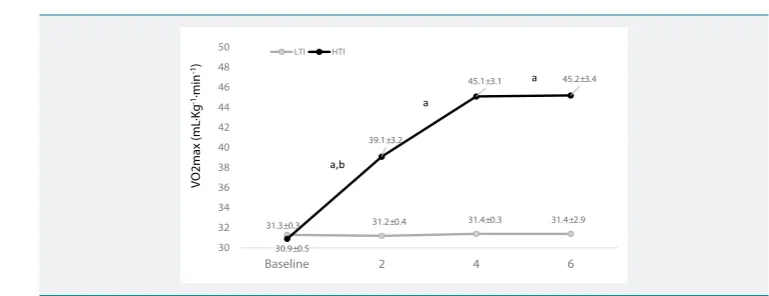
All subjects completed the exercise challenge without dif iculties or abnormal symptoms. Mean descriptive data for the two experimental groups are presented in tables 1,2. Figure 1 discloses that s-Klotho i n the HTI group, increased signi icantly (p≤0.05) from baseline to 2 months, and from 2 months to 4 months, yet insigni icantly increased thereafter. The LTI group as a control did not signi icantly differ in their values from pretesting values to the end of the study; namely the 6th month. Figure 2, reveals that VO2 in the HTI group, increased signi icantly (p≤0.05) and gradually following the 2nd and 4th training months without signi icant differences from the 4th month to the 6th. The LTI group as a control did not signi icantly (p>0.05) differ in their VO2 values from pretesting to the end of the study; namely the 6th month.
Table 1:Subjects’ Physical Characteristics at Rest. (mean ± S.D).
Variables LTI HTI
N of subjects 30 30
Age (years) 26.9±1.1 27.2±1.1
Weight (kg) 71.3±1.7 70.1±1.8
Height (cm) 180.4±2.0 179.8±2.1
Fat (%) 12.1± 1.6 12.4± 1.7
VO2 (mL∙kg-1∙min-1) 3.4±0.3 3.4±0.3
Table 2:Physiological Responses at Rest and Pre-Post Training at Maximal Exercise in Both Groups (mean S.D).
Variables HTI HTI LTI LTI
Rest Exercise Rest Exercise
Lactic acid
(mmol∙L-1) 1.3±0.3 12.8±1.2 1.4±0.3 12.1± 1.1
Heart Rate
(beats∙min-1) 67.1±9.3 198±7.2 79.6±8.4 a 194.4±8.2
Systolic BP
(mmHg) 109.2±6.8 180.4±7.6 110±8.0 182±6.4
Diastolic BP
(mmHg) 70.6±2.7 68.0±2.2 72.2±3.3 71.0±2.4
HTI = Exercising group; LTI = Inactive group; a = signifi cant (p>0.05) differences between both groups.
432±90 433±79 436±90 437±85
430±87
546±91
590±88 593±87
400 450 500 550 600
Baseline 2 4 6
LTI HTI
S-Klotho (pg
·mL
-1)
a,b
a a
Figure 1: S-Klotho levels in HTI and LTI (mean±SD); a. = Signifi cant differences between HTI and LTI (p>0.05); b =
Discussion
This study demonstrated that aerobic exercise training induced a signi icant increase in plasma s-Klotho concentration following six months of aerobic training. Additionally, it revealed that following four months of aerobic training S-Klotho values leveled off. In the LTI group no signi icant changes in s-Klotho and VO2max were found. S-K lotho was increased in the HTI group, suggesting that circulating s-Klotho levels were increased in response to long-lasting aerobic exercise training, and that the response depends on the subjects’ itness level, since s-Klotho increased in a similar pattern as VO2max. Maximal oxygen uptake ( Vo2m ax), is largely used as the best single physiological variable to evaluate the cardiopulmonary function ability. Furthermore, the noninvasively breath-by-breath method to determine the value of exercise training effectiveness in health and disease by measuring Vo2max is powerful tool for investigational and clinical evaluations. However, the mechanism by which Vo2 level in luence blood s-Klotho values is yet unclear.
Previously it has been reported that oxidizing free radical species (ORS) are generated during aerobic bouts [13]. Skeletal muscles generate Super Oxide and Nitric Oxide which is increased by muscle contraction activity [14]. ROS is essential for skeletal muscle force generation, however, ROS in high values may reduce muscle contraction properties and thus, result in early exhaustion [15,16]. The increase in s-Klotho following aerobic exercise training may be a response to ROS that increase in muscle cells as a result of aerobic training.
Although the association between s-Klotho and aerobic exercise training is not clear, s-Klotho reduces programmed cell death (apoptosis) through Nitric Oxide production and thus, supp resses ROS [17]. Previously,a parallel increase of circulating s-Klotho was also observed in response to an acute exercise in young and old mice, suggesting that this may be a good model for mechanistically probing the role of aerobic exercise on Klotho expression [18]. Endurance exercise such as running, cycling and swimming appear to bene it and minimize the physiological alterations that occur during aging and may contribute to improvements in health and well-being [19]. Previously it has been suggested that aerobic training above the anaerobic threshold intensities increase VO2max [20]. The unchanged VO2max in the LTI group and s-Klotho, is probably due to the low training intensity; namely below the anaerobic threshold and below of heart rate reserve [21]. Prolonged or high-intensity exercise result in oxidative damage to macromolecules in both blood and skeletal muscles. However, low intensity aerobic training does not produce ROS thus, S-Klotho levels did not increase in the LTI group, since s-Klotho protein suppresses oxidative stress [17,22].
Study's limitations
There are limitations in the present study that should be well-thought-out when
31.3±0.3 31.2±0.4 31.4±0.3 31.4±2.9
30.9±0.5
39.1±3.2
45.1±3.1 45.2±3.4
30 32 34 36 38 40 42 44 46 48 50
Baseline 2 4 6
LTI HTI
VO2max (mL
·Kg
-1·min -1)
a
a
a,b
Figure 2: VO2 levels in HTI and LTI (mean±SD); a. = Signifi cant differences between HTI and LTI (p>0.05); b =
understanding the results. a) Only s-Klotho in the plasma was measure and, b) mechanisms causal of the effects of aerobic exercise on Klotho were not investigated in this study.
In conclusion, data suggest that increases in s-Klotho is close associated with VO2max levels. In addition, the s-Klotho increase levels-off following 4 months of aerobic training. Exercising below the anaerobic threshold does not increase VO2max and thus, does not increase s-Klotho.
References
1. Maekawa Y, Ohishi M, Ikushima M, Yamamoto K, Yasuda O, et al. Klotho Protein Diminishes Endothelial Apoptosis and Senescence via a Mitogen-Activated Kinase Pathway. Geriatr Gerontol Int. 2011; 11: 510-516. Ref.: https://goo.gl/T2kFua
2. Kim JH, Hwang KH, Park KS, Kong ID, Cha SK. Biological Role of Anti-Aging Protein Klotho. J. Lifestyle Med. 2015; 5: 1-6. Ref.:https://goo.gl/3ywkYX
3. Navarro-González JF, Donate-Correa J, Muros de Fuentes M, Pérez-Hernández H, Martínez-Sanz R, et al. Reduced Klotho is Associated with the Presence and Severity of Coronary Artery Disease. 2014; Heart. 2014; 100: 34-40. Ref.:https://goo.gl/8mRBnH
4. Kingsley D. Aging and Health Care Costs: Narrative versus Reality. Poverty & Public Policy. 2015; 7:3-21. Ref.:https://goo.gl/uZHt4t
5. Martín-Núñez E, Donate-Correa J, Muros-de-Fuentes M, Mora-Fernández C, Navarro-González JF. Implications of Klotho in Vascular Health and Disease. World J Cardiol. 2014; 6: 1262-1269. Ref.:
https://goo.gl/8ruuoV
6. Saghiv MS, Sira DB, Goldhammer E, Sagiv M. The Effects of Aerobic and Anaerobic Exercises on Circulating Soluble-Klotho and IGF-I in Young and Elderly Adults and in CAD Patients. J Circ Biomark. 2017; 6:1849454417733388. Ref.:https://goo.gl/VKkH6z
7. Saghiv M. Effects of Aerobic Exercise Training on S-Klotho in Young and Elderly. JJ Physiology. 2015; 1: 001. Ref.:https://goo.gl/8W69JV
8. Behenke AR, Wilmore J. Evaluation and regulation of Body Build and Composition. Englewood Cliffs, N.J. Prentile Hall, Inc. 1974;
9. American College of Sports Medicine. ACSM's Guidelines for Exercise Testing and Prescription, 10th edition, Philadelphia, PA: Lippincott Williams & Wilkins; 2014; 145-147 and 165-199. Ref.:
https://goo.gl/ZVQKaC
10. Yamazaki Y, Imura A, Urakawa I, Shimada T, Murakami J, et al. Establishment of Sandwich ELISA for Soluble Alpha-Klotho Measurement: Age-Dependent Change of Soluble Alpha-Klotho Levels in Healthy Subjects. Biochem Biophys Res Commun 2010; 398: 513–518. Ref.:https://goo.gl/81XzNR
11. Pedersen L, Pedersen SM, Brasen CL, Rasmussen LM. Soluble Serum Klotho Levels in Healthy Subjects. Comparison of Two Different Immunoassays. Clin Biochem 2013; 46:1079–1083. Ref.:
https://goo.gl/yjgXPg
12. Heijboer AC, Blankenstein MA, Hoenderop J, de Borst MH, Vervloet MG, et al. Laboratory Aspects of Circulating Alpha-Klotho. Nephrol Dial Transplan. 2013; 28: 2283-2287. Ref.:https://goo.gl/RymBJD
13. McArdle A, Pattwell D, Vasilaki A, Griffi ths RD, Jackson MJ. Contractile Activity-Induced Oxidative Stress: Cellular Origin and Adaptive Responses. Am J Physiol Cell Physiol. 2001; 280: C621-627.
Ref.:https://goo.gl/rHtB8f
14. McArdle A, Pollock N, Staunton CA, Jackson MJ. Aberrant Redox Signalling and Stress Response in Age-Related Muscle Decline: Role in Inter and Intra-Cellular Signalling. Free Radic Biol Med. 2019; 132: 50-57. Ref.:https://goo.gl/7x7EuQ
15. Powers KS, Jackson JM. Exercise-Induced Oxidative Stress: Cellular Mechanisms and Impact on Muscle Force Production. Physiol Rev. 2008; 88: 1243-1276. Ref.:https://goo.gl/7UtBHB
16. Powers KS, Nelson WB, Hudson BM. Exercise-Induced Oxidative Stress in Humans: Cause and Consequences. Free Radical Biology and Medicine. 2011; 51: 942-950. Ref.:https://goo.gl/ruxq6D
17. Carracedo J, Buendía P, Merino A, Madueño JA, Peralbo E, et al. Klotho Modulates the Stress Response in Human Senescent Endothelial Cells. Mech Ageing Dev. 2012; 133: 647-654.Ref.:
18. Avin KG, Coen PM, Huang W, Stolz DB, Sowa GA, et al. Skeletal Muscle as a Regulator of the Longevity Protein, Klotho Front. Physiol. 2014; 5: 189. Ref.:https://goo.gl/oEawDv
19. American College of Sports Medicine, Chodzko-Zajko WJ, Proctor DN, Fiatarone Singh MA, Minson CT, et al. American College of Sports Medicine Position Stand. Exercise and Physical Activity for Older Adults. Med Sci Sports Exerc. 2009; 41: 1510-1530. Ref.:https://goo.gl/NcmTcx
20. Carter H, Jones AM, Doust JH. Effect of Six weeks of Endurance Training on the Lactate Minimum Speed. J Sports Sci. 1999; 17: 957-967. Ref.: https://goo.gl/hKSVXv
21. Weltman A. The Blood Lactate Response to Exercise. Champaign, IL: Human Kinetics. 1995; Ref.:
https://goo.gl/RjXWat