R E S E A R C H A R T I C L E
Open Access
Three-dimensional quantitative structure activity
relationship (QSAR) of cytotoxic active
3,5-diaryl-4,5-dihydropyrazole analogs: a comparative
molecular field analysis (CoMFA) revisited study
Abdel-Sattar S Hamad Elgazwy
1*, Dalia H S Soliman
2, Saad R Atta-Allah
1and Diaa A Ibrahim
3Abstract
In vitro antitumor evaluation of the synthesized 46 compounds of 3,5-diaryl-4,5-dihydropyrazoles against EAC cell lines and 3D QSAR study using pharmacophore and Comparative Molecular Field Analysis(CoMFA) methods were described. CoMFA derived QSAR model shows a good conventional squared correlation coefficient r2and cross validated correlation coefficient r2cv 0.896 and 0.568 respectively. In this analysis steric and electrostatic field contribute to the QSAR equation by 70% and 30% respectively, suggesting that variation in biological activity of the compounds is dominated by differences in steric (van der Waals) interactions. To visualize the CoMFA steric and electrostatic field from partial least squares (PLS) analysis, contour maps are plotted as percentage contribution to the QSAR equation and are associated with the differences in biological activity.
Background:Pyrazole derivatives exhibit a wide range of biological properties including promising antitumor activity. Furthermore, Aldol condensation assisted organic synthesis has delivered rapid routes toN-containing heterocycles, including pyrazoles. Combining these features, the use of chalconisation-assisted processes will provide rapid access to a targeted dihydropyrazoles library bearing a hydrazino 3D QSAR study using
pharmacophore and Comparative Molecular Field Analysis(CoMFA) methods were described for evaluation of antioxidant properties.
Results:Chalcones promoted 1 of the 2 steps in a rapid, convergent synthesis of a small library of hydrazinyl pyrazole derivatives, all of which exhibited significant antitumor activity against Ehrlich Ascites Carcinoma (EAC) human tumor cell line comparable to that of the natural anticancer doxorubicin, as a reference standard during this study. In order to understand the observed pharmacological properties, quantitative structure-activity relationship (3D QSAR) study was initiated.
Conclusions:Chalcones heating provides a rapid and expedient route to a series of pyrazoles to investigate their chracterization scavenging properties. Given their favorable properties, in comparison with known anticancer, these pyrazole derivatives are promising leads for further development and optimization.
Keywords:3,5-diaryl-4,5-dihydropyrazole-1-carbothioamide, 1-(3,5-diaryl-4,5-dihydropyrazol-1-yl)ethanethione, Antitumor, QSAR, CoMFA
* Correspondence:[email protected]
1
Department of Chemistry Faculty of Science, Ain Shams University, Abbassia 11566, Cairo, Egypt
Full list of author information is available at the end of the article
Introduction
The cancer chemotherapy has entered a new era of mo-lecularly targeted therapeutics, that highly selective and not associated with the serious toxicities of conventional cytotoxic drugs [1]. The urea and thiourea derivatives played an important role in anticancer agents because of their good inhibitory activity against receptor tyrosine kinases (RTKs), protein tyrosine kinases (PTKs), and NADH oxidase which played also critical roles in many aspects of tumorigenesis [2-4]. Many pyrazole deriva-tives were found to possess a wide range of bioactivities, such as anti-viral and anti-tumor [5-7].
Much attention was paid to pyrazoles as a potential anti-microbial agent after the discovery of natural pyrazole C-glycoside, pyrazofurin that demonstrated a broad spectrum of antimicrobial activity [8]. To the best of our knowledge, few reports have been dedicated to the synthesis of 3, 5-diaryl-4, 5-dihydropyrazole-carbothioamide and 1-(3, 5-diaryl-4, 5-dihydropyrazol-1-yl) ethanone as EAC cells inhibitory activities.
In continuation to anticancer compounds with EAC cells [9], the biological evaluation indicated that some of synthesized compounds are potent inhibitors of cytotoxic activity. The compoundD5displayed the most potent in-hibitory activity with IC50of 0.09μM, which was
compar-able to the positive control doxorubicin (IC50= 7.36 μM)
as reference drug. In the present work, we report the syn-thesis of pyrazole derivatives containing thiourea and urea skeletonsD1-30andF1-16with CoMFA study, not only for validating the observed pharmacological properties but also, for investigating the most important parameters con-trolling these properties as anticancer agents.
Results and discussion
Chemistry
The synthetic Pyrazoles are provided to be useful syn-thetic intermediates for synthesis of biologically active deazafolates ring system and pyrimidine nucleosides, which are reported to be significantly active, both
in vitro and in vivo [10,11] as dihydrofolate reductase inhibitors [12]. Their cytotoxicity against various ex-perimental tumors were found to be as potentially methotrexate [13,14], one of the most effective anti-metabolites currently used in treatment of various solid tumors [15,16]. Owing to our plane to develop an efficient and simple procedure for the synthesis of new antimetabolites [17,18], in this part of new synthesis and due to the development of an efficient and simple method for synthesis of new antitumor agents for some of 3, 5-diaryl-4, 5-dihydropyrazole-1-carbothioamide
D1-30and 1-(3, 5-diaryl-4, 5-dihydropyrazol-1-yl)etha-noneF1-16analogs we presented here this research art-icle. Firstly, Chalcones can be prepared by an aldol condensation between a substituted benzaldehyde and substituted acetophenone in the presence of sodium hy-droxide as a catalyst.
This reaction has been found to work without any solvent at all a solid-state reaction [19]. The reaction be-tween substituted benzaldehydes and acetophenones has been used to demonstrate green chemistry in under-graduate chemistry education [20]. In a study investigat-ing green chemistry synthesis, chalcones were also synthesized from the same starting materials in high temperature (200 to 350°C) [21].
Thus, it has been found that chalcones C reacted
with thiosemicarbazide to give the corresponding 3, 5-diaryl-4, 5-dihydropyrazole-1-carbothioamide D1-30 while reacted with hydrazine hydrate in the presence of acetic acid gave 1-(3, 5-diaryl-4, 5-dihydropyrazol-1-yl)
ethanone F1-16 in good yields. The synthesis of
compounds D1–30 and F1–16 followed the general
pathway as outlined in Scheme 1, gave satisfactory analytical and spectroscopic data, which were in full accordance with their depicted structures.
Anti-tumor properties
The synthesized compounds (D1-30 and F1-16) were
screened for their anti-tumor properties against Ehrlich
N N
N N 40% NaOH
EtOH
NH2NHCSNH2
NH2-NH2 . 4H2O CH3CO2H/EtOH
reflux, 12 h. reflux
D1-30
F1-16 R3
R4 R1
R2
S NH2
O
O CHO
+
A B
R1 R2
C O R1
R2
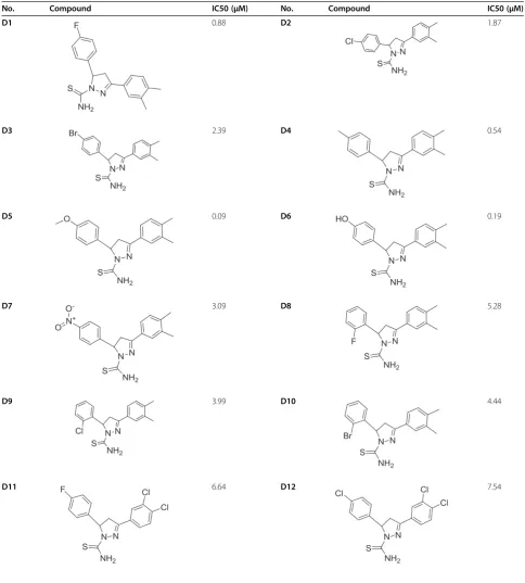
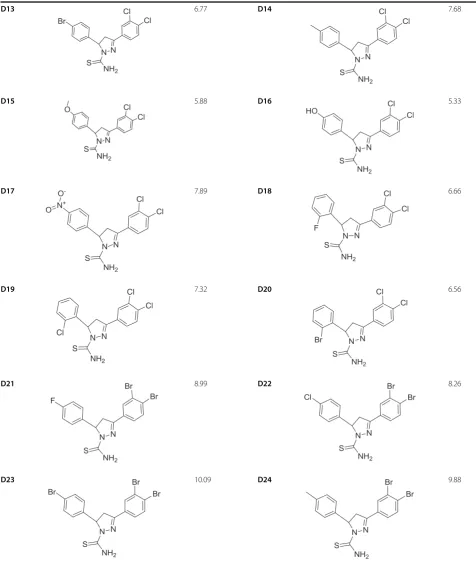
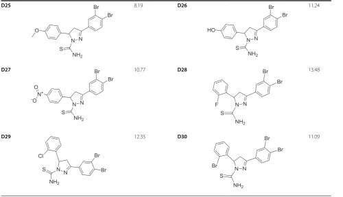
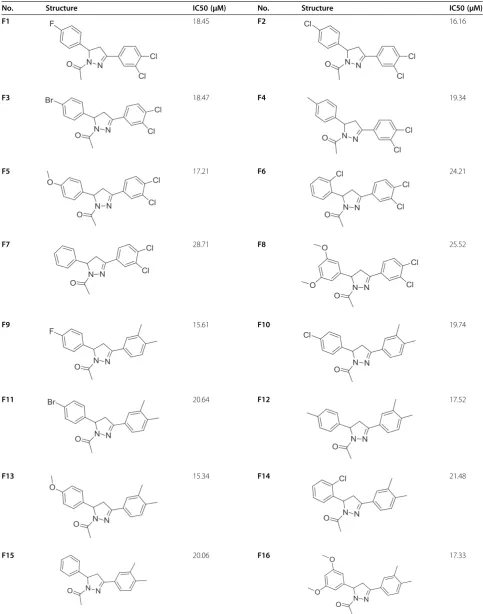
Ascites Carcinoma (EAC) human tumor cell line. From the observed results presented in Tables 1 and Table 2 it has been noticed that most of the synthesized com-pounds reveal mild to potent anti-tumor properties against (EAC) human tumor cell lines. Meanwhile,
compound D5 exhibit high potent anti-tumor
activ-ity with potency (IC50, concentration required to
produce 50% inhibition of cell growth compared to
control experimental) = 0.09 μM, compared with
Doxorubicin (IC50= 7.36 μM), which was used as a
reference standard during this study. In order to understand the observed pharmacological properties, quantitative structure-activity relationship (3D QSAR) study was initiated.
Table 1 Structure and biological activities of training set molecules (D1-30)
No. Compound IC50 (μM) No. Compound IC50 (μM)
D1 0.88 D2 1.87
D3 2.39 D4 0.54
D5 0.09 D6 0.19
D7 3.09 D8 5.28
D9 3.99 D10 4.44
Table 1 Structure and biological activities of training set molecules (D1-30)(Continued)
D13 6.77 D14 7.68
D15 5.88 D16 5.33
D17 7.89 D18 6.66
D19 7.32 D20 6.56
D21 8.99 D22 8.26
QSAR study
Pharmacophore modeling
All compounds were built using the Discovery Studio 2.5 software (Accelrys Inc., San Diego, CA, USA), and optimized by using a CHARMm-like force field [22]. The Hip Hop model treats molecular structures as tem-plates consisting of strategically positioned chemical functions that will bind effectively with complementary functions on receptor (Table 1).
The biologically most important binding functions are deduced from a small set of compounds that cover a broad range of activity. In this paper, considering the relatively high flexibility of ligands under investigation, the confor-mational analyses are carried out with an energy window high enough to include the bioactive conformation.
DS 2.5 automatically generated conformational models for each compound. Diverse conformational models for each compound were generated such that the confor-mers covered accessible conformational space defined within 20 kcal/mol of the estimated global minimum. The estimation of conformational energy was performed based on the CHARMm force field. CATALYST pro-vides two types of conformational analysis: fast and best quality. The best option was used, specifying 250 as the maximum number of conformers. Hypotheses approxi-mating the 3D QSAR pharmacophore were described as a set of features distributed within 3D space with the
correlation to the biological results. Five kinds of surface-accessible functions were considered, including two hydrogen bond acceptors (HBA) and 3 hydrophobic centers, as shown in Figure 1.
Furthermore, to emphasize the importance of an aro-matic group of the studied molecules, aroaro-matic ring (AR), which consists of directionality, was also included in the subsequent run. Considering the complexity of the studied molecules, the hypothesis generator was restricted to select only five or less features. During a hy-pothesis generation run, it attempts to minimize a score function consisting of two terms. One term penalizes the deviation between the estimated activities of the molecules in data set and their experimental values; the other term penalizes the complexity of the hypothesis. After assessing all generated hypotheses, the most plausible one is considered the best, as presented in Table 3 and displayed in Figure 2.
Molecular alignment
However, for most studies, the reliability and efficiency of a CoMFA analysis depend on the correct molecular alignment of the ligands. So we performed a pharmaco-phore mapping study to select both a proposed bioactive conformer and a superposition rule [23]. It is naturally deduced that the pharmacophore model thus obtained provides a good starting point for CoMFA studies. In
Table 1 Structure and biological activities of training set molecules (D1-30)(Continued)
D25 8.19 D26 11.24
D27 10.77 D28 13.48
Table 2 Structure and biological activities of test set molecules (F1-16)
No. Structure IC50 (μM) No. Structure IC50 (μM)
F1 18.45 F2 16.16
F3 18.47 F4 19.34
F5 17.21 F6 24.21
F7 28.71 F8 25.52
F9 15.61 F10 19.74
F11 20.64 F12 17.52
F13 15.34 F14 21.48
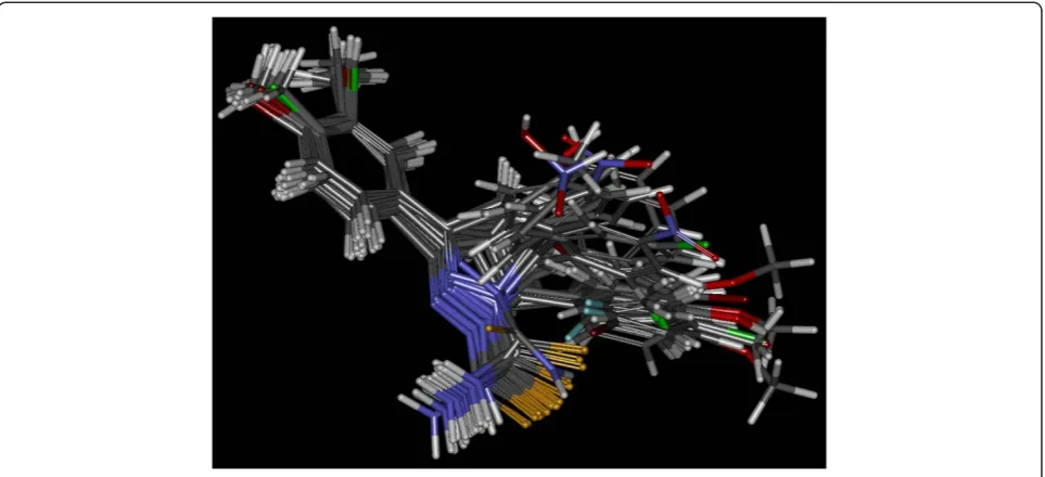
CATALYST, the studied molecules were automatically superimposed on the best pharmacophore model in gen-eration hypothesis workbench using the generated con-formers. For each ligand, one aligned conformer based on the lowest RMS deviation of feature atom coordi-nates from those of the corresponding reference features was superimposed on the hypothesis. In the studying process, we found that the best alignment based on the lowest RMS sometimes was not the most appropriate for a subsequent CoMFA analysis, and artificial adjust-ment was usually needed to get the reasonable align-ment, as shown in Figure 3. In most cases of the CoMFA calculations, the alignment from the simple atom-by-atom fits may be the most widely used proce-dure. So a rigid alignment was applied to superimpose D-5 compounds onto an unsubstituented template shown in Figure 2 using an atom-by-atom least-squares fit as implemented in the SYBYL FIT option, and com-pound D-5 with the best biological activity was treated as the reference molecule. A conventional CoMFA was performed with the usually used steric and electrostatic fields implemented in SYBYL. The atomic Gasteiger charges were applied in the determination of the electro-static field. All CoMFA calculations were performed with SYBYL standard setup (steric and electrostatic fields with Lennard-Jones and Coulomb-type potentials, di-electric constant 1/r, cutoff 30 kcal/mol) using an sp3 carbon atom with a charge of +1.0. The extent and the
orientation of the grids surrounding the tested molecules were the same as the grid spacing was set to 2 Å.
GRID size
Once the molecules are aligned a grid or lattice is estab-lished which surrounds the set of analogs in potential re-ceptor space. Current CoMFA studies seldom use grid resolution less than 1 Å and, most often, 2 Å. The choice of grid resolution represents a compromise be-tween computational practicality and detailing of the fields. If the grid resolution is too small, the number of field–points (cells) becomes too large to perform a timely analysis. Moreover spatial information on field preference can be lost, through a‘smearing out’effect, if the cells become too small. The grid resolution in the 1 Å to 2 Å range corresponds to, at best, differentiating single carbon-carbon (1.54 Å) from one another.
Figure 1Superimposition of compound D5 with the best pharmacophore model.
Figure 2Correlation line of log actual activity vs log estimated activity (r 0.695) of the best pharmacophore model.
Table 3 Summary of 3D QSAR pharmacophor model results
r2Conventional 0.695
Standard error of estimate 2.963
F Value 47.491
P Value 0.363
CoMFA interaction energy
The steric and electrostatic (potential fields) energies were calculated at each lattice intersection of a regularly spaced grid box. The lattice spacing was set a value of 2.0 Å. CoMFA region was defined automatically which extends the lattice walls beyond the dimensions of each structure by 4.0 Å in all directions. The Lennard-Jones potential and coulomb potential energy which represent, respectively steric and electrostatic fields, were calcu-lated using the TRIPOS force fields. A sp3 carbon atom with a van der Waals radius of 1.52 Å and a +1.0 charge served as the probe atom to calculate steric and electro-static fields. The default value 30.0 kcal/mole was used as the maximum electrostatic and steric energy cutoff.
Partial least squares (PLS) and cross-validation in CoMFA
The last step in a CoMFA is a partial least square ana-lysis to determine the minimal set of grid points which is necessary to explain the biological activities of the compounds. Partial least–square is an iterative proced-ure that applies two criteria to produce its solution. First, to extract a new component, the criterion is to maximize the degree of commonality between all of the structural parameter columns (independent variable) collectively and the experimental data (dependent vari-able). Second, in the evaluation phase of a PLS iteration, the criterion for acceptance of the principal component just generated is an improvement in the ability to pre-dict, not to reproduce, the dependent variable, as out-lined in Figure 4.
The technique used in PLS to assess the predictive ability of a QSAR is cross-validation [24]. Cross-validation is based on the idea that the best way to assess predictive
performance is to predict. When cross-validating, one pre-tends that one or more of the unknown experimental value is, infect, unknown. The analysis being cross-validated is repeated, excluding the temporarily‘unknown’compounds and then using the resulting equation to predict the experi-mental measurement of the omitted compound(s). The cross-validation cycle is repeated until each compound has been excluded and predicted exactly once. The results of cross-validation are the sum of the squared prediction errors, sometimes called the predicted residual sum of squares (PRESS). For evaluation of the overall analysis, the PRESS is commonly expressed as a cross–validated correl-ation coefficient r2, or xv - r2value, as outlined in Figure 5.
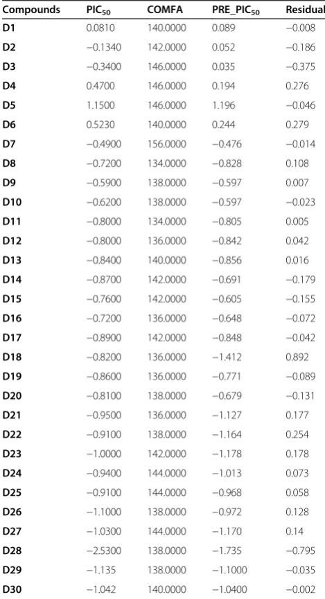
The results of the CoMFA studies are summarized in Table 4. From this table it is evident that the CoMFA derived 3D QSAR shows a good cross validated r2, (0.568)
Figure 3Alignment of compounds D1-30 with the best 3D QSAR pharmacophore.
and conventional r2, 0.895, therefore indicates a consider-able predictive and correlative capacity of the model as shown in Table 5. In this analysis both steric and electro-static field contribute to the 3D QSAR equation by 73.8% and 26.2%, respectively, suggesting that variation in bio-logical activity of compounds is dominated by differences in steric (van der Waals) interactions.
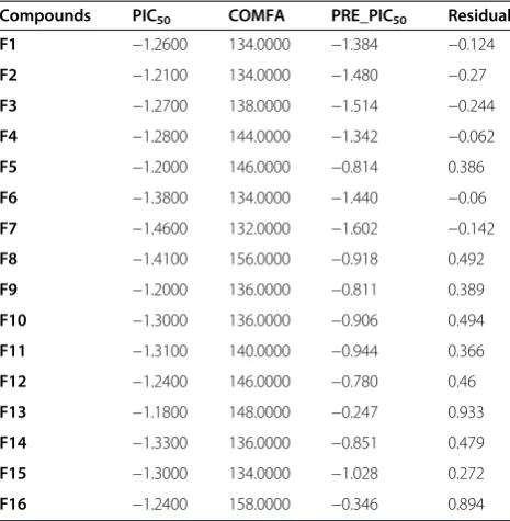
The real test for model predictiveness is to predict the activity of ligands, which were not used in the model generation. Our test set has 16 compounds, which were randomly kept with bias given to both chemical and bio-logical diversity in both the training set and the test set molecules, as shown in Tables 6.
The CoMFA models exhibited a good predictiveness on these ligands with conventional r2 0.469 and standard error of estimation 0.372 as the data described in Table 7. The contour maps are plotted as percentage contribution to the QSAR equation and are associated with the differ-ences in biological activity.
In Figures 6(a) the regions of high and low steric toler-ance are shown in green and yellow polyhedral, respect-ively. The areas of high bulk tolerance (80% contribution) are observed near 4-methoxy phenyl of the ligands . The active analogue (5) outlined in Figure 6(b), shows that 4-methoxy phenyl ring embedded in the green region. The antitumor activity shown by the different training com-pounds was due to the presence of bulky groups at position 5 and 3, 4-disbstituted phenyl at position 3 surrounded by green contours. In the present sterically unflavored yellow regions were observed near the 3, 6-substituted phenyl at position 3. The steric bulk in this region has a negative ef-fect on the antitumor activity, suggesting that there is a
Figure 5Correlation line of PIC50 vs COMFA_PIC50 of the pharmacophore model.
Table 4 Summary of COMFA results*
r2Conventional 0.895
Standard error of estimate 0.163
F Value (n1 = 6, n2 = 23) 24.478
P Value 0.000
r2cross-validated 0.568
Standard error of predictions 0.21
No. of components 6
Steric contribution 0.454
Electrostatic contribution 0.546
Mean PIC50 −0.8896
Mean predicted PIC50 −0.799
*
Results from leave one out (LOO) cross validation analysis using six components.
Table 5 Data from partial least squares (PLS) cross-validated analysis of training set
Compounds PIC50 COMFA PRE_PIC50 Residual
D1 0.0810 140.0000 0.089 −0.008
D2 −0.1340 142.0000 0.052 −0.186
D3 −0.3400 146.0000 0.035 −0.375
D4 0.4700 146.0000 0.194 0.276
D5 1.1500 146.0000 1.196 −0.046
D6 0.5230 140.0000 0.244 0.279
D7 −0.4900 156.0000 −0.476 −0.014
D8 −0.7200 134.0000 −0.828 0.108
D9 −0.5900 138.0000 −0.597 0.007
D10 −0.6200 138.0000 −0.597 −0.023
D11 −0.8000 134.0000 −0.805 0.005
D12 −0.8000 136.0000 −0.842 0.042
D13 −0.8400 140.0000 −0.856 0.016
D14 −0.8700 142.0000 −0.691 −0.179
D15 −0.7600 142.0000 −0.605 −0.155
D16 −0.7200 136.0000 −0.648 −0.072
D17 −0.8900 142.0000 −0.848 −0.042
D18 −0.8200 136.0000 −1.412 0.892
D19 −0.8600 136.0000 −0.771 −0.089
D20 −0.8100 138.0000 −0.679 −0.131
D21 −0.9500 136.0000 −1.127 0.177
D22 −0.9100 138.0000 −1.164 0.254
D23 −1.0000 142.0000 −1.178 0.178
D24 −0.9400 144.0000 −1.013 0.073
D25 −0.9100 144.0000 −0.968 0.058
D26 −1.1000 138.0000 −0.972 0.128
D27 −1.0300 144.0000 −1.170 0.14
D28 −2.5300 138.0000 −1.735 −0.795
D29 −1.135 138.0000 −1.1000 −0.035
definite requirement of a substructure with appropriate shape to exhibit high activity.
CoMFA applications in drug design
There are now a few hundred practical applications of CoMFA in drug design. Most applications are in the field of ligand protein interactions, describing affinity or inhibition constants. In addition, CoMFA has been used to correlate steric and electronic parameters [25]. Less appropriate seems the application of CoMFA to in vivo data, even if lipophilicity is considered as an additional parameter. As most CoMFA applications in drug design have been comprehensively reviewed in three books [26,27] and in some reviews [28,29].
Conclusion
This work demonstrated that the 3D QSAR pharmaco-phore modeling and MFA technique (CoMFA) could be effectively combined. The best model identified by 3d QSAR pharmacophore search served as suitable modes of superimposition for subsequent CoMFA utilizing compounds (D1-30), as training set. The better results were obtained by superimposing the molecules onto the 3D QSAR pharmacophore model than onto the com-mon structure used in the energy-lowest conformers. Moreover, from our calculations, it can be found that the active conformers and the 3D QSAR pharmacophore mapping automatically afforded by CATALYST are usu-ally not the optimal ones. The manual adjustments are usually needed to obtain the best results. As illustrated by the contour maps, our 3D-QSAR model, which is based on a homogeneous set of ligands, is expected to correctly predict affinities of structurally related com-pounds. These results will provide useful information in understanding the structural and chemical features required for EAC cells cytotoxicity and in designing new potential compounds.
Experimental
Chemistry
Separation of the compounds by column chromatography was carried out with silica gel 60 (200–300 mesh ASTM, E. Merck). The quantity of silica gel used was 50–100 times the weight charged on the column. Then, the eluates were monitored using TLC. Melting points (uncorrected) were determined on a XT4 MP apparatus (Ain Shams University, Egypt). Mass spectra were recorded on a Var-ian MAT 112 spectrometer. Analytical data were obtained from the Microanalytical Data Center at Cairo University and some compounds at Ain Shams University. The1H NMR spectra were recorded on a Varian 400 MHz spec-trometer at 25°C with TMS and solvent signals allotted as internal standards. Chemical shifts were reported in ppm (δ). Elemental analyses were performed on a CHN-O-Rapid instrument and were within ±0.4% of the theoretical values obtained from the Microanalytical Data Center at Cairo University.
General synthetic procedure of chalcones
Equimolar portions of the appropriately substituted aro-matic aldehydes (10 mmol, 1 equiv) and ketones (10 mmol, 1 equiv) were dissolved in approximately 15 mL of ethanol. The mixture was allowed to stir for several minutes at 5–10°C. A 10 mL aliquot of a 40% aqueous sodium hydroxide solution was then slowly added dropwise to the reaction flask via a self-equalizing addition funnel. The reaction solution was allowed to stir at room temperature for approximately 4 h. Most Table 6 Data from PLS cross-validated analysis of test set
Compounds PIC50 COMFA PRE_PIC50 Residual
F1 −1.2600 134.0000 −1.384 −0.124
F2 −1.2100 134.0000 −1.480 −0.27
F3 −1.2700 138.0000 −1.514 −0.244
F4 −1.2800 144.0000 −1.342 −0.062
F5 −1.2000 146.0000 −0.814 0.386
F6 −1.3800 134.0000 −1.440 −0.06
F7 −1.4600 132.0000 −1.602 −0.142
F8 −1.4100 156.0000 −0.918 0.492
F9 −1.2000 136.0000 −0.811 0.389
F10 −1.3000 136.0000 −0.906 0.494
F11 −1.3100 140.0000 −0.944 0.366
F12 −1.2400 146.0000 −0.780 0.46
F13 −1.1800 148.0000 −0.247 0.933
F14 −1.3300 136.0000 −0.851 0.479
F15 −1.3000 134.0000 −1.028 0.272
F16 −1.2400 158.0000 −0.346 0.894
Table 7 Data from partial least squares (PLS) cross-validated analysis of test set
r2Conventional 0.469
Standard error of estimate 0.372
P Value 0.0713
r2cross-validated 0.268
No. of components 6
Steric contribution 0.454
Electrostatic contribution 0.546
Mean PIC50 −1.2856
Mean predicted PIC50 −1.0254
*
commonly, a precipitate formed and was then collected by suction filtration.
General synthetic procedure of pyrazole derivatives C1–C30
A mixture of chalcone (0.01 mol), thiosemicarbazide (0.01 mol), and NaOH (0.025 mol) was refluxed in ethanol (25 mL) for 8 h. The solution was poured into ice-water. The precipitate was filtered and crystallized from methanol to give white powder compounds in good to excellent yields.
N N
Ha Hb
Ar'
Hc
Ar
J
vicinal-cisJ
vicinal-Transdd
dd
dd
J
GeminalR
3-(3,4-Dimethylphenyl)-5-(4-fluorophenyl)-4,5-dihydro-1 H-pyrazole-1-carbothioamide (D1)
Appearance: white powder. Melting Point: 223–225°C.
1
H NMR (300 MHz, DMSO-d6): 2.29 (s, 6 H); 3.12–
3.18 (dd, J1 = 3.66, J2 = 17.58 Hz, 1Ha); 3.74–3.83 (dd, J1 = 11.52, J2 = 17.76 Hz, 1Hc); 5.92–5.97 (dd, J1 = 3.66, J2 = 11.56 Hz, 1Hb); 7.09–7.18 (m, 3 H); 7.42–7.46 (m, 2 H); 7.46–7.48 (m, 1 H); 7.50 (s, 1 H). MS (ESI): 328.1 (C18H19FN3S, [M + H]+). Anal. Calcd for C18H18FN3S: C, 66.03; H, 5.54; N, 12.83. Found: C, 66.17; H, 5.72; N, 12.65.
5-(4-Chlorophenyl)-3-(3,4-dimethylphenyl)-4,5-dihydro-1 H-pyrazole-1-carbothioamide (D2)
Appearance: white powder. Melting Point: 216–217°C.
1
H NMR (300 MHz, DMSO-d6): 2.30 (s, 6 H); 3.11–
3.18 (dd, J1 = 3.66, J2 = 17.73 Hz, 1Ha); 3.76–3.86 (dd, J1 = 11.52, J2 = 17.73 Hz, 1Hc); 5.96–6.01 (dd, J1 = 3.66, J2 = 11.34 Hz, 1Hb); 7.14–7.19 (m, 3 H); 7.27–7.30 (m, 2 H); 7.41–7.44 (m, 1 H); 7.50 (s, 1 H). MS (ESI): 344.1 (C18H19ClN3S, [M + H]+). Anal. Calcd for C18H18ClN3S:
C, 62.87; H, 5.28; N, 12.22. Found: C, 62.69; H, 5.42; N, 12.41.
5-(4-Bromophenyl)-3-(3,4-dimethylphenyl)-4,5-dihydro-1 H-pyrazole-1-carbothioamide (D3)
Appearance: white powder. Melting Point: 231–233°C.
1
H NMR (300 MHz, DMSO-d6): 2.30 (s, 6 H); 3.11–
3.18 (dd, J1 = 3.66, J2 = 17.55 Hz, 1Ha); 3.76–3.86 (dd, J1 = 11.52, J2 = 17.76 Hz, 1Hc); 5.94–5.99 (dd, J1 = 3.66, J2 = 11.52 Hz, 1Hb); 7.08–7.19 (m, 3 H); 7.41–7.44 (m, 2 H); 7.45–7.47 (m, 1 H); 7.51 (s, 1 H). MS (ESI): 388.1 (C18H19BrN3S, [M + H]+). Anal. Calcd for C18H18BrN3S: C, 55.67; H, 4.67; N, 10.82. Found: C, 55.78; H, 4.81; N, 10.64.
3-(3,4-Dimethylphenyl)-5-p-tolyl-4,5-dihydro-1 H-pyrazole-1-carbothioamide (D4)
Appearance: white powder. Melting Point: 198–200°C.
1
H NMR (300 MHz, DMSO-d6): 2.30 (s, 9 H); 3.14–3.21 (dd, J1 = 3.48, J2 = 17.55 Hz, 1 H); 3.74–3.84 (dd, J1 = 11.34, J2 = 17.55 Hz, 1 H); 5.96–6.01 (dd, J1 = 3.45, J2 = 11.34 Hz, 1 H); 7.08–7.13 (m, 3 H); 7.14–7.18 (m, 2 H); 7.41–7.44 (m, 1 H); 7.51 (s, 1 H). MS (ESI): 324.1 (C19H22N3S, [M + H]+). Anal. Calcd for C19H21N3S: C, 70.55; H, 6.54; N, 12.99. Found: C, 70.74; H, 6.42; N, 12.76.
3-(3,4-Dimethylphenyl)-5-(4-methoxyphenyl)-4,5-dihydro-1 H-pyrazole-3-(3,4-Dimethylphenyl)-5-(4-methoxyphenyl)-4,5-dihydro-1-carbothioamide (D5)
Appearance: white powder. Melting Point: 207–209°C.
1
H NMR (300 MHz, DMSO-d6): 2.31 (s, 6 H); 3.11–
3.19 (dd, J1 = 3.47, J2 = 17.56 Hz, 1 H); 3.71 (s, 3 H); 3.72–3.81 (dd, J1 = 11.42, J2 = 17.55 Hz, 1 H); 5.97–6.03 (dd, J1 = 3.45, J2 = 11.34 Hz, 1 H); 7.06–7.11 (m, 3 H); 7.15–7.18 (m, 2 H); 7.42–7.45 (m, 1 H); 7.51 (s, 1 H). MS (ESI): 340.1 (C19H22N3OS, [M + H]+). Anal. Calcd for C19H21N3OS: C, 67.23; H, 6.24; N, 12.38. Found: C, 67.48; H, 6.46; N, 12.17.
3-(3,4-Dimethylphenyl)-5-(4-hydroxyphenyl)-4,5-dihydro-1 H-pyrazole-3-(3,4-Dimethylphenyl)-5-(4-hydroxyphenyl)-4,5-dihydro-1-carbothioamide (D6)
Appearance: white powder. Melting Point: 218–219°C.
1
H NMR (300 MHz, DMSO-d6): 2.30 (s, 6 H); 3.12–3.18 (dd, J1 = 3.67, J2 = 17.55 Hz, 1 H); 3.74–3.83 (dd, J1 = 11.53, J2 = 17.76 Hz, 1 H); 5.93–5.99 (dd, J1 = 3.68, J2 = 11.53 Hz, 1 H); 7.09–7.19 (m, 3 H); 7.41–7.44 (m, 2 H); 7.44–7.46 (m, 1 H); 7.50 (s, 1 H). MS (ESI): 326.1 (C18H20N3OS, [M + H]+). Anal. Calcd for C18H19N3OS: C, 66.43; H, 5.88; N, 12.91. Found: C, 66.65; H, 5.69; N, 12.75.
3-(3,4-Dimethylphenyl)-5-(4-nitrophenyl)-4,5-dihydro1H-pyrazole-1-carbothioamide(D7)
Appearance: white powder. Melting Point: 202– 203°C.
1
H NMR (300 MHz, DMSO-d6): 2.31 (s, 6 H); 3.13–3.19 (dd, J1 = 3.66, J2 = 17.55 Hz, 1 H); 3.76–3.85 (dd, J1 = 11.51, J2 = 17.73 Hz, 1 H); 5.92–5.97 (dd, J1 = 3.65, J2 = 11.51 Hz, 1 H); 7.07–7.16 (m, 3 H); 7.40–7.43 (m, 2 H); 7.45–7.47 (m, 1 H); 7.50 (s, 1 H). MS (ESI): 355.1 (C18H19N4O2S, [M + H]+). Anal. Calcd for C18H18N4O2S: C, 61.00; H, 5.12; N, 15.81. Found: C, 61.18; H, 5.32; N, 15.64.
3-(3,4-Dimethylphenyl)-5-(2-fluorophenyl)-4,5-dihydro-1 H-pyrazole-1-carbothioamide (D8)
Appearance: white powder. Melting Point: 211–213°C.
1
H NMR (300 MHz, DMSO-d6): 2.30 (s, 6 H); 3.08–3.15 (dd, J1 = 4.04, J2 = 17.89 Hz, 1 H); 3.88–3.97 (dd, J1 = 11.53, J2 = 17.96 Hz, 1 H); 6.33–6.38 (dd, J1 = 4.09, J2 = 11.43 Hz, 1 H); 7.08–7.12 (m, 1 H); 7.16–7.26 (m, 3 H); 7.39–7.45 (m, 2 H); 7.51 (s, 1 H). MS (ESI): 328.1 (C18H19FN3S, [M + H]+). Anal. Calcd for C18H18FN3S: C, 66.03; H, 5.54; N, 12.83. Found: C, 66.19; H, 5.31; N, 12.74.
5-(2-Chlorophenyl)-3-(3,4-dimethylphenyl)-4,5-dihydro-1 H-pyrazole-1-carbothioamide (D9)
Appearance: white powder.Melting Point:208–210°C.1H NMR (300 MHz, DMSO-d6): 2.29 (s, 6 H); 3.06–3.14 (dd, J1 = 4.02, J2 = 17.94 Hz, 1 H); 3.85–3.95 (dd, J1 = 11.52, J2 = 17.91 Hz, 1 H); 6.30–6.35 (dd, J1 = 4.05, J2 = 11.52 Hz, 1 H); 7.04–7.07 (m, 1 H); 7.15–7.23 (m, 3 H); 7.37–7.44 (m, 2 H); 7.50 (s, 1 H). MS (ESI): 344.1 (C18H19ClN3S, [M + H]+). Anal. Calcd for C18H18ClN3S: C, 62.87; H, 5.28; N, 12.22. Found: C, 62.63; H, 5.38; N, 12.46.
5-(2-Bromophenyl)-3-(3,4-dimethylphenyl)-4,5-dihydro-1 H-pyrazole-1-carbothioamide (D10)
Appearance: white powder. Melting Point: 217–219°C.
1
H NMR (300 MHz, DMSO-d6): 2.31 (s, 6 H); 3.05–3.12 (dd, J1 = 4.06, J2 = 17.96 Hz, 1 H); 3.83–3.94 (dd, J1 = 11.51, J2 = 17.93 Hz, 1 H); 6.28–6.32 (dd, J1 = 4.03, J2 = 11.52 Hz, 1 H); 7.06– 7.09 (m, 1 H); 7.16–7.23 (m, 3 H); 7.38–7.45 (m, 2 H); 7.50 (s, 1 H). MS (ESI): 388.1 (C18H19BrN3S, [M + H]+). Anal. Calcd for C18H18BrN3S: C, 55.67; H, 4.67; N, 10.82. Found: C, 55.83; H, 4.54; N, 10.74.
3-(3,4-Dichlorophenyl)-5-(4-fluorophenyl)-4,5-dihydro-1 H-pyrazole-1-carbothioamide (D11)
Anal. Calcd for C16H12Cl2FN3S: C, 52.18; H, 3.28; N, 11.41. Found: C, 52.35; H, 3.41; N, 11.34.
5-(4-Chlorophenyl)-3-(3,4-dichlorophenyl)-4,5-dihydro-1 H-pyrazole-1-carbothioamide (D12)
Appearance: white powder. Melting Point: 211–213°C.
1
H NMR (300 MHz, DMSO-d6): 3.11–3.17 (dd, J1 =
3.68, J2 = 17.75 Hz, 1 H); 3.75–3.86 (dd, J1 = 11.51, J2 = 17.73 Hz, 1 H); 5.94–6.02 (dd, J1 = 3.65, J2 = 11.37 Hz, 1 H); 7.13–7.17 (m, 3 H); 7.28–7.32 (m, 2 H); 7.40–7.44 (m, 1 H); 7.51 (s, 1 H). MS (ESI): 383.0 (C16H13Cl3N3S, [M + H]+). Anal. Calcd for C16H12Cl3N3S: C, 49.95; H, 3.14; N, 10.92. Found: C, 49.82; H, 3.26; N, 10.78.
5-(4-Bromophenyl)-3-(3,4-dichlorophenyl)-4,5-dihydro-1 H-pyrazole-1-carbothioamide (D13)
Appearance: white powder. Melting Point: 214–216°C.
1
H NMR (300 MHz, DMSO-d6): 3.12–3.18 (dd, J1 =
3.68, J2 = 17.53 Hz, 1 H); 3.74–3.86 (dd, J1 = 11.53, J2 = 17.76 Hz, 1 H); 5.92–5.98 (dd, J1 = 3.66, J2 = 11.53 Hz, 1 H); 7.07–7.17 (m, 3 H); 7.42–7.45 (m, 2 H); 7.47–7.48 (m, 1 H); 7.51 (s, 1 H). MS (ESI): 427.0 (C16H13BrCl2N3S, [M + H]+). Anal. Calcd for C16H12BrCl2N3S: C, 44.78; H, 2.82; N, 9.79. Found: C, 44.96; H, 2.66; N, 9.98.
3-(3,4-Dichlorophenyl)-5-p-tolyl-4,5-dihydro-1 H-pyrazole-1-carbothioamide (D14)
Appearance: white powder.Melting Point:223–225°C.1H
NMR (300 MHz, DMSO-d6): 2.30 (s, 3 H); 3.12–3.21
(dd, J1 = 3.46, J2 = 17.55 Hz, 1 H); 3.76–3.84 (dd, J1 = 11.37, J2 = 17.53 Hz, 1 H); 5.94–6.01 (dd, J1 = 3.46, J2 = 11.34 Hz, 1 H); 7.06–7.13 (m, 3 H); 7.13–7.17 (m, 2 H); 7.41–7.43 (m, 1 H); 7.50 (s, 1 H). MS (ESI): 364.0 (C17H16Cl2N3S, [M + H]+). Anal. Calcd for C17H15Cl2N3S: C, 56.05; H, 4.15; N, 11.53. Found: C, 56.18; H, 4.26; N, 11.38.
3-(3,4-Dichlorophenyl)-5-(4-methoxyphenyl)-4,5-dihydro-1 H-pyrazole-1-carbothioamide (D15)
Appearance: white powder. Melting Point: 209–210°C.
1
H NMR (300 MHz, DMSO-d6): 3.12–3.19 (dd, J1 = 3.46, J2 = 17.53 Hz, 1 H); 3.71 (s, 3 H); 3.74–3.81 (dd, J1 = 11.43, J2 = 17.53 Hz, 1 H); 5.94–6.01 (dd, J1 = 3.46, J2 = 11.34 Hz, 1 H); 7.07–7.11 (m, 3 H); 7.14–7.18 (m, 2 H); 7.42–7.45 (m, 1 H); 7.51 (s, 1 H). MS (ESI): 380.0 (C17H16Cl2N3OS, [M + H]+). Anal. Calcd for C17H15Cl2N3OS: C, 53.69; H, 3.98; N, 11.05. Found: C, 53.78; H, 3.81; N, 11.24.
3-(3,4-Dichlorophenyl)-5-(4-hydroxyphenyl)-4,5-dihydro-1 H-pyrazole-1-carbothioamide (D16)
Appearance: white powder.Melting Point:p 235–237°C.
1
H NMR (300 MHz, DMSO-d6): 3.12–3.17 (dd, J1 =
3.67, J2 = 17.53 Hz, 1 H); 3.72–3.83 (dd, J1 = 11.56, J2 = 17.76 Hz, 1 H); 5.92–5.98 (dd, J1 = 3.68, J2 = 11.51 Hz, 1 H); 7.08–7.19 (m, 3 H); 7.42–7.44 (m, 2 H); 7.44–7.47
(m, 1 H); 7.50 (s, 1 H). MS (ESI): 366.0 (C16H14Cl2N3OS, [M + H]+). Anal. Calcd for C16H13Cl2N3OS: C, 52.47; H, 3.58; N, 11.47. Found: C, 52.62; H, 3.74; N, 11.59.
3-(3,4-Dichlorophenyl)-5-(4-nitrophenyl)-4,5-dihydro1H-pyrazole-1-carbothioamide (D17)
Appearance: white powder. Melting Point: 203– 205°C.
1
H NMR (300 MHz, DMSO-d6): 3.13–3.18 (dd, J1 = 3.65, J2 = 17.53 Hz, 1 H); 3.74–3.83 (dd, J1 = 11.53, J2 = 17.73 Hz, 1 H); 5.94–5.97 (dd, J1 = 3.65, J2 = 11.53 Hz, 1 H); 7.07–7.14 (m, 3 H); 7.40–7.43 (m, 2 H); 7.46–7.48 (m, 1 H); 7.50 (s, 1 H). MS (ESI): 395.0 (C16H13Cl2N4O2S, [M + H]+). Anal. Calcd for C16H12Cl2N4O2S: C, 48.62; H, 3.06; N, 14.17. Found: C, 48.76; H, 3.24; N, 14.34.
3-(3,4-Dichlorophenyl)-5-(2-fluorophenyl)-4,5-dihydro-1 H-pyrazole-1-carbothioamide (D18)
Appearance: white powder. Melting Point: 235–236°C.
1
H NMR (300 MHz, DMSO-d6): 3.09–3.15 (dd, J1 =
4.16, J2 = 17.86 Hz, 1 H); 3.85–3.96 (dd, J1 = 11.53, J2 = 17.96 Hz, 1 H); 6.31–6.38 (dd, J1 = 4.21, J2 = 11.43 Hz, 1 H); 7.08–7.14 (m, 1 H); 7.18–7.26 (m, 3 H); 7.36–7.43 (m, 2 H); 7.51 (s, 1 H). MS (ESI): 368.0 (C16H13Cl2FN3S, [M + H]+). Anal. Calcd for C16H12Cl2FN3S: C, 52.18; H, 3.28; N, 11.41. Found: C, 52.32; H, 3.39; N, 11.58.
5-(2-Chlorophenyl)-3-(3,4-dichlorophenyl)-4,5-dihydro-1H-pyrazole-1-carbothioamide (D19)
Appearance: white powder. Melting Point: 241–243°C.
1
H NMR (300 MHz, DMSO-d6): 3.06–3.12 (dd, J1 =
4.14, J2 = 17.92 Hz, 1 H); 3.87–3.95 (dd, J1 = 11.53, J2 = 17.91 Hz, 1 H); 6.31–6.35 (dd, J1 = 4.26, J2 = 11.52 Hz, 1 H); 7.03–7.07 (m, 1 H); 7.17–7.23 (m, 3 H); 7.36–7.44 (m, 2 H); 7.50 (s, 1 H). MS (ESI): 383.0 (C16H13Cl3N3S, [M + H]+). Anal. Calcd for C16H12Cl3N3S: C, 49.95; H, 3.14; N, 10.92. Found: C, 49.76; H, 3.27; N, 10.81.
5-(2-Bromophenyl)-3-(3,4-dichlorophenyl)-4,5-dihydro-1 H-pyrazole-1-carbothioamide (D20)
Appearance: white powder.Melting Point:229–230°C.1H NMR (300 MHz, DMSO-d6): 3.07–3.12 (dd, J1 = 4.16, J2 = 17.93 Hz, 1 H); 3.86–3.94 (dd, J1 = 11.53, J2 = 17.91 Hz, 1 H); 6.26–6.32 (dd, J1 = 4.13, J2 = 11.51 Hz, 1 H); 7.04– 7.09 (m, 1 H); 7.17–7.23 (m, 3 H); 7.36–7.45 (m, 2 H); 7.50 (s, 1 H). MS (ESI): 427.0 (C16H13BrCl2N3S, [M + H]+). Anal. Calcd for C16H12BrCl2N3S: C, 44.78; H, 2.82; N, 9.79. Found: C, 44.92; H, 2.69; N, 9.93.
3-(3,4-Dibromophenyl)-5-(4-fluorophenyl)-4,5-dihydro-1 H-pyrazole-1-carbothioamide (D21)
Appearance: white powder. Melting Point: 246–247°C.
1
H NMR (300 MHz, DMSO-d6): 3.14–3.17 (dd, J1 =
1 H); 7.05–7.16 (m, 3 H); 7.42–7.45 (m, 2 H); 7.46–7.48 (m, 1 H); 7.51 (s, 1 H). MS (ESI): 455.0 (C16H13Br2FN3S, [M + H]+). Anal. Calcd for C16H12Br2FN3S: C, 42.04; H, 2.65; N, 9.19. Found: C, 42.17; H, 2.74; N, 9.35.
5-(4-Chlorophenyl)-3-(3,4-dibromophenyl)-4,5-dihydro-1 H-pyrazole-1-carbothioamide (D22)
Appearance: white powder. Melting Point: 216–218°C.
1
H NMR (300 MHz, DMSO-d6): 3.12–3.17 (dd, J1 =
3.66, J2 = 17.75 Hz, 1 H); 3.73–3.86 (dd, J1 = 11.53, J2 = 17.71 Hz, 1 H); 5.96–6.04 (dd, J1 = 3.65, J2 = 11.45 Hz, 1 H); 7.15–7.17 (m, 3 H); 7.26–7.32 (m, 2 H); 7.41–7.44 (m, 1 H); 7.51 (s, 1 H). MS (ESI): 471.0 (C16H13Br2ClN3S, [M + H]+). Anal. Calcd for C16H12Br2ClN3S: C, 40.58; H, 2.55; N, 8.87. Found: C, 40.46; H, 2.68; N, 8.69.
5-(4-Bromophenyl)-3-(3,4-dibromophenyl)-4,5-dihydro-1 H-pyrazole-1-carbothioamide (D23)
Appearance: white powder. Melting Point: 239–241°C.
1
H NMR (300 MHz, DMSO-d6): 3.14–3.18 (dd, J1 =
3.69, J2 = 17.53 Hz, 1 H); 3.77–3.86 (dd, J1 = 11.51, J2 = 17.76 Hz, 1 H); 5.91–5.98 (dd, J1 = 3.66, J2 = 11.57 Hz, 1 H); 7.09–7.17 (m, 3 H); 7.42–7.46 (m, 2 H); 7.47–7.48 (m, 1 H); 7.50 (s, 1 H). MS (ESI): 515.0(C16H13Br3N3S, [M + H]+). Anal. Calcd for C16H12Br3N3S: C, 37.09; H, 2.33; N, 8.11. Found: C, 37.18; H, 2.45; N, 8.32.
3-(3,4-Dibromophenyl)-5-p-tolyl-4,5-dihydro-1 H-pyrazole-1-carbothioamide (D24)
Appearance: white powder.Melting Point:211–213°C.1H
NMR (300 MHz, DMSO-d6): 2.30 (s, 3 H); 3.13–3.21
(dd, J1 = 3.46, J2 = 17.55 Hz, 1 H); 3.74–3.84 (dd, J1 = 11.31, J2 = 17.57 Hz, 1 H); 5.96–6.01 (dd, J1 = 3.46, J2 = 11.37 Hz, 1 H); 7.08–7.13 (m, 3 H); 7.13–7.17 (m, 2 H); 7.41–7.43 (m, 1 H); 7.50 (s, 1 H). MS (ESI): 451.0 (C17H16Br2N3S, [M + H]+). Anal. Calcd for C17H15Br2N3S: C, 45.05; H, 3.34; N, 9.27. Found: C, 45.21; H, 3.48; N, 9.36.
3-(3,4-Dibromophenyl)-5-(4-methoxyphenyl)-4,5-dihydro-1 H-pyrazole-1-carbothioamide (D25)
Appearance: white powder.Melting Point:236–238°C.1H NMR (300 MHz, DMSO-d6): 3.11–3.17 (dd, J1 = 3.43, J2 = 17.53 Hz, 1 H); 3.71 (s, 3 H); 3.77–3.81 (dd, J1 = 11.46, J2 = 17.53 Hz, 1 H); 5.97–6.01 (dd, J1 = 3.43, J2 = 11.34 Hz, 1 H); 7.08– 7.11 (m, 3 H); 7.16–7.18 (m, 2 H); 7.42–7.45 (m, 1 H); 7.51 (s, 1 H). MS (ESI): 467.0 (C17H16Br2N3OS, [M + H]+). Anal. Calcd for C17H15Br2N3OS: C, 43.52; H, 3.22; N, 8.96. Found: C, 43.68; H, 3.37; N, 8.84.
3-(3,4-Dibromophenyl)-5-(4-hydroxyphenyl)-4,5-dihydro-1 H-pyrazole-1-carbothioamide (D26)
Appearance: white powder. Melting Point: 221–223°C.
1
H NMR (300 MHz, DMSO-d6): 3.11–3.16 (dd, J1 =
3.63, J2 = 17.51 Hz, 1 H); 3.75–3.83 (dd, J1 = 11.53, J2 =
17.76 Hz, 1 H); 5.93–5.98 (dd, J1 = 3.68, J2 = 11.53 Hz, 1 H); 7.09–7.19 (m, 3 H); 7.42–7.45 (m, 2 H); 7.46–7.48 (m, 1 H); 7.50 (s, 1 H). MS (ESI): 453.0 (C16H14Br2N3OS, [M + H]+). Anal. Calcd for C16H13Br2N3OS: C, 42.22; H, 2.88; N, 9.23. Found: C, 42.45; H, 2.69; N, 9.38.
3-(3,4-Dibromophenyl)-5-(4-nitrophenyl)-4,5-dihydro1H-pyrazole-1-carbothioamide (D27)
Appearance: white powder. Melting Point: 246– 248°C.
1
H NMR (300 MHz, DMSO-d6): 3.12–3.18 (dd, J1 = 3.69, J2 = 17.53 Hz, 1 H); 3.76–3.83 (dd, J1 = 11.51, J2 = 17.73 Hz, 1 H); 5.92–5.97 (dd, J1 = 3.63, J2 = 11.53 Hz, 1 H); 7.09–7.14 (m, 3 H); 7.41–7.43 (m, 2 H); 7.46–7.48 (m, 1 H); 7.50 (s, 1 H). MS (ESI): 482.0 (C16H13Br2N4O2S, [M + H]+). Anal. Calcd for C16H12Br2N4O2S: C, 39.69; H, 2.50; N, 11.57. Found: C, 39.78; H, 2.41; N, 11.69.
3-(3,4-Dibromophenyl)-5-(2-fluorophenyl)-4,5-dihydro-1 H-pyrazole-1-carbothioamide (D28)
Appearance: white powder. Melting Point: 236–237°C.
1
H NMR (300 MHz, DMSO-d6): 3.07–3.13 (dd, J1 =
4.18, J2 = 17.86 Hz, 1 H); 3.83–3.92 (dd, J1 = 11.51, J2 = 17.96 Hz, 1 H); 6.35–6.38 (dd, J1 = 4.36, J2 = 11.43 Hz, 1 H); 7.12–7.14 (m, 1 H); 7.19–7.26 (m, 3 H); 7.36–7.43 (m, 2 H); 7.51 (s, 1 H). MS (ESI): 455.0 (C16H13Br2FN3S, [M + H]+). Anal. Calcd for C16H12Br2FN3S: C, 42.04; H, 2.65; N, 9.19. Found: C, 42.16; H, 2.43; N, 9.31.
5-(2-Chlorophenyl)-3-(3,4-dibromophenyl)-4,5-dihydro-1 H-pyrazole-1-carbothioamide (D29)
Appearance: white powder. Melting Point: 219–221°C.
1
H NMR (300 MHz, DMSO-d6): 3.08–3.12 (dd, J1 =
4.26, J2 = 17.92 Hz, 1 H); 3.89–3.95 (dd, J1 = 11.53, J2 = 17.91 Hz, 1 H); 6.33–6.35 (dd, J1 = 4.26, J2 = 11.52 Hz, 1 H); 7.04–7.07 (m, 1 H); 7.17–7.26 (m, 3 H); 7.36–7.44 (m, 2 H); 7.50 (s, 1 H). MS (ESI): 471.0 (C16H13Br2ClN3S, [M + H]+). Anal. Calcd for C16H12Br2ClN3S: C, 40.58; H, 2.55; N, 8.81. Found: C, 40.69; H, 2.38; N, 8.72.
5-(2-Bromophenyl)-3-(3,4-dibromophenyl)-4,5-dihydro-1 H-pyrazole-1-carbothioamide (D30)
Appearance: white powder. Melting Point: 214–216°C.
1
H NMR (300 MHz, DMSO-d6): 3.09–3.15 (dd, J1 =
4.21, J2 = 17.91 Hz, 1 H); 3.84–3.91 (dd, J1 = 11.52, J2 = 17.93 Hz, 1 H); 6.29–6.32 (dd, J1 = 4.26, J2 = 11.51 Hz, 1 H); 7.06–7.09 (m, 1 H); 7.19–7.23 (m, 3 H); 7.34–7.45 (m, 2 H); 7.50 (s, 1 H). MS (ESI): 515.0 (C16H13Br3N3S, [M + H]+). Anal. Calcd for C16H12Br3N3S: C, 37.09; H, 2.33; N, 8.11. Found: C, 37.19; H, 2.42; N, 8.32.
General synthetic procedure of pyrazole derivatives F1–F16
The mixture was then poured onto ice-water, the crude pyrazole derivatives D1–D16 were obtained by vacuum filtration, which were crystallized from ethanol to give white powder compounds in good to excellent yields.
1-(3-(3,4-Dichlorophenyl)-5-(4-fluorophenyl)-4,5-dihydro-1 H-pyrazol-1-yl)ethanone (F1)
Appearance: white powder.1HNMR (300 MHz,
DMSO-d6): 2.30 (s, 3 H); 3.17–3.22 (dd, J1 = 2.91, J2 = 10.98 Hz, 1 H); 3.80–3.86 (dd, J1 = 7.32, J2 = 10.98 Hz, 1 H); 5.55– 5.58 (dd, J1 = 2.94, J2 = 7.14 Hz, 1 H); 7.12–7.16 (t, J = 5.31 Hz, 2 H); 7.22–7.25 (m, 2 H); 7.72–7.78 (m, 2 H); 7.97 (d, J = 11.10 Hz, 1 H). MS (ESI): 351.0 (C17H14Cl2FN2O, [M + H]+). Anal. Calcd for C17H13Cl2FN2O: C, 58.14; H, 3.73; N, 7.98. Found: C, 58.16; H, 3.71; N, 7.84.
1-(5-(4-Chlorophenyl)-3-(3,4-dichlorophenyl)-4,5-dihydro-1 H-pyrazol-1-yl)ethanone (F2)
Appearance: white powder.1HNMR (300 MHz,
DMSO-d6): 2.30 (s, 3 H); 3.17–3.22 (dd, J1 = 2.90, J2 = 10.96 Hz, 1 H); 3.81–3.87 (dd, J1 = 7.30, J2 = 10.91 Hz, 1 H); 5.54– 5.57 (dd, J1 = 2.96, J2 = 7.14 Hz, 1 H); 7.21–7.23 (d, J = 5.13 Hz, 2 H); 7.37–7.39 (d, J = 4.95 Hz, 2 H); 7.72–7.78 (m, 2 H); 7.96–7.97 (d, J = 11.10 Hz, 1 H). MS (ESI):
367.0 (C17H14Cl3N2O, [M + H]+). Anal. Calcd for
C17H13Cl3N2O: C, 55.54; H, 3.56; N, 7.62. Found: C, 55.52; H, 3.58; N, 7.53.
1-(5-(4-Bromophenyl)-3-(3,4-dichlorophenyl)-4,5-dihydro-1 H-pyrazol-1-yl)ethanone (F3)
Appearance: white powder.1HNMR (300 MHz,
DMSO-d6): 2.30 (s, 3 H); 3.17–3.22 (dd, J1 = 2.91, J2 = 10.96 Hz, 1 H); 3.81–3.87 (dd, J1 = 7.34, J2 = 10.98 Hz, 1 H); 5.52– 5.56 (dd, J1 = 2.96, J2 = 7.17 Hz, 1 H); 7.21–7.23 (d, J = 5.13 Hz, 2 H); 7.37–7.39 (d, J = 4.95 Hz, 2 H); 7.72–7.78 (m, 2 H); 7.96–7.97 (d, J = 11.10 Hz, 1 H). MS (ESI): 410.9 (C17H14BrCl2N2O, [M + H]+). Anal. Calcd for C17H13BrCl2N2O: C, 49.55; H, 3.18; N, 6.80. Found: C, 49.57; H, 3.17; N, 6.68.
1-(3-(3,4-Dichlorophenyl)-5-p-tolyl-4,5-dihydro-1 H-pyrazol-1-yl)ethanone (F4)
Appearance: white powder. 1H NMR (300 MHz,
DMSO-d6): 2.26 (s, 3 H); 2.29 (s, 3 H); 3.13–3.17 (dd, J1 = 2.76, J2 = 10.80 Hz, 1 H); 3.79–3.85 (dd, J1 = 7.32, J2 = 10.98 Hz, 1 H); 5.49–5.52 (dd, J1 = 2.76, J2 = 7.14 Hz, 1 H); 7.05–7.13 (dd, J1 = 4.74, J2 = 17.37 Hz, 4 H); 7.71–7.77 (m, 2 H); 7.96–7.97 (d, J = 11.1 Hz, 1 H).
MS (ESI): 347.1 (C18H17Cl2N2O, [M + H]+). Anal.
Calcd for C18H16Cl2N2O: C, 62.26; H, 4.64; N, 8.07. Found: C, 62.27; H, 4.62; N, 8.21.
1-(3-(3,4-Dichlorophenyl)-5-(4-methoxyphenyl)-4,5dihydro-1 H-pyrazol-1-yl)ethanone (F5)
Appearance: white powder. 1H NMR (300 MHz,
DMSO-d6): 2.25 (s, 3 H); 3.11–3.16 (dd, J1 = 2.73, J2 = 10.80 Hz, 1 H); 3.68 (s, 3 H); 3.74–3.80 (dd, J1 = 7.14, J2 = 10.80 Hz, 1 H); 5.45–5.49 (dd, J1 = 2.73, J2 = 6.93 Hz, 1 H); 6.83–6.85 (d, J = 5.31 Hz, 2 H); 7.07–7.08 (d, J = 5.31 Hz, 2 H); 7.69–7.70 (d, J = 5.13 Hz, 1 H); 7.73–7.75 (dd, J1 = 1.29, J2 = 5.13 Hz, 1 H); 7.94 (d, J = 11.1 Hz, 1 H). MS (ESI): 363.1 (C18H17Cl2N2O2, [M + H]+). Anal. Calcd for C18H16Cl2N2O2: C, 59.52; H, 4.44; N, 7.71. Found: C, 59.53; H, 4.42; N, 7.85.
1-(5-(2-Chlorophenyl)-3-(3,4-dichlorophenyl)-4,5-dihydro-1 H-pyrazol-1-yl)ethanone (F6)
Appearance: white powder.1HNMR (300 MHz,
DMSO-d6): 2.30 (s, 3 H); 3.15–3.24 (dd, J1 = 2.91, J2 = 10.96 Hz, 1 H); 3.82–3.89 (dd, J1 = 7.34, J2 = 10.98 Hz, 1 H); 5.51– 5.58 (dd, J1 = 2.96, J2 = 7.17 Hz, 1 H); 7.21–7.25 (d, J = 5.13 Hz, 2 H); 7.35–7.39 (d, J = 4.95 Hz, 2 H); 7.72–7.78 (m, 2 H); 7.94–7.97 (d, J = 11.10 Hz, 1 H). MS (ESI):
367.0 (C17H14Cl3N2O, [M + H]+). Anal. Calcd for
C17H13Cl3N2O: C, 55.54; H, 3.56; N, 7.62. Found: C, 55.56; H, 3.53; N, 7.78.
1-(3-(3,4-Dichlorophenyl)-5-phenyl-4,5-dihydro-1Hpyrazol-1-yl)ethanone (F7)
Appearance: white powder. 1H NMR (300 MHz,
DMSO-d6): 2.31 (s, 3 H); 3.14–3.22 (dd, J1 = 4.74, J2 = 18.27 Hz, 1 H); 3.79–3.89 (dd, J1 = 11.88, J2 = 18.30 Hz, 1 H); 5.53–5.58 (dd, J1 = 4.77, J2 = 11.91 Hz, 1 H); 7.17– 7.35 (m, 5 H); 7.70–7.79 (m, 2 H); 7.70–7.72 (s, 1 H).
MS (ESI): 333.1 (C17H15Cl2N2O, [M + H]+). Anal.
Calcd for C17H14Cl2N2O: C, 61.28; H, 4.23; N, 8.41. Found: C, 61.26; H, 4.25; N, 8.56.
1-(3-(3,4-Dichlorophenyl)-5-(3,5-dimethoxyphenyl)4,5-dihydro-1 H-pyrazol-1-yl)ethanone (F8)
Appearance: white powder. 1H NMR (300 MHz,
DMSO-d6): 2.26 (s, 3 H); 3.12–3.16 (dd, J1 = 2.73, J2 = 10.80 Hz, 1 H); 3.68 (s, 6 H); 3.73–3.83 (dd, J1 = 7.14, J2 = 10.80 Hz, 1 H); 5.45–5.49 (dd, J1 = 2.73, J2 = 6.93 Hz, 1 H); 6.81–6.85 (d, J = 5.31 Hz, 2 H); 7.06–7.08 (d, J = 5.31 Hz, 2 H); 7.69–7.70 (d, J = 5.13 Hz, 1 H); 7.72–7.75 (dd, J1 = 1.29, J2 = 5.13 Hz, 1 H); 7.95 (d, J = 11.1 Hz, 1 H). MS (ESI): 393.1 (C19H19Cl2N2O3, [M + H]+). Anal. Calcd for C19H18Cl2N2O3: C, 58.03; H, 4.61; N, 7.12. Found: C, 58.05; H, 4.59; N, 7.26.
1-(3-(3,4-Dimethylphenyl)-5-(4-fluorophenyl)-4,5-dihydro-1 H-pyrazol-1-yl)ethanone (F9)
Appearance: white powder. 1H NMR (300 MHz,
10.80 Hz, 1 H); 5.50–5.54 (dd, J1 = 2.76, J2 = 6.96 Hz, 1 H); 7.09–7.23 (m, 5 H); 7.48–7.50 (d, J = 7.86, 1 H); 7.56 (s, 1 H); MS (ESI): 311.2 (C19H20FN2O, [M + H]+). Anal. Calcd for C19H19FN2O: C, 73.53; H, 6.17; N, 9.03. Found: C, 73.55; H, 6.16; N, 9.16.
1-(5-(4-Chlorophenyl)-3-(3,4-dimethylphenyl)-4,5-dihydro-1 H-pyrazol-1-yl)ethanone (F10)
Appearance: white powder.1H NMR (300 MHz,
DMSO-d6): 2.25 (s, 6 H); 2.29 (s, 3 H); 3.06–3.14 (dd, J1 = 2.76, J2 = 10.86 Hz, 1 H); 3.77–3.87 (dd, J1 = 7.32, J2 = 10.80 Hz, 1 H); 5.49–5.55 (dd, J1 = 2.76, J2 = 6.96 Hz, 1 H); 7.18–7.23 (m, 3 H); 7.36–7.39 (d, J = 8.22, 2 H); 7.47–7.50 (d, J = 8.04, 2 H); 7.56 (s, 1 H); MS (ESI): 327.1 (C19H20ClN2O, [M + H]+). Anal. Calcd for C19H19ClN2O: C, 69.83; H, 5.86; N, 8.57. Found: C, 69.84; H, 5.84; N, 8.64.
1-(5-(4-Bromophenyl)-3-(3,4-dimethylphenyl)-4,5-dihydro-1 H-pyrazol-1-yl)ethanone (F11)
Appearance: white powder.1H NMR (300 MHz,
DMSO-d6): 2.25 (s, 6 H); 2.29 (s, 3 H); 3.07–3.14 (dd, J1 = 4.56, J2 = 18.09 Hz, 1 H); 3.76–3.86 (dd, J1 = 11.88, J2 = 18.09 Hz, 1 H); 5.47–5.53 (dd, J1 = 4.38, J2 = 11.70 Hz, 1 H); 7.12–7.15 (d, J = 8.43, 2 H); 7.20–7.23 (d, J = 7.86, 1 H); 7.47–7.55 (m, 4 H). MS (ESI): 371.1 (C19H20BrN2O, [M + H]+). Anal. Calcd for C19H19BrN2O: C, 61.47; H, 5.16; N, 7.55. Found: C, 61.49; H, 5.13; N, 7.69.
1-(3-(3,4-Dimethylphenyl)-5-p-tolyl-4,5-dihydro-1Hpyrazol-1-yl)ethanone (F12)
Appearance: white powder. 1H NMR (300 MHz,
DMSO-d6): 2.22 (s, 6 H); 2.25 (s, 6 H); 3.01–3.06 (dd, J1 = 2.73, J2 = 10.80 Hz, 1 H); 3.73–3.79 (dd, J1 = 6.96, J2 = 10.80 Hz, 1 H); 5.42–5.46 (dd, J1 = 2.55, J2 = 8.14 Hz, 1 H); 7.01–7.02 (d, J = 4.77, 2 H); 7.07–7.09 (d, J = 4.74, 2 H); 7.17–7.19 (d, J = 4.59, 1 H);7.45–7.46 (d, J = 4.74, 1 H); 7.53 (s, 1 H). MS (ESI): 307.2 (C20H23N2O, [M + H]+). Anal. Calcd for C20H22N2O: C, 78.40; H, 7.24; N, 9.14. Found: C, 78.43; H, 7.22; N, 9.25.
1-(3-(3,4-Dimethylphenyl)-5-(4-methoxyphenyl)-4,5dihydro-1 H-pyrazol-1-yl)ethanone (F13)
Appearance: white powder. 1H NMR (300 MHz,
DMSO-d6): 2.25 (s, 6 H); 2.27 (s, 3 H); 3.04–3.11 (dd, J1 = 3.84, J2 = 12.09 Hz, 1 H); 3.71 (s, 3 H); 3.76–3.82 (dd, J1 = 11.68, J2 = 18.06 Hz, 1 H); 5.44–5.47 (dd, J1 = 4.26, J2 = 10.91 Hz, 1 H); 6.85–6.87 (d, J = 7.24, 2 H); 7.07–7.10 (d, J = 7.22, 2 H); 7.20–7.22 (d, J = 4.86, 2 H); 7.47–7.56 (m, 2 H). MS (ESI): 323.2 (C20H23N2O2, [M + H]+). Anal. Calcd for C20H22N2O2: C, 74.51; H, 6.88; N, 8.69. Found: C, 74.53; H, 6.85; N, 8.76.
1-(5-(2-Chlorophenyl)-3-(3,4-dimethylphenyl)-4,5dihydro-1 H-pyrazol-1-yl)ethanone (F14)
Appearance: white powder.1H NMR (300 MHz,
DMSO-d6): 2.24 (s, 6 H); 2.34 (s, 3 H); 2.98–3.06 (dd, J1 = 4.74, J2 = 17.91 Hz, 1 H); 3.86–3.96 (dd, J1 = 11.88, J2 = 17.91 Hz, 1 H); 5.70–5.76 (dd, J1 = 4.59, J2 = 11.73 Hz, 1 H); 7.02–7.05 (m, 1 H); 7.19–7.22 (d, J = 7.86 Hz, 1 H); 7.27–7.30 (m, 2 H); 7.47–7.55 (m, 3 H). MS (ESI): 327.1 (C19H20ClN2O, [M + H]+). Anal. Calcd for C19H19ClN2O:
C, 69.83; H, 5.86; N, 8.57. Found: C, 69.85; H, 5.84; N, 8.71.
1-(3-(3,4-Dimethylphenyl)-5-phenyl-4,5-dihydro-1Hpyrazol-1-yl)ethanone (F15)
Appearance: white powder.1H NMR (300 MHz,
DMSO-d6): 2.25 (s, 6 H); 2.30 (s, 3 H); 3.05–3.12 (dd, J1 = 4.38, J2 = 18.12 Hz, 1 H); 3.77–3.87 (dd, J1 = 11.91, J2 = 18.12 Hz, 1 H); 5.49–5.54 (dd, J1 = 4.23, J2 = 11.73 Hz, 1 H); 7.15– 7.34 (m, 6 H); 7.48–7.56 (m, 2 H). MS (ESI): 293.2 (C19H21N2O, [M + H]+). Anal. Calcd for C19H20N2O: C,
78.05; H, 6.89; N, 9.58. Found: C, 78.07; H, 6.87; N, 9.67.
1-(5-(3,5-Dimethoxyphenyl)-3-(3,4-dimethylphenyl)4,5-dihydro-1 H-pyrazol-1-yl)ethanone (F16)
Appearance: white powder. 1H NMR (300 MHz,
DMSO-d6): 2.26 (s, 6 H); 2.27 (s, 3 H); 3.02–3.10 (dd, J1 = 3.84, J2 = 12.09 Hz, 1 H); 3.71 (s, 6 H); 3.74–3.81 (dd, J1 = 11.68, J2 = 18.06 Hz, 1 H); 5.42–5.47 (dd, J1 = 4.26, J2 = 10.91 Hz, 1 H); 6.84–6.87 (d, J = 7.24, 2 H); 7.06–7.10 (d, J = 7.22, 2 H); 7.20–7.22 (d, J = 4.86, 2 H); 7.45–7.56 (m, 2 H). MS (ESI): 353.2 (C21H25N2O3, [M + H]+); Anal. Calcd for C21H24N2O3: C, 71.57; H, 6.86; N,
7.95. Found: C, 71.55; H, 6.87; N, 7.84.
Anti-tumor cytotoxicity bioassay in vitro
In vitro antitumor assay
All chemicals and reagents are supplied from Sigma-Aldrich (SIGMA-ALDRICH Chemie GmbH, Steinheim, Germany) [29]. Animal house and biochemical equip-ments have been made available by the Cairo University, Egypt. Female Swiss albino mice weighing 25–30 g were used in this study (The Holding Company for Biological Products and Vaccines National Cancer institute, Cairo, Egypt. Mice were housed at a constant temperature (24 ± 2°C) with alternating 12 h- light and dark cycles and fed standard laboratory food and water. Tests were made in consideration of the internationally valid guide-lines. The Medical Centre for Research, National Cancer Institute, Cairo, Egypt is concerned with biological and animal studies which have an approval of an institution responsible for animal ethics.
Cell growth inhibition assay
evaluation depends on using the standard 48 h exposure assay [30]. Ehrlich Ascites Carcinoma (EAC) cells were obtained by needle aspiration of ascetic fluid from the pre-inoculated mice under aseptic conditions. Tumor cells suspension (2.5 × 10⁶per ml) was prepared in sa-line. The parent line was kindly supplied by the National Cancer institute (NCI), Cairo, Egypt, Diagnostics Lab. Cairo University, Egypt. The tumor cells were main-tained by weekly intra-peritoneal transplantation of cells.
Bioassay in vitro
EAC (Ehrlich Ascites Carcinoma cell line) was obtained from the Pharmacology Unit, Cancer Biology Depart-ment, National Cancer Institute, Cairo University, Egypt. Cells were maintained in DMEM medium with 10% foetal calf serum, sodium pyruvate, 100 U/ml penicillin and 100 mg/ml streptomycin at 37οC and 5% CO2.
Po-tential cytotoxicity of D1-30 and F1-16 were tested using the method of Skeha et al. [31,32] briefly, 104 cells/well were plated onto 96-well dishes overnight before the treatment with the tested compounds to allow the at-tachment of cells to the wall of the plate. Different con-centrations of each tested compound (0.1, 2.5, 5, 10 mg/ml) were added to the cell monolayer; triplicate wells were used for each individual dose. Monolayer cells were incubated with the tested agent(s) for 48 h at 37°C and 5% CO2. At
the end of the incubation period, the cells were fixed and stained with sulforhodamine B dissolved in acetic acid. Un-bound stain was removed by washing four times with 1% acetic acid and the protein bound dye was extracted with tris–EDTA buffer. Absorbance was measured in an ELISA reader. The relation between surviving fraction and com-pound concentration was plotted to get the survival curve of each tumor cell line and IC50, the concentration of an
agent that causes a 50% growth inhibition for each tested agent using each cell line was obtained from the survival curve.
Competing interests
The authors declare that they have no competing interests.
Authors’contributions
ASE developed the study concept and aims and initiated the project. All authors assisted in further development of the protocol. ASE and SRA-A were responsible for drafting the manuscript. ASSHE, DHSS, SRA-A, DAI implement the protocol and oversee collection of the data. All authors contributed to the final manuscript.
Disclosure
The authors report no conflicts of interest in this work.
Acknowledgment
This work was financially supported by the grant from Scientific Research program funded by Higher Education Commission (HEC) (Project 43/2011) of Egypt.
Author details 1
Department of Chemistry Faculty of Science, Ain Shams University, Abbassia 11566, Cairo, Egypt.2Department of pharmaceutical chemistry, Faculty of
Pharmacy (girls' branch), Al-Azhar University, Nasser city, Cairo, Egypt.
3
National Organization for Drug Control & Research, P.O. Box: 29, Cairo, Egypt.
Received: 19 January 2012 Accepted: 26 April 2012 Published: 30 May 2012
References
1. Seymour L:Novel anti-cancer agents in development: exciting prospects and new challenges.Cancer Treat Rev1999,25:301–312.
2. Ullrich A, Schlessinger J:Signal transduction by receptors with tyrosine kinase activity.Cell1990,61(2):203–212.
3. Hubbard SR, Till JH:Protein tyrosine kinase structure and function.Annu Rev Biochem2000,69:373–398.
4. Dai Y, Guo Y, Frey RR, Ji Z, Curtin ML, Ahmed AA, Albert DH, Arnold L, Arries SS, Barlozzari T, Bauch JL, Bouska JJ, Bousquet PF, Cunha GA, Glaser KB, Guo J, Li J, Marcotte PA, Marsh KC, Moskey MD, Pease LJ, Stewart KD, Stoll VS, Tapang P, Wishart N, Davidsen SK, Michaelides MR:Thienopyrimidine ureas as novel and potent multitargeted receptor tyrosine kinase inhibitors. J Med Chem2005,48(19):6066–6083.
5. Manfredini S, Bazzanini R, Baraldi PG, Guarneri M, Simoni D, Marongiu ME, Pani A, Tramontano E, La Colla P:Pyrazole-related nucleosides. Synthesis and antiviral/antitumor activity of some substituted pyrazole and pyrazolo[4,3-d]-1,2,3-triazin-4-one nucleosides.J Med Chem1992, 35(5):917–924.
6. Manfredini S, Bazzanini R, Baraldi PG, Bonora M, Marangoni M, Simoni D, Pani A, Scintu F, Pinna E, Pisano L, La Colla P:Pyrazole-related nucleosides. 4. Synthesis and antitumor activity of some 1-tetrahydropyranyl-4-substituted pyrazoles.Anticancer Drug Des1996,11(3):193–204. 7. Park HJ, Lee K, Park SJ, Ahn B, Lee JC, Cho H, Lee KI:Identification of
antitumor activity of pyrazole oxime ethers.Bioorg Med Chem Lett2005, 15(13):3307–3312.
8. Comber RN, Gray RJ, Secrist JA III:Acyclic analogues of pyrazofurin: syntheses and antiviral evaluation.Carbohydr Res1991,216:441–452. 9. Elgazwy AS, Ismail NS, Elzahabi HS:A convenient synthesis and molecular
modeling study of novel purine and pyrimidine derivatives as CDK2/ cyclin A3 inhibitors.Bioorg Med Chem2010,18(21):7639–7650. 10. Temple C Jr, Elliot RD, Montgomery JA:Pyrido[2,3-d]pyrimidines. The
synthesis of the 5-deaza analogs of aminopterin, methotrexate, folic acid, and N10-methylfolic acid.J Org Chem1982,47:761–764. 11. Taylor EC, Palmer DC, George TJ, Fletcher SR, Tseng CP, Harrington PJ,
Beardsley GP, Dumas DJ, Rosowsky A, Wick M:Synthesis and biological activity of L-5-deazafolic acid and L-deazaaminopterin: synthetic strategies to 5-deazapteridines.J Org Chem1983,48:4852–4860. 12. Stone SR, Montgomery JA, Morrison JF:Inhibition of dihydrofolate
reductase from bacterial and vertebrate sources by folate, aminopterin, methotrexate and their 5-deaza analogues.Biochem Pharmacol1984, 33(2):175–179.
13. Grivsky EM, Lee S, Sigel CW, Duch DS, Nichol CA:Synthesis and antitumor activity of 2,4-diamino-6-(2,5-dimethoxybenzyl)-5-methylpyrido [2,3-d] pyrimidine.J Med Chem1980,23(3):327–329.
14. Ensminger WD, Grindey GB, Hoglind JA:Advances. InCancer Chemotherapy. Vol. 1st edition. Edited by Rosowsky A. New York: Marcel Dekker; 1979:61–109. 15. DeGraw JI, Christie PH, Kisliuk RL, Gaumont Y, Sirotnak FM:Synthesis and
antifolate properties of 10-alkyl-5,10-dideaza analogues of methotrexate and tetrahydrofolic acid.J Med Chem1990,33(2):673–677.
16. Taylor EC, Harrington PJ, Fletcher SR, Beardsley GP, Moran RG:Synthesis of the antileukemic agents 5,10-dideazaaminopterin and 5,10-dideaza-5,6,7,8-tetrahydroaminopterin.J Med Chem1985,28(7):914–921. 17. Havrylyuk D, Kovach N, Zimenkovsky B, Vasylenko O, Lesyk R:Synthesis and
anticancer activity of isatin-based pyrazolines and thiazolidines conjugates.Arch Pharm (Weinheim)2011,344(8):514–22.
18. Elgemeie GH, Mohamed MA, Jones PG: N-(3-Cyano-2-oxo-2,5,6,7,8,9-hexahydro-1 H-cyclohepta[b]pyridin-1-yl)-4-methylbenzenesulfonamide. Acta Crystallogr E2002,58:1293–1295.
19. Toda F, Tanaka K, Hami K:Aldol condensation in the absence of solvent: acceleration of the reaction and enhancement of the stereochemistry. J Chem Soc Perkin Trans1990,1:3207–3209.
20. Palleros DR:Solvent-free synthesis.J Chem Ed2004,81:1345–1347. 21. Comisar CM, Savage PE:Kinetics of crossed aldol condensations in
22. Brooks BR, Bruccoleri RE, Olafson BD, States DJ, Swaminathan S, Karplus M: CHARMM: a program for macromolecular energy, minimization, and dynamics calculations.J Comp Chem1983,4(2):187–217.
23. Martin YC, Bures MG, Danaher EA, DeLazzer J, Lico I, Pavlik PA:A fast new approach to pharmacophore mapping and its application to dopaminergic and benzodiazepine agonists.J Comput Aided Mol Des 1993,7(1):83–102.
24. Cramer RD, Bunce JD, Patterson DE:Frank. . .QSAR studies.Quant. Struct. Act. Relat.1988,7:18–25.
25. In3D QSAR in Drug Design. Theory, Methods and Applications. Edited by Kubinyi H. Leiden: ESCOM; 1993.
26. In3D QSAR in Drug Design. Vols. 2. 3rd edition. Edited by Kubinyi H, Folkers G, Martin YC. Dordrecht: Kluwer; 1998.
27. Blankley CJ,et al:Structure Property Correlations in Drug Research. Austin: Academic; 1996:111–177.
28. Martin YC, Kim K-H, Lin CT: InAdvances in Quantitative Structure Property Relationships. Vol. Ith edition. Edited by Charton M. Greenwich: JAI Press; 1996:1–52.
29. Monks A, Scudiero D, Skehan P, Shoemaker R, Paull K, Vistica D, Hose C, Langley J, Cronise P, Vaigro-Wolff A, Gray-Goodrich M, Campbell H, Mayo J, Boyd M:Feasibility of a high-flux anticancer drug screen using a diverse panel of cultured human tumor cell lines.J Natl Cancer Inst1991, 83(11):757–766.
30. Paull KD, Shoemaker RH, Hodes L, Monks A, Scudiero DA, Rubinstein L, Plowman J, Boyd MR:Display and analysis of patterns of differential activity of drugs against human tumor cell lines: development of mean graph and COMPARE algorithm.J Natl Cancer Inst1989,81(14):1088–1092. 31. Skehan P, Storeng R, Scudiero D, Monks A, McMahon J, Vistica D, Warren JT,
Bokesch H, Kenney S, Boyd MR:New colorimetric cytotoxicity assay for anticancer-drug screening.J Natl Cancer Inst1990,82(13):1107–1112. 32. Al-Abd AM, Lee JH, Kim SY, Kun N, Kuh HJ:Novel application of
multicellular layers culture for in situ evaluation of cytotoxicity and penetration of paclitaxel.Cancer Sci2008,99(2):423–431.
doi:10.1186/1752-153X-6-50
Cite this article as:Hamad Elgazwyet al.:Three-dimensional quantitative structure activity relationship (QSAR) of cytotoxic active 3,5-diaryl-4,5-dihydropyrazole analogs: a comparative molecular field analysis (CoMFA) revisited study.Chemistry Central Journal20126:50.
Open access provides opportunities to our colleagues in other parts of the globe, by allowing
anyone to view the content free of charge.
Publish with
Chemistry
Central and every
scientist can read your work free of charge
W. Jeffery Hurst, The Hershey Company.
available free of charge to the entire scientific community peer reviewed and published immediately upon acceptance cited in PubMed and archived on PubMed Central yours you keep the copyright
Submit your manuscript here: