N A R R A T I V E R E V I E W
Open Access
Diffusion tensor imaging in the
musculoskeletal and peripheral nerve
systems: from experimental to clinical
applications
Vito Chianca
1, Domenico Albano
2, Carmelo Messina
7, Claudia Maria Cinnante
3, Fabio Maria Triulzi
3,5,
Francesco Sardanelli
4,6and Luca Maria Sconfienza
6,7*Abstract
Magnetic resonance imaging (MRI) is a well-established imaging modality which is used in all districts of the musculoskeletal and peripheral nerve systems. More recently, initial studies have applied multiparametric MRI to evaluate quantitatively different aspects of musculoskeletal and peripheral nerve diseases, thus providing not only images but also numbers and clinical data. Besides1H and31P magnetic resonance spectroscopy, diffusion-weighted imaging (DWI) and blood oxygenation level-dependent imaging, diffusion tensor imaging (DTI) is a relatively new MRI-based technique relying on principles of DWI, which has traditionally been used mainly for evaluating the central nervous system to track fibre course. In the musculoskeletal and peripheral nerve systems, DTI has been mostly used in experimental settings, with still few indications in clinical practice. In this review, we describe the potential use of DTI to evaluate different musculoskeletal and peripheral nerve conditions,
emphasising the translational aspects of this technique from the experimental to the clinical setting.
Keywords:Magnetic resonance imaging, Diffusion tensor imaging, Tractography, Muscle, Tendon
Key points
Diffusion tensor imaging (DTI) is feasible in the musculoskeletal and peripheral nervous systems
Most studies on musculoskeletal DTI were performed in experimental settings
Clinical data on DTI in the musculoskeletal system are still sparse
Standardisation of protocols in musculoskeletal DTI is a major issue
Quantitative evaluation of musculotendinous damage is the most promising clinical application
Introduction
Magnetic resonance imaging (MRI) is a well consoli-dated modality in all districts of the musculoskeletal sys-tem. Until a few years ago, most musculoskeletal and peripheral nerve disorders were qualitatively assessed with MRI using standard morphological sequences [1]. More recently, initial studies have applied multipara-metric MRI to quantitatively evaluate different aspects of musculoskeletal and peripheral nerve diseases, thus providing not only images but also numbers and clinical data [2]. 1H and 31P magnetic resonance spectroscopy have been used for soft-tissue lesion characterisation [3], to measure muscular fat content [4] and to evaluate the effect of physical exercise in muscles [5]. Diffusion-weighted imaging (DWI) has been applied mainly to dif-ferentiate benign from malignant lesions [6]. Blood oxy-genation level-dependent imaging has been used for evaluating muscle structural and functional changes [7].
Diffusion tensor imaging (DTI) is a relatively new MRI-based technique relying on principles of DWI,
* Correspondence:[email protected] 6
Department of Biomedical Sciences for Health, Università degli Studi di Milano, Via Mangiagalli 31, 20133, 20122 Milano, Italy
7Unit of Diagnostic and Interventional Radiology, IRCCS Istituto Ortopedico
Galeazzi, Via Riccardo Galeazzi 4, 20161 Milano, Italy
Full list of author information is available at the end of the article
The concept of anisotropy and water diffusion
The principle of water molecule diffusion evaluated using MRI in the human body was firstly reported in 1985 [9] to assess microstructural changes in fibre archi-tecture involved in pathologic conditions of the central nervous system [10]. Molecular diffusion refers to the random translational motion of molecules, also called Brownian motion, as a result from the thermal energy they carry [11]. Diffusion can be studied by using spin-echo single-shot spin-echo-planar imaging sequences [12] with adequate fat suppression. DWI sequences have technical parameters not different from those of conven-tional sequences, such as time of echo (TE) and time of repetition (TR), but also an additional parameter, i.e. the b-value [11]. This parameter represents the degree of diffusion weighting of the sequence, determined by the application of specific magnetic field gradients, and is measured in s/mm2. The amount of water molecule dif-fusion is quantified by the apparent difdif-fusion coefficient (ADC)—obtained by interpolating the results given with different applied b-values—which provides indirect in-formation also about the arrangement of the surround-ing structures.
Molecular mobility in human tissues is usually non-isotropic, which means that diffusion does not occur equally in all directions; protein fibres, cell membranes and myelin sheath tend to hinder water diffusion [13].
The effect of diffusion anisotropy can be easily de-tected by evaluating variations in the diffusion measure-ments when the direction of gradient pulses is changed [14]. Fractional anisotropy (FA) is a parameter used to quantify the directional orientation of water molecules within the tissue. FA values are in the range of 0–1. When a tissue is intact, water is forced to move in a spe-cific direction and the FA value is close to 1. When tis-sues have micro- or macro-structural damages, the water molecules are directed in multiple directions and the FA value decreases toward 0 [15].
DTI is derived from the concept of water molecule dif-fusion. In DTI, it is possible to calculate FA and evaluate the preferred spatial movement of molecules. Hence, since structural arrangement makes the water diffusion prevalent along the major axis of fibrillary tissues, DTI
ered. Acquisition time is largely variable, as standardised protocols are still lacking. As an example, six directions may be enough for the median nerve, about 15–25 vec-tor directions are needed for the brachial plexus, while 10–12 different vector directions are needed for muscu-lar structures [13]. Regarding the b-values, there is no unanimous agreement on what to use in musculoskeletal imaging [17]. Higher b-values increase the power of the gradients and diffusion weighting of the sequence, but reduce the signal-to-noise ratio (SNR). Oudeman et al. suggest optimising the SNR using voxel volumes in the range of 20–30 mm3, short TE, b-values in the range of 400–500 s/mm2 and at least ten gradient directions [18]. Clearly, the higher the B0magnetic field strength, the
higher the SNR [19]. However, Alexander et al. reported that FA and ADC do not significantly change in correl-ation with field strength [20].
Tractography
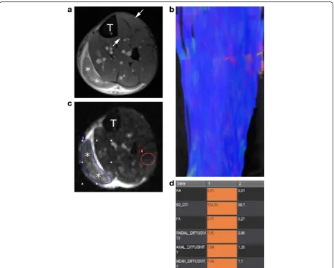
Tractography represents an application of DTI [21] that allows the analysis of muscle architecture aspects such as pennation angle, curvature of fibres, fibre length and possible muscle fibrosis (Fig. 1) [22]. Generally, muscle fibres are aligned parallel and do not diverge or curve as myelin sheath tracts; for this reason, it is possible to use less gradient directions than in the study of nervous structures. Accurate FA maps are necessary to manually or automatically draw a region of interest within the muscle. The features of the analysed tract may depend on some intrinsic muscles properties such as fatty infiltration, muscle atrophy, fibres tracking algorithm setting and the possible presence of partial volume arte-facts [23].
Specific applications in the musculoskeletal system
Experimental applications
chick embryonic skeletal muscle development in ovo (dur-ing incubation days 5–18 under a 3 T scanner) and investi-gated the correlation between FA and fibre length, finding that the result of DTI-tracked fibres during incubation cor-responds to the development of chick embryonic skeletal muscle [26]. McMillan et al. [27] found increased axial and radial diffusivity on ADC maps and decreased FA of muscle in dystrophic wild-type mice vs. normal controls.
Normal muscle tissue
Feasibility of DTI in the evaluation of normal muscle tis-sue has been widely demonstrated, with high reproduci-bility [16]. In different studies, capareproduci-bility of DTI in evaluating muscle cross-sectional area, fibre length and pennation angle has been demonstrated. Some studies
demonstrated feasibility of DTI to assess changes of fibre orientation according to positional variations of different body segments [28]. DTI was also used to measure fibre orientation, which has been reported to be a parameter that potentially predicts the pattern of tearing after muscle strain and evaluates the presence of a muscular lesion. An example of DTI of normal muscle tissue is reported in Fig. 1a and b.
Muscle contraction and muscle injury
average magnitude of molecular displacement by diffu-sion, were significantly increased in the biceps femoris muscle; also, MD was significantly increased in the semi-tendinosus and gracilis muscles two days after the mara-thon, while there were no changes on fat-suppressed T2-weighted sequences [31]. Zaraiskaya et al. [32] studied four patients with gastrocnemius and soleus muscles in-juries comparing them to eight healthy controls. Authors found significant differences in FA and ADC values in injured skeletal muscle which presented very low values (0.08 ± 0.02 vs. 0.23 ± 0.02 in healthy controls). Since diffusivity increases only in the muscles that are more injured after running, these data imply that DTI parame-ters might become a powerful diagnostic tool for prog-nosis and response assessment to treatment of sports-related muscle injuries (Fig. 1a, c, d). Further future ap-plication of DTI might be the monitor of restitution ad integrum of torn muscles in athletes.
Muscular dystrophy
Under the name of muscular dystrophy, a group of gen-etic, progressive and degenerative muscular diseases is included, whose primary symptom is muscle weakness [33]. These pathologic conditions may occur at any age with variable clinical features [34]. The early-onset muscu-lar dystrophies may be associated with profound loss of muscle function, affecting ambulation and posture, and may lead to respiratory and cardiac impairment. Con-versely, late-onset muscular dystrophies or myopathies may be less symptomatic and associated with slight weak-ness and inability to increase muscle mass [34].
Duchenne muscular dystrophy (DMD) is the most com-mon form of inherited muscular dystrophy in children. It is caused by mutations in the X-linked dystrophin gene and is characterised by systemic muscle weakness due to the pro-gressive destruction of skeletal muscle [35]. In this setting, quantitative evaluation of muscle status may be very im-portant, both to reliably aiding the diagnosis and to moni-tor the effect of treatments [33]. Until a few years ago, disease progression was only evaluated quantitatively on MRI. Mercuri et al. developed a four-point grading system based on fatty tissue infiltration to categorise disease sever-ity [36]. Ponrartana et al. [35] focused their attention on
tractography images showed the expected decrease in fibre length, number and architecture, suggesting that fibre tracking may provide further quantitative information (Fig. 2). Li et al. [38] showed that damage to thigh muscles in DMD patients can be evaluated by ADC and FA values and can be applied to assess quantitatively disease severity.
Ligaments
Arthroscopic grafting for ligament tears is common in active population [39] and MRI is routinely used to as-sess graft integrity. Despite the high diagnostic performance of conventional MRI [40], it cannot allow advanced quantitative evaluation. Graft ligaments pro-gress through four stages: avascular necrosis; revasculari-sation; cellular proliferation; and remodelling [41], with different signal intensities at conventional MRI. The po-tential use of DTI was firstly reported by Yang et al. [42], who performed DTI sequences in 40 healthy volun-teers and in 15 patients with anterior cruciate ligament (ACL) reconstruction. He found that ACL grafts in dif-ferent stages present difdif-ferent FA and ADC values, with significantly higher FA value at ten years from surgery. More recently, Van Dyck et al. [43] found lower FA and higher MD values of ACL graft in comparison to the study by Yang et al. [42]. Differences could be related to a different reconstruction software and a shorter time between surgery and MRI [43]. Indeed, after four months, thick synovial tissue starts to envelop the graft providing its vascular supply [44]. Future research may be aimed to test feasibility and reliability of DTI to quantitatively assess the fibrillar structure, especially in case of a partially torn ligament (Fig. 3).
Peripheral neuropathies
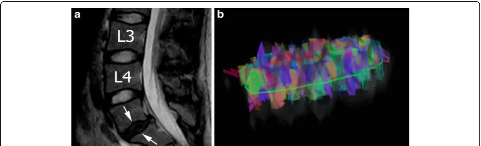
Torg-Pavlov ratio and potential signal changes of the compressed spinal tract [47]. However, all these findings are not always seen in symptomatic patients and there are often discrepancies between clinical and imaging features [48]. DTI and tractography can detect micro-architectural changes of CSC [29]. Studies reported an increase in MD values and a decrease in FA values around compressed CSC [49]. The diminished FA value seems to be a more sensitive parameter of cord injury than T2-weighted signal hyperintensity [50] and appears to be strictly correlated to symptom severity [51]. Trac-tography can detect abnormalities of the annulus fibro-sus. The normal annulus has a consecutive and regular ring configuration composed by multilayer fibres, while the degenerative annulus shows irregular and disordered aspect (Fig. 4), becoming thinner and disrupted [52].
Brachial plexus
Evaluation of brachial plexus abnormalities represents a diagnostic challenge due to anatomical complexity and to overlapping presentation of both benign and malignant
conditions. There is a wide range of disease which may involve the brachial plexus, including tumours, radiation fibrosis, trauma and inflammatory processes. MRI is the imaging modality of choice for the evaluation of the brachial plexus [53]. To date, DTI of the brachial plexus at 3.0 T has been reported as feasible and reproducible in a series of healthy volunteers [54].
Cubital tunnel syndrome
Cubital tunnel syndrome is the second most common peripheral compression neuropathy after carpal tunnel syndrome (CTS) [55]. It is related to a combination of compression and friction of the ulnar nerve in the cubital tunnel [56]. Breitenseher et al. [57] showed significant reduction of ulnar nerve FA values at the cubital groove and when passing the deep flexor fascia. At tractography, they showed complete or par-tial discontinuity of the ulnar nerve in 65% of pa-tients. However, they conclude that T2 neurography is more sensitive than DTI in the detection of cubital tunnel syndrome [57].
Fig. 3Magnetic resonance of the knee performed on a 24-year-old male patient two months after soccer injury.aSagittal T1-weighted image shows that the anterior cruciate ligament is remarkably inhomogeneous due a partial tear (arrows).Ffemur,Ttibia.bThe corresponding tractography image shows that fibres are partially interrupted and architecture is disrupted (arrows)
Carpal tunnel syndrome
CTS is the most common compressive peripheral neur-opathy, affecting 3–4% of the general population [58]. It is caused by an entrapment of the median nerve at the level of the carpal tunnel of the wrist. The diagnosis of CTS is usually established by clinical features, clinical tests, electrophysiological tests, and high-resolution ultrasonography [59]. However, electrophysiology is as-sociated with pain and conspicuous rate of false-negative and false-positive results [60].
Conventional MRI of CTS gives variable qualitative re-sults [61]. DTI may potentially improve the diagnosis of peripheral nerve disorders, optimise lesion localisation, and determine the extent of neural dysfunction [62]. FA tends to increase distally in healthy participants and to de-crease in patients with CTS for the presence of intrafasci-cular oedema. Kabakci et al. reported that FA values of two patients (FA = 0.41 and 0.44) were significantly lower than that of the control group (FA = 0.709 ± 0.046). Diffu-sion parameters are not influenced by the patients’gender, while changes in diffusion and FA similar to those ob-served in CTS may appear with age, also without clinical symptoms [63]. Indeed, FA measured at the carpal tunnel inlet seems to have the highest accuracy for the diagnosis of CTS (sensitivity of 62%, specificity of 82%, positive pre-dictive value of 80%, negative prepre-dictive value of 75%) [64] compared to other imaging modalities, and it showed the highest correlation with sensory and motor amplitude (r = 0.54, p< 0.001), even though it always needs to be correlated with clinical symptoms.
Sciatic nerve and piriformis syndrome
Sciatic neuropathy is a common cause of lower extrem-ity pain, which can be related to several causes affecting any level of the nerve and resulting in different symp-toms [65]. The diagnosis is made through the patient history, clinical findings, and electrophysiological tests
[66]. Routine MRI has been widely used to study the normal anatomy and pathology of the sciatic nerve and the associated muscle oedema or denervation, especially on T2-weighted sequences [67]. DTI may implement conventional MRI examination, providing additional in-formation regarding the myelinic structures [68]. DTI does not show significant differences in either FA or ADC values at any level between sciatic nerve in the dominant or non-dominant lower limb. Wata et al. eval-uated ten patients with sciatica symptoms, reporting lower FA and higher ADC values in the affected sciatic nerve compared with healthy controls [69].
Piriformis syndrome (PS) is a not easily recognisable disorder, characterised by buttock and leg pain due to compression of the sciatic nerve through or around the piriformis muscle in patients with usually normal neuro-logical examination [70]. Manoeuvres of flexion, adduc-tion and internal rotaadduc-tion of the hip, and direct palpation of the piriformis, cause severe pain. CT and MRI may help to diagnose PS and to differentiate it from other possible causes of lower lumbar pain; however, im-aging diagnosis remains challenging [68].
Nerve tumours
Previous studies reported higher frequency of destruction or massive fibre disorganisation in malignant peripheral nerve tumours, whereas benign tumours are often associated with dislocation or partial interruption of fibres. Chhabra et al. reported that tractography may provide three-dimensional visualisation of fibre dislocation or destruction. On the other hand, FA and MD show low values that indicate malig-nancy extension in neural structures [71].
Conclusion
these tools are mainly applied in experimental settings and are uncommonly translated to clinical practice.
Since application of DTI and tractography in the mus-culoskeletal and peripheral nerve systems is different from application in the central nervous system, some is-sues still need to be addressed, such as: standardisation of acquisition protocols with optimised parameters and duration times; optimisation of post-processing software for DTI to be used in the musculoskeletal system; inves-tigation of the possibility of using DTI around metallic implants, used in clinical practice more and more frequently; and analysis of reproducibility of DTI quanti-tative measurements (inter-study reproducibility; com-parison among MR units from different vendors and between 1.5 and 3 T magnets; comparison among differ-ent DTI post-processing software).
At any rate, DTI and tractography seem to be promising tools and are able to provide useful quantitative informa-tion about muscular tissue and peripheral nerves as an ad-junct to morphological MRI sequences. The definition of the real clinical added value of this approach requires well designed studies on large population samples, possibly with clinical end-points potentially showing a benefit to the pa-tients which is not limited to diagnostic performance.
Abbreviations
ACL:Anterior cruciate ligament; ADC: Apparent diffusion coefficient; CSM: Cervical spondylotic myelopathy; CTS: Carpal tunnel syndrome; DMD: Duchenne muscular dystrophy; DTI: Diffusion tensor imaging; DWI: Diffusion-weighted imaging; FA: Fractional anisotropy; MD: Mean diffusivity; MRI: Magnetic resonance imaging; PS: Piriformis syndrome; SNR: Signal-to-noise ratio; TE: Time of echo; TR: Time of repetition
Authors’contributions
All authors read and approved the final manuscript.
Competing interests
FS is the Editor-in-chief of European Radiology Experimental. For this reason, he was not involved in any way in the revision/decision process, which was completely managed by the Deputy Editor. The other authors have no competing interests related to the present paper to disclose.
Publisher’s Note
Springer Nature remains neutral with regard to jurisdictional claims in published maps and institutional affiliations.
Author details 1
Department of Advanced Biomedical Sciences, Università Federico II, Via Pansini 5, 80131 11 Napoli, Italy.2Department of Radiology, DIBIMED, Università di Palermo, Via del Vespro 127, 90127 Palermo, Italy.3Unit of Neuroradiology, Fondazione IRCCS Ca’Granda Ospedale Maggiore Policlinico, Via Francesco Sforza 35, 20122 Milano, Italy.4Unit of Radiology, IRCCS Policlinico San Donato, Via Morandi 30, 20097 San Donato Milanese, Italy.5Department of Pathophysiology and Transplantation, Università degli Studi di Milano, Via Festa del Perdono 7, 20122 Milano, Italy.6Department of Biomedical Sciences for Health, Università degli Studi di Milano, Via Mangiagalli 31, 20133, 20122 Milano, Italy.7Unit of Diagnostic and Interventional Radiology, IRCCS Istituto Ortopedico Galeazzi, Via Riccardo Galeazzi 4, 20161 Milano, Italy.
Received: 22 May 2017 Accepted: 1 August 2017
References
1. Coté C, Hiba B, Hebert LJ et al (2011) MRI of tibialis anterior skeletal muscle in myotonic dystrophy type 1. Can J Neurol Sci 38:112–118
2. Sardanelli F (2017) Trends in radiology and experimental research. Eur Radiol Exp. 1:1.
3. Patni RS, Boruah DK, Sanyal S et al (2017) Characterisation of musculoskeletal tumours by multivoxel proton MR spectroscopy. Skeletal Radiol 46:483–495 4. Torriani M (2007) Measuring muscle lipids with 1H-MR spectroscopy.
Skeletal Radiol 36:607–608
5. ValkovičL, Chmelík M, Krššák M (2017) In-vivo 31P-MRS of skeletal muscle and liver: A way for non-invasive assessment of their metabolism. Anal Biochem 529:193–215
6. Pozzi G, Albano D, Messina C et al (2017) Solid bone tumors of the spine: Diagnostic performance of apparent diffusion coefficient measured using diffusion-weighted MRI using histology as a reference standard. J Magn Reson Imaging. doi:10.1002/jmri.25826.
7. Noseworthy MD, Davis AD, Elzibak AH (2010) Advanced MR imaging techniques for skeletal muscle evaluation. Semin Musculoskelet Radiol 14:257–268
8. Teixeira PA, Beaumont M, Gabriela H et al (2015) Advanced techniques in musculoskeletal oncology: perfusion, diffusion, and spectroscopy. Semin Musculoskelet Radiol 19:463–474
9. Le Bihan D, Breton E (1985) Imagerie de diffusion in vivo par resonance magnetique nucleaire. CR Acad Sci Paris 301:1109–1112
10. Abhinav K, Yeh FC, Pathak S et al (2014) Advanced diffusion MRI fiber tracking in neurosurgical and neurodegenerative disorders and neuroanatomical studies: a review. Biochim Biophys Acta 1842:2286–2297 11. Le Bihan D, Clark CA, Poupon C et al (2001) Diffusion tensor imaging:
concepts and applications. J Magn Reson Imaging 13:534–546
12. Albano D, La Grutta L, Grassedonio E et al (2016) Pitfalls in whole body MRI with diffusion weighted imaging performed on patients with lymphoma: what radiologists should know. Magn Reson Imaging 34:922–931 13. Cotten A, Haddad F, Hayek G, Lefebvre G, Dodré E, Budzik JF (2015)
Tractography: possible applications in musculoskeletal radiology. Semin Musculoskelet Radiol 19:387–395
14. Le Bihan D (1991) Molecular diffusion nuclear magnetic resonance imaging. Magn Reson Q 7:1–30
15. Oudeman J, Nederveen AJ, Strijkers GJ, Maas M, Luijten PR, Froeling M (2016) Techniques and applications of skeletal muscle diffusion tensor imaging: a review. J Magn Reson Imaging 43:773–788
16. Soares JM, Marques P, Alves V, Sousa N (2013) A hitchhiker’s guide to diffusion tensor imaging. Front Neurosci 7:31
17. Guggenberger R, Eppenberger P, Markovic D et al (2012) MR neurography of themedian nerve at 3.0T: optimization of diffusion tensor imaging and fiber tractography. Eur J Radiol 81:e775–e782
18. Schlaffke L, Rehmann R, Froeling M et al (2017) Diffusion tensor imaging of the human calf: Variation of inter- and intramuscle-specific diffusion parameters. J Magn Reson Imaging. doi:10.1002/jmri.25650.
19. Choi S, Cunningham DT, Aguila F et al (2011) DTI at 7 and 3 T: systematic comparison of SNR and its influence on quantitative metrics. Magn Reson Imaging 29:739–751
20. Alexander AL, Lee JE, Wu YC, Field AS (2006) Comparison of diffusion tensor imaging measurements at 3.0T versus 1.5T with and without parallel imaging. Neuroimaging Clin N Am 16:299–309
21. Qin W, Yu CS, Zhang F, Du XY, Yan YX, Li KC (2009) Effects of echo time on diffusion quantification of brain white matter at 1.5 T and 3.0 T. Magn Reson Med 61:755–760
22. Damon BM, Heemskerk AM, Ding Z (2012) Polynomial fitting of DT-MRI fiber tracts allows accurate estimation of muscle architectural parameters. Magn Reson Imaging 30:589–600
23. Hori M, Ishigame K, Shiraga N, Kumagai H, Aoki S, Araki T (2008) Mean diffusivity, fractional anisotropy maps, and three-dimensional white-matter tractography by diffusion tensor imaging. Comparison between single-shot fast spin-echo and single-shot echo-planar sequences at 1.5 Tesla. Eur Radiol 18:830–834
Fractional anisotropy values of calf muscles in normative state after exercise: preliminary results. Magn Reson Med Sci 7:157–162
31. Froeling M, Oudeman J, Strijkers GJ et al (2015) Muscle changes detected with diffusion-tensor imaging after long-distance running. Radiology 274:548–562
32. ZaraiskayaT KD, Noseworthy MD (2006) Diffusion tensor imaging in evaluation of human skeletalmuscle injury. J Magn Reson Imaging 24:402–408
33. Angelini C, Tasca E (2012) Fatigue in muscular dystrophies. Neuromuscul Disord 22:S214–S220
34. McNally EM, Pytel P (2007) Muscle diseases: the muscular dystrophies. Annu Rev Pathol 2:87–109
35. Ponrartana S, Ramos-Platt L, Wren TA et al (2015) Effectiveness of diffusion tensor imaging in assessing disease severity in Duchenne muscular dystrophy: preliminary study. Pediatr Radiol 45:582–589
36. Mercuri E, Bushby K, Ricci E et al (2005) Muscle MRI findings in patients with limb girdle muscular dystrophy with calpain 3 deficiency (LGMD2A) and early contractures. Neuromuscul Disord 15:164–171
37. Williams SE, Heemskerk AM, Welch EB, Li K, Damon BM, Park JH (2013) Quantitative effects of inclusion of fat on muscle diffusion tensor MRI measurements. J Magn Reson Imaging 38:1292–1297
38. Li GD, Liang YY, Xu P, Ling J, Chen YM (2016) Diffusion-tensor imaging of thigh muscles in duchenne muscular dystrophy: correlation of apparent diffusion coefficient and fractional anisotropy values with fatty infiltration. AJR Am J Roentgenol 206:867–870
39. Csintalan RP, Inacio MC, Funahashi TT (2008) Incidence rate of anterior cruciate ligament reconstructions. Perm J 12:17–21
40. Adriaensen ME, Hogan B, Al-Bulushi HI, Kavanagh EC (2012) Double-bundle depiction of the anterior cruciate ligament at 3 Tesla. Skeletal Radiol 41:831–834
41. Abe S, Kurosaka M, Iguchi T, Yoshiya S, Hirohata K (1993) Light and electron microscopic study of remodeling and maturation process in autogenous graft for anterior cruciate ligament reconstruction. Arthroscopy 9:394–405 42. Yang X, Li M, Chen D et al (2014) Diffusion tensor imaging for anatomical
and quantitative evaluation of the anterior cruciate ligament and ACL grafts: a preliminary study. J Comput Assist Tomogr 38:489–494
43. Van Dyck P, Froeling M, De Smet E, Pullens P, Torfs M, Verdonk P et al (2017) Diffusion tensor imaging of the anterior cruciate ligament graft. J Magn Reson Imaging. doi:10.1002/jmri.25666
44. Janssen RP, Scheffler SU (2014) Intra-articular remodelling of hamstring tendon grafts after anterior cruciate ligament reconstruction. Knee Surg Sports Traumatol Arthrosc 22:2102–2108
45. Gore DR, Sepic SB, Gardner GM (1986) Roentgenographic findings of the cervical spine in asymptomatic people. Spine (Phila Pa 1976) 11:521–524 46. Wang K, Chen Z, Zhang F et al (2017) Evaluation of DTI parameter ratios and
diffusion tensor tractography grading in the diagnosis and prognosis prediction of cervical spondylotic myelopathy. Spine (Phila Pa 1976) 15:E202–E210
47. Fernandez de Rota JJ, Meschian S, Fernandez de Rota A, Urbano V, Baron M (2007) Cervical spondylotic myelopathy due to chronic compression: the role of signal intensity changes in magnetic resonance images. J Neurosurg Spine 6:17–22 48. Lee JW, Kim JH, Park JB et al (2011) Diffusion tensor imaging and fiber tractography in cervical compressive myelopathy: preliminary results. Skeletal Radiol 40:1543–1551
49. Facon D, Ozanne A, Fillard P, Lepeintre JF, Tournoux-Facon C, Ducreux D (2005) MR diffusion tensor imaging and fiber tracking in spinal cord compression. AJNR Am J Neuroradiol 26:1587–1594
tunnel syndrome. J Hand Surg [Am] 40:1897–1904
57. Breitenseher JB, Kranz G, Hold A et al (2015) MR neurography of ulnar nerve entrapment at the cubital tunnel: a diffusion tensor imaging study. Eur Radiol 25:1911–1918
58. Ibrahim I, Khan WS, Goddard N et al (2012) Carpal tunnel syndrome: a review of the recent literature. Open Orthop J 6:69–76
59. Mondelli M, Filippou G, Gallo A, Frediani B (2008) Diagnostic utility of ultrasonography versus nerve conduction studies in mild carpal tunnel syndrome. Arthritis Rheum 59:357–366
60. Nathan PA, Keniston RC, Meadows KD, Lockwood RS (1993) Predictive value of nerve conduction measurements at the carpal tunnel. Muscle Nerve 16:1377–1382 61. Martins RS, Siqueira MG, Simplicio H, Agapito D, Medeiros M (2008)
Magnetic resonance imaging of idiopathic carpal tunnel syndrome: correlation with clinical findings and electrophysiological investigation. Clin Neurol Neurosurg 110:38–45
62. Razek AA, Shabana AA, El Saied TO, Alrefey N (2016) Diffusion tensor imaging of mild-moderate carpal tunnel syndrome: correlation with nerve conduction study and clinical tests. Clin Rheumatol. doi:10.1007/s10067-016-3463-y 63. Kabakci N, Gürses B, Firat Z et al (2007) Diffusion tensor imaging and tractographyofmedian nerve: normative diffusion values. AJR Am J Roentgenol 189:923–927
64. Kwon BC, Koh SH, Hwang SY (2015) Optimal parameters and location for diffusion-tensor imaging in the diagnosis of carpal tunnel syndrome: a prospective matched case-control study. AJR Am J Roentgenol 204:1248–1254 65. Chhabra A, Faridian-Aragh N, Chalian M, Soldatos T, Thawait SK, Williams EH et al (2012) High-resolution 3-T MR neurography of peroneal neuropathy. Skeletal Radiol 41:257–271
66. Gutmann L (2003) Pearls and pitfalls in the use of electromyography and nerve conduction studies. Semin Neurol 23:77–82
67. Thawait SK, Chaudhry V, Thawait GK et al (2011) High-resolution MR neurography of diffuse peripheral nerve lesions. AJNR Am J Neuroradiol 32:1365–1372
68. Bernabeu Á, López-Celada S, Alfaro A, Mas JJ, Sánchez-González J (2016) Is diffusion tensor imaging useful in the assessment of the sciatic nerve and its pathologies? Our clinical experience. Br J Radiol 89:20150728 69. Wada K, Hashimoto T, Miyagi R, Sakai T, Sairyo K (2017) Diffusion tensor
imaging and tractography of the sciatic nerve: assessment of fractional anisotropy and apparent diffusion coefficient values relative to the piriformis muscle, a preliminary study. Skeletal Radiol 46:309–314 70. Lee EY, Margherita AJ, Gierada DS, Narra VR (2004) MRI of piriformis
syndrome. AJR Am J Roentgenol 183:63–64