7. Troubleshooting 31
Wound deterioration 31
Patient experiencing pain 31
Safety alarms 32
8. Appendices 33
Appendix 1 – Wound sealing kit guides 33
Appendix 2 – Preparing the wound bed 34
Appendix 3 – Quick reference dressing guidlines 35
Appendix 4 – Specialist dressing techniques 36
Appendix 5 – Quick reference clinical guidance 37
6. Healing progress – knowing when
to stop or change treatment 29
Wound appearance and dimensions 29
Volume and appearance of exudate 29
Length of treatment 29
Discontinuing NPWT therapy 30
4. Specialist applications 19
Undermined wounds 19
Tunnelled wounds 19
Options for treating multiple wounds 20
The bridging technique 20
Skin grafts 21
Dermal Substitutes 22
Skin flaps 23
Fistulae 24
Abscess dressing technique 25
3. Application 13
Patient preparation 13
Preparing the wound for dressing application 15
Using RENASYS™ G 15
Connecting NPWT devices and commencing therapy 17
Disconnecting NPWT devices 18
Dressing changes 18
Changing the canister 18
2. NPWT consumables 12
Choosing an appropriate drain 12
1. Introducing NPWT 4 Indications 6 Precautions 6 Contraindications 9 General guidelines 10 NPWT pressure settings 11
1. Introducing NPWT
The Smith & Nephew Negative Pressure Wound Therapy (NPWT) systems are designed to apply localised negative pressure (suction) to a patient’s wound site. It consists of a suction pump to generate negative pressure and a variety of wound sealing kits to deliver the therapy to the wound site.
The purpose of this document is to describe the most appropriate use of NPWT as an integral part of wound bed preparation to ensure optimal healing and cost-effective woundcare. Wound bed preparation has been defined as the process of removing the barriers to healing. Removal of these barriers is thought to allow the wound repair process to progress normally. Wound bed preparation represents a combination of both scientific knowledge and practical skill; its application can help correct abnormalities in chronic wounds and stimulate the healing process (Schultz, 2003). To optimise the use of NPWT it is essential that clinicians ensure wound bed
preparation is achieved prior to, during and after therapy.
Optimise healing
• Removes excess fluid oedema • Assists in wound contraction • Stimulates granulation tissue • Protects from outside contaminants • Increases vascular perfusion • May reduce wound bioburden
Debride wound
Effective debridement may: • Reveal extent of tissue damage
• Reduce biochemical imbalance, senescent cells • Reduce bacterial burden
• Reduce odour
• Optimise healing potential
Prepare
the wound
environment
Know when to stop or change treatment
• Initial therapy objectives have been met • 100% granulation tissue in the wound bed • Granulation tissue level with the surrounding skin • Patient’s overall condition/wound is improving • Wound bed ready to take a skin graft/flap • Exudate levels less than 20–50mls per day • No improvement/reduction in size is seen in the
wound bed following two consecutive dressing changes
Know when
to stop
or change
treatment
Optimal healing
Cost-effective woundcare
Indications, precautions, contraindications and
discontinuation of NPWT
Successful NPWT depends on conditions which are essential to wound resolution, for example:
• The wound bed must have sufficient blood flow
• The patient must receive adequate nutrition/ nutritional supplements, if they are malnourished
• Pressure relief, if appropriate, should be used • Other co-morbidities must be optimised
NPWT indications
NPWT is indicated in a number of different wounds: • Post-operative and dehisced surgical wounds • Pressure ulcers
• Diabetic/neuropathic ulcers • Traumatic wounds
• Skin flaps and grafts • Venous insufficiency ulcers • Explored fistulae
Certain wounds with particular attributes may be ideal for NPWT, for example:
• Wounds that are resistant to traditional wound care and showing signs of chronicity or little progress towards healing
• Deep / cavity wounds with high levels of wound exudate
NPWT precautions
Precautions should be taken when using NPWT in the following cases: • Malnourished patients who have not received adequate nutrition/nutritional
supplements
• Children (Please see page 8 for additional information)
• Extra caution should be used for patients with neuropathic aetiologies or circulatory compromise
• Bone fragments or sharp edges could puncture protective barriers, vessels, or organs causing injury. Any injury could cause bleeding, which, if uncontrolled, could be
potentially fatal. Beware of possible shifting in the relative position of tissues, vessels or organs within the wound that might increase the possibility of contact with sharp edges. Sharp edges or bone fragments must be eliminated from the wound area or covered to prevent them from puncturing blood vessels or organs before the application of NPWT. Where possible, completely smooth and cover any residual edges to decrease the risk of serious or fatal injury, should shifting of structures occur. Use caution when removing dressing components from the wound so that wound tissue is not damaged by unprotected sharp edges.
• Enteric fistulae require special precautions to optimise therapy (See specialist dressing section for options)
• Defibrillation: Remove the dressing if defibrillation is required in the area of dressing placement. Failure to remove the dressing may inhibit transmission of electrical energy and/or patient resuscitation.
• Magnetic Resonance Imaging (MRI) – Therapy Unit: The Therapy Unit is MR Unsafe. Do not take the Therapy Unit into the MR environment.
• Magnetic Resonance Imaging (MRI) – Dressings: Dressings can typically remain on the patient with minimal risk in an MR environment.
• Hyperbaric Oxygen Therapy (HBO): Do not take the Therapy Unit into a hyperbaric oxygen chamber. The Therapy Unit is not designed for this environment, and should be considered a fire hazard. After disconnecting the Therapy Unit, either (i) replace the dressing with another HBO compatible material during the hyperbaric treatment, or (ii) cover the unclamped end of the tubing with moist cotton gauze.
Although not contraindicated with NPWT, extra care should be taken in patients with bleeding problems, including:
• Haemostatic Agents applied at the wound site: Non-sutured haemostatic agents for example, bone wax, absorbable gelatin sponge, or spray wound sealant may, if disrupted, increase the risk of bleeding, which, if uncontrolled, could be potentially fatal. Protect against dislodging such agents. Consideration should be given to the negative pressure setting and therapy mode used when initiating therapy.
• Patients receiving long-term anticoagulant therapy • Patients with haemophilia
• Patients with haemoglobinopathies, such as sickle cell disease
• Patients receiving anticoagulant therapy or platelet aggregation inhibitors, or those who are actively bleeding or have weakened irradiated blood vessels or organs.
If significant bleeding develops, immediately discontinue the use of the
RENASYS™; take measures to stop the bleeding. Do not remove the dressing until the treating clinician or surgeon is consulted. Do not resume the use of NPWT until adequate haemostasis has been achieved, and the patient is not at risk for continued bleeding.
• Spinal Cord Injury: In the event a patient experiences autonomic hyperreflexia (sudden elevation in blood pressure or heart rate in response to stimulation of the sympathetic nervous system), discontinue NPWT to help minimize sensory stimulation and seek immediate medical assistance.
• Patient Size and Weight: The size and weight of the patient should be considered when prescribing NPWT. Infants, children, certain small adults and elderly patients should be closely monitored for fluid loss and dehydration. Also, patients with highly exudating wounds or large wounds in relation to the patient size and weight should be closely monitored, as they may have a risk of excessive fluid loss and dehydration. When monitoring fluid output, consider the volume of fluid in both the tubing and canister. • 800mL Canister Size: DO NOT USE the 800mL canister on patients with a high risk of
bleeding or on patients unable to tolerate a large loss of fluid volume, including children and the elderly. Consider the size and weight of the patient, patient condition, wound type, monitoring capability and care setting when using this canister. This canister is recommended for acute care (hospital) use only.
NPWT and wound infection
NPWT is safe to use on colonised, critically-colonised and infected wounds. Most wounds, particularly chronic wounds, such as pressure ulcers and leg ulcers, are colonised by bacteria. These can be grown on a wound swab, but do not necessarily delay healing or create undue problems with the healing environment. When a wound becomes critically colonised or clinically infected, a host response occurs. Patients receiving NPWT should be monitored for clinical signs and symptoms of wound infection, and additional treatments instigated accordingly, as and when such conditions arise.
• Closely monitor and replace any fluid loss into canister with neonates/infants and with younger children as per lead clinician instructions. Children always use a small canister and consider partially pre filling the volume with sterile saline/sterile water to ensure canister full alarm alerts user to excess fluid loss.
• Progress should be seen in the first week of therapy • Average treatment time is 1–2 weeks
NPWT contraindications
NPWT is contraindicated in certain cases:
• Untreated osteomyelitis: Suspected or confirmed osteomyelitis must be treated as per local guidelines prior to and during any NPWT, until resolved.
• Malignancy (except palliative care): NPWT is not recommended in malignant wounds, but can be used post-tumour removal, if tissue margins are clear of disease (if unsure, please ask for guidance or clarification from lead clinician).
• Unexplored fistulae: Fistulae must be investigated to establish the location and extent of communication between organs and establish causation, e.g. a small bowel fistulae with a distal obstruction will not seal unless the distal obstruction is cleared. However, if there is no distal obstruction, it may heal over with appropriate management. As such, the endpoints of care with NPWT will differ depending on the results of investigations.
• Exposed arteries, veins or organs: Negative pressure, directly applied to exposed arteries or veins, could result in vessel compromise. The patient’s consultant must approve use of NPWT prior to use.
• Necrotic tissue with eschar/ dry wounds: If a dry, necrotic eschar is covering the wound, or if there is a large amount of necrotic tissue present, NPWT is not appropriate. Best results are obtained if the wound is debrided prior to NPWT application. Small amounts of soft tissue slough will autolyse during therapy. As long as haemostasis
is achieved, NPWT should be applied as soon after debridement as possible, if not immediately.
• Severe peripheral arterial disease: Ankle Brachial Pressure Index ≤ 0.5 needs investigation and, if appropriate, revascularisation prior to commencement of NPWT. • Any cavity/ sinus, of which the origin is not clearly visible, or cannot be gently probed to
gain an indication of origin: Complications are rare, but can include infection, desiccation, pain, erosions, odour, and tissue in growth, maceration, and bullae.
General NPWT dressing guidelines and precautions
• Dressing Placement: Always use dressings from sterile packages that have not been opened or damaged. Do not place any RENASYS™ F dressings into blind/unexplored tunnels. The RENASYS G Channel drain may be more appropriate for use with explored tunnels. Do not force dressings into any area of the wound, as this may damage tissue, alter the delivery of negative pressure, or hinder exudate and dressing removal.
Always count the total number of pieces of dressing used in the wound and document that number on the drape and in the patient’s chart. Also document the dressing change date on the drape.
• Dressings are not bio-absorbable: Always count the total number of pieces of
dressing removed from the wound and ensure the same number of pieces was removed as placed. Dressings left in the wound for greater than the recommended time period may foster in growth of tissue, and create difficulty in removing them from the wound, or lead to infection or other adverse events.
• Standard Dressing Precautions: To reduce the risk of transmission of blood borne pathogens, apply standard precautions for infection control with all patients, per institutional protocol, regardless of their diagnosis or presumed infection status. In addition to gloves, use gown and goggles if exposure to body fluid is likely.
• Protect Periwound Skin: Consider use of a skin preparation product to protect periwound skin. Do not allow RENASYS™ F to overlap onto intact skin. Protect fragile/friable
periwound skin with additional Drape, hydrocolloid or other transparent film. Multiple layers of the drape may decrease the moisture vapour transmission rate, which may increase the risk of periwound maceration. To avoid trauma to the periwound skin, do not pull or stretch the drape over dressings during drape application.
• Acrylic Adhesive: The Drape has an acrylic adhesive coating, which may present a risk of an adverse reaction in patients who are allergic or hypersensitive to acrylic adhesives. If a patient has a known allergy or hypersensitivity to such adhesives, do not use the RENASYS. If any signs of allergic reaction or hypersensitivity develop, such as redness, swelling, rash, urticaria, or significant pruritus, discontinue use and consult a physician immediately.
Negative pressure settings
The guidelines on therapy settings in this booklet are general recommendations. You may wish to vary the pressure settings to optimise NPWT therapy based on individual patient need and the lead clinician’s guidance.
Negative pressure may be increased by 10mmHg increments where: • High levels of exudate/excessive drainage
• Large wound volume/size
• There is a tenuous seal/positional difficulties maintaining the seal
• High pressure may cause tissue necrosis and increase pain for patients. Careful
consideration and consultation with the lead clinician should be sought prior to exceeding -120 mmHg
Negative pressure may be decreased by 10mmHg increments where: • Pain or discomfort is not relieved by appropriate analgesia
• The patient is elderly and/or nutritionally compromised
• There is a risk of excessive bleeding (for example, patients on anticoagulation therapy) • The circulation is compromised (for example, peripheral arterial disease)
• There is excessive granulation tissue growth
• With pressures lower than -40 consider discontinuing therapy
• Continuous versus Intermittent Therapy: Continuous, rather than intermittent therapy
is recommended over unstable structures, such as an unstable chest wall or non-intact fascia, in order to help minimise movement and stabilise the wound bed. Continuous therapy is also generally recommended for patients at increased risk of bleeding, highly exudating wounds, fresh flaps and grafts, and wounds with acute enteric fistulae.
2. NPWT consumables
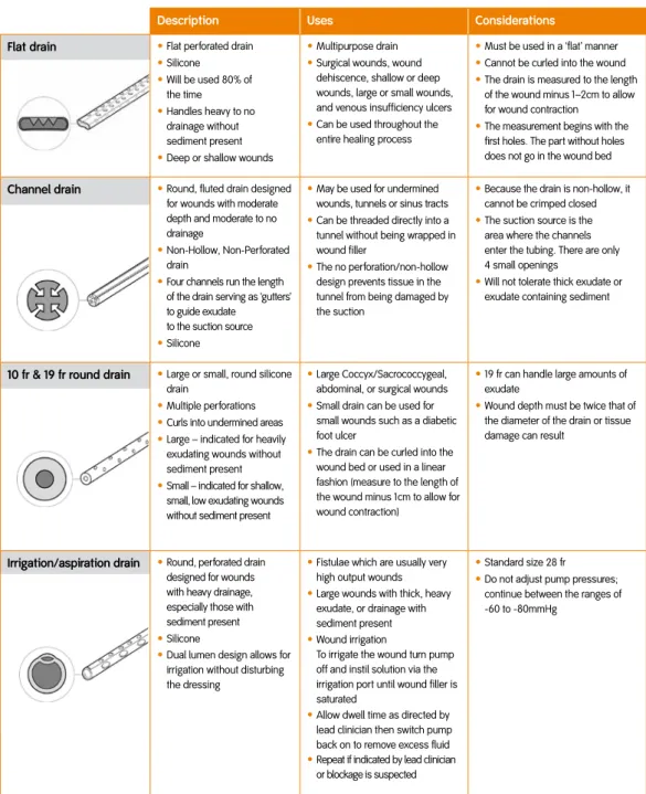
Description Uses Considerations
Flat drain • Flat perforated drain
• Silicone • Will be used 80% of the time • Handles heavy to no drainage without sediment present
• Deep or shallow wounds
• Multipurpose drain
• Surgical wounds, wound dehiscence, shallow or deep wounds, large or small wounds, and venous insufficiency ulcers
• Can be used throughout the entire healing process
• Must be used in a ‘flat’ manner
• Cannot be curled into the wound
• The drain is measured to the length of the wound minus 1–2cm to allow for wound contraction
• The measurement begins with the first holes. The part without holes does not go in the wound bed
Channel drain • Round, fluted drain designed for wounds with moderate depth and moderate to no drainage
• Non-Hollow, Non-Perforated drain
• Four channels run the length of the drain serving as ‘gutters’ to guide exudate to the suction source
• Silicone
• May be used for undermined wounds, tunnels or sinus tracts
• Can be threaded directly into a tunnel without being wrapped in wound filler
• The no perforation/non-hollow design prevents tissue in the tunnel from being damaged by the suction
• Because the drain is non-hollow, it cannot be crimped closed
• The suction source is the area where the channels enter the tubing. There are only 4 small openings
• Will not tolerate thick exudate or exudate containing sediment
10 fr & 19 fr round drain • Large or small, round silicone drain
• Multiple perforations
• Curls into undermined areas
• Large – indicated for heavily exudating wounds without sediment present
• Small – indicated for shallow, small, low exudating wounds without sediment present
• Large Coccyx/Sacrococcygeal, abdominal, or surgical wounds
• Small drain can be used for small wounds such as a diabetic foot ulcer
• The drain can be curled into the wound bed or used in a linear fashion (measure to the length of the wound minus 1cm to allow for wound contraction)
• 19 fr can handle large amounts of exudate
• Wound depth must be twice that of the diameter of the drain or tissue damage can result
Irrigation/aspiration drain • Round, perforated drain designed for wounds with heavy drainage, especially those with sediment present
• Silicone
• Dual lumen design allows for irrigation without disturbing the dressing
• Fistulae which are usually very high output wounds
• Large wounds with thick, heavy exudate, or drainage with sediment present
• Wound irrigation
To irrigate the wound turn pump off and instil solution via the irrigation port until wound filler is saturated
• Allow dwell time as directed by lead clinician then switch pump back on to remove excess fluid
• Repeat if indicated by lead clinician or blockage is suspected
• Standard size 28 fr
• Do not adjust pump pressures; continue between the ranges of -60 to -80mmHg
RENASYS™ G Dressings/Drains
Table 1 – Choosing an appropriate drainThe following recommendations offer a step-by-step guide to dressing application. Universal precautions should be observed. Use clean/aseptic or sterile techniques for application depending on local guidelines.
Prior to use of NPWT, the clinician(s) treating the wound must assess how to best use the system for an individual wound. It is important to carefully assess the wound and patient to ensure clinical indications for NPWT are met.
Patient preparation
In addition to considering the use of NPWT to benefit the patient and the wound, the clinician must first undertake a holistic assessment of the patient and consider the following points:
• Is the therapy appropriate for the wound, and also for the patient?
• The practitioner should consider the patient’s general wellbeing and state of health, as well as product indications and precautions, and concomitant therapies/ medication • What type of wound is to be treated, where is the wound located, and what type of
tissue is present?
Patient information
It is important to prepare the patient carefully before any procedure, as explanations are more likely to result in a relaxed patient, which makes any procedure much safer. If any pain is experienced, the procedure should be stopped immediately and analgesia reviewed before re-commencement, if appropriate.
There are many methods of treating wounds, and it is important that patients are involved in the decision, so that they are able to give informed consent. To make effective clinical decisions and ensure that patients are fully informed, it is essential that clinicians are aware of the different methods of treating wounds. The clinician should also be aware of the professional requirements for competence and the risks and benefits of each method.
Informed consent
It is vital that the patient understands what he or she has consented to, and it is an important role of the clinician to check and ensure that this happens (Cable, Lumsdaine and Semple, 2003). Effective communication is vital in gaining consent. Patients have a right to know exactly what they are consenting to, and information
should empower them and enable them to make a decision. The clinician needs to be able to provide a balanced perspective in a form that is appropriate for each individual patient.
In order to make an informed decision, the patient should know: • what equipment is proposed for use
• how the equipment works
• what the objective of the treatment is
• the likely impact of the treatment on the progression of wound healing • the likely outcome if the treatment is not given
• what alternatives are available
• any possible side effects (including pain), and how these will be managed
• any possible impact of the therapy on the patient’s quality of life or length of stay in hospital, and how long the treatment is likely to take
Some patients may be particularly interested in the mode of action of the treatment and how their wound is progressing. Patients should be encouraged to be as involved as possible in their treatment, and in their journey to wound healing.
Clinicians are accountable for their actions, and should always ensure that they are competent in obtaining consent from the patient. This means ensuring that you have the appropriate skills to undertake an assessment of the patient’s capacity to give consent, and the communications skills to ensure that they are adequately informed.
The following parameters should be noted at the outset of therapy: • Start date and duration of wound pre therapy
• Dressings, materials and supplies used including any adjunctive therapy or dressings • Pressure setting(s) to be used
• Frequency of dressing changes
• Wound type, location, size (length, width and depth). Tissue types present at the wound bed if necrotic tissue or slough is present consider debridement prior to
commencement of therapy to optimise outcomes.
Dressings should be changed 48 hours after initial application if no leak is present. If the patient is comfortable and the wound is progressing, dressing changes should occur 2–3 times a week there after. In the event of heavy exudate, more frequent dressing changed may be needed.
Preparing the wound for dressing application
• Appropriate debridement of eschar or hardened slough if present • Achieve haemostasis
• Thoroughly clean and irrigate wound according to local guidelines using normal saline or solution as directed by the lead clinician
NB: For more information on wound bed preparation see Appendix 2 Prepare the periwound area
• Dry the periwound tissue
• If required you may apply a skin preparation such as a liquid surgical adhesive or liquid barrier film to the periwound tissue
• For patients with fragile or excoriated periwound tissue, a protective, thin-layered dressing such as a moisture vapour-permeable adhesive film dressing may be applied to the periwound area, eg, cut thin strips of spare drape
• Document the wound dimensions in the patient’s notes at the beginning of treatment • If appropriate, trim a single layer of non-adherent wound contact layer and lay across
the wound bed
Using RENASYS™ G (gauze) see Appendix 3 & 4
• Moisten the wound filler with saline and gently place it in the wound cavity, covering the entire wound base and sides, tunnels and undermined areas. Ensure the filler stands slightly above the top of the wound
Applying the drain
• If required, cut the drain so that it fits within the wound margins if using the flat or irrigation aspiration drain, or coil the drain to fit if using the channel or round drains, these drains may also be cut to fit if appropriate.
• Place your chosen drain into the wound sandwiched between the layers of wound filler
• Ensure the drain does not come into contact with the wound bed • Secure the drain at the wound margin with strip paste
The channel drain can be placed directly into the wound bed – please see tunnelling section on page 19 for application technique.
Preparing the drape
• Size and trim the drape to cover the dressing as well as an additional 3–5cm border • Apply drape over the entire wound, including the wound filler dressing and about 3–5cm of surrounding intact skin. Cutting the drape into smaller pieces may aid application, ensuring that each piece overlaps
• If the skin surrounding the wound site is excessively moist or oily, use of a SKIN-PREP™ wipe may improve adhesion
• Seal around the tubing and drain using the strip paste
NB: Do not stretch the drape or place onto skin under tension.
Do not discard excess drape as you may need it to patch the dressing if a leak becomes apparent when the suction is applied.
Connecting the NPWT device and commencing therapy
• Place the NPWT device unit on a level surface
• Remove the canister from the packaging and place it into the NPWT device • For RENASYS™ EZ, connect the filter to the device (when using GO, the canister has an integrated filter)
• For RENASYS EZ, Connect the canister tubing to the canister, uncurl and ensure clamp is open (when using GO, the canister has integrated tubing)
• Attach the dressing drain to the canister tubing. Make sure any clamps are open on the tubing
• Press the power button to turn on the NPWT device
NB: Setting the suction level is a decision that the healthcare provider must make based on individual assessment of the wound.
• -80mmHg is the recommended pressure setting and should be sufficient for most wound types. This can be adjusted if required for specific wound types in accordance with local guidelines or in line with the lead clinician instructions
• Press the START button to activate NPWT. In less than 1 minute the dressing should collapse, unless leaks are present
• If you hear or suspect a leak (small leaks may create a whistling noise), you can often fix it by gently pressing around the drain and/or wrinkles in the drape. If this is a large area, gently lifting and cutting excess or wrinkled drape back to the level of the wound margin and resealing section by section is generally the best method for achieving
Disconnecting the NPWT device
In the event that the patient needs to be disconnected from the pump (eg, rehabilitation, ambulation, etc) and exudate is controlled, follow the steps below: • Close the clamp on the dressing drain
• Separate canister tube and dressing drain by disconnecting them at the connector • For RENASYS™ EZ, connect the canister tubing to the canister, uncurl and
ensure clamp is open (when using GO, the canister has integrated tubing)
• Allow the therapy unit to pull the exudate in the canister drain into the canister then close the cap on the canister tubing
• Switch the vacuum off by pressing the POWER button
NB: Patients should be disconnected from the unit only for short periods of time and for no more than a total of 2 hours a day.
Dressing changes
Follow above steps listed for disconnecting the NPWT device:
• Gently stretch the drape horizontally and slowly remove from the skin. Do not peel • Gently remove wound filler from the wound
• Consider simultaneous saline irrigation if required • Discard disposables in accordance with local guidelines
NB: If the dressing is accidentally removed and no trained staff are available to reapply, apply an appropriate dressing and notify the trained staff for reapplication as soon as possible.
Changing the canister
A new disposable canister kit should be used with each patient and changed a
minimum of once per week, when the device alarms to suggest it is full or if the odour from the wound is offensive.
4. Specialist applications
Undermined wounds
(see Appendix 4)
Undermining is a tunnel or pocket under the edge(s) of a wound. An undermined wound is smaller at the surface than it is at the base.
There are two options for undermined wounds:
Option 1 – RENASYS™ G
• Cover the wound bed and fill the undermined space with either RENASYS G saline-moistened wound filler or RENASYS F
• Insert drain as per described previously in the application technique section • Attach to machine, set and start the therapy
Option 2 – RENASYS G
• Wrap a round or channel drain with saline-moistened wound filler • Curl the wrapped drain into the undermined space
• Fill the defect to skin level with saline-moistened wound filler and seal as described previously in the application technique section
• Attach to machine, set and start the therapy
N.B.: Please ensure the undermined area is larger than the diameter of the drain plus the wound filler.
Tunnelled wounds
(see Appendix 4)
Tunnelling is the extension of the wound bed into adjacent tissue, also known as a sinus tract.
NB: Care should be taken to ensure that the sinus does not communicate with internal structures/organs prior to commencement of treatment.
There are two options to choose from when dressing wounds with a tunnel present:
Option 1 – RENASYS G
When the tunnel is only slightly larger than the drain circumference: • Measure the sinus – Cut the channel drain 1-2cm smaller than the sinus • Insert a channel drain completely into the tunnel and then retract the drain
approximately 1–2cm to allow for closure If present fill the remaining defect to skin level with saline-moistened wound filler and seal as described previously in the application technique section
• Fill the defect to skin level with saline-moistened wound filler and seal as described previously in the dressing application section
• Wrap the remaining drain that exits the tunnel in saline-moistened wound filler • Attach to machine, set control panel to desired pressure and start the therapy The above techniques leave the distal end of the tunnel clear of dressing/drain and
enables the distribution of higher pressures to collapse the edges together, allowing the wound to granulate together from the distal portion forward.
Options for treating multiple wounds
For treatment of wounds that are of differing aetiology or are some distance apart it may be best to use a Y connector.
Using a Y connector:
• Dress both wounds as per instructions in section 3 • Connect both dressing tubing sets to the Y connector • Connect the Y connector to the canister tubing
• Attach to machine, set control panel to desired pressure and start the therapy
The bridging technique
(see Appendix 4)
Wounds that are in close proximity to one another on the same patient and of similar pathologies may also be treated with one NPWT device using a technique known as bridging. The advantage of bridging is that it requires only one dressing kit.
There are two options for using the bridging technique:
Option 1 – RENASYS™ G
• Using transparent film or thin hydrocolloid, protect the intact skin creating a bridge at least 5cm in width of dressing between the two wounds that links the intact skin between the two wounds together
• Fill both wounds to skin level with wound filler
• Connect the two wounds by extending the wound filler from the first wound to the second wound on top of the dressing bridge in a continuous line (if multiple pieces are used then please ensure each piece is in contact with each other and there are
Option 2 – RENASYS G
Where only one wound is present this technique can be used to move the drain tubing from a wound’s periphery to a non-weight bearing area.
• Protect the intact periwound skin using either drape or a thin hydrocolloid
• Choose the non-weight bearing area where you want to place the drain and apply a dressing bridge at least 5cm in width running from the wound to this area in a continual strip (if multiple pieces of drape or thin hydrocolloid are used please ensure they overlap). Ensure this is as close as possible to the wound
• Fill the wound to skin level with wound filler
• Cover the dressing bridge with wound filler to connect the wound to the non-weight bearing area with a continual strip of wound filler ensuring complete contact with the wound filler in the wound bed
• Place the drain at the non-weight bearing end of your dressing bridge on top of the wound filler, secure the drain using strip paste or drape. You may cut the drain to size if necessary
NB: Length of drain should be no less than 3cm and if perforated include at least one set of perforations
• Seal all dressing components using drape as for all dressing applications • Attach to machine, set and start the therapy
Skin grafts
(see Appendix 4)
The goals for using NPWT with grafts include keeping the area free from excess moisture, holding the graft in place, and improving graft take.
• Cover the entire graft with a non-adherent wound contact layer extending at least 1–2cm beyond the suture/staple line
• Place a layer of wound filler over the non-adherent contact layer
• Place the drain as close to the middle of the wound as possible. Cut to fit the wound dimensions if required
• If using RENASYS™ G place a layer of saline-moistened wound filler over the drain in the same fashion as the first layer. Seal both layers and the drain using the drape and/or strip paste as for all dressing applications.
• Attach to machine, set control panel to desired pressure and start the therapy
Use over skin substitutes/Dermal replacements
• Depending on the severity and depth of a skin defect, the body can only heal itself to a certain degree. This has led to the development of dermal substitution materials to be developed. Examples of these are INTEGRATM and PELNACTM.
• They help in the reconstruction of the natural skin architecture where the body is unable to build destroyed or surgically removed dermis on its own. Tissue substitute materials have seen widespread use in recent years with the primary areas of use include burns, cancer removals and plastic and reconstructive procedures to rebuild the damaged skin and achieve optimal aesthetic outcomes.
• Following surgery and excision the dermal implant is laid on top of the freshly prepared wound bed and secured in place. This may be achieved by the use of staples, fibrin glues or just firm compression. It is very important that the implant is in very close (‘fixed’) proximity to the wound surface and does not move once placed. This is because new dermal cells need to invade the scaffold of the implant, in a moist environment and form new granulation tissue. This usually occurs over a couple of weeks and then the dermal surface can be either left to epithelialise or a split thickness skin graft applied.
• It is important that the wound environment is moist, not saturated and a degree of bacterial control is in place. One major cause of dermal implant failure is the development of infection, haematoma or seroma which can result in the complete or partial loss of the dermal replacement, which in turn can lead to further surgical procedures to remove it and replace it if appropriate.
Dermal Replacement Synergies with NPWT
• In the management of grafted wounds it is important to achieve a light and uniform pressure over the graft, in order to prevent dead spaces where haematomas and seromas may form. Uniform pressure and a good fixation of the graft are the main mechanical elements to ensure that a skin graft takes.
• Both gauze and foam wound contact layers in NPWT systems have been successfully used to stabilise and fix these replacements in close proximity to the wound bed. They also facilitate exudate management and contribute towards the management of bacterial burden.
• NPWT used in this indication is used in the same way as described above for managing skin graft sites. Following initial fixation pressures of -50 to -80mmHg may
Skin flaps
(see Appendix 4)
The benefits of using NPWT with flaps include keeping the area free from excess moisture, removing any oedema holding the flap in place, and improving flap take. • Cover the intact skin of the flap with transparent film to within approximately 1–2cm of
the suture line
• Cover the opposite side of the suture line with transparent film to within approximately 1–2cm of the suture line (creating a ‘ring’ of exposed skin and suture line)
• Cover the exposed sutures with non-adherent contact layer
• Place a layer of wound filler to extend just beyond the non-adherent wound filler that is covering the sutures
• If using RENASYS™ G position the drain in the centre of the wound, trim to fit if required • If using RENASYS G place a second layer of saline-moistened wound filler over the
drain in the same fashion as the first layer
• Seal wound filler and the drain using the drape and/or strip paste as for all dressing applications
• Attach to machine, set control panel to desired pressure and start the therapy For skin grafts/dermal replacements and skin flaps:
• Set the machine at negative pressure of between -50 and -80mmHg
• Monitor exudate level – it should begin to decrease after the first 24 hours of treatment
• Duration of treatment with NPWT is up to the lead clinician but as a general guide the time lapse between dressing changes should not exceed 5 days
Fistulae
Definition – an abnormal opening or track between two or more structures or spaces.
Fistulae can be internal or external:
• Internal – communication is between a body cavity or hollow organ to another body cavity or hollow organ
• External – communication between a hollow organ and the skin
Fistula terminology
Described by the anatomic location or the site of origin and the site of termination.
Fistulae may also be described by amount of output: • High output – 500ml or more / 24 hours
• Moderate output – 200–500ml / 24 hours • Low output – less than 200ml / 24 hours
In small bowel enteric fistulae the aim is to convert high output fistulae to low output (less than 200ml per 24 hours) fistulae. Low residue diets and reduced oral intake are examples of approaches to achieve this, however, advice and guidance must be
sought from the lead clinician.
Dressing application:
Option 1 RENASYS™ G – Incorporating the fistulae
• Use thin hydrocolloid and apply in thin strips around the wound to protect the periwound skin
• Cover entire wound bed with a single layer of moistened AMDTM KerlixTM roll
• Place large bore perforations of irrigation/aspiration drain in close proximity and distal
Name From To Type
Enterocutaneous Intestine Skin External Recto-vaginal Rectum Vagina Internal Vesicocutaneous Bladder Skin External
Option 2 RENASYS™ G – To isolate the fistulae
• Cut a single layer of wound contact layer to fit wound bed leaving fistulae mouth exposed
• Cut/insert chosen wound filler to wound bed
• If using RENASYS G apply drain between layers of moistened filler • Isolate fistulae using a circle of strip paste/stoma paste circumferentially
• Apply drape to wound to seal filler at wound edges, ensure drape is applied up to the edge of the fistulae but not covering it
• If using RENASYS F insert and seal drain through the drape • Attach drain to device and switch on to start negative pressure • Look for leaks and patch as necessary
• Apply stoma bag to fistulae mouth to collect efficient
Abscess dressing technique – RENASYS G channel drain
This can be used if the entrance site to the abscess cavity is the same size or only slightly larger than the channel drain diameter (10fr).
• After cleaning the wound, pat periwound skin dry and apply SKIN-PREP • Protect periwound skin by applying hydrocolloid or other protective dressing • Cut the channel drain to length of the abscess minus 1cm to allow for contraction • Insert drain into abscess ensuring the ‘hub’ or the suction source of the channel drain
is secure inside the wound bed • Secure drain with strip paste
• Cover entire area, including strip paste, with transparent dressing • Attach to machine, set and start the therapy
5. Successful outcomes – hints
and tips
Using NPWT
• Ensure that the patient/wound is suitable
• Ensure accurate diagnosis of wound aetiology and that all underlying and associated causes have been addressed
• Ensure appropriate wound debridement prior to treatment
• To ensure a good seal has been achieved, cut drape into small pieces and apply so they overlap piece by piece to avoid wrinkles that can facilitate air entry at the wound margins
• Ensure accurate drain selection and indication-specific dressing techniques are used as appropriate
• Do not pack the wound filler; place gently into the wound and accurately record the number of pieces used in the patient’s notes and, if possible, on the drape
• Do not place directly over exposed organs/and or exposed major blood vessels. If appropriate, protect with non-adherent contact layer prior to application of wound filler • Monitor the patient and check the device for alarms if they occur
• If no response/improvement in the wound is observed within the first 2 weeks, reconsider treatment/reassess the patient to establish reasons
• Therapy should remain ON for the duration of the treatment. NPWT dressings should not be left in-situ without active NPWT
• Care must be taken to ensure that the drain is placed without any kinks to avoid occlusion of the dressing and alarms
• Care must be taken to position the drain tubing to avoid the risk of the patient lying on the tubing, and to position the device appropriately to avoid the risk of causing a trip hazard
• When bathing/showering, the patient must disconnect from the device
• Do not apply No-Sting Skin Prep wipes supplied in the RENASYS™ G dressing packs directly onto open wounds
• Seek advice/support from local Smith & Nephew personnel if you experience problems during therapy
Preventing adherence
Successful outcomes using NPWT on skin grafts dermal
replacement and flaps
• Begin therapy as soon as possible after graft/flap placement
• Expect more drainage in the canister in the initial 24 hours of NPWT post-graft/flap,
after which drainage usually tapers off significantly
• In general, significant drainage in the tubing 24 hours plus post-graft/flap may indicate a complication underneath the dressing, please seek advice from the lead clinician
• If there is any sign of infection, remove the dressing and assess the wound
Successful outcomes for tunnelled wounds – RENASYS™ G
• If the tunnel is larger than the channel drain, wrap the drain in wound filler prior toinsertion
• Be sure to mark on the dressing and record in the patient’s notes the exact number of
pieces of wound filler that have been placed into all aspects of the wound to ensure that all pieces are removed during dressing changes
• Ensure the tunnel fill material is visible in the wound bed to avoid the risk of material
being ‘lost’ or missed at the next dressing change
Successful outcomes using the bridging technique
• Wounds must be in relatively close proximity• The intact skin between the wounds must be protected using transparent film or thin hydrocolloid
• It is important to place the drain in a central location to ensure that exudate from one
wound is not drawn across the other wound
• All pieces of dressing must be in direct contact with each other
Successful outcomes for undermined wounds
• Always fill the defect to skin level with saline-moistened wound filler• Choose the appropriate drain for the exudate type and volume
• Ensure the drain size is not larger than the space created by the undermining. A drain
too large for the space will potentially cause pressure necrosis to the fragile skin bridges if they are overextended when the drain and wound filler are inserted
Managing dressing adherence
Maintaining a seal
Maintaining a seal around the dressing is the key to successful NPWT. The following are among the best ways to maintain the integrity of the seal:
• Dry the periwound area thoroughly after cleansing. You may use a skin preparation agent to prepare the skin for the drape application (for example, a liquid surgical adhesive or liquid barrier film)
• For delicate periwound tissue or in areas that are difficult to dress, frame the wound
with a skin barrier to enhance the seal
• Try to position the drain on flat surfaces and away from the perineal area, bony
prominences or pressure areas
• Secure or anchor the drain with an additional piece of drape or tape several centimetres away from the dressing/wound. This prevents it from pulling on the wound area, which can cause leaks
Wound appearance and dimensions
The wound appearance should begin to improve within two dressing changes
• Wound dimensions should begin to decrease as the active state of healing progresses • Weekly wound measurements should be taken and documented according to local
guidelines for subsequent comparison and to effectively assess the progression of healing
• As the wound continues to form granulation tissue, new epithelial growth should be seen at the wound edges
Volume and appearance of exudate
• The exudate volume should gradually decrease over time
• The colour of the exudate may change from serous to serosanguineous and, whilst rare, some sanguineous drainage may also be noted during NPWT. This can be due to the increased blood perfusion and disruption of capillary buds at dressing changes as granulation tissue formation increases, or may happen if NPWT has been applied immediately post wound debridement and complete haemostasis has not been achieved. This should be minimal and revert to serosanguineous loss soon after commencement of therapy
If large volumes of frank blood drain or this type of drainage persists at low volumes for a number of hours after the dressing change, therapy should be stopped and pressure applied on top of the dressing. The lead clinician should be contacted prior to removal of the dressing and the wound bed assessed to identify the underlying cause of the bleeding. Contact the lead clinician for advice about ongoing management prior to restarting the therapy. Consider dressing any small bleeding points with a haemostatic dressing and or applying pressure, follow local guidelines on achieving haemostasis. A 24-hour rest period is recommended prior to recommencing therapy.
Length of treatment
The length of treatment depends on the lead clinician’s goal of therapy, wound
pathology and size, and the management of patient co-morbidities. For chronic wound types, NPWT may be used for an extended period of time as long as satisfactory progress continues.
6. Healing progress – knowing
when to stop or change treatment
Discontinuing NPWT
Treatment should be discontinued when the goal of therapy has been met. In some cases this will be full closure of the wound; in others the wound may be closed surgically.
Some measures of progress include:
• Decreased wound size (length, width, depth, undermining, tunnelling) • Increased granulation tissue
• Increased epithelialisation • Decreased wound odour • Decreased wound pain
• Decreased volumes of exudate
As a general rule we advocate that this therapy should be reviewed against the outcome criteria set at the beginning of therapy, at each dressing change or, as a minimum, every 2 weeks.
Although individual circumstances will vary, therapy should be stopped if the wound shows no progress for one to two consecutive dressing changes and all efforts to encourage wound healing have failed.
7. Troubleshooting
Wound deterioration
If a wound has been progressing well from dressing change to dressing change but then deteriorates rapidly within 48 hours, consider the following interventions and where necessary seek the guidance/expertise of the lead clinician and contact your Smith & Nephew representative for specialist advice.
• Check for small leaks by listening for a whistling noise or moving your hand around the edges of the dressing while applying light pressure. Patch if necessary
• Clean the wound more thoroughly during dressing changes • Increase the frequency of dressing changes
• Assess the wound for clinical signs and symptoms of wound infection as per local guidelines, and if necessary obtain a microbiology culture or biopsy and treat accordingly
• Consider debridement of the wound edges if they appear non-viable or rolled under as this may inhibit the formation of granulation tissue and migration of epithelial cells over the wound bed
• If appropriate, assess for osteomyelitis. Examine any exposed bone and debride as necessary
If the wound assessment reveals dark discolouration:
• Rule out mechanical trauma with specific reference to weight bearing wound sites or during dressing changes caused by dressing adherence
• Relieve wound of excessive pressure due to prolonged sitting, excess dressing in the wound, or pulling or stretching of the drape over the dressing
• Avoid over packing of the wound, the dressing should feel firm to the touch and be level with the wound margins when the vacuum is on and the dressing has had all of the air expelled
• Decrease the NPWT target pressure by 10mmHg
• Check whether the patient is taking anticoagulant medication, and if so evaluate recent clotting times of laboratory values
If the wound appears white, excessively moist or macerated:
• Consider increasing the pressure in increments of 10mmHg to encourage the removal of excessive exudate
• Consider using a larger drain to facilitate removal of fluid
Patient experiencing pain
• If the patient reports discomfort at the wound site with the dressing in place and the suction on, that cannot be rectified by administering analgesia, the pressure should be reduced in 10mmHg reductions to a minimum pressure of -40mmHg. Below this,
Safety alarms
All devices are equipped with alarms for the following errors. Errors are indicated through an audible signal and flashing LED.
• Low Vacuum Alarm
If the vacuum level is low or there is a leak in the system, the alarm will signal after 2 minutes. Pressing the Alarm Suppress button will silence the alarm for
approximately 5 minutes. Once the system is sealed, the alarm will automatically reset • High Vacuum Alarm
This will only activate if pressure at the machine exceeds safe levels. The machine will alarm and stop active therapy immediately – please contact Smith & Nephew for advice/new device
• Canister full alarm
If the canister is full an alarm will sound to alert user to change the canister • Line blockage alarm
If there is a blockage in the tubing an alarm will alert user. Identify blockage and flush line with sterile saline or water. If blockage cannot be resolved this way then changing the canister should resolve alarm state.
• Low battery alarm
Will indicate that the device is running low on battery power – plug into mains power to rectify
All devices have an alarm silence function which will mute the alarm whilst you try to rectify the problem. If not rectified the device will re-alarm after 2 minutes.
Appendix 1
Wound sealing kit guides
Kit contents Small Medium Large X–Large Wooding – Scott
Drain Flat or 10 fr Flat or Channel Flat or 19 fr 19 fr Irrigation aspiration Non-adherent
wound filler 1 – 3 x 3″ 1 – 3 x 8″ 3 – 3 x 8″ 3 – 3 x 8″ 3 – 3 x 8″ Antimicrobial
wound filler 1 – package 4 x 4s 1 – package 6 x 6s 1 – Kerlix roll 2 – Kerlix roll 2 – Kerlix roll Transparent film 2 – 4 x 4.75″
sheets 2 – 6 x 8″ sheets 2 – 8 x 12″ sheets 2 – 8 x 12″ sheets 2 – 8 x 12″ sheets Saline bullet 1 bullet 2 bullets 3 bullets 4 bullets 4 bullets SKIN-PREP 1 – packet 1 – packet 2 – packets 2 – packets 3 – packets Strip paste 1 strip 1 strip 1 strip 1 strip 1 strip Wound ruler 1 each 1 each 1 each 1 each 1 each Waterproof tape 2 strips 2 strips 4 strips 4 strips 4 strips
Appendix 2
Preparing the wound bed
The principles of TIME have been formulated to help clinicians gain a better understanding of the pathogenesis of chronic wounds and can be used as a framework to guide clinical practice (Schultz, 2003).
Tissue, non-viable or deficient
Tissue management is defined as the removal of non-viable tissue and the
encouragement of viable, well vascularised tissue to grow. The removal of non-viable tissue can be achieved by the process of debridement. This involves the removal of dead or necrotic tissue and foreign material from the wound and is an important step in wound bed preparation. Debridement enhances wound assessment, decreases the likelihood of infection and removes necrotic tissue, which otherwise would delay the formation of granulation and epithelial tissue. Devitalised tissues (eschar and slough) in the wound bed reduce the clinician’s ability to assess the wound’s depth, or the condition of tissue and surrounding structures. Necrotic tissue may mask signs of infection (it supports significant bacterial growth and is a physical barrier to healing). Devitalised tissue may also cause excessive amounts of proteases to be released, which have an extremely detrimental effect on healing. Natural mechanisms facilitate debridement, but wounds heal more rapidly if this process is accelerated.
Infection or inflammation
Wound bed preparation also identifies infection or inflammation as a barrier to
healing. When preparing the wound bed it is critical to assess the nature and extent of the infection or inflammation component (also referred to as bioburden) so that these can be corrected to optimise wound healing.
Bacteria present in the wound may prevent healing, even when there are no obvious signs of infection. The clinician needs to identify when bioburden is acceptable, and when it reaches a level where bacteria contribute to impaired healing and intervene appropriately.
Moisture imbalance
The management of moisture within the wound environment is the third consideration for wound bed preparation. The goal is to identify factors which maintain the moisture equilibrium within the wound environment, in order to facilitate wound healing. These include addressing systemic factors highlighted during holistic patient assessment and local factors identified at the wound bed.
1. Clean wound bed with normal saline
if indicated 2.periwound skin Apply skin protection wipe to 3.wound contact layer to fit Cut a single layer of non-adherent wound dimensions
4. Lay non-adherent wound contact layer into wound bed
5. Moisten gauze with saline 6. Place a layer of moistened gauze into wound bed
7. If required, cut drain to fit wound dimensions. As a guide the drain should be at least 1–2cm shorter than the wound size
8. Secure drain using strip paste
beneath and on top of drain as shown 9.with additional moistened gauze Fill remaining defect to skin level
10. Cover wound and drain with drape with approx. 3–5cm overlap onto surrounding skin
11. Attach canister and filter to the device, secure tubing to the canister, connect patient tubing to canister tubing
12. Ensure all tubing clamps are open
Appendix 3
Quick reference dressing guidelines
Appendix 4
Undermining
Option 1 – Follow instructions overleaf making sure that all undermined areas are filled with saline moistened gauze. The drain should also be inserted as described overleaf
Option 2 – Wrap your selected drain in moistened gauze and coil into the wound. Fill remaining defect with saline-moistened gauze, secure drain with strip paste and seal with drape
Tunnelled wounds
Option 1 – When the tunnel is only slightly larger than the drain circumference – insert channel drain directly into the tunnel until resistance is felt and then draw back at least 1cm to prevent excess pressure at the wound base. Coil the remaining drain into the wound bed then fill the defect with saline moistened gauze, secure drain with strip paste and seal with drape
Option 2 – When the tunnel is larger than the drain circumference, wrap your chosen drain in saline moistened gauze, insert into the tunnel until resistance is felt and then draw back at least 1cm to prevent excess pressure at the wound base. Coil the remaining drain into the wound bed then fill the defect with saline moistened gauze, secure drain with strip paste, and seal with drape
Bridging technique
Option 1 – treating two wounds in close proximity with one pump – fill both wounds with saline moistened gauze, protect intact skin between the wounds with drape then connect both wounds with a strip of gauze, place drain between the wounds on the strip of gauze connecting the two wounds, secure the drain with strip paste, seal all wounds and the drain with drape
Option 2 – Where only one wound is present this technique can be used to move the drain tubing from wound’s periphery to a non-weight bearing area. Fill the wound defect with gauze, apply drape onto the skin to create a protective bridge between the wound and the upper body surface. Run a strip of gauze on top of the drape in a continuous strip from the wound to the non-weight bearing surface. Secure the drain with strip paste, cover the wound and all gauze and the drain with drape
Skin grafts Skin flaps
Specialist dressing techniques
Appendix 5
Wound Type Pressure Setting Wound Contact Layer Special Considerations
Acute / Traumatic -80 to -120mm/Hg If tendon, nerves, blood
vessels, bone, viscera, organs exposed
Infected wounds, blood vessels and structures should be protected and care taken to avoid desiccation of tendon and nerve sheaths if exposed
Cardiothoracic (Partial thickness)
-80 to -120mm/Hg If bone / heart is exposed,
the lead clinician must take responsibility for treatment choices
If heart is exposed ensure that the cavity is packed sufficiently to ensure no movement of the heart – but not excessively
Partial thickness Abdominal (Muscle intact)
-80 to -120mm/Hg Not required unless adhesion
occurs
Layer the filler into the wound to ensure it fits the cavity from the bottom up to ensure contact with the wound margins Full thickness Abdominal (Muscle intact) Decompression & closure
-60 to -120mm/Hg A single piece of wound
contact layer large enough to cover all exposed viscera should be used
The lead clinician must take full responsibility for treatments choices and materials/ method of NPWT and pressure setting used
Healing by secondary intention
-60 to -120 mm/Hg Essential to protect exposed
viscera and organs
Use a single layer of wound contact layer to ensure any exposed viscera is protected and to ensure it is removed and replaced at each dressing change. Extra care should be taken when patients have inflammatory bowel disorders/infected/inflamed bowel and lead clinician must be consulted prior to commencement of therapy
Pressure Ulcers -60 to -80 mm/Hg Yes if tendon / bone exposed Always address underlying aetiology
and address factors affecting healing – if slough or necrosis present debride prior to commencement of NPWT or consider using foam
Diabetic foot ulcers post surgery -60 to -80 mm/Hg Yes if tendon / bone exposed Dressing should be placed as soon
after surgery as is practical once haemostasis is achieved
Diabetic foot ulcers -60 to -80 mm/Hg Yes if tendon / bone exposed Sharp debridement of any devitalised
tissue should occur prior to placement of NPWT
Meshed grafts /bioengineered tissue
-50 to -80 mm/Hg Yes to avoid adherence of
filler to the graft
Dressings are typically removed after 5 days or as per clinician instructions.
Flaps -50 to -80 mm/Hg Yes to avoid adherence of
filler to the graft
Dressings are typically removed after 5 days or as per clinician instructions.
Dehisced surgical -80 to -120mm/Hg Yes if tendon, bone, internal
organs are exposed
Consideration should be taken to debride any devitalised tissue prior to commencement of NPWT
Chronic wounds -80mm/Hg Yes if tendon / bone exposed Always address underlying aetiology
and address factors affecting healing Enteric Fistulae
(incision of fistulae in dressing)
-80mm/Hg To protect exposed viscera
and or organs
See specialist dressing techniques