YAG laser iridotomy treatment for primary angle
closure in east Asian eyes
Winifred P Nolan, Paul J Foster, Joe G Devereux, Davaatseren Uranchimeg, Gordon J Johnson, Jamyanjav Baasanhu
Abstract
Aim—To assess the eYcacy of Nd:YAG laser iridotomy as initial treatment for primary angle closure in a community setting in rural Mongolia.
Methods—Subjects with occludable
drainage angles in two glaucoma preva-lence surveys in Mongolia (carried out in 1995 and 1997) were treated with YAG laser iridotomy at the time of diagnosis. These patients were re-examined in 1998. Patency of iridotomy, intraocular pres-sure (IOP), visual acuity, and gonioscopic findings were recorded. Iridotomy was classified unsuccessful in eyes where fur-ther surgical intervention was required or in which there was a loss of visual acuity to <3/60 from glaucomatous optic neu-ropathy.
Results—164 eyes of 98 subjects were
examined. Patent peripheral iridotomies were found in 98.1% (157/160) of eyes that had not undergone surgery. Median angle width increased by two ShaVer grades fol-lowing iridotomy. Iridotomy alone failed in 3% eyes with narrow drainage angles and either peripheral anterior synechiae or raised IOP, but normal optic discs and visual fields. However, in eyes with estab-lished glaucomatous optic neuropathy at diagnosis iridotomy failed in 47%. None of the eyes with occludable angles that were normal in all other respects, and under-went iridotomy, developed glaucomatous optic neuropathy or symptomatic angle closure within the follow up period. Conclusions—Nd: YAG laser iridotomy is eVective in widening the drainage angle and reducing elevated IOP in east Asian people with primary angle closure. This suggests that pupil block is a significant mechanism causing closure of the angle in this population. Once glaucomatous optic neuropathy associated with synechial angle closure has occurred, iridotomy alone is less eVective at controlling IOP. (Br J Ophthalmol2000;84:1255–1259)
Primary angle closure glaucoma (PACG) probably accounts for half the cases of primary glaucoma worldwide and is the commonest type of glaucoma in eastern Asia.1
A prevalence rate of 1.4% of PACG and 6.5% of occludable angles was reported in the Mongolian province of Hövsgol in 1995.2
A second survey has been carried out in the southern Ömnögobi prov-ince to investigate potential methods of screen-ing for angle closure glaucoma.3
In Mongolia and other developing countries resources are limited and access to primary eye care is diYcult. In many cases it is not practical to examine patients regularly or use topical medication. A “once only” treatment of PACG and primary angle closure (PAC) in the form of laser iridotomy may oVer an ideal alterna-tive. Prophylactic treatment of eyes at risk of angle closure may also be worthwhile. The portable Nd:YAG laser is robust, and easy to transport and set up in rural areas.4
Laser iridotomy has been shown to be an eVective primary therapy for early PACG although advanced cases require further treat-ment with topical medication or filtration surgery.5 6
UnaVected fellow eyes of patients who have had angle closure have a 50% risk of developing an episode of angle closure if not treated with prophylactic iridotectomy.7 8
Iri-dotomy acts by eliminating relative pupil block which is one mechanism underlying the devel-opment of angle closure. Non-pupil block mechanisms may also contribute to angle closure in Asians.9–11
The relative importance of these factors in diVerent Asian populations, and consequently the degree of benefit con-ferred by iridotomy, is not clear.
In this population based study we examined the eYcacy of YAG laser iridotomy in the management of people with occludable drain-age angles, and those with established glauco-matous optic neuropathy in rural Mongolia. Factors that may aVect the outcome of iridotomy were evaluated.
Materials and methods
Ethical approval for this project was obtained from the Mongolian Ministry of Health. During glaucoma prevalence surveys of 1995 and 1997 in the Mongolian provinces of Hövs-göl and Ömnögobi, respectively, subjects with occludable angles were treated with YAG laser iridotomy in the aVected eye using a portable laser (Carl Zeiss Visulas II, Carl Zeiss Ltd, Germany). In addition, people with occludable angles referred for assessment by local doctors were also treated with iridotomy. All these peo-ple were advised to seek regular follow up with the local ophthalmologist.
SELECTION AND SAMPLING OF SUBJECTS
Subjects treated with laser iridotomy in 1995 and 1997 were traced and invited for re-examination in 1998. Details of the sampling in the prevalence surveys have been described elsewhere.2 3
In both surveys the samples were selected randomly from the over 40 year old population. Department of Epidemiology and International Eye Health, Institute of Ophthalmology, University College, London, UK W P Nolan P J Foster J G Devereux G J Johnson Department of Ophthalmology, Medical University Central Hospital, Ulaanbaatar, Mongolia D Uranchimeg G J Johnson J Baasanhu Correspondence to: Gordon J Johnson, International Centre for Eye Health, Institute of Ophthalmology, Bath Street, London EC1V 9EL, UK w.nolan@ucl.ac.uk Accepted for publication 3 May 2000
CLINICAL ASSESSMENT
Snellen visual acuities were measured on all subjects, and pinhole acuities were measured if less than 6/12. Visual fields were assessed using a static, semiautomated field analyser (Henson 3000, Tinsley Medical Instruments, Newbury, UK).2
A slit lamp examination was carried out on every subject, looking for ischaemic sequelae of angle closure and signs of secondary glaucoma. The laser iridotomy site was examined for pat-ency by retroillumination and direct visualisa-tion of structures posterior to the hole. Intraocular pressure (IOP) was measured with a tonometer (Goldmann model, Haag-Streit, Bern, Switzerland). The median of three tono-meter readings was recorded as the final IOP for each eye. Gonioscopy was performed with a two mirror Goldmann gonioscopy lens. The width of the irido-trabecular recess was re-corded in the four quadrants. The angle was graded occludable or open (see diagnostic cri-teria). Dynamic (indentation) gonioscopy using either the two mirror Goldmann lens or Sussman four mirror lens was used to assess the presence or absence of peripheral anterior synechiae (PAS) in each quadrant.
All eyes with angles judged not occludable were dilated with tropicamide 1%. The optic disc was examined with the central lens of the Goldmann gonioprism. The vertical cup:disc ratio was measured with the aid of a graticule (measuring eyepiece, Haag-Streit, Bern, Swit-zerland). The fundus was examined for any other pathology.
All slit lamp examinations at follow up were done by the same observer. Interobserver agreement was measured between the field investigator of this study (WN) and the two investigators on the previous surveys (PF, 1995 and JD, 1997) for measurements of IOP, goni-oscopy angle grading, and optic disc assess-ment. To calculate interobserver agreement measurements were taken by WN and PF on 40 eyes of 20 people from a random sample of Chinese Singaporeans who were taking part in a glaucoma survey. WN and JD recorded these measurements on 32 eyes of 16 Mongolian people, some with normal eyes and the remainder who were a mix of normal cases and individuals with occludable angles presenting outside the study. The gonioscopic angle width of the eyes examined for interobserver measurement varied over the range of ShaVer grades 0–4.
In four subjects follow up examinations were conducted as home visits. In these cases a hand held slit lamp was used for anterior segment examination, gonioscopy was not done and IOP was measured using a Tonopen. A direct ophthalmoscope was used for fundus examina-tion.
DIAGNOSTIC CRITERIA
An occludable angle was defined as one in which three quarters of the posterior pig-mented trabecular meshwork was not visible on viewing with a Goldmann two mirror lens in the primary position of gaze without indenta-tion.
Primary angle closure glaucoma (PACG) was diagnosed in eyes with an occludable angle and glaucomatous optic neuropathy. Evidence of glaucomatous optic neuropathy was defined as a cup:disc ratio (CDR) of >0.7 or >0.2 CDR asymmetry. In cases of early structural damage (0.7–0.8 CDR or 0.2 asymmetry) a reproducible visual field defect was required to confirm the diagnosis. In cases with advanced damage (>0.9 CDR or>0.3 asymmetry) peri-metric testing was not an absolute requirement in making the diagnosis. PACG was also diag-nosed in eyes where it was not possible to visu-alise the optic disc, but which had ischaemic sequelae of primary angle closure (iris stromal atrophy with sectoral distortion of radially ori-ented musculature) and visual acuity < 3/60.
Primary angle closure (PAC) (previously termed “latent angle closure glaucoma”) was diagnosed in eyes with an occludable angle, normal optic discs and visual fields and any of the following: raised IOP (>19 mm Hg), PAS, pigment smearing in the superior angle, or sequelae of acute angle closure (iris whorling or glaucomflecken). A positive darkroom/ prone provocation test (rise in IOP = 8 mm Hg) was another finding used to make this diagnosis in 1995. This diagnosis was also made in fellow eyes of eyes with PACG. An eye with an occludable angle but no other abnor-mality was classified as primary angle closure suspect (PACS); 19 mm Hg was used as the “upper limit of normal IOP” in both the origi-nal glaucoma surveys2 3
and in this study. Mean IOP in east Asian populations has been reported as lower than in white people.12
The value of 19 mm Hg was based on the mean IOP (+ 2 SD) from studies in Chinese and Eskimo populations13 14
which share a genetic heritage with the people of Mongolia.
Laser peripheral iridotomy was deemed to have failed on its own as the primary treatment, when glaucoma filtering surgery was required to control IOP either between the laser procedure and follow up examination or at the time of follow up. Eyes with a reduction in visual acuity to <3/60 with no other cause apart from glaucomatous optic disc damage were also classified as failures.
MANAGEMENT OF UNCONTROLLED GLAUCOMA
In the six eyes with non-patent or small iridotomies and occludable angles a further YAG laser treatment was given. Subjects with eyes that had evidence of glaucoma and persistently elevated IOP (>19 mm Hg) with a patent iridotomy were advised on manage-ment. Filtering surgery in the form of a trabeculectomy was oVered and in cases where the subject refused surgery or it was not appro-priate (visual loss due to glaucoma was advanced and there was unlikely to be any ben-efit) topical medication was prescribed.
DATA ANALYSIS
SPSS (Statistical Package for Social Sciences)
version 6.1 was used for statistical analysis. McNemar’s test was used to assess numbers of eyes before and after iridotomy treatment, in two IOP measurement categories - >19 mm
Hg and <19 mm Hg. Univariate and multi-variate logistic regression analysis was used to assess predictors for outcome.
Results
Sixty four subjects were treated with YAG laser iridotomy in 1995 and 84 subjects were treated in 1997. Re-examination was possible on 39 (64 eyes) of the 64 subjects treated in 1995 (60.9%), and 59 (100 eyes) of 84 treated in 1997 (70.2%). In 1995, 16 patients were referred from outside the sampling frame of the survey whereas in 1997 only five cases were outside the sampling frame. The median follow up time was 35 months (range 33–37) for the 1995 group and 11 months (range 10–11) for the 1997 group. The age and sex distributions of the subjects examined are shown in Table 1 and the distribution of diagnosis by eye (at ini-tial examination) is set out in Table 2.
During the follow up interval (between laser iridotomy and re-examination) four eyes had glaucoma surgery. These are excluded from analysis of slit lamp findings including patency of iridotomy and gonioscopy. They are classi-fied as failures in the analysis of overall outcome of iridotomy and as having IOP >19 mm Hg in the analysis of eVect of iridotomy on IOP.
Patent iridotomies were present in 157 of 160 (98.1%) eyes treated (excluding the four eyes mentioned above). In two eyes of the 1995
group and one of the 1997 group the iridotomies had closed over. In three eyes of the 1997 group the angles were still occludable or borderline occludable. It had been recorded in three of these six cases at the time of treatment that the iridotomy holes were small. Thirteen of 164 (7.9%) eyes treated were classed as failures at follow up; 10 eyes from the 1995 group and three of the 1997 group. Of the 13 eyes, four had been operated on with either surgical iridectomy or trabeculectomy during the follow up period, five were referred for trabeculectomy at the time of follow up, and four had end stage glaucoma with “light perception” or worse visual acuity in the aVected eye. All but two of the eyes classified as failures had evidence of glaucomatous optic neuropathy before iridotomy. The two remain-ing cases had PAC, one of which also had pseudoexfoliation. Both these PAC cases had elevated IOPs at follow up despite open angles and no PAS. Apart from two cases which were classified as failures, none of the patients was using topical medication at follow up. The fail-ure rate in PACG eyes was 11/23 (47.8%) and in PAC eyes was 2/67 (3%).
None of the 74 PACS eyes that had been treated with iridotomy had developed glau-coma or ischaemic sequelae of primary angle closure between treatment and follow up.
INTRAOCULAR PRESSURE
Twenty seven eyes had an IOP recorded as >19 mm Hg before iridotomy. Of these, 15 (55.5%) had IOP<19 mm Hg at follow up on no medi-cation (McNemar’s test for matched pairs
÷2
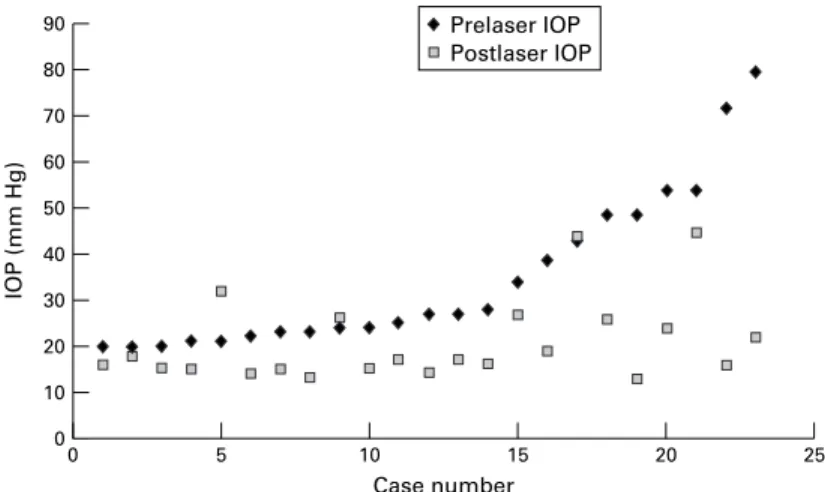
=15, p <0.01) (Table 3). In 8/27 eyes the IOP was >19 mm Hg at follow up and in 4/27 eyes, as mentioned above, glaucoma surgery was performed during the follow up period. Surgery was required in these four cases as laser iridotomy had failed to control the IOP. Therefore for the purposes of this analysis they are defined as having IOP >19 mm Hg at follow up. Extent of change in IOP after iridotomy is demonstrated in Figure 1 (exclud-ing the four operated eyes).
GONIOSCOPY
In all, 148 of 151 (98%) iridotomy treated eyes had angles that were no longer classified as occludable (excluding the four operated eyes, eight eyes examined at home visits, and one eye with dense corneal scarring). Of the three eyes with angles that remained occludable one had a non-patent iridotomy, another had a small iridotomy, and the third had extensive PAS covering the trabecular meshwork.
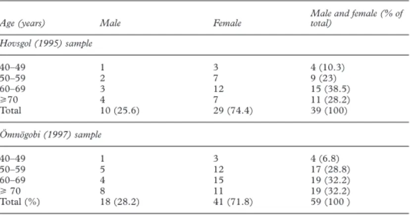
Table 1 Age and sex distributions of study populations
Age (years) Male Female
Male and female (% of total) Hovsgol (1995) sample 40–49 1 3 4 (10.3) 50–59 2 7 9 (23) 60–69 3 12 15 (38.5) >70 4 7 11 (28.2) Total 10 (25.6) 29 (74.4) 39 (100) Ömnögobi (1997) sample 40–49 1 3 4 (6.8) 50–59 5 12 17 (28.8) 60–69 4 15 19 (32.2) >70 8 11 19 (32.2) Total (%) 18 (28.2) 41 (71.8) 59 (100 )
Table 2 Initial diagnosis in eyes treated with laser iridotomy
Diagnosis Hövsgol (1995) Ömnögobi (1997) Total
PACG 13 (20.3%) 10 (10%) 23 (14%)
PAC 44 (68.8%) 23 (23%) 67 (40.9%)
PACS 7 (10.9%) 67 (67%) 74 (45.1%)
Total 64 (100%) 100 (100%) 164 (100%)
PACG = primary angle closure with glaucomatous optic neuropathy. PAC = primary angle closure.
PACS = primary angle closure suspect.
Table 3 Pre and post laser intraocular pressures (IOP) greater than 19 mm Hg
IOP >19 mm Hg
PACG (no of eyes) PAC (no of eyes)
1995 group 1997 group Total 1995 group 1997 group Total
Before laser 9/13 7/10 16/23 8/44 3/23 11/67
After laser 7/13 3/10 10/23 2/44 0/23 2/67
McNemar’s test for matched pairs
÷2=15, p<0.01
PACG = primary angle closure glaucoma with glaucomatous optic neuropathy. PAC = primary angle closure.
The median change in angle width between initial examination before iridotomy and follow up examination was an increase of two ShaVer grades. All four angle quadrants had a similar degree of change in angle width. The number of quadrants of the angle containing PAS remained the same in 96 (64%) eyes, de-creased in 28 (18.8%), and inde-creased in 25 (16.8%). There were 10 (6.7%) eyes in which a decrease of two quadrants of PAS was seen and four (2.7%) in which an increase of two quad-rants of PAS was seen.
FACTORS INFLUENCING OUTCOME OF LASER IRIDOTOMY
Among the 13/90 eyes with an initial diagnosis of PAC or PACG that were classified as failures seven had a combination of CDR>0.8, three
or four quadrants containing PAS, and IOP >19 mm Hg at the pre-laser examination (Table 4). Results of univariate and multivari-ate logistic regression analysis of predictive factors for a failure outcome after iridotomy are shown in Table 5. The nine fellow eyes classified as PAC but with occludable angles only are excluded from this analysis. The adjusted odds ratios are lower in the multiple regression model and in the case of PAS do not reach significance, demonstrating the relation between the three factors. The results of this analysis should be interpreted with caution in view of the large confidence intervals which are due to the small number of eyes in the failure outcome group.
INTEROBSERVER AGREEMENT
Good agreement between the field investigator on this study (WN) and the investigators of the 1995 study (PF) and the 1997 study (JD) for gonioscopic ShaVer grading was achieved (weighted kappa = 0.62, 0.73 respectively). The mean diVerence in IOP measurements between WN and PF was 0.75 mm Hg (SD 2.74) with WN reading lower measurements. Good agreement was achieved for optic disc grading (weighted kappa = 0.84, WN and PF).
Discussion
The outcome of the cases in this study was related to the stage of disease at diagnosis. Forty seven per cent of eyes with glaucomatous optic neuropathy were classed as treatment failures on grounds that IOP was poorly controlled or visual acuity had deteriorated due to progression of glaucoma. Three per cent of PAC eyes were found to have glaucomatous optic neuropathy at follow up, but there were other features such as pseudoexfoliation to explain a poor prognosis. None of the PACS eyes suVered symptomatic angle closure or glaucomatous optic neuropathy. It appears that once extensive synechial angle closure and glaucomatous optic neuropathy have devel-oped, an iridotomy will be less likely to reduce IOP and protect visual function than in eyes treated at an earlier stage. However, it must be acknowledged that this finding could be explained by the phenomenon of “lead time bias”. This form of bias can result in an appar-ent increase in survival or, in the case of glau-coma, increased time to loss of visual function in a case detected and treated at an early stage of the disease process. The real time from inci-dence of disease to visual function loss or blindness in such a case may be no diVerent from that of a case detected at a more advanced stage.15
Longer follow up or a randomised con-trolled trial would be required to overcome the possible influence of this bias.
Long term patency rates of YAG laser iridotomy were very good in these dark Asian irides in line with other studies in white and Afro-Caribbean eyes.16–18
Closure, or reduced eVectiveness of the iridotomy, seemed to be related to a documented “small” initial hole.
Our results show normalisation of IOP with-out medication in 55.5% (15/27) of eyes with elevated pressure before iridotomy. None of
Figure 1 Pre and post iridotomy intraocular pressures (IOP) in eyes with IOP >19 mm Hg before iridotomy (excluding four eyes that underwent glaucoma surgery during follow up interval). 90 70 80 60 40 50 30 20 0 10 25 20 Case number IOP (mm Hg) 15 10 0 5 Prelaser IOP Postlaser IOP
Table 4 Relation between pre-iridotomy examination findings and outcome of iridotomy in eyes with diagnosis of PAC or PACG
Pre-laser examination findings
Outcome of iridotomy
Failure (no of eyes) Stable (no of eyes)
(CDR>0.8) + (PAS 3-4) + (IOP>19) 7 1 (CDR>0.8) + (PAS 3-4) + (IOP<19) 1 1 (CDR>0.8) + (PAS 0-2) + (IOP>19) 1* 1 (CDR<0.8) + (PAS 3-4) + (IOP>19) 3 5 (CDR>0.8) + (PAS 0-2) + (IOP<19) 0 1 (CDR<0.8) + (PAS 3-4) + (IOP<19) 0 7 (CDR<0.8) + (PAS 0-2) + (IOP>19) 1† 7 (CDR<0.8) + (PAS 0-2) + (IOP<19) 0 45 Total 13 68
*Pseudoexfoliation; †mixed mechanism glaucoma; PAS = number of angle quadrants containing peripheral anterior synechiae; IOP = intraocular pressure (mm Hg); CDR = cup:disc ratio. Numbers after PAS (peripheral anterior synechiae) indicate number of angle quadrants contain-ing PAS.
Table 5 Logistic regression analysis of predictive findings for outcome of failure with iridotomy Factor OR 95% CI Significance Univariate analysis PAS (3–4) 7.78 1.87–32.33 p = 0.005 CDR>0.8 26.43 5.56–125.62 p <0.0001 IOP >19 31.7 3.7–266.93 p = 0.002 Multivariate analysis PAS (3–4) 2.47 0.38–15.97 p = 0.34 CDR>0.8 15.04 2.28–99.29 p = 0.005 IOP >19 19.8 1.85–212.49 p = 0.01
PAS = number of quadrants containing perpheral anterior synechiae. CDR = cup:disc ratio.
IOP = intraocular pressure (mm Hg). OR = odds ratio.
the eyes with an IOP<19 mm Hg before treat-ment had elevated IOP at follow up. Other studies have shown rates of IOP normalisation ranging from 9% without medication and 51% with medication to 90% in a mixture of cases with and without maintenance medication.5 19
CDR >0.8, PAS involving three to four angle quadrants, and IOP >19 mm Hg at diag-nosis were all significantly associated with future failure of iridotomy. Table 4 shows that in eyes where two of these factors were present, approximately half were controlled with iri-dotomy, although there were small numbers of eyes with CDR >0.8 with one other factor; 15/16 eyes with only one of these factors had a successful outcome after iridotomy. Eyes with all three factors present appear to be at highest risk of failure after iridotomy. Quigley found no association between any examination findings before treatment and outcome of argon laser iridotomy,20
but several studies demonstrate a relation between the extent of angle closure by PAS and failure of iridotomy to control IOP and progression of glaucoma.5 6 21
Iridectomy or iridotomy is less eVective in eyes with glau-comatous visual field loss and further surgical or medical treatment is often required to control IOP.22 23
In the absence of sophisticated resources, CDR is probably more useful than visual fields in predicting outcome of treatment. Visual field testing in a rural population is fraught with problems related to reliability and the need for multiple tests to achieve a baseline result. Our results suggest that in PACG eyes with advanced optic disc damage and three to four quadrants of PAS, trabeculectomy would be a better option than iridotomy. However, we do not know the success rate of filtering surgery in this popula-tion. It is likely that early detection of eyes at risk of angle closure will increase the probability of success of an iridotomy.
Despite the subjective nature of gonioscopy we have demonstrated good agreement be-tween observers. It may be more diYcult to reliably assess PAS in the presence of an occludable angle at initial examination. PAS were reassessed a day after iridotomy when the angle had widened.
The development of new PAS in the presence of a patent iridotomy suggests that irido-trabecular apposition is still occurring. IOP rises are known to occur in some eyes with patent iridectomies subjected to the dark prone provocative tests.9
As pupil block is eliminated by an iridotomy, ciliolenticular block or peripheral iris crowding are presumed to play a part in these pressure rises. Despite this, it does appear that pupil block is the main mechanism in the development of angle closure in the Mongolian population. This is demonstrated by reduction in IOP, widening of the angle, and recorded regression of PAS in some of the cases of PACG and PAC.
In summary, this is the first comprehensive study of the outcome of peripheral iridotomy in Mongolia, and may have relevance to neigh-bouring populations. It is documented that glaucoma and, particularly, PACG are a major cause of blindness in the east Asian region.2 24
This fact demands that we investigate strategies for the prevention of PACG. Laser peripheral iridotomy appears to oVer a simple, low risk method of achieving this goal. However the appropriate timing for interven-tion remains uncertain.
To answer this question, we have begun a randomised controlled trial of screening for primary angle closure in Mongolia using bilat-eral laser periphbilat-eral iridotomy as a prophylac-tic treatment.
This study was funded by the National Lottery Charities Board, through Fight for Sight with additional funding from the Inter-national Glaucoma Association, the British Council for the Pre-vention of Blindness, and the Danish Association for the Blind. The YAG laser used in this study was donated by the Velux Foundation, Copenhagen.
We thank Pak Sang Lee, MSc, BSc, technical adviser, Institute of Ophthalmology, Catey Bunce, MSc, medical statis-tician, Moorfields Eye Hospital, and Dr Poul Helge Alsbirk, MD, Dr Med, Department of Ophthalmology, Hillerød Hospi-tal, Hillerød, Denmark.
Proprietary interests: None.
1 Quigley HA. Number of people with glaucoma worldwide. Br J Ophthalmol1996;80:389–93.
2 Foster PJ, Baasanhu J, Alsbirk PH,et al. Glaucoma in
Mongolia—a population-based survey in Hövsgöl
Prov-ince, northern Mongolia.Arch Ophthalmol1996;114:1235–
41.
3 Devereux JG, Foster PJ, Baasanhu J,et al. Anterior chamber
depth measurement as a screening tool for primary
angle-closure in an East Asian population. Arch Ophthalmol
2000;118:257–63.
4 Robin AL, Arkell S, Gilbert SM. Q-Switched neodymium-YAG laser iridotomy. A field trial with a portable laser sys-tem.Arch Ophthalmol1986;104:526–30.
5 Salmon JF. Long-term intraocular pressure control after Nd-YAG laser iridotomy in chronic angle-closure
glau-coma.J Glaucoma1993;2:291–6.
6 Yamamoto T, Shirato S, Kitazawa Y. Treatment of primary angle-closure glaucoma by argon laser iridotomy: a
long-term follow-up.Jpn J Ophthalmol1985;29:1–12.
7 Snow JT. Value of prophylactic peripheral iridectomy on the
second eye in angle-closure glaucoma.Trans Ophthalmol
Soc UK1977;97:189–91.
8 Lowe RF. Acute angle-closure glaucoma. The second eye:
an analysis of 200 cases.Br J Ophthalmol1962;46:641–50.
9 Hung PT, Chou LH. Provocation and mechanism of
angle-closure glaucoma after iridectomy.Arch Ophthalmol1979;
97:1862–4.
10 Lowe RF, Ritch R. Angle-closure. Clinical types. In: Ritch
R, Shields MB, Krupin T. eds.The glaucomas.St Louis,
MO: CV Mosby; 1989:839–53.
11 Sakuma T, Sawada A, Yamamoto T,et al.Appositional angle
closure in eyes with narrow angles: an ultrasound
biomicroscopic study.J Glaucoma1997;6:165–9.
12 Shiose Y. Intraocular pressure. New perspectives.Surv
Oph-thalmol1990;34:413–35.
13 Hu Z, Zhao ZL, Dong FT. An epidemiological investigation
of glaucoma in Beijing and Shun-yi county. Chin J
Ophthalmol1989;25:115–8.
14 Arkell SM, Lightman DA, Sommer A,et al.The prevalence
of glaucoma among eskimos of Northwest Alaska.Arch
Ophthalmol1987;105:482–5.
15 Wormald R. Screening in ophthalmology. In: Johnson GJ,
Minassian DC, Weale R, eds.The epidemiology of eye disease.
London: Chapman and Hall, 1998:83–100.
16 Schwartz LW, Moster MR, Spaeth GL,et al.
Neodynium-YAG laser iridectomies in glaucoma associated with closed
or occludable angles.Am J Ophthalmol1986;102:41–4.
17 Canning CR, Capon MRC, Sherrard ES,et al.Neodymium:
YAG laser iridotomies—short-term comparison with cap-sulotomies and long-term follow-up. Graefes Arch Clin Exp Ophthalmol1988;226:49–54.
18 Del-Priore LV, Robin AL, Pollack IP. Neodymium:YAG and argon laser iridotomy. Long-term follow-up in a
prospec-tive, randomized clinical trial. Ophthalmology 1988;95:
1207–11.
19 Tomey KF, Traverso C, Shammas IV. Neodymium-YAG laser iridotomy in the treatment and prevention of angle
closure glaucoma. A review of 373 eyes.Arch Ophthalmol
1987;105:476–81.
20 Quigley HA. Long-term follow-up of laser iridotomy. Ophthalmology1981;88:218–24.
21 Kim YY, Jung HR. Dilated miotic-resistant pupil and laser
iridotomy in primary angle-closure glaucoma.
Ophthalmo-logica1997;211:205–8.
22 Gelber EC, Anderson DR. Surgical decisions in chronic
angle-closure glaucoma.Arch Ophthalmol1976;94:1481–4.
23 Richardson P, Cooper RL. Laser iridotomy.Aust NZ J
Oph-thalmol1987;15:119–23.
24 Baasanhu J, Johnson GJ, Burendei G,et al.Prevalence and
causes of blindness and visual impairment in Mongolia: a survey of populations aged 40 years and older. Bull World Health Organ1994;72:771–6.