DOI: 10.12928/TELKOMNIKA.v14i1.2675 630
Leaf Morphological Feature Extraction of Digital Image
Anthocephalus Cadamba
Fuzy Yustika Manik*1, Yeni Herdiyeni2, Elis Nina Herliyana3 1,2Department of Computer Science, Bogor Agricultural University,
Jl. Meranti, Wing 20 Level 5, Darmaga, Bogor 16680 3Departement of Silvikultur
, Bogor Agricultural University, Jl. Meranti, Wing 20 Level 5, Darmaga, Bogor 16680
*Corresponding author, e- mail: [email protected], [email protected], [email protected]
Abstract
This research implemented an image feature extraction method using morphological techniques. The goal of this proccess is detecting objects that exist in the image. The image is converted into a grayscale image format. Then, grayscale image is processed with tresholding method to get initial segmentation. Furthermore, image from segmentation results are calculated using morphological methods to find the mapping of the original features into the new features. This process is done to get better class separation. Research conducted on two Antocephalus cadamba (Jabon) leaf diseased seedlings data set image that contained leaf spot disease and leaf blight. The results obtained morphological features such as rectangularity, roundness, compactness, solidity, convexity, elongation, and eccentricity able to represent the characteristic shape of the symptoms of the disease. All properties form the symptoms can be quantitatively explained by the features form. So it can be used to represent type of symptoms of two diseases in Antocephalus cadamba (Jabon).
Keywords: antocephalus cadamba, feature extraction, morphology
Copyright © 2016 Universitas Ahmad Dahlan. All rights reserved.
1. Introduction
Anthocephalus cadamba (Jabon) is a type of commercial plantations of fast-growing local people (fast growing species) and can grow well on the acreage used for cultivation, shrubs, and swamp forests are widespread in forest areas in Indonesia. Jabon can be used for reforestation and afforestation in order to increase productivity of the land, and should be developed in industrial forest plantations, as demand for wood is increasing [1].
Plantation development that has implications for the kind of tree planting (monoculture) on a large scale requires the availability of high-quality seeds in sufficient quantities [2]. On the other hand this trend impact on the emergence of the disease [3] that causes harm, among others, reduces the quantity and quality of the results as well as increase production costs [4]. According to Anggraeni and Wibowo [5] the success of forest plantation development starts from seedlings produced from the nursery.
During this period leaf diseases receive less attention because they do not cause significant losses, except on the seedlings in the nursery. Jabon leaf disease that attacks the nursery phase in Bogor reported by Herliyana et al. [6] and Aisah [7], namely dieback, leaf spot disease and leaf blight. These three diseases are caused by fungi. Fungi cause local symptoms or systemic symptoms in its host. Generally, fungi cause local necrosis, tissue necrosis common or kill plants [8].
Symptoms and signs of disease have an important role to diagnose the disease. With symptoms dan signs, we can also determine morphology and characteristic of the causative pathogen. Experts can identify the type of disease based on the visible symptoms and signs. With image processing techniques, the image of symptoms and signs is processed to obtain the characteristics for identification process. On AnthocephalusCadamba (Jabon) plant seedlings, the blotches on leaves (leaf spot) are indicated that leaf has disease symptoms. The leaf spot is the death of necrotics that have sharp margins and it is the result of local infection by a pathogen. The color of spots is colored from yellow to brownish. The spots can either be round-shaped, oval-shaped or shapeless. If the shape is round, the disease is called leaf spot disease. If spotting or death occurred rapidly in whole or in part, the disease is called blight [9].
Digital image processing techniques today have grown very rapidly with a fairly extensive application in various fields. In image processing, in order to make the process of with drawal of information or description of the object or object recognition that exists in the image, feature extraction process is required. Feature extraction is the process for finding the mapping of original features to new features in which it is expected to result in better class separation [10]. Feature extraction is an important step in the classification [11], because well-extracted features will be able to increase the level of accuracy, while features that are not well-extracted will tend to exacerbate the level of accuracy [12].
To identify or classify objects in the image, first we must extract features from an image and then use this feature in a pattern to obtain a final grade classifier. Feature extraction is used to identify features that can make a better representation of the object. Not only color and texture, shape or morphology can also be used to extract features. Mathematical morphology is a tool to extract image components that are useful to represent and describe shape of region, such as boundaries, skeletons, and the convex hull. Morphological is related to certain operations that are useful for analyzing the shape of the image so that shape of the object can be recognized.
Zinove et al. [13] has predicted the radiologist assessment of Lung Image Database Consortium (LIDC) nodules using 64 feature images of the four categories (forms, intensity, texture, and size). Putzu et al. [14] has been doing research for identifying and classifying white blood cells (leukocytes) based on morphological features, colors and textures. Gartner et al., [15] has used morphological features such as roundness and elongation to classify zircon grains from sediment.
The problem in this research is how to identify features that can make a good representation of the object based on its shape. So, it can be used to find significant features area in the image.
2. Research Method 2.1. Data Set
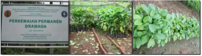
The data used are the image of two types of leaves Jabon affected by leaf diseases. The diseases that are focused in this research are leaf spot and leaf blight ± 4 months. Data in the form of symptomatic leaves Jabon obtained from the observation of the symptoms and making Jabon examples that show symptoms of leaf spot and leaf blight of 2 nursery locations around the campus of Dramaga are one nursery located in Situ Gede area and one nursery in IPB show in Figure 1. The number of plant samples taken from the location of the nursery is adjusted to the nursery condition.
Figure 1. Data collection sites 2.2. Methodology
Methodology in general can be described in Figure 2.
2.2.1. Image Acquisition
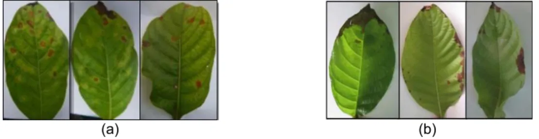
The Symptomatic leaf photograph was taken by using a digital camera for every kind of disease. Example of this photo is shown in Figure 3.
Leaf Spot Disease: Symptoms and signs of leaf spot disease are generally the same on each affected plants, which are sores or blemishes that are local to the host leaf consists of dead cells (necrosis) in the leaves [8]. The area of necrosis ranging from small to large with a form of irregular until uniform [16]. Symptoms of leaf spot disease Jabon seeds can be seen in Figure 3(a).
Leaf Blight Disease: Symptoms and signs that happen in the organ leaves, branches, twigs and flowers turn brown very quickly and thoroughly are the causes of death [8]. There are spots on the leaves opaque, dark brown surrounded by a chlorotic halo [9]. Symptoms of leaf spot disease Jabon seeds can be seen in Figure 3(b).
(a) (b)
Figure 3. The image of the diseased leaf (a) Leaf spot disease (b) Leaf blight disease 2.2.2. Preprocessing
At preprocess stage, there are two steps to process the captured image. The processes are cutting off (cropping) and segmentation the image. Cropping technique is done to cut and take diseases part in the images, just like shown below (Figure 4(a)). Meanwhile, the object image segmentation process is done to obtain a binary image of the object image by using the concept of morphology consists of thresholding, edge detection, and opening. A process of feature extraction is followed after the process of image segmentation [17]. Segmentation is a very important step in object recognition. There are various segmentation methods that can be used [18].
(a) (b) Figure 4 Preprocessing (a) Cropping (b) Segmentation
As shown in Figure 4(b), Thre sholding process based on Otsu method was applied to the original images [12]. Process is continued by object edge detection using canny edge detection technique to get the edge lines of the object that will be used to calculate objects perimeter features [12]. 'Holes' that are found in the binary image objects from applying the segmentation process is filled with applying the morphological opening process so it becomes a fully binary image object area.
2.2.3. Morphological Feature Extraction
To recognize an object in the image, some features must be extracted first. Morphology of the digital image is the fact that digital image containing series of pixels that make collection of two-dimensional data. Certain math equations on a series of pixels can be used to improve aspects of the form and structure so it can be easier to recognize.
There are several features of the shape that can be calculated, like area which is calculated based on the number of pixels that occupies the object image, the perimeter (boundary object) is calculated based on the number of pixels around the object image. Based on area and perimeter features, values of other morphology features can also be calculated as well. The following equations are formulas that can be used to extract morphological features [13-15].
Table 1. Formula Morphological Features Morphological
Features Formula Description
Rectangularity major axis x minor axisarea Technique to illustrate similarity of object shape with rectangular shape. Elongation 1 minor axis
major axis Measuring the length of the object.
Solidity area
convex_area
Measuring the density of the object, ratio of area to the full convex object.
Roundness 4 x π x area
convex_perimeter
Technique to illustrate the level of determination object. Value 1 for circular object is greater than one for not circular object.
Convexity convex_perimeter
parimeter
The relative amount that the object is different from convex hull. This value is the perimeter ratio between convex full of object and the object itself. If the value is1, the object is called convex hull, when the value is bigger than 1 the object is not convex hull or object with irregular boundaries.
Compactness 4 x π x area
perimeter
The ratio between the object area with circle area uses the same perimeter.
Eccentricity major axis minor axis
major axis
The ratio of distance between the ellipse focal and major axis. The value is between 0-1.
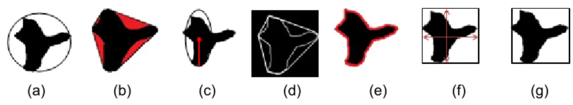
This research is based on analysis using symptomic spots’ shape of leaf spot disease. The extracted shape feature is a feature that has numerical data as shown in Figure 5. Area is the wide of spots (Figure 5(b)), perimeter is the perophery or limit of spots (Figure 5(c)), major axis is the length of spots measured from the base of the leaf to the tip, mean-while minor axis is the width of spots measured from the widest leaf surface (Figure 5d), the convex hull (Figure 5(e)), convex area (Figure 5(f)), and the convex perimeter (Figure 5(g)).
(a) (b) (c) (d) (e) (f) (g)
Figure 5. (a) The original image, (b) Area, (c) Perimeter, (d) Mayor Axis and Minor Axis, (e) Convex Hull, (f) Convex Area, (g) Convex Perimeter
The feature is also used to calculate roundness, solidity, elongation, eccentricity, compactness, convexity, and rectangularity. Illustration for all features can be seen in Figure 6 below:
(a) (b) (c) (d) (e) (f) (g) Figure 6. (a) Roundness, (b) Solidity, (c) Eccentricity, (d) Convicity, (e) Compactness,
(f) Elongation, (g) Rectangularity 3. Results and Analysis
Table 2. Features Extraction Leaf Diseases Jabon Symptoms image Features Extraction
Roundness Rectangularity Compactness Convicity Solidity Elongation Eccentricity
Leaf Spot Disease
0.506 0.738 0.733 1.203 0.918 0.157 0.537 0.456 0.781 0.747 1.280 0.951 0.142 0.514 0.523 0.744 0.734 1.185 0.933 0.010 0.142 0.516 0.765 0.824 1.263 0.952 0.016 0.180 0.539 0.748 0.787 1.208 0.952 0.058 0.337 0.570 0.786 0.829 1.206 0.964 0.099 0.434 0.475 0.793 0.751 1.258 0.952 0.008 0.122 0.431 0.790 0.762 1.329 0.961 0.189 0.586 0.439 0.805 0.766 1.321 0.932 0.032 0.251
Leaf Blight Diseases
0.172 0.369 0.169 0.497 1.049 0.626 0.927 0.204 0.480 0.198 0.636 0.910 0.778 0.975 0.208 0.523 0.149 0.731 1.069 0.688 0.950 0.197 0.348 0.188 0.444 0.910 0.628 0.928 0.127 0.265 0.174 0.341 0.893 0.546 0.891 0.142 0.410 0.101 0.542 1.106 0.617 0.924 0.171 0.307 0.163 0.469 1.050 0.647 0.936 0.194 0.202 0.237 0.292 0.877 0.483 0.856 0.188 0.302 0.169 0.456 1.027 0.653 0.938 0.199 0.298 0.225 0.432 0.921 0.526 0.881
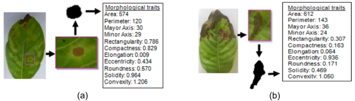
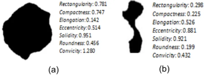
(a) (b)
Figure 7. Calculation morphological features (a) Leaf spot disease (b) Leaf blight disease In experiments using 100 images of each class Jabon symptomatic leaf seedlings were tested to determine which features are capable to represent the image so that to be able to get useful information and can be used to get good accurate results during the classification process. Matrix resulted from the extraction of morphological traits the entire image in the form of a matrix measuring 7 x 100 which is a representation of 100 images for each type of disease with every image has a vector which is composed of 7 elements.
Features such as area, perimeter, major axis and minor axis as discussed can not be used independently as object identification features. Such feature is influenced by the size of the object. In order not to depend on scaling, some of the features that can be derived from these features are rectangularity, compactness, elongation, eccentricity, convicity, roundness and solidity.
Roundness and rectangularity shows how well an object can be described by a circle and a rectangle. While the compactness measures the ratio between the object area and circle area using perimeter. Based on the Table 2 above, blight has a roundness value (<0.207) and a rectangularity (<0.523), while leaf spot has a roundness highest resolution (>0.569) and rectangularity (>0.738). The average value compactness leaf spot is larger (>0.73) than the average value of blight (<0.237). Elongation shows elongated polygon level. It is known as late blight elongation values greater (> 0.483) of the value of elongation at leaf spot (<0.007). Eccentricity is the ratio between the major axis and a minor axis, from the table above shows that the value of eccentricity blight (<0.856) is greater than the value of eccentricity leaf spot (> 0.585). Convexity and solidity are able to describe convex of a polygon. The difference between these two metrics is that convecity using the ratio of the perimeter while the solidity use area ratios. If the convexity calculate the relative amounts that differ from the convex hull object while the object density counting solidity.Leaf spot has the highest convexity value (>1.184) and the highest solidity (>0.917) when compared with the value of convexity blight (<1.106) and solidity leaf blight (>0.731).
(a) (b)
As seen in Figure 8, a convex polygon that seems like having a complex detail that the perimeter could be huge compared to the perimeter of the convexhull, this seems to be the symptoms of a blight of leaves. One point at the convexity is a convex hull (8a), if it is greater than one point the object is not convex full or irregular boundaries. Based on the table above 2, it is known that the convex leaf spot is not convex full or irregular boundary objects, while late blight has a value approaching convexhull. While the result of solidity is known that the density of object leaf spot is higher than the density of object leaf blight. Solidity value obtained according to the symptom that occurs as both symptoms of leaf blight and leaf spot. When the form becomes less subtle (or rough) or has intricate details that perimeter can be very large compared to its convex hull perimeter, so the value could be lower convexity while the value of high solidity indicates that solidity is better while convexity is more sensitive.
(a) (b)
Figure 9. Morphological features jabon leaf disease symptoms (a) Leaf spots (b) Blight From the results of Figure 9, it is known that the two Jabon leaf disease symptoms and seen that the value of the convexity is greater than the solidity. Leaf spot has the highest convexity and solidity value compared to the value of convexity and solidity leaf blight. If it is seen from the value of elongation blight and leaf spot, it is also known that the symptoms of leaf blight have an elongated shape. It is known that leaf blight elongation value is greater than the value of elongation at leaf spot. Elongation showed elongated polygon level. If viewed from the eccentricity blight and leaf spot, it is also known that the symptoms of leaf blight have an elongated shape. It is known as late blight eccentricity value greater than the value of the eccentricity leaf spot. Leaf spot has a roundness value and the highest rectangularity. This according to the symptoms seen that leaf spot has a shape like a circle. The value of the leaf spots for compactness value is greater than the average value of blight. This indicates that the leaf spots have a more compact when compared with leaf blight. This is consistent with the visible symptoms.
4. Conclusion
Utilizing the shape as a component in the image analysis to investigate the shape features can be used to measure the characteristics of the shape of the object image. To measure the basic geometric attributes, there are seven morphological features selected for analysis. Seven of the morphological features namely convexity, solidity, elongation, roundness, rectangularity, eccentricity, and compactness. Each is used to measure the convexity, solidity, elongation, roundness, rectangle, ellipse, and measure the complexity of the form. The morphological features were able to describe the characteristics of the shape of the same aspect. These features are very good and appropriate for use in characterizing classes of Jabon leaf disease symptoms ie, leaf spot and leaf blight. The results showed that all symptoms shape properties can be quantitatively explained by the features of shape. Overall seven morphological features are capable to extract characteristic symptom forms contained in the leaves Jabon gets useful information from an image. Leaf spot has a value roundnees, compactenes and rectangularity is greater than the blight. These results are consistent with the symptoms seen in fisual that leaf spot symptoms have a more complex shape, while late blight symptoms have elongated shape, it is proved by the value of elongation and leaf blight
eccentricity greater than leaf spot. While the solidity and convexity able to describe the symptoms of a more convex leaf spots and solid from the blight.
References
[1] Wahyudi. Analisis Pertumbuhan Dan Hasil Tanaman Jabon (Anthocephallus Cadamba). Jurnal Perennial. 2012; 8(1): 19-24.
[2] Prananda R, Indriyanto, Riniarti M. Respon Pertumbuhan Bibit Jabon (Anthocephalus Cadamba) dengan Respon Pertumbuhan Bibit Jabon Pemberian Kompos Kotoran Sapi Pada Media Penyapihan. Jurnal Sylva Lestari. 2014; 2(3): 29-38.
[3] Widyastuti SM, Harjono, Surya ZA. Infeksi Awal Jamur Uromycladium tepperianum pada Daun Falcataria moluccana dan Acacia mangium di Laboratorium. Jurnal Manajemen Hutan Tropika. 2013. [4] Anggraeni I, Lelana NE. Penyakit Karat Tumor Pada Sengon. Badan Penelitian dan Pengembangan
Kehutanan. Jakarta. 2011.
[5] Anggraeni, Wibowo. Pengendalian Cylindrocladium Sp. Penyebab Penyakit Lodoh Pada Bibit Acacia Mangium Wild. Dengan Fungi Antagonis Trichoderma Sp. dan Gliocladium Sp. Jurnal Penelitian Hutan Tanaman, Pusat Litbang Hutan Tanaman. 2009; 6(4).
[6] Herliyana EN, Achmad, Putra A. Pengaruh pupuk organik cair terhadap pertumbuhan bibit Jabon (Anthocephalus cadamba miq.) dan ketahanannya terhadap penyakit. Jurnal Silvikultur Tropika. 2012; 3(3): 168-173.
[7] Aisah AR. Klasifikasi dan Patogenisitas Cendawan Penyebab Primer Penyakit Mati Pucuk pada Bibit Jabon (Anthocephalus Cadamba (Roxb.) Miq). Tesis. Bogor: Institut Pertanian Bogor; 2014.
[8] Yunasfi. Faktor-Faktor yang Mempengaruhi Perkembangan Penyakit dan Penyakit yang Disebabkan oleh Jamur. Medan: USU Digital Library; 2002.
[9] Agrios GN. Plant Pathology. Fifth edition. New York (US): Elsevier Academic Pr. 2005.
[10] Gue L, Riveno D, Derado J, Munteanu CR, Pazos A. Automatic feature extraction using genetic programming: An application to epileptic EEG classification. Expert Systems with Applications. 2011; 38: 10425–10436
[11] Ahsan M, M Dzulkifli. Features Extraction for Object Detection Based on Interest Point. TELKOMNIKA Telecommunication, Computing, Electronics and Control. 2013; 11: 2716-2722. [12] Gonzalez R, Woods, R. Digital Image Processing. Third edition. New Jersey, USA: Pearson Prentice
Hall. 2008.
[13] Putzu L, Caocci G, Di Ruberto C. Leucocyte classification for leukaemia detection using image processing techniques. 2014.
[14] Zinovev D, Raicu D, Furst J, Armato SG. Predicting Radiological Panel Opinions Using a Panel of Mechine Learning Classifiers. Algorithms. 2009; 2: 1473-1500.
[15] Gartner A, Linnemann U, Sagawe A, Hofmann M, Ullrich B, Kleber A. Morphology of zircon crstal grains in sediments- characteristics, classifications, definitions. Journal of central European Geology. 2013; 59: 65-73.
[16] Anggreini I. Colletotrichum Sp., Penyebab Penyakit Bercak Daun Pada Beberapa Bibit Tanaman Hutan Di Persemaian. Pusat Litbang Hutan Tanaman. 2011.
[17] G Patil B, Mane N, Subbaraman S. Iris Feature Extraction and Classification using FPGA. International Journal of Electrical and Computer Engineering (IJECE). 2012: 214-222.
[18] Fanani A, Yuniarti A, Suciati N. Geometric Feature Extraction of Batik Image Using Cardinal Spline Curve Representation. TELKOMNIKA Telecommunication, Computing, Electronics and Control. 2014.