Aliakbar Bahreman,
DDS
,
MS
Clinical Professor
Orthodontic and Pediatric Dentistry Programs
Eastman Institute for Oral Health
University of Rochester
Rochester, New York
EARLY-AGE
ORTHODONTIC
TREATMENT
Quintessence Publishing Co, Inc
Chicago, Berlin, Tokyo, London, Paris, Milan, Barcelona, Beijing,
Istanbul, Moscow, New Delhi, Prague, São Paulo, Seoul, Singapore,
and Warsaw
Bahreman_FM.indd iii
Foreword
by J. Daniel Subtelny
vii
Preface
and
Acknowledgments
viii–ix
Introduction
x
Part I
Clinical and Biologic Principles of Early-Age
Orthodontic Treatment
1
1
Rationale for Early-Age Orthodontic Treatment
3
2
Development of the Dentition and Dental Occlusion
15
3
Examination, Early Detection, and Treatment
Planning
41
Part II
Early-Age Orthodontic Treatment of Nonskeletal
Problems
71
4
Space Management in the Transitional Dentition
73
5
Management of Incisor Crowding
105
6
Management of Deleterious Oral Habits
131
7
Orthodontic Management of Hypodontia
157
8
Orthodontic Management of Supernumerary Teeth
189
9
Diagnosis and Management of Abnormal Frenum
Attachments
205
10
Early Detection and Treatment of Eruption
Problems
225
Contents
Bahreman_FM.indd v
Part III
Early-Age Orthodontic Treatment of Dentoskeletal
Problems
291
11
Management of Sagittal Problems
(Class II and Class III Malocclusions)
293
12
Management of Transverse Problems
(Posterior Crossbites)
355
13
Management of Vertical Problems
(Open Bites and Deep Bites)
377
Index
417
Bahreman_FM.indd vi
vii
This book is a compendium of signifi cant and pertinent in-formation related to early-age orthodontic treatment, a sub-ject that seems to have evolved into one of considerable controversy, with as many orthodontists expressing a nega-tive reaction as a posinega-tive reaction to its benefi ts. Dr Bahre-man is a believer in early-age orthodontic treatment, and he expresses some cogent arguments founded in years of ex-perience in practice and teaching to back up his beliefs. In developing his treatise, Dr Bahreman outlines the develop-ment of the occlusion and/or malocclusion from the embry-onic stages, when the foundation of the jaws and thereby the position of the dentition is fi rst established.
Early-age orthodontics is not about the time it takes to orthodontically treat a problem; it is a story of growth, of variation in anatomy, and of muscle function and infl uenc-es, a realization that it is the jaws that contain the teeth and that where the jaws go, the teeth will have to go, and
both undergo varying infl uences as well as grow in varying directions. Early-age orthodontics necessitates recognition of this process and aims to alter and redirect it whenev-er feasible and possible. Dr Bahreman has undwhenev-ertaken a monumental effort in directing efforts along this path. An extensive exploration of the literature is an added bonus, as the mechanical approaches are based on this literature. In fact, the extensive review of the literature and its applica-tion to diagnosis and varying forms of therapy are worth a veritable fortune.
You may or may not agree with the basic premises, but you will have access to important information that will wid-en your scope of vision and thereby widwid-en your treatmwid-ent horizons. To my mind, an ounce of prevention, if possible, is worth a pound of cure. The reality of prevention can exist at the earliest stages of development.
J. Daniel Subtelny,
DDS, MS, DDSc(Hon)Professor Emeritus
Interim Chair and Director of Orthodontic Program Eastman Institute for Oral Health
University of Rochester Rochester, New York
Foreword
Bahreman_FM.indd vii
viii
After obtaining a master’s degree in orthodontics in 1967, I began my career at a newly founded dental school in Tehran. My responsibilities included teaching and administrative du-ties at the university and maintenance of a very busy private practice. In addition, I established both the orthodontic and pediatric dentistry departments at the university.
Many patients were being referred to the orthodontic de-partment, and there were no qualifi ed faculty members to help me provide care. To rectify the situation, I designed an advanced level, comprehensive curriculum in orthodontics for undergraduate students, including classroom instruction, laboratory research, and clinical demonstrations. Once the students completed the course, they could work in the clinic, thus temporarily solving the issue of the heavy patient load in the orthodontic clinic. With additional staff now available, I could select patients, mostly children in the primary or mixed dentition, for some interceptive treatment.
Despite my diffi culties in performing all of the aforemen-tioned duties, this situation had a fortunate outcome. It helped me to understand and discover the advantages of early-age orthodontic treatment, which was not common in those years. During my more than 40 years of practice and teaching, especially in early orthodontic treatment, I have accumulated a considerable amount of educational data for teaching pur-poses. I would like to share this experience and information with readers.
The public’s growing awareness of and desire for dental services, especially at an early age, have encouraged our pro-fession to treat children earlier. Despite the recommendation by the American Association of Orthodontists that orthodon-tic screening begin by the time a child is 7 years old, many orthodontists still do not treat children prior to the complete eruption of the permanent teeth. I believe that this inconsis-tency is due to the educational background of orthodontists as well as a lack of familiarity with recent technical advance-ments and the various treatment options that are available for young patients.
The therapeutic devices available for this endeavor are not complex, but deciding which ones to use and when to employ them are important steps. As we make these decisions, we should also remember not to treat the symptom but rather to treat the cause. My goal is to present the basic information necessary to understand the problems, to differentiate among various conditions, and to review different treatment options. Case reports are examined to facilitate clinical application of the theory in a rational way.
To understand the morphogenesis of nonskeletal and skel-etal occlusal problems, to detect problems early, and to inter-vene properly, we must look at all areas of occlusal develop-ment, including prenatal, neonatal, and postnatal changes of the dentoskeletal system, and explore all genetic and envi-ronmental factors that can affect occlusion at different stages of development. In other words, we must have a profound understanding of the fundamental basis and morphogenesis of each problem and then apply this knowledge to clinical practice. Thus, the goals of this book are:
• To provide a comprehensive overview of all areas of dental development, from tooth formation to permanent occlu-sion, to refresh the reader’s memory of the fundamentals necessary for diagnosis and treatment planning.
• To emphasize all the important points of the developmen-tal stages that must be recognized during examination of the patient to facilitate differential diagnosis. Each tooth can become anomalous in a number of ways and to different degrees. Occlusion and maxillomandibular relationships can vary in the sagittal, transverse, and vertical directions. • To discuss the application of basic knowledge to practice by
presenting several cases with different problems and differ-ent treatmdiffer-ent options.
• To demonstrate the benefi ts of early-age orthodontic treat-ment, achieved by intervention in developing malocclusion and guidance of eruption.
Materials are presented in three parts: In Part I, “Clinical and Biologic Principles of Early-Age Orthodontic Treatment,” three chapters introduce and explain the concept of early-age treat-ment, describe its necessity and advantages, and discuss the controversies surrounding this topic; discuss the basic foun-dation of occlusal development, empowering the practitioner to detect anomalies and intervene as necessary; and illustrate the procedures, tools, and techniques available for diagnosis, emphasizing differential diagnosis and treatment planning for early-age treatment.
Part II, “Early-Age Orthodontic Treatment of Nonskeletal Problems,” consists of seven chapters describing the non-skeletal problems that might develop during the primary and mixed dentitions. The chapters explain the ontogeny, diagno-sis, and early detection of, and intervention for, these prob-lems. Topics include space management, crowding, abnormal oral habits, abnormal frenum attachment, hypodontia, super-numerary teeth, and abnormal eruption problems.
Preface
Bahreman_FM.indd viii
ix
Part III, “Early-Age Orthodontic Treatment of Dentoskeletal Problems,” consists of three chapters on early intervention for the dentoskeletal problems that might arise during the pri-mary and mixed dentitions in the three dimensions: sagittal problems (anterior crossbite and Class II and Class III maloc-clusions); transverse problems (posterior crossbites); and ver-tical problems (open bites and deep bites).
This book will provide the reader with a fi rm foundation of the basic science and case examples with various treatment options. It is my hope that the information provided will pro-mote a better understanding of abnormalities and their causes and enable readers to recognize the clues for early detection and intervention.
Acknowledgments
First and foremost, I would like to gratefully acknowledge the valuable opportunity that was afforded me as a student in Dr Daniel Subtelny’s orthodontic program. Between 1964 and 1967, I completed both my orthodontic specialty and master degree programs with Dr Subtelny as my mentor. As chairman and program director, researcher, and mentor, Dr Subtelny has dedicated over 57 years of his life to teaching, personally infl uencing the lives of over 350 students from around the world, myself included. In 1999, after over 32 years of teaching, practicing, and administrating in Tehran, I was fortunate enough to return to the Eastman Institute for Oral Health to work alongside Dr Subtelny as a faculty mem-ber in the Orthodontic and Pediatric Dentistry Programs.
In addition to Dr Subtelny, there are several individuals to whom I would like to express my deep gratitude for their help and encouragement in preparation of this book: the late Dr Estepan Alexanian, head of the Department of His-tology at the Shahid Beheshti University Dental School in
Tehran, whose dedication as an educator and preparation of superb histologic slides is remarkable and who allowed me to use his slides in my publication; Mr Aryan Salimi for scanning some of the slides and radiographs in this book; and Ms Elizabeth Kettle, Program Chair of the Dental Sec-tion of the Medical Library AssociaSec-tion, head of Eastman’s library, for her sincere help in editing this publication.
Finally, I wish to acknowledge the constant support of my family: Malahat, Nasreen, Saeid, Alireza, Tannaz, and Peymann Motevalei. Especially high gratitude goes to my wife, Malahat, for her tolerance, support, and encourage-ments. I also want to thank my son Alireza for his technical help and guidance in computer skills and my granddaughter Tannaz Motevalei for drawing some of the illustrations.
This publication is the product of 17 years spent orga-nizing materials derived from my 45 years of practice and teaching as well as reviewing hundreds of articles and books. I herewith dedicate this book to the teachers, practi-tioners, residents, and students who are dedicated to treat-ing malocclusion earlier in children, before it becomes more complicated and costly.
Bahreman_FM.indd ix
x
Occlusal development is a long process starting around the sixth week of intrauterine life and concluding around the age of 20 years. This long developmental process is a sequence of events that occur in an orderly and timely fashion under the control of genetic and environmental factors. Dental oc-clusion is an integral part of craniofacial structure and coordi-nation of skeletal growth changes. Occlusal development is essential for establishing a normal and harmonious arrange-ment of the occlusal system.
As we learn about craniofacial growth changes, the poten-tial infl uences of function on the developing dentition, and the relationships of basal jawbones and head structure, we acquire a better understanding of when and how to inter-vene in the treatment guidance for each patient. It is more effective to intervene during the primary or mixed dentition period to reduce or, in some instances, avoid the need for multibanded mechanotherapy at a later age.
Untreated malocclusions can result in a variety of prob-lems, including susceptibility to dental caries, periodontal disease, bone loss, temporomandibular disorders, and un-desirable craniofacial growth changes. Moreover, the child’s appearance may be harmed, which can be a social handicap. The benefi ts of improving a child’s appearance at an early age should not be undervalued. The goals of many clinicians who provide early treatment are not only to reduce the time and complexity of comprehensive fi xed appliance therapy but also to eliminate or reduce the damage to the dentition and supporting structures that can result from tooth irregu-larity at a later age. In short, early intervention of skeletal and dental malocclusions during the primary and mixed dentition stages can enable the greatest possible control over growth changes and occlusal development, improving the function, esthetics, and psychologic well-being of children.
For many decades, orthodontists have debated about the best age for children to start orthodontic treatment. While we agree on the results of high-quality orthodontic treatment, we often differ in our opinions as to how and when to treat the patient. Some practitioners contend that starting treat-ment in the primary dentition is the most effective means of orthodontic care. Others prefer to begin the treatment in the mixed dentition. There is also controversy about whether the early, middle, or late mixed dentition is preferable.
Despite the fact that the American Association of Ortho-dontists recommends that orthodontic screening be started by the age of 7 years, many orthodontists do not treat chil-dren prior to the eruption of permanent teeth, and some postpone the treatment until the full permanent dentition
has erupted, at approximately 12 years. The controversy sur-rounding early versus late treatment is often confusing to the dental community; therefore, clinicians must decide on a case-by-case basis when to provide orthodontic treatment. Indeed, there are occasions when delaying treatment until a later age may be advisable.
The long-term benefi ts of early treatment are also con-troversial. The majority of debates seem to revolve around early or late treatment of Class II malocclusions. There is less controversy regarding many other services that can be per-formed for the benefi t of young patients during the primary or mixed dentition, such as treatment of anterior and poste-rior crossbite, habit control, elimination of crowding, space management, and management of eruption problems.
Practitioners who are in favor of early treatment of Class II problems contend that early intervention is the best choice for growth modifi cation when the problem is skeletal and especially when it results from mandibular retrusion. On the other hand, opponents believe that there is no difference in the fi nal result and that a single-phase treatment approach is preferable because of the advantages that accompany the reduced treatment time.
Unfortunately, some practitioners, without a profound evaluation of the indications for early treatment, conclude that late treatment is always preferable. However, broad conclusions drawn from narrowly focused research can be misleading. One cannot conclude that no birds can fl y by considering the fl ight characteristics of the ostrich.
To evaluate and demonstrate the benefi ts of early treat-ment, I aim to discuss and clarify available treatments and services and discuss cases with different problems and dif-ferent treatment options. An understanding of all aspects of early treatment requires a thorough knowledge of the basics of embryology, physiology, and growth and development. This includes development of the dentition, tooth formation, eruption, exfoliation, and all transitional changes. Therefore, my other goal is to integrate the basic science and the clini-cal, in order to refresh the reader’s memory on important points about the bases of nonskeletal and skeletal problems that can arise during the transitional stages of occlusion.
Each patient who enters our practice represents a new chapter and a new lesson that we can learn from. A thorough knowledge of the basis for early-age orthodontic treatment, an understanding of the proper treatment techniques, and a willingness to consider their appropriateness for each in-dividual patient will allow us to intervene in ways that will provide the maximum benefi t for a young and growing child.
Introduction
Bahreman_FM.indd x
CLINICAL AND
BIOLOGIC PRINCIPLES
OF EARLY-AGE
ORTHODONTIC
TREATMENT
I
PART
Bahreman_CH01.indd 1 Bahreman_CH01.indd 1 3/18/13 10:26 AM3/18/13 10:26 AMRationale for Early-Age
Orthodontic Treatment
1
3
In the past, orthodontic treatment has been focused mainly on juvenile and adult treatment. Treatment options for patients in these age groups often are limited by complex dental and orthodontic problems and the lack of suffi cient future cranio-facial growth.
During the later part of the 18th century, orthodontic treatment of Class II malocclusion was limited primarily to retrac-tion of the maxillary anterior teeth to decrease excessive overjet. In 1880, Norman Kingsley1 published a description of
techniques for addressing protrusion. He was among the fi rst to use extraoral force to retract the maxillary anterior teeth after extraction of the maxillary fi rst premolars; the extraoral force was applied with headgear. Later, Case2 continued to
refi ne these methods.
Angle’s classifi cation3 of malocclusion, published in the 1890s, provided a simple defi nition of normal occlusion and was
an important step in the development of orthodontic treatment. Angle opposed the extraction of teeth and favored the preservation of the full dentition. His position against tooth extraction led him to depend on extraoral force for the expan-sion of crowded dental arches and retraction of the anterior segment. Later he discontinued the use of extraoral force and advocated the use of intraoral elastics to treat sagittal jaw discrepancies.
Because of Angle’s dominating belief that treatment with Class II elastics was just as effective as extraoral force, the use of headgear was abandoned by the 1920s. Then, in 1936, Oppenheim4 reintroduced the concept of extraoral anchorage,
employing extraoral traction to treat maxillary protrusion. Accepting the position of the mandible in Class II malocclusions, Oppenheim attempted to move the maxillary dentition distally by employing a combination of occipital anchorage and an E-arch, allowing the mandible to continue its growth. This resulted in an improved relationship with the opposing jaw. In 1947, Silas Kloehn5 reintroduced extraoral force, in the form of cervical headgear, for the treatment of skeletal Class II
relationships.
In 1944,another student of Angle’s, Charles Tweed,6 was discouraged by the prevalence of relapse in many of his
pa-tients treated without extraction, so he decided to oppose the conventional wisdom of nonextraction.
In the early part of the 20th century, there was optimism about the infl uence of orthopedic force on skeletal growth. An almost universal belief was that orthodontic forces, if applied to the growing face, could alter the morphologic outcome. In the United States, headgear was the principal appliance used for facial orthopedic treatment, whereas in Europe the functional appliance was predominantly used.
In 1941, Alan Brodie,7 one of Angle’s students, concluded that the growing face could not be signifi cantly altered from
its genetically predetermined form and that the only option for the orthodontist in cases of skeletal malocclusion would be dental camoufl age, or the movement of teeth within their jaws. This idea led to tooth extraction.
Bahreman_CH01.indd 3
Examination, Early Detection, and Treatment Planning
3
60
Panoramic radiographs
The panoramic radiograph is a common diagnostic tool in today’s dental practice. It is a kind of radiograph that pro-vides a full picture of the dentition and the complete maxilla and mandible.
Panoramic radiographs do not show the fi ne detail captured on intraoral radiographs and are not as specifi c as other intraoral radiographs, but in a single radiograph it provides a useful general view of all dentition, the maxilla and mandible, the sinuses, and both TMJs. This type of radiograph is very useful, especially during the mixed dentition, for early detection and prevention of all problems disturbing the normal development of occlusion.
Especially during the mixed dentition as a diagnostic tool for early-age orthodontic treatment, the following are important aspects that should be carefully evaluated on a panoramic radiograph before any orthodontic treatment: • Position and pattern of fully emerged as well as emerging
permanent teeth
• Sequence of permanent tooth eruption • Asymmetric eruption
• Comparison of crown height levels on the left and right sides
• Obstacles preventing eruption
• Abnormal tooth malformations (gemination, fusion, dens in dente, or dilaceration)
• Exfoliation and pattern of primary teeth root resorption • Tooth number and supernumerary teeth or congenitally
missing teeth
• Eruption problems, such as impaction, ectopic, transposi-tion, or ankylosis
• Bone density and trabeculation
• Cysts, odontomas, tumors, and other bone defects or pathologic lesions
• Third and second molar positions, inclinations, and rela-tionships to the fi rst molars and ramus edge
• Shape of the condylar head and ramus height
• Comparison of the left and right condylar heads and rami The characteristics and management of these problems are discussed in their related chapters in part 2 of this book. Chapter 10 introduces a simple and practical technique for application of panoramic radiographs to assess canine im-paction.
Longitudinal Panoramic
Radiograph Monitoring
Over many years of teaching and practice, in both pediat-ric dentistry and orthodontic departments, the author be-came interested in conducting a retrospective evaluation of patients who were referred for some type of orthodontic problem and who had previous panoramic radiographs avail-able. This retrospective evaluation led to the conclusion that the longitudinal monitoring of panoramic radiographs dur-ing the mixed dentition is a very valuable, easy technique that enables detection of developmental anomalies during the transitional dentition. Today the author strongly recom-mends this easy and very useful technique to all practitio-ners, especially pediatric dentists and orthodontists.
The transitional dentition is one of the most critical stages of the dentition, and many eruption problems, whether hereditary or environmental, emerge during this stage. Longitudinal panoramic radiograph monitoring is a careful serial monitoring technique that any practitioner can perform for young patients during transitional dentition to watch for developmental anomalies that may arise at these ages.
The technique the author recommends is to take one panoramic radiograph when the patient is around the age of 6 years (during the eruption of the permanent fi rst molar) and then two more panoramic radiographs at 8 and 10 years of age. Careful comparison of two or three consecutive radiographs of a patient at this stage of the dentition can easily reveal any abnormal developmental processes emerging between radiographs and therefore can enable early detection and intervention. The following three cases illustrate the advantages of longitudinal monitoring of panoramic radiographs and proper intervention.
Bahreman_CH03.indd 60
61
Longitudinal Panoramic Radiograph Monitoring
This case confi rms the importance of longitudinal radiographic evaluation, indicating how early interven-tion could have helped this little girl. Figures 3-23a to 3-23c are three consecutive radiographs found in her record. A periapical radiograph reveals the fi rst sign of a problem, that is, asymmetric eruption of the central incisors at age 7 years. A panoramic radiograph taken about 15 months later shows the eruption of both central incisors and the asymmetric position of the lateral incisors. A third radiograph, a panoramic radiograph taken about 7 months later, reveals that the left lateral incisor had erupted while the right lateral incisor remained unerupted.
The important, detectable abnormal sign in this radiograph is the abnormal position of the maxillary permanent right canine in relation to the unerupted lateral incisor; unfortunately, no intervention was performed at this point, and the patient did not return until 3 years later. Figures 3-23d and 3-23e present the last panoramic and occlusal views, showing the complete resorption of the permanent lateral incisor root.
Possible intervention:
Assessment of the available serial radiographs indicates that the best treatment option was early inter-vention and extraction of the maxillary primary right canine when the fi rst (see Fig 3-23b), or even the second (see Fig 3-23c), panoramic radiograph was taken. Extraction of the maxillary primary right canine would have facilitated and accelerated eruption of the permanent lateral incisor, moving this tooth away from the canine forces and preventing root resorption (see Figs 3-23d and 3-23e).
Fig 3-23 (a) Periapical radiograph showing asymmetric eruption of the maxillary central incisors. (b) Panoramic radiograph taken about 15 months later, showing the eruption of both central incisors and the asymmetric position of the lateral incisors. (c) Panoramic radiograph taken 7 months after the fi rst panoramic radio-graph, revealing that the right lateral incisor remains unerupted. Panoramic (d)
and occlusal (e) radiographs taken 3 years later. In the absence of treatment, the permanent lateral incisor has undergone complete root resorption.
a c b d e
Case 3-1
Bahreman_CH03.indd 61 Bahreman_CH03.indd 61 3/18/13 2:33 PM3/18/13 2:33 PMSpace Management in the Transitional Dentition
4
88
This type of unilateral regainer is recommended in cases where the force is to be directed only to the molar in the maxillary dentition.
Sliding loop and lingual arch. This appliance is designed similarly to the sliding loop regainer, but it includes a lingual holding arch connected to the opposite molar band to pro-vide anchorage and prevent adverse effects on the anterior component (Fig 4-21).
Pendulum appliance (molar distalizer). The pendulum appliance is a fi xed bilateral or unilateral molar distalizer. It is designed with two bands cemented to the primary fi rst molars or the premolars and an acrylic resin button touch-ing the palate to provide good anchorage. One end of a
β-titanium spring is embedded in acrylic and the other end
is inserted in the palatal tube, making the spring removable (Fig 4-22). The appliance can be activated at each appoint-ment. This type of distalizer is indicated for the permanent dentition, in cases of space loss or Class II molar correc-tion.
Distal jet appliance. The distal jet appliance is also a fi xed unilateral or bilateral distalizer with an acrylic resin button for anchorage. Bands are cemented to the anterior abut-ment, and two bars with open coil spring slide to embed-ded tubes for activation. The bars connected to the molar palatal tube can be removed, and the push coil can be re-activated (Fig 4-23).
2 × 4 bonding. Molar distalization and space regaining can be achieved as a part of 2 × 4 bonding in patients who need Fig 4-17 Fixed unilateral sliding loop space
regainer.
Fig 4-18 Gurin lock space regainer. Fig 4-19 Band and U-loop space regainer. (Courtesy of Great Lakes Orthodontics.)
Fig 4-20 Molar distalizer with Nance anchorage. (a) Space loss at the time of appliance placement. (b) Space regained at the end of treatment.
a b
Fig 4-21 Mandibular molar distalizer. (Courtesy of Great Lakes Orthodontics.)
Fig 4-23 Distal jet appliance for molar distal-ization. (Courtesy of Great Lakes Ortho-dontics.)
Fig 4-22 Pendulum distalizer with spring activation on the right molar. The distalizer in this image also includes a screw for expansion.
Bahreman_CH04.indd 88
89
Space Regaining
incisor alignment (such as space closure, crossbite correc-tion, or midline shift) during the early or middle mixed denti-tion. A light force can be applied to molars by a push coil inserted between lased incisors and the permanent molar tube (Fig 4-24).
Sectional bracketing. In patients with normal occlusion and space loss in one quadrant, minor tooth movement and space regaining can be achieved by sectional bracketing. Figure 4-25 shows a patient with a good Class I mandibular and maxillary left dentition. The problem is space loss at the maxillary right second premolar site that has resulted from mesial tipping of the molar and distal tipping of the fi rst
pre-molar. Sectional bracketing of this segment, leveling with a sectional archwire, and placement of a push coil between the tipped molar and premolarcan open space and upright the adjacent teeth.
Removable space regainers
Removable appliances can also be used for space regaining as well as space maintenance. This can be accomplished by incorporating different springs or screws in the appliance, either unilaterally or bilaterally. A Hawley appliance with different modifi cations is a simple, effective appliance that can be used for all of these purposes (Fig 4-26).
Fig 4-24 (a to d) Push coil and 2 × 4 bonding to regain space for the maxillary second premolars.
a b
c d
Fig 4-25 Sectional bracketing to open space for the maxillary right premolar.
Fig 4-26 Hawley removable space regainers with jackscrews. (a and b) Bilateral removable regainers for the maxilla. (c) Bilateral removable regainer for the mandible. (d) Unilateral removable regainer for the maxilla.
a b
d c
Bahreman_CH04.indd 89
Orthodontic Management of Supernumerary Teeth
8
196
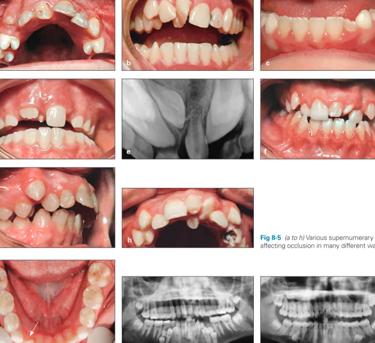
Fig 8-7 (a) Parapremolar supernumerary teeth preventing eruption of mandibular premo-lars. (b) Paramolar supernumerary teeth damaging the permanent fi rst molar roots. Fig 8-6 Supplemental mandibular
supernu-merary tooth (arrow) causing crowding, mid-line shift, and arch asymmetry.
a b
Early Recognition and
Clinical Signs of Hyperdontia
Development of supernumerary teeth can occur any time during the primary dentition, mixed dentition, and the per-manent dentition. They are almost always harmful to adja-cent teeth and to the occlusion. Most cases of supernumer-ary teeth are asymptomatic and are usually found during routine clinical or radiologic investigations. Therefore, early recognition of and treatment planning for supernumerary teeth are important components of the preliminary assess-ment of a child’s occlusal status and oral health, which is based on careful clinical and paraclinical examinations.
Clinical examination
Clinical examination of children during the primary or mixed dentition is discussed in detail in chapter 3. When assess-ing supernumerary teeth in the developassess-ing occlusion of a child, the clinician must consider the number, size, and form of teeth, the eruption time, the sequence of eruption, the position of each tooth, and local and general factors that can affect occlusion during transitional changes. The following are clinical signs of the presence of supernumer-ary teeth:
• Abnormal pattern and abnormal sequence of eruption • Delayed eruption
• Absence of eruption
Fig 8-5 (a to h) Various supernumerary teeth, affecting occlusion in many different ways. a d f b e g c h Bahreman_CH08.indd 196 Bahreman_CH08.indd 196 3/19/13 8:49 AM3/19/13 8:49 AM
Diagnosis and Management of Abnormal Frenum Attachments
9
218
Case 9-2
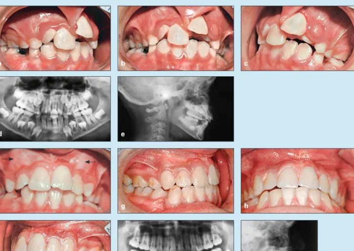
A 10-year, 8-month-old girl exhibited a Class II division 1 malocclusion and maxillary and mandibular incisor protrusion. In addition, an invasive frenum attachment caused severe maxillary incisor crowding, displacement, and cystic formation (Figs 9-19a to 9-19e).
Treatment:
The treatment plan included removal of the frenum, the cyst, and all abnormal soft tissue attachment and extrac-tion of the four fi rst premolars, carried out as a serial step-by-step extracextrac-tion.
After the surgical procedure and tissue healing, a removable maxillary Hawley appliance was inserted to achieve slow, minor incisor alignment, and use of a lower holding arch for about 1 yearwas followed by step 1 of the extraction series: removal of the maxillary primary canines, both maxillary primary fi rst molars, and both mandibular primary fi rst molars. Figure 9-19f shows alignment of the maxillary incisors and the canine bulges before serial extraction.
Step 2 was extraction of all four fi rst premolars. Maxillary anchorage was prepared with a Nance appliance, and the lower holding arch was removed as reciprocal anchorage.
Step 3 of the extraction sequence was removal of the remaining primary second molars. This was followed by maxillary and mandibular bonding to start maxillary canine retraction. Then mandibular and later anterior retrac-tion and space closure were accomplished. Some mesial movement of the mandibular molars was allowed, in order to achieve a Class I molar relationship (Figs 9-19g to 9-19k).
a f i d b g j e c h k
Fig 9-19 Treatment of a 10-year, 8-month-old girl with a Class II division 1 malocclusion and maxillary and mandibular protru-sion. An invasive frenum attachment has caused tooth displacement, maxillary incisor crowding, and formation of a cyst. (a to c) Pretreatment occlusion. (d) Pretreatment panoramic radiograph. (e) Pretreatment cephalometric radiograph. (f) Tissue heal-ing and some incisor alignment. The arrows show canine bulge. (g to i) Posttreatment occlusion. (j) Posttreatment panoramic radiograph. (k) Posttreatment cephalometric radiograph.
Bahreman_CH09.indd 218
Early Detection and Treatment of Eruption Problems
1 0
244
Fig 10-18 Management of an ectopic maxillary canine that has caused resorption of the permanent central incisor root and subsequent exfoliation. (a to c) Pretreatment occlusion. (d) Pretreatment panoramic radiograph. (e to h) Occlusion during active treatment and level-ing. The canine bracket has a higher K distance to achieve elongation. (i to l) Posttreatment occlusion, after end of active treatment and reshaping of the canine to mimic the central incisor. 1—permanent central incisor; 2—permanent lateral incisor; 3—permanent canine; C—primary canine. a d g j b e h k c f i l
Tooth Transposition
Another kind of eruption disturbance is tooth transposition, or positional interchange of two adjacent teeth, especially their roots. Tooth transposition is a rare but clinically diffi -cult developmental anomaly. Depending on the transposed teeth and their position, normal eruption of adjacent teeth can be affected, root anatomy can be damaged, and erup-tion of the affected teeth can be delayed. This eruperup-tion disturbance was fi rst defi ned in 1849 by Harris,50 who
de-scribed tooth transposition as an “aberration in the position of the teeth.”
Transposed teeth are classifi ed into two types of tooth displacement: complete transposition and incomplete
transposition (Fig 10-19). In complete transposition, both the crowns and the entire root structures of the involved teeth are displaced to abnormal positions. In incomplete transposition, only the crown of the involved tooth is trans-posed, and the root apices remain in place.
Transposition is sometimes accompanied by other dental anomalies, such as peg-shaped lateral incisors, congenitally missing teeth, crowding, overretained primary teeth, dilac-erations, and rotation of adjacent teeth.
Displacement of one tooth from one quadrant across the midline to the other side of the arch has very rarely been re-ported, but according to Shapira and Kuftinec51 these types
of anomalies should be considered ectopically erupted teeth, not transposed teeth.
C 2 3
1
Bahreman_CH10.indd 244
321
Simple Dental Crossbite
Case 11-9:
Anterior dental crossbite
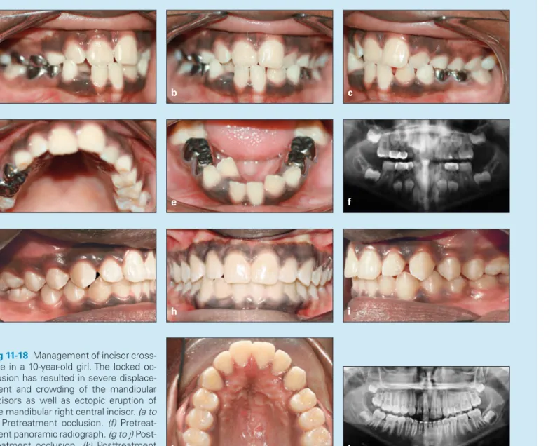
A 10-year-old girl in the middle mixed dentition presented with a Class III molar relationship on the right side because of space loss, 0- to 1-mm overbite and overjet, and three maxillary incisors in crossbite. Treatment had been delayed, causing severe crowding of the mandibular incisors and ectopic eruption of the mandibular right lateral incisor (Figs 11-18a to 11-18f).
Treatment:
Because of the severe crowding and displacement of incisors, the treatment plan incorporated fi xed appliances with maxillary and mandibular 2 × 6 bonding. The fi rst step in treatment was 2 × 4 maxillary bonding, mandibu-lar fi rst momandibu-lar occlusal bonding to disocclude the anterior segment, and placement of 0.016-inch nickel-titanium maxillary arches (cinched back) for leveling and release of abnormal anterior contact. The second step was placement of 0.016-inch stainless steel maxillary arches with an open U-loop mesial to the molar tube (extended arch length) to procline the maxillary incisors out of crossbite. The third step was mandibular 2 × 4 bonding: fi rst with 0.014-inch nickel-titanium archwire because of severe crowding and later with 0.016-inch nickel-titanium archwire for further leveling.
The fourth step was use of an open U-loop to place an extended-length stainless steel archwire against the mandibular molar tube to achieve minor mandibular incisor proclination in order to gain space and align the man-dibular incisors.The fi nal step was bonding the permanent canines after eruption for fi nal anterior alignment. Figures 11-18g to 11-18k show the treatment outcome.
a d g j b e h k c f i
Fig 11-18 Management of incisor cross-bite in a 10-year-old girl. The locked oc-clusion has resulted in severe displace-ment and crowding of the mandibular incisors as well as ectopic eruption of the mandibular right central incisor. (a to e) Pretreatment occlusion. (f) Pretreat-ment panoramic radiograph. (g to j) Post-treatment occlusion. (k) Posttreatment panoramic radiograph.
Bahreman_CH11.indd 321
Index
417 A Acellular cementum, 23 Achondrodysplasia, 230 Acrodynia, 236Active holding arch, 82, 83f Active lingual arch, 92 Adenoid facial type, 146 Age of patient
midline diastema and, 210 for orthodontic screening, 7 serial extraction considerations,
117
space loss affected by, 76 Agranulocytosis, 236 Alginate, 51
Alkaline phosphatase, 20, 235 Allergies
hypodontia and, 162 mouth breathing and, 147 Alveolar bone, 24
Alveolar process development of, 225 function of, 26 growth of, 26
maxilla and mandible relationship to, 37 Alveolar ridge, 233 Ameloblasts, 18, 19f, 20 Amelogenesis, 20 Amelogenesis imperfecta, 19 Amelogenin, 21
Anchored space regainers, 87–89, 88f Angle’s classifi cation of
malocclusion, 3, 150 Ankyloglossia, 215f, 215–216 Ankylosis
case studies of, 285f–286f defi nition of, 281
dentition effects of, 282, 282f–283f diagnosis of, 283
etiology of, 281–282
lateral tongue thrust and, 141 management of, 283–284 permanent teeth, 31 prevalence of, 281 primary teeth, 31, 165, 281 treatment of, 283–284 Anodontia, 158
Anterior Bolton discrepancy, 209 Anterior crossbite
case studies of, 320f–322f cephalometric evaluation of, 316 Class III malocclusion and, 316 clinical examination of, 316 differential diagnosis of, 316 Hawley appliance for, 319 illustration of, 49f, 257f incisor, 317, 318f, 321f
maxillary canine impaction and, 257f
in mixed dentition, 321f simple
defi nition of, 316 etiology of, 317, 318f incidence of, 316 signs of, 317 single-incisor, 320f
treatment of, 319, 347f–351f Anterior open bite
anterior tongue thrust and, 142 illustration of, 49f
lisping caused by, 50
thumb sucking as cause of, 133, 134f Anterior provisional partial denture,
84–85, 85f Anterior teeth
early loss of, 84 protrusion of, 90
Anterior tongue thrust, 141, 141f Apposition, 21
Arch
collapse of, 6, 6f, 339, 344f crowding in, 52
dental cast evaluation of, 52 development of, 28
form of, 52 length of
defi nition of, 53 incisor proclination for
increasing, 91 loss of, 282
palatal canine impaction and, 255 primary dentition’s role in, 30 reduction of, during transitional
dentition, 115
tooth size and, discrepancy between, 106–107
transitional dentition changes in, 38
physiologic changes in, 29 required space in, 53 symmetry of, 52, 53f Arnold expander, 363, 363f
Asymmetric tooth eruption, 240–241 Atavism theory, 192
Autotransplantation
canine impaction treated with, 265 disadvantages of, 171
lateral incisor hypodontia treated with, 171
mandibular second premolar hypodontia treated with, 174
B
Band and loop space maintainer, 82, 83f
Band and occlusal bar, 84, 84f Band and pontic, 84, 84f
Band and U-loop space regainer, 87, 88f
Behavioral evaluation, 43 Behavioral modifi cation, for
non-nutritive sucking, 135 Bipupillary plane, 56 Bite guards, 152 Bite plate, 403–404, 404f Bitewing radiographs, 58 Blanching test, 211, 211f Bluegrass appliance, 136, 137f Bolton analysis, 54, 78, 79f Bolton discrepancy, 115, 127, 128f, 209
Bone morphogenetic protein 2, 23 Bone remodeling, 228
Brachycephalic head shape, 45 Brodie syndrome, 360f, 360–361,
372f–373f Bruxism, 151–152
Buccal canine impaction, 254, 257f, 263–264
Buccal crossbite, 360, 372f–374f Bud stage, 17f–18f, 17–18 Page numbers with “t” denote tables; those with “f” denote fi gures; those with “b” denote boxes
Bahreman_Index.indd 417
Index
418 D
C
Calcospherites, 21 Camoufl age treatment
for Class II malocclusion, 297 for open bite, 380
Canines
crescent moon–shaped root resorption of, 118, 119f eruption of
ectopic, 165, 243, 244f before premolar eruption,
239–240
mandibular. See Mandibular canines.
maxillary. See Maxillary canines. permanent
ectopic eruption of, 165, 243, 244f eruption of, 37, 38b
primary
early loss of, 403 extraction of, 261–262 overretained, 246
premature exfoliation of, 118 serial extraction of, 120 transposition of, 245–246, 255 unerupted, bulging of, 118 Cap stage, 18, 18f
Cartilage calcifi cation, 21 Casts, dental, 51–54 Cellular cementum, 23 Cementoblasts, 22f, 23 Cementogenesis, 23 Cementum acellular, 23 cellular, 23 formation of, 23 Central diastema, 36 Central incisors
eruption of, before maxillary lateral incisor eruption, 240 maxillary
anterior crossbite caused by, 317, 318f diastema between, 205 overretained, 317 supernumerary, 199f–200f Cephalometric radiographs, 66–67, 68b, 101f, 116–117, 258, 296 Cervical headgear, 3 Cervical loop, 22, 22f Chemotherapy, 162–163 Chin cap with spurs, 332, 332f Clark’s rule, 258
Class I malocclusions, serial extraction in, 119–122, 121f, 124f–126f
Class II malocclusion case studies of, 302f–315f cephalometric analysis of, 296 characteristics of, 294
diagnosis of, 295–296 division 1, 301–302, 407f division 2, 302, 410f early treatment of, 9, 294 facial height effects on, 294 growth patterns, 294 historical background of, 3 jaw characteristics in, 295b morphologic characteristics of,
295, 295b
panoramic radiograph of, 62f prevalence of, 294
serial extraction in, 122–123 transverse dimension considerations, 294 treatment of camoufl age, 297 early, 294 extraoral traction, 298–299 functional appliances, 298 growth modifi cation and occlusal
guidance, 297–299 headgear, 298–300 HLH technique, 299–302,
308f–309f, 314f lip bumper, 300–301, 301f modifi ed Hawley appliance, 300,
300f
one-phase, 302, 310f–315f orthognathic surgery, 297 two-phase, 301–302, 302f–309f variations of, 295f
Class III malocclusion anterior crossbite and, 316 case studies of, 333f–351f causes of, 329
classifi cation of, 331 crossbite and, comparisons
between, 331b
dentofacial characteristics of, 329 hereditary, 334f, 342f–343f mandibular prognathism with,
330, 341f, 346f
pretreatment evaluation of, 329 prevalence of, 329–330
pseudo–
case studies of, 325f–329f defi nition of, 323
delayed treatment of, 324, 330 multiple incisor involvement in,
323
removable appliances for, 324, 324f
signs of, 323
treatment of, 323–324, 324f serial extraction in, 123 skeletal, 329–330 treatment of
after incisor eruption, 338, 338f–346f
chin cap with spurs, 332, 332f in early mixed dentition, 333 early strategies for, 331–332 face mask–chin cap combination,
332, 332f
factors that affect, 331b interceptive, 335f–336f in late mixed dentition, 344f in primary dentition, 333, 334f Cleft lip and palate, 163
Cleidocranial dysostosis, 231 Clinical examination
ankylosed primary molars, 283 anterior crossbite, 316
delayed tooth eruption, 232 description of, 44
differential diagnosis of, 142–143 hyperdontia, 196–197
posterior crossbite, 361 before serial extraction, 116 tongue thrust, 142–143
Closing the drawbridge, 383, 383f Computed tomography scans, 59,
59f, 258–259 Concave profi le, 47, 47f Concomitant hypodontia and
hyperdontia, 165
Condylar hypertrophy, 361, 361f Condylar hypotrophy, 361 Congenital hypothyroidism, 230
Convex facial profi le, 47f, 117 Coronoid process, 26
Corrective orthodontic treatment, 4 Craniofacial growth
dentition development and, 15, 25–27
description of, 5, 116 genetic infl uences on, 5 mouth breathing effects on, 148 occlusion affected by, 116 Crossbite
anterior. See Anterior crossbite. central incisor, 179f
functional. See Pseudo–Class III malocclusion.
posterior. See Posterior crossbite. skeletal Class III malocclusion and,
comparisons between, 331b thumb sucking as cause of, 133,
134f unilateral, 6, 7f Crowding
arch, 52 degree of
space analysis and, 79 space loss affected by, 76 of incisors. See Incisor(s),
crowding of.
of mandibular incisors, 38–39 of molars, 91f
Crown
epithelial coverage of, 24 permanent, primary root
resorption and, 32 Crown and bar, 84, 84f Crown and pontic, 84, 84f Curve of Spee, 53–54, 79, 404 Curve of Wilson, 361
Cuspal height, 400
Cusps, enamel knot’s role in formation of, 20
Cyst formation, 18
D
Deep bite
case studies of, 405f–413f cuspal height effects on, 400 defi nition of, 397
degree of, 397
dental, 397–398, 402–403 development of, 397
differential diagnosis of, 399–400 etiology of, 397–399
factors that affect, 400
impinging, 6, 6f, 202f, 206, 209, 310f, 314f, 405f
mandibular forward growth and, 397
morphologic characteristics of, 399 periodontal disease and, 401 relapse of, 399 reverse, 336f skeletal, 398–399, 403–404 treatment of appliances for, 404–405 delayed, 400 early, 401 in mixed dentition, 403–404, 412f in permanent dentition, 401–402 in primary dentition, 403 strategies for, 402–405 Deep overbite, 303f Deglutition, 139–140 De-impactor spring, 242, 243f Bahreman_Index.indd 418 Bahreman_Index.indd 418 3/19/13 11:09 AM3/19/13 11:09 AM
Index
419
Delayed exfoliation, of primary dentition, 31
Dental caries, 31 Dental casts
arch form and symmetry evaluations using, 52 description of, 51–52
occlusion evaluations using, 52 before serial extraction, 116 Dental follicle
anatomy of, 22–23 fi broblasts of, 24
permanent, congenital absence of, 31
tooth eruption affected by, 227 Dental history, 43–44
Dental lamina
development of, 16f, 16–17 magnifi cation of, 16f
Dental occlusion. See Occlusion. Dental retrusion, 56 Dentigerous cyst, 193, 194f Dentin apposition of, 21 formation of, 20 hypoplasia of, 21 interglobular, 21 mineralization of, 20–21, 23 Dentin fl uorosis, 21
Dentin matrix protein-2, 23 Dentinogenesis, 20
Dentinogenesis imperfecta, 19 Dentition
ankylosis effects on, 282, 282f–283f bruxism effects on, 152
intraoral examination of, 48–49, 49f monitoring of, during early-age
orthodontic treatment, 9 Dentition development
craniofacial growth and, 15, 25–27 description of, 15
neonatal, 27f, 27–28 permanent, 19 postnatal, 28
primary. See also Primary dentition.
bud stage of, 17f–18f, 17–18 calcifi cation stage of, 20–21 cap stage of, 18, 18f crown stage of, 21, 21f early bell stage of, 18f, 18–19 initiation stage of, 16f–17f, 16–17 late bell stage of, 19f–20f, 19–20 molecular level of, 22–23 morphodifferentiation stage of,
19f–20f, 19–21 root formation, 22, 22f studies of, 22–23 retarded, 238–239 Dentogingival junction development of, 24 tissues of, 24 Desmosomes, 18 Developmental spaces, 28–29 Diagnostic database, 42 Diagnostic process description of, 51 goal of, 41 interview, 42–44 questionnaire, 42–44
schematic diagram of, 42, 42f steps involved in, 42
Diastema, 36, 94, 179f central, 94, 179f
midline. See Midline diastema. Dichotomy theory, 192
Digit sucking, 132–136, 134f Digital imaging, 59
Distal drift, 76–77
Distal jet appliance, 88, 88f Distal shoe, 82
Distal step terminal plane, 29f, 29–30, 33f
Distraction osteogenesis, 360 Divergence of the face, 47
Dolichocephalic head shape, 45, 146f Down syndrome, 163, 231, 245 Drift, 26, 76–77 “Dual bite,” 51 Dwarfi sm, 230 E E space, 110, 110f
Early exfoliation, of primary dentition, 31
Early-age orthodontic treatment advantages of, 66
benefi ts of, 11–12
clinical evidence about, 10 controversy associated with, 9–11 costs of, 11
current interest in, 6–7 defi nition of, 4
dentition monitoring during, 9 goals of, 8
growth patterns and, 10–11 lack of training in, 12 misconceptions about, 10–11 modern views on, 41 objectives of, 4 one-phase, 8 patient benefi ts, 11 phases of, 8–9
practitioner benefi ts, 12
professional encouragement of, 12 rationale for, 7
reasons for, 4–7 results with, 11–12 single phase of, 8, 10 strategy of, 4, 8 timing of, 7–8, 298 two-phase, 9–10 Ectoderm, 22 Ectodermal dysplasia, 163 Ectomesenchymal cells, 19–20, 23–24 Ectomesenchyme, 16–17 Ectopic eruption defi nition of, 241
permanent canines, 243, 244f permanent fi rst molars, 241–242,
242f
prevalence of, 241
Ectopic impacted canines, 260 Ellis lingual arch, 82, 83f Embryonic period, 15 Enamel
apposition of, 21 formation of, 20 mineralization of, 20–21 tetracycline discoloration of, 21 Enamel hypoplasia, 21
Enamel knot
in cusp formation, 20 defi nition of, 17 illustration of, 18f Enamel matrix, 20–21 Enamel organ, 18, 18f Epithelial cuff, 24 Epithelial thickening, 16, 16f Examination(s)
clinical. See Clinical examination. extraoral. See Extraoral
examination.
photographic evaluation. See Photographic evaluation. radiographic. See Radiographs. Exfoliation, of primary dentition
description of, 30–32, 229 early, 235–236
External enamel epithelium, 18 Extraction. See also Serial extraction.
early-age orthodontic treatment effects on need for, 11–12 space creation through, 90 Extraoral anchorage, 3 Extraoral examination elements of, 44–45
frontal facial evaluation, 45–46, 46f lateral facial evaluation, 46–47 Extraoral photography facial esthetics, 55–56, 57f frontal view, 54–57, 55f lateral view, 55–56 oblique view, 55 Extraoral radiographs, 58–59 Extraoral traction, for Class II
malocclusion, 298–299
F
Face
description of, 131
embryologic development of, 15 vertical growth of, 380
Face mask–chin cap combination, 332, 332f
Facial asymmetry, 56, 57f, 361f Facial esthetics
composition of, 45
early-age orthodontic treatment benefi ts for, 11
evaluation of, 44
malocclusion effects on, 9
photographic evaluation of, 55–56, 57f
primary dentition’s role in, 30 Facial evaluation
frontal, 45–46, 46f lateral, 46–47 Facial form, 44 Facial height, 294 Facial profi les, 47, 47f Facial proportion
evaluation of, 46, 46f frontal, 56, 57f head posture and, 148 lateral, 56, 57f
Facial symmetry, 45–46, 46f Facial trauma, 162
Facial typing, 45
Family medical history, 43–44 Fiber-reinforced composite resin
fi xed partial denture, 170 Fibroblast growth factors, 23 Fibroblasts, 228
Finger sucking, 132–136, 134b, 134f, 378, 378f, 381–382
Bahreman_Index.indd 419
Index
420 I
First molars
distalization of, 91, 91f
ectopic eruption of, 241–242, 242f mandibular
maxillary fi rst molar and, 37 mesial shift of, 38
permanent, 32 maxillary
ectopic eruption of, 119, 119f, 243f mandibular fi rst molar and, 37 vertical palisading of, 119, 119f, 124 Fixed expanders, 93–94, 94f, 362–
364, 363f–364f
Fixed orthodontic appliances, 136, 137f Fluorosis, dentin, 21
Flush terminal plane, 29f, 29–30, 33, 33f
Fourth germ layer, 22 Frenectomy, 214, 215f, 217f Frenotomy, 216 Frenum maxillary labial, 207 morphogenesis of, 206–207 structure of, 206–207
Frenum attachment abnormalities ankyloglossia, 215–216
case studies of, 216f–222f differential diagnosis of, 210–211 in infants, 214 management of in adults, 211–212 delayed, 212 frenectomy, 214, 215f, 217f in infants, 214 in mixed dentition, 212–213, 213f in primary dentition, 213–214 results of, 213f two-phase, 213
midline diastema. See Midline diastema.
occlusion affected by, 210 radiographs of, 211, 211f signs of, 211, 211f
Frontal cephalometric radiographs, 67 Frontal view, 54–55, 55f
Functional crossbite, 359–360.
See also Pseudo–Class III malocclusion.
Functional matrix, 6, 131
G
Genetic theory, of maxillary canine impaction, 255–256
Gingival groove, 27 Glossectomy, 49 Glycosaminoglycans, 18
Groper fi xed anterior prosthesis, 85 Growth modifi cation techniques
Class II malocclusion treated with, 297–299
open bite treated with, 382–383, 383f
Growth patterns
Class II malocclusion, 294 early-age orthodontic treatment
and, 10–11
incisor position and crowding affected by, 111
mixed dentition space analysis, 53 sagittal expansion and, 90–91 serial extraction considerations,
117–118
space analysis and, 79 Growth status evaluation, 43
Gubernaculum dentis, 227
Guidance theory, of maxillary canine impaction, 254–255
Gum pads, 27, 27f, 140 Gurin lock regainer, 87, 88f
H
Haas expander, 93, 94f, 363, 363f Halterman appliance, 242, 243f Hand-wrist radiographs, 59 Hard tissues. See Dentin; Enamel. Hawley appliance
anterior crossbite treated with, 319 bruxism treated with, 152
Class II malocclusion treated with, 299–300
as habit breaker, 136, 137f modifi ed, 300, 300f, 324f, 406f as removable distalizer, 92 space maintenance using, 86, 86f space regaining using, 89, 89f tongue thrust treated with, 145f Headgear
Class II malocclusion treated with, 298–300
high-pull, 299
historical background of, 3 J-hook, 299
patient’s cooperation in using, 299 sagittal expansion using, 92 Hemifacial microsomia, 163 Hereditary crowding, of incisors,
118–119, 119f
Hertwig’s epithelial root sheath, 22, 22f, 24
Histodifferentiation description of, 18, 19f
developing abnormalities during, 19 Holoprosencephaly, 207
Homeobox genes, 22, 161 Hyperactivity theory, 192 Hyperdontia. See also
Supernumerary teeth. case studies of, 198f–202f clinical examination of, 196–197 defi nition of, 17
hypodontia and, 165–166, 192 management of, 197–198 occlusion affected by, 196, 197f prevalence of, 189–190, 190t–191t radiographic examination of, 197 Hypodontia
autotransplantation for, 171, 174 case studies of, 174f–185f central incisors, 180f–181f clefts associated with, 163 clinical signs of, 167 defi nition of, 17, 158
dental anomalies associated with, 164–165
dentoskeletal patterns affected by, 166
description of, 157 distribution of, 160t in Down syndrome, 163 early recognition of, 167 environmental factors, 161–163 ethnicity and, 159t–160t etiology of, 160–163 sex and, 159t–160t genetic factors, 160–161 in hemifacial microsomia, 163 hyperdontia and, 165–166, 192 lateral incisors autotransplantation for, 171 canine substitution for space
closure, 168–169 case studies of, 176f–178f,
184f–185f
impaction caused by, 274 management of, 168–171 maxillary, 208f
midline diastema caused by, 208, 208f
prosthesis for, 169–171, 171f management of, 167–168 mandibular second premolars,
172–174, 180f–181f microdontia and, 164
occlusion affected by, 157, 166 partial, 163
prevalence of, 158, 159t, 162 soft tissue affected by, 166 space closure, 168–169, 173 syndromes associated with,
163–164
systemic diseases associated with, 162 treatment of, 167–168 Hypophosphatasia, 235–236 Hypopituitarism, 230 Hypoplasia dentin, 21
enamel. See Enamel hypoplasia. Hypothyroidism, 230
Hyrax expander, 93–94, 94f, 363, 363f
I
Image shift principle, 258
Impinging deep bite, 6, 6f, 202f, 206, 209, 310f, 314f, 405f Implant-supported restorations, 173–174 Incisor(s) anterior crossbite, 317, 318f, 321f crowding of acquired, 118 Bolton discrepancy, 115, 127, 128f causes of, 106–107 characteristics of, 107–108 in Class I malocclusions, 119–122, 121f, 124f–126f in Class II malocclusions, 122–123 in Class III malocclusions, 123 classifi cation of, 107–108 description of, 95, 105 environmental, 118 hereditary, 118–119, 119f intervention for, 107 measurement of, 117 minor, 108 in mixed dentition, 95, 105–106 moderate, 108–114, 109f–114f prediction of, 106 prevention of, 107
serial extraction for. See Serial extraction.
severe, 115, 117, 120, 121f, 264 tooth size–arch length
discrepancy as cause of, 106–107, 123
in transitional dentition, 110 transverse expansion for, 93 eruption of
asymmetric, 36
central diastema persistence during, 36
Bahreman_Index.indd 420
Index
421
Class III malocclusion treatment after, 338, 338f–346f
mandibular central incisors, 34, 34f mandibular lateral incisors, 34, 34f maxillary central incisors, 35, 35f maxillary lateral incisors, 35–36, 36f permanent, 34f–36f, 34–36 problems during, 36, 36b impaction of
case studies of, 275f–280f early detection and diagnosis of,
273 etiology of, 273
interceptive treatment of, 274 odontoma as cause of, 273 supernumerary teeth as cause of,
273, 274f, 279f trauma as cause of, 273 inclination of, 53, 56, 79, 91, 117 intrusion of, 402
labial movement of, 91 lateral
mandibular, 34, 34f maxillary, 35–36, 36f splaying of, 118, 119f lip position and, 53 mandibular
crowding of
Bolton discrepancy as cause of, 127, 128f
description of, 38–39, 91, 105, 107, 114f, 207, 406f eruption of, 34, 34f gingival recession at, 118 maxillary
eruption of, 35–36, 36f space closure with, 94 overretained, 32f
periodontal condition of, 91 primary
early loss of, 81f, 85 overretained, 274, 317, 318f roots, delayed resorption of, 109f sequential stripping of, 109 spaces between, 28 proclination of, 34f, 91–92 root resorption of delayed, 34f, 109f description of, 256 splaying of, 118, 119f Incisor liability, 33–34, 36 Inconstant swallowing, 142 Infantile swallowing, 140, 142 Initiation stage, 16f–17f, 16–17 Intercanine arch width, 34 Interceptive treatment
defi nition of, 4
of incisor impaction, 275 of maxillary canine impaction,
260–262
patient expectations about, 43 Interdental fi bers, 207
Interdental spacing, 79 Interglobular dentin, 21 Interincisal angle, 400 Intermolar width, 92
Interproximal wedging technique, 242, 242f Intertransitional periods, 28 Interview, 42–44 Intraoral examination components of, 116 dentition, 48–49, 49f description of, 47, 116 paraclinical evaluation, 51 soft tissues, 49–51
temporomandibular joint function, 51 tongue, 49–51, 50f Intraoral photography, 58 Intraoral radiographs, 58 Irradiation, 162–163 J Jaw fracture of, 246 ontogenesis of, 25, 25f Jaw muscles, 26 J-hook headgear, 299 Jumping the bite, 298 Juvenile hypothyroidism, 230 Juvenile rheumatoid arthritis, 26
L
Lasers, 214
Lateral cephalometric radiographs, 4, 258
Lateral expansion, 93
Lateral facial evaluation, 46–47 Lateral facial proportion, 56, 57f Lateral incisors
hypodontia of
autotransplantation for, 171 canine substitution for space
closure, 168–169 case studies of, 176f–178f management of, 168–171 palatally displaced maxillary
canines associated with, 255 prosthesis for, 169–171, 171f mandibular eruption of, 34, 34f transposition of, 245, 249f maxillary eruption of, 35–36, 36f, 240 microdontia of, 208, 208f supernumerary, 194f transposition of, 250f microdontia of, 208, 208f peg-shaped, 274 proclination of, 36, 36f supernumerary, 198f transposition of, 245, 249f–250f, 252f–253f
Lateral jaw radiographs, 58–59 Lateral tongue thrust, 141f, 141–142 Lateral view, 55–56
Leeway space, 78, 80, 95, 276f Ligand for receptor activator for
nuclear factor κB, 30 Lingual crossbite, 360, 360f Lip bumper, 91–92, 92f, 299, 300–301, 301f Lip dysfunction, 209, 209f, 398 Lip line, 169 Lip position, 56, 57f Lip proportion, 56, 57f Lip seal, 382 Lip strain, 55f Lisping, 50 Locked occlusions, 6f, 6–7, 113f Longitudinal panoramic radiographs,
60, 61f–65f, 116
Lower holding arch, 82, 83f, 109f Lower lip dysfunction, 403
M
Macroglossia
description of, 49, 49f
tongue thrust associated with, 141, 141f
Malocclusions
Angle’s classifi cation of, 3, 150 Class I, 119–122, 121f, 124f–126f Class II. See Class II malocclusion. Class III. See Class III malocclusion. environmental factors associated
with, 293 etiology of, 42
facial esthetics affected by, 9 speech problems and, 50, 150–151 thumb/fi nger sucking as cause of,
133, 134f, 137f treatment of, 115
untreated, problems secondary to, 9 Mandible
anatomy of, 25, 25f anterior shift of, 325f–326f displacement of, 247
masticatory muscle attachment to, 26
normal closure pattern of, 400 positions of, 297 retrusion of, 412f Mandibular arch, 49f Mandibular canines eruption of, 37 impaction of, 265
permanent, eruption of, 37 primary
early extraction of, 36
premature loss of, 35, 98f, 102f Mandibular condyle
ankylosis of, 26 growth of, 26–27 Mandibular fi rst molars
extraction of, 247f
maxillary fi rst molar and, 37 mesial shift of, 38
permanent, 32
Mandibular fi rst premolar eruption before canine eruption, 239–240 description of, 37
Mandibular growth asymmetric, 7f, 25, 46f direction of, 26
impinging deep bite effects on, 6 insuffi cient, 19
malocclusions caused by problems with, 25
at mandibular condyle, 26 occlusion affected by, 106 temporomandibular joint-related
factors that affect, 26 Mandibular incisors. See also
Incisor(s). central, 34, 34f crowding of
Bolton discrepancy as cause of, 127, 128f description of, 38–39, 49, 91, 105, 107, 114f, 207 inclination of, 79 lateral, 34, 34f proclination of, 350f relative position of, 397
Mandibular molar distalizer, 88, 88f Mandibular plane–occlusal plane
angle, 400
Bahreman_Index.indd 421
Index
422 N
Mandibular prognathism, 330, 341f, 346f
Mandibular second molars eruption of, maxillary second
molar eruption before, 240 impaction of, 119, 119f
terminal plane, 32, 33f
Mandibular second premolars, 172– 174, 180f–181f Mastication, 30 Masticatory muscles, 26 Maternal rubella, 162 Maxillary arch collapse of, 6, 6f, 339, 344f constriction of, 355, 357, 359 Maxillary bone, 25
Maxillary canines. See also Canines. displacement of, 266f–267f eruption before premolars, 239 impaction of
autotransplantation of, 265 buccal, 254, 257f, 263–264 case studies of, 266f–272f clinical examination of, 257–258 consequences of, 256–257 early detection of, 257–260 ectopic, 260, 262f, 268f etiology of, 254–256
interceptive treatment of, 260–262 labial, 256
odontoma as cause of, 269f–271f orthodontic procedures for, 264 palatal, 254–256, 263
panoramic radiographs of, 259f, 259–260
position of, 262 prevalence of, 254
proximity of, to adjacent teeth, 259 radiographic evaluation of,
258f–259f, 258–260 signs of, 258
space defi ciency as cause of, 266f–267f
step-by-step management of, 264b surgical exposure of, 262–264, 263f treatment of, 260–265
Maxillary fi rst molars
ectopic eruption of, 119, 119f, 243f mandibular fi rst molar and, 37 vertical palisading of, 119, 119f, 124 Maxillary incisors. See also
Incisor(s). central, 35, 35f
delayed treatment of midline diastema until complete eruption of, 212
fl aring of, 133
labial migration of, 401 lateral description of, 35–36, 36f eruption of, 35–36, 36f, 240 microdontia of, 208, 208f supernumerary, 194f liability, 36 permanent, 319 protrusion of, 36f secondary spacing, 36, 36f Maxillary intercanine distance, 29 Maxillary second molars
eruption of, before mandibular second molar eruption, 240 terminal plane, 32, 33f
Mechanotherapy
incisor impaction treated with, 277f with selective extraction, for open
bite, 383
tongue thrust treated with, 143, 143f Meckel’s cartilage, 25
Medical history, 43–44 Mentolabial sulcus, 56, 57f Merrifi eld analysis, of space, 78 Mesial drift, 26, 76–77
Mesial occlusion. See Class III malocclusion, skeletal. Mesial shift, 38, 347f
Mesial step terminal plane, 29f, 29–30, 33, 33f
Mesiodens, 194, 208, 208f Mesocephalic head shape, 45 Microdontia
hypodontia and, 164 illustration of, 163f lateral incisors, 208, 208f Midline diastema
case studies of, 216f–222f causes of, 207–210
anterior Bolton discrepancy, 209 impinging deep bite, 209 lateral incisor hypodontia, 208,
208f
lip dysfunction, 209, 209f mesiodens, 208, 208f odontoma, 208 overview of, 207–208
pathologic tooth migration, 210, 210f
defi nition of, 205
differential diagnosis of, 210–211 etiology of, 206–210 sex and, 206 management of in adults, 211–212 delayed, 212 in infants, 214 in mixed dentition, 212–213, 213f in primary dentition, 213–214 results of, 213f two-phase, 213
occlusion affected by, 210 prevalence of, 206 radiographs of, 211, 211f shape of, 211, 211f
Mineralization, of hard tissues, 20–21, 23
Mixed dentition
anterior crossbite in, 317, 318f, 321f deep bite in, 403–404, 412f
early, Class III malocclusion treatment in, 333
frenum attachment abnormalities in, 212–213, 213f
incisor crowding in
description of, 95, 105–106 serial extraction for, 115. See also
Serial extraction. Moyers analysis of, 78 open bite management in, 382 posterior crossbite in, 367f space analysis of, 52–54, 78 transverse expansion during, 93 Modifi ed Hawley appliance, 406f Molar(s)
distalization of, 91 fi rst. See First molars. mesially tipped, 91 permanent, 53 primary ankylosis of, 165, 281, 282f, 285f extraction of, 74, 120–121, 121f, 173
long-term retention of, 172 submerged, 285f
second. See Second molars. Molar distalizer
with Nance anchorage, 87–88, 88f pendulum appliance as, 92, 92f Morphodifferentiation stage, 19f–20f,
19–21 Mouth breathing
adenoid tissue location evaluations, 150
clinical examination of, 149–150 dentofacial characteristics of, 146,
146f, 148–149 etiology of, 147 evaluation of, 149–150
general body growth associated with, 147–148
lip incompetence associated with, 150
maxillofacial complex affected by, 146–147
occlusion effects of, 146–147 open bite caused by, 378 orthodontic management of, 150 posterior crossbite secondary to,
356–357
postural changes associated with, 146–148
problems associated with, 149, 149t signs of, 146f, 147–149
treatment of, 150
Moyers mixed dentition analysis, 78
MSX1, 23 MSX2, 23 MSX genes, 161 Multirooted teeth, 22 Muscular dystrophy, 26 N
Nance analysis, of space, 54, 77–78 Nance holding arch, 83, 84f Nasal obstruction, 147, 149 Nasolabial angle, 56, 57f Nasomaxillary complex, 27, 146 Natal teeth, 27–28 Neonatal dentition development of, 27f, 27–28 gum pads, 27, 27f, 140 natal teeth, 27–28 Neural crest cells, 15, 22 Non-nutritive sucking
case studies of, 137f–138f clinical examination of, 134 defi nitions of, 132
etiology of, 132–133 fi nger, 132–136, 134b, 134f midline diastema caused by, 209,
209f
occlusion effects, 133
open bite caused by, 378, 378f, 381–382, 384f
pacifi ers, 136, 139
posterior crossbite caused by, 356 prevalence of, 133
thumb, 132–136, 134b, 134f treatment of
age of intervention, 135 behavioral modifi cation
Bahreman_Index.indd 422
Index
423 techniques, 135 description of, 135–138, 137f–138f orthodontic appliances, 138, 139f OObstructive sleep apnea syndrome, 148
Occlusal bite plate, 152 Occlusal development
craniofacial growth effects on, 116 environmental factors, 5–6 factors that affect, 37
form and function in, 6, 24–25 genetic factors, 5
hypodontia effects on, 157 locked occlusions effect on, 6f, 6–7 long process of, 5
mechanisms that affect, 5–6 prenatal stage of, 16–27 tongue’s role in, 27
Occlusal interferences, 6, 6f–7f Occlusal radiographs, 58, 258, 258f Occlusal system, 47
Occlusion Class I, 47
dental cast evaluation of, 52 frenum attachment abnormalities
effect on, 210
hyperdontia effects on, 196, 197f hypodontia effects on, 166 importance of, 25
locked, 6f, 6–7, 113f
mandibular growth effects on, 106 midline diastema effects on, 210 mouth breathing effects on,
146–147 normal, 397
pacifi er sucking effects on, 139 preparation of, for prosthetics, 174 sagittal evaluation of, 52
serial extraction and, 11