DEPARTMENT OF MEDICINE SOLNA,
DIVISION OF CLINICAL EPIDEMIOLOGY
Karolinska Institutet, Stockholm, Sweden
BREAST CANCER – BRAIN METASTASES AND
TREATMENT ASPECTS
Gabriella Frisk
All previously published papers were reproduced with permission from the publisher. Published by Karolinska Institutet.
Printed by E-print AB 2019 © Gabriella Frisk, 2019 ISBN 978-91-7831-405-8
Breast cancer – brain metastases and treatment aspects
THESIS FOR DOCTORAL DEGREE (Ph.D.)
By
Gabriella Frisk
Principal Supervisor:
Senior Lecturer
Karin Ekström Smedby MD. PhD. Karolinska Institutet
Department of Medicine Solna, Division of Clinical Epidemiology
Co-supervisor(s):
Magnus Bäcklund MD. PhD. Karolinska Institutet
Department of Medical Epidemiology and Biostatistics (MEB)
Elisabet Lidbrink MD. PhD. Karolinska Institutet
Department of Oncology and Pathology
Opponent:
Associate Professor Johan Ahlgren MD. PhD. Örebro University
Department of Medical science
Examination Board:
Senior Lecturer
Anna Milberg MD. PhD. Linköping University
Department of Medical and Health Science Division of Community Medicine
Associate Professor Sven Törnberg MD. PhD. Karolinska Institutet
Department of Oncology and Pathology
Associate Professor Nick Tobin BSc. PhD. Karolinska Institutet
ABSTRACT
Background: In Sweden breast cancer is the most common malignant cancer disease among women, with 8000 new individual cases each year. The prognosis is generally very good, but nevertheless many patients still die of breast cancer every year. The general aim of this thesis is to gain increased knowledge of brain metastases due to breast cancer, including incidence, predictors and treatment aspects and better knowledge of the potential benefit of low-dose aspirin among women with breast cancer in different stages.
Patients, methods and results: In study I and II we aimed to assess if the incidence of brain metastases have increased in Sweden over time. In study I, all Swedish patients with breast cancer during 1998-2006 were identified from the Swedish National Cancer Register. These individuals were matched to the National Patient Register to get information on admissions to hospital due to distant metastases. In the cohort of 50 528 identified breast cancer patients, 696 (1.4%) had admissions to hospital due to brain metastases. Patients were at 44%
increased risk of being admitted to hospital with brain metastases if diagnosed with a primary breast cancer in 2004-2006 compared with 1998-2000. In study II we used the BcBaSe cohort (based on three quality-of-care registers in the Stockholm-Gotland, Uppsala-Örebro and the North region). Here, we identified all women with a first breast cancer 2002-2012
(N=30 996) and used ICD-codes for distant metastases from both non-primary outpatient care and hospital admissions. Overall, 789 (2.5 %) patients were registered with brain metastases at diagnosis or during follow-up. According to preliminary results, patients diagnosed with breast cancer in 2009-2012 were at a 37% increased risk of developing brain metastases compared with the period 2002-2004.
In study III we aimed to evaluate survival and level of care following whole brain radiotherapy due to brain metastases among breast cancer patients in Stockholm. We identified 241 patients treated at the Karolinska University hospital radiotherapy units 1999 to 2012. We gathered data on outcome and prognostic factors including level of care before and after the radiotherapy treatment through reviews of the patients’ medical files. Median survival following whole brain radiotherapy was 2.9 months and 57 (24%) of the patients could never be discharged from hospital-care. Patients with poor performance status (WHO 3-4) had a median survival of 0.9 months and women with triple-negative primary tumors a median survival of 2.0 months. Poor performance status and being admitted to hospital before radiotherapy were associated with increased risk of not coming home.
In study IV we aimed to evaluate if low-dose aspirin use may have a role in the treatment of breast cancer, accounting for clinical characteristics. In this study we used the BcBaSe linkage to identify a cohort of 21 414 women diagnosed with a primary stage I-III breast cancer and 621 women diagnosed in stage IV 2006 to 2012. We analysed information from Swedish health-care registers on dispensings of low-dose aspirin, comorbidity and dates and causes of death. We found no clear association between low-dose aspirin use and breast-cancer specific death overall, nor with risk of recurrence in a subgroup analysis. A possible benefit was however noted in women with smaller breast cancer tumors, stage I, which warrants further study.
Discussion: The incidence of brain metastases in breast cancer appears to have increased in Sweden in recent years perhaps due to improved disease control outside of the brain. When a decision is made of treating brain metastases in breast cancer with whole brain radiotherapy, we should take into account the patient’s need of hospital care before treatment, performance status and choice of level of care in the late palliative stage of disease and the end-of-life period, since the median survival is short and many patients can never be discharged from the hospital after whole brain radiotherapy. Low-dose aspirin use in breast cancer does not seem to have any clear role in improving outcomes for breast cancer patients.
LIST OF SCIENTIFIC PAPERS
I. Incidence and time trends of brain metastases admissions among breast cancer patients in Sweden
Frisk G, Svensson T, Bäcklund LM, Lidbrink E, Blomqvist P, Smedby KE. British Journal of Cancer. 2012 May 22;106(11):1850-3
II. Update on incidence of brain metastases by tumor characteristics in breast
cancer 2002-2012 in Sweden
Frisk G,Ekberg S, Lidbrink E, Pettersson A, Bäcklund LM, Sund M, Fredriksson I, Lambe M, Smedby KE.
Manuscript
III. Survival and level of care among breast cancer patients with brain metastases treated with whole brain radiotherapy
Frisk G, Tinge B, Ekberg S, Eloranta S, Bäcklund LM, Lidbrink E, Smedby KE.
Breast Cancer Res Treat. 2017 Dec;166(3):887-896
IV. No association between low-dose aspirin use and breast cancer outcomes overall - a Swedish population-based study
Frisk G, Ekberg S, Lidbrink E, Eloranta S, Sund M, Fredriksson I, Lambe M, Smedby KE.
CONTENTS
1 Background... 1
1.1 Epidemiology ... 1
1.2 Breast cancer tumor characteristics ... 2
1.2.1 Histological type ... 2
1.2.2 Stage ... 3
1.2.3 Grade ... 4
1.2.4 Hormonal receptors ... 4
1.2.5 Human epidermal growth factor receptor 2 (HER2) ... 4
1.2.6 Proliferation ... 5
1.2.7 Molecular subtypes ... 5
1.3 Breast cancer treatment ... 6
1.3.1 Neoadjuvant chemotherapy ... 6 1.3.2 Surgery ... 6 1.3.3 Chemotherapy ... 7 1.3.4 Radiotherapy ... 7 1.3.5 Anti-HER2 treatment ... 7 1.3.6 Endocrine therapy ... 8
1.3.7 Aspirin (acetyl-salicylic acid, ASA) ... 9
1.4 Metastatic disease ... 9
1.5 Performance status ... 11
1.6 Brain metastases treatment ... 13
1.6.1 Whole brain radiotherapy (WBRT) ... 14
1.7 Palliative treatment and care ... 15
2 Aims of the thesis ... 17
3 Material and methods ... 18
3.1 Data sources ... 18
3.1.1 The Swedish National Cancer Register (NCR) ... 18
3.1.2 The National Breast Cancer Quality Register ... 18
3.1.3 The Swedish Cause of Death Register ... 18
3.1.4 The Swedish National Patient Register ... 19
3.1.5 Longitudinal Integration database for health insurance and labor market studies (LISA) ... 19
3.1.6 The Swedish Prescribed Drug Register ... 19
3.1.7 ARIA ... 19
3.1.8 BcBaSe ... 19
3.2 Study design and statistics ... 20
3.2.1 Cohort study design ... 20
3.2.2 Study I ... 20
3.2.3 Study II ... 21
3.2.5 Study IV ... 22 4 Results ... 25 4.1 Study I ... 25 4.2 Study II ... 26 4.3 Study III ... 28 4.4 Study IV ... 29 5 Discussion ... 31 5.1 Methodological considerations ... 32 5.1.1 Selection bias ... 32 5.1.2 Information bias ... 32 5.1.3 Confounding ... 32 5.1.4 Confounding by indication ... 33
5.1.5 Immortal time bias ... 33
5.1.6 Reverse causality ... 34 5.1.7 Missing data ... 34 5.2 Study I ... 34 5.3 Study II ... 35 5.4 Study III ... 35 5.5 Study IV ... 36 6 Conclusions ... 39 7 Future perspectives ... 41 8 Svensk sammanfattning ... 43 10 Acknowledgements ... 45 11 References ... 47
LIST OF ABBREVIATIONS
AJCC American Joint Committee for Cancer
ASA Acetyl-Salicylic Acid
CI COX Confidence Interval Cyclooxygenase ER ECOG Estrogen Receptor
Eastern Cooperative Oncology Group
GPA Graded Prognostic Assessment
HER2 Human Epidermal growth factor Receptor 2
HR Hazard Ratio
ICD International Classification of Disease
LISA Longitudinal Integration database for health insurance and labor market Studies
NCR National Cancer Register
NPR National Patient Register
OR Odds Ratio
RPA Recursive Partitioning Analysis
PR Progesterone Receptor
RTOG WBRT
Radiation Therapy Oncology Group Whole Brain Radiotherapy
1 BACKGROUND
1.1 EPIDEMIOLOGY
Breast cancer is the most common malignant disease in women, accounting for about 25% of all cancers worldwide and about 30% of all female cancers in Sweden. There are around 8000 patients, who get the diagnosis breast cancer, in Sweden each year. The median age at breast cancer diagnosis in 2016 was 65 years [1, 2]. The breast cancer incidence has increased by 1.4 % each year in Sweden during the last 20 years, and the increase is most evident in women between the ages of 50-69 years. A moderate increase is however seen in all other age groups. Previously, the incidence peak was amongst the oldest women, more recently the 60-69 year age group has the highest incidence [3]. The 10-year relative survival has gone from almost 50% to over 80%, similarly, 5-year relative survival has increased from 60% to about 90% from 1970 until 2016 [3]. Survival is dependent on the stage of breast cancer at primary diagnosis. The 5-year relative survival for women with a stage 0-I tumor is close to 100%, for stage II about 80%, for stage III approximately 60% and for stage IV only 20% [4]. Breast cancer mortality varies between countries. Sweden has had a relatively low mortality for several decades [5] despite a good survival approximately 1500 patients die each year due to breast cancer in Sweden and metastatic breast cancer is the over-all most common cause of death in Swedish females up to the age of 65.
Figure 1. Incidence (red) of and mortality (green) due to breast cancer over time in women age 0-85+ in Sweden, (NORDCAN, Association of the Nordic Cancer Registries)
Although the treatment for breast cancer with metastases have improved the prognosis [6], this disease is still considered an incurable [7]. Symptoms of metastases in breast cancer can be presence of new lumps in the breast or axilla, pain from the bone, abdomen or chest, dyspnea or headache [8]. Common metastatic sites are bone, liver, lung, skin and brain.
Brain metastases are the most common type of tumors in the brain [9] and breast cancer is, after lung cancer, the second most common cause of brain metastases [10]. Brain metastases due to breast cancer may have increased in more recent years [10, 11]. This may be due to several factors, including technical advances in neuroimaging and modifications of systemic treatment schedules leading to an increased survival [9, 12].
1.2 BREAST CANCER TUMOR CHARACTERISTICS
Breast cancer is a heterogeneous type of cancer, with different histological and molecular clinical characteristics. The different characteristics lead to different clinical behavior and response to oncological treatment. In clinical practice, the categorization of breast cancer is the basis for decisions about oncological treatments, clinical trials and planning for follow-up schedules. The last decades there have been great progress in the molecular classification of the breast cancer disease.
1.2.1 Histological type
Breast cancer has historically only been sub-classified into subgroups based on histology [13, 14]. The main histological subtypes are the ductal and lobular types, constituting cancer, accounting for about 75% and 15%, respectively [15, 16], referring to the origin of the tumor (see Figure 2). Ductal breast cancer is also called “no special type” according to the latest WHO-classification from 2012 [17]. There are also other more uncommon histological types including the tubular, medullary and mucinous (colloid) types and sarcoma in the breast. The histological breast cancer types are not crucial for decisions about treatment, although the lobular breast cancer type can easily be underestimated in size clinically and radiologically before surgery.
Figure 2. Breast cancer tissue. Ductal and Lobular histological types are most common. Reprinted with permission from the publisher [18].
1.2.2 Stage
The TNM staging classification system was introduced in 1959, developed by the American Joint Committee for Cancer (AJCC). TNM is the acronym for primary tumor (T), regional lymph nodes (N), distant metastases (M) and used for staging the majority of the solid tumors [19]. In breast cancer, tumor size (T) is a measure of the largest tumor in the breast, the presence and size of lymph node metastases (N) and the presence or not of distant metastases (M). The TNM-status in breast cancer is also a prognostic marker [20].
Table 1. The TNM Classification and staging of breast cancer [19]
Stage T N M 0 Tis N0 M0 IA T1 N0 M0 IB T0 N1* M0 T1 N1* M0 IIA T0 N1 M0 T1 N1 M0 T2 N0 M0 IIB T2 N1 M0 T3 N0 M0 IIIA T0 N2 M0 T1 N2 M0 T2 N2 M0 T3 N1 M0 T3 N2 M0
IIIB T4 N0 M0
T4 N1 M0
T4 N2 M0
IIIC Any T N3 M0
IV Any T Any N M1
* There are cancer cells (0.2 mm-2 mm) in the lymph nodes 1.2.3 Grade
Grade is based on morphological characteristics of the breast cancer cells, including scores on tubular formation, nuclear polymorphism and mitotic count I-III [21]. To obtain the tumor grade, the scores for each category are summed up, giving scores from 3-9 points.
Grade I (3-5 points): These breast cancer cells are well differentiated and look very much like normal breast cells and they grow in well-organized patterns. Not that many cells are in cell division to make new breast cancer cells.
Grade II (6-7 points): These breast cancer cells are moderately differentiated and do not look like normal breast cells and are growing and dividing faster than normal breast cells.
Grade III (8-9 points): These breast cancer cells are poorly differentiated and look different from normal breast cells. They grow in irregular patterns, with many cells in cell division to make new breast cancer cells.[21]
1.2.4 Hormonal receptors
The estrogen receptor (ER) is an intracellular receptor expressed in several tissues, including in breast, endometrium, ovarian stroma and hypothalamus [22]. ER positivity in the breast cancer cells is a strong predictor of response to oncological endocrine therapy [23, 24]. Guidelines in Sweden recommend the cut-off to be less than 10% of the breast cancer cells to be stained positive for the hormone receptors to be classified as negative [5, 25, 26]. The progesterone receptor (PR) is also an intracellular receptor and its expression is induced by estrogen signaling via ER [27]. PR is therefore lower in breast cancer cells in postmenopausal women as the estrogen levels decreases [28]. In Sweden, about 85% of the breast cancer patients have cancer tumors that express hormone receptors ER and/or PR.
1.2.5 Human epidermal growth factor receptor 2 (HER2)
The Human epidermal growth factor receptor 2 (HER2), is a transmembrane receptor and member of the Epidermal Growth Factor Receptor Tyrosine Kinase group. It is coded by the erytroblastic oncogene B (ERBB2), which is located on the long arm of chromosome 17 [29]. This gene is coding for a network of proteins in signaling pathways controlling cellular proliferation, apoptosis and capacity to metastasize [30]. Amplification of this gene produce high HER2 protein levels, which are seen in 12-20 % of primary breast tumors [31]. HER2 positive breast cancer is associated with worse prognosis [32], but also with the possibility to administer treatments targeting the receptor [29, 33]. HER2 is routinely analysed on the primary
breast cancer tumors, either by HER2 protein quantity measurement or by determining gene amplification [34]. Treatment with anti-HER-2 antibodies, was introduced in year 2000 in Sweden and gradually introduced in oncological clinical practice.
Figure 3. A HER2 amplified breast cancer cell and a normal cell. Reprinted with permission from the publisher [35].
1.2.6 Proliferation
Proliferation is a prognostic marker in most cancer types [36]. Proliferation is often measured in clinical practice by the biomarker Ki-67, which is a protein expressed during all the active dividing phases of the cell cycle, but not in the resting phase (G0) [37, 38]. Ki-67 acts as a surfactant and prevents the chromosome to collapse during cell division [39]. Tumor
proliferation rate is assessed by the number of nuclei positively stained for Ki-67-antibodies divided by the total number of analysed breast cancer cells, presented in percentage [40]. This percentage level of Ki-67 is used in clinical practice as a prognostic marker and to separate breast cancer subtype Luminal A from Luminal B [41].
1.2.7 Molecular subtypes
The molecular subtypes in breast cancer were first described in 2000 by Perou and Sorlie et al. [42]. They found four subtypes with distinct gene expression patterns using frozen tumor material: Luminal, HER2-enriched, Basal-like and normal-breast-like. Later studies led to the Luminal subtype being divided into Luminal A and Luminal B [43]. The St Gallen International Breast Cancer Conference suggested in 2011 the following definition of subtypes of breast cancer: Luminal A (ER + and/or PR+, Ki-67 low and HER2-), Luminal B (ER + and/or PR+, Ki67 high and/or HER2+), HER2-positive (ER-, and HER2+) and triple negative (ER-, PR-, and HER2-) [44]
Figure 4. Molecular subtypes of breast cancer 1.3 BREAST CANCER TREATMENT
Based on the analysis of the breast cancer subtype and stage, patients are treated with surgery and individualized therapy (radiotherapy, chemotherapy, antibodies and/or endocrine therapy). The oncological treatments added to surgery are called adjuvant therapies.
1.3.1 Neoadjuvant chemotherapy
The majority of adjuvant breast cancer therapy is given post-operatively. When it instead is given pre-operatively, it is defined as neoadjuvant therapy. Neoadjuvant chemotherapy is offered to breast cancer patients with locally advanced tumors, i.e., T3-T4 or fixed lymph nodes [45], and to breast cancer patients who may benefit from breast tumor shrinkage before surgery. Neoadjuvant treatment in breast cancer has become more common in the last years [46]. There are regional differences in Sweden in the use of neoadjuvant chemotherapy, but the proportion is generally increasing. In 2016, 8,6 % of all breast cancer patients in Sweden received
neoadjuvant chemotherapy before surgery [1]. 1.3.2 Surgery
Surgery is one of the treatments in breast cancer and still considered as the most important one. Several randomized trials have compared breast preservation surgery with mastectomy [47, 48]. Breast preservation surgery (partial mastectomy followed by postoperative radiation therapy) is a safe medical alternative to mastectomy for unifocal breast tumors regardless of adjuvant treatment [47-49]. The upper size limit of the tumor for breast-preserving surgery has not been established, but studies have included few patients with tumors larger than 4 cm [48]. In 2016, 80% of all breast cancer patients were treated with breast-preserving surgery in Sweden [1].The proportion of mastectomies has gradually decreased due to reduced tumor size at diagnosis, possibly due to mammographic screening. Mastectomy is a good alternative to breast-preserving surgery and postoperative radiotherapy, if there are any contraindications to radiotherapy after surgery, in multifocal tumors, after local recurrence after previous partial
All breast tumors ER/PR + HER2 -Ki-67 <15 % Luminal A Ki-67 >15 % Luminal B, HER2 -HER2 + Luminal B, HER2 + ER/PR -HER2 + HER2-positive
mastectomy followed by postoperative radiotherapy, in inflammatory tumors or other T4 tumors after neoadjuvant treatment.
Surgery of the axillary lymph nodes are performed if there are known axillary metastases or macro metastasis in the sentinel node at the time of surgery. The sentinel node is the first lymph node reached from the breast and it has been shown that it is safe to not surgically remove the lymph nodes from the axilla, if the sentinel node is free from breast cancer. It is routine surgical practice to leave the lymph nodes if the sentinel node biopsy contains a minimal number of breast cancer tumor cells (isolated tumor cells or micrometastases < 2 mm). There is however a debate about whether it is safe to leave the axillary lymph nodes if there are macrometastases in the sentinel node. The rationale for not undergoing extensive axillary surgery is to avoid post-operative morbidity such as lymph edema, nerve sensations or pain in the arm. There are two ongoing studies in Sweden, SenoMic and SENOMAC, which are investigating the best way to handle micro- and macrometastases in sentinel nodes. SenoMic is a Swedish national cohort study, which investigates survival and axillary lymph node recurrences in breast cancer patients with sentinel node micometastasis, who not have undergone axillary surgery [50]. SENOMAC is a randomized trial, including several centers in Sweden and other European countries. The hypothesis in this study is that axillary surgery can be avoided in breast cancer patients with 1 or 2 sentinel nodes with macrometastases without making the breast cancer specific survival worse [51, 52].
1.3.3 Chemotherapy
Adjuvant treatment with chemotherapy after primary surgery is used to treat micro metastasis and has been associated with an improved 5-year survival in breast cancer [53]. Results from 40 randomized trials with over 13,000 women further show that combination therapy is more effective than treatment with a single cytotoxic drug and that chemotherapy with 8-24 months duration was not beneficial compared to 4-6 months of treatment [53]. In 2016, 77% of the breast cancer patients with stage I-III received adjuvant chemotherapy after surgery in Sweden[1].
1.3.4 Radiotherapy
Postoperative radiotherapy reduces the risk of local breast cancer recurrence and increases breast cancer-specific survival, after breast-preserving surgery and after mastectomy [54]. In 2016 in Sweden 93% of the patients who had breast-preserving surgery received adjuvant treatment with radiation therapy, 90% among patients > 65 years of age [1].
1.3.5 Anti-HER2 treatment
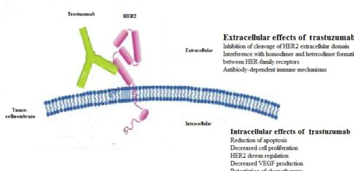
As specified above, HER2 is an onco-protein overexpressed in 12-20% of primary breast cancer tumors, often referred to as HER2-positive tumors. The presence of overexpression of HER2 is associated with a worse prognosis [31, 32]. The most commonly used anti-HER2 therapy is trastuzumab, a monoclonal antibody binding to the extracellular part of HER2. Patients with a HER2-positive breast cancer should be offered with adjuvant anti-HER2 therapy. In Sweden
2016, 14% of the diagnosed breast cancer patients had HER2-positive tumors, of which 95% were treated with adjuvant Trastuzumab for one year [1].
Figure 5. Extracellular and intracellular mechanisms of anti-HER2 treatment, trastuzumab 1.3.6 Endocrine therapy
Of all diagnosed breast cancer tumors, about 85% express estrogen receptors (ER) [1]. Through these receptors, the female sex hormone estrogen can bind to the tumor cell nuclei and stimulate cell division which leads to tumor growth. The options for endocrine therapy today is tamoxifen, mainly offered to premenopausal patients, or aromatase inhibitors mainly offered to postmenopausal patients. If aromatase inhibitors is to be used in the premenopausal setting, it needs to be combined with gonadotropin-releasing-hormone (GnRH) analogues for ovarian function suppression. When breast cancer patients with ER + tumors are treated with adjuvant tamoxifen for 5 years it is estimated to result in an approximately 13% absolute risk reduction in breast cancer recurrence and 9% absolute reduction in breast cancer mortality at fifteen year follow-up. Prolonged treatment with tamoxifen for a total of 10 years may
provide a further 3% decrease in recurrence and just over 2% improved breast cancer survival. Adjuvant treatment with aromatase inhibitors as monotherapy for 5 years gives a reduced risk of recurrence by almost 4% compared to tamoxifen, and decreases breast cancer mortality after 10 years by 2.1% and overall mortality by 2.7% [55-60]. In Sweden about 89% of all who were diagnosed with an ER positive tumor received adjuvant endocrine therapy in 2016 [1].
1.3.7 Aspirin (acetyl-salicylic acid, ASA)
Even though we have a generally good prognosis overall in breast cancer, many patients still die every year due to metastatic breast cancer and therefore we still need new and cost-effective treatments. There are several recent studies indicating that low dose aspirin use and potentially also other non-steroidal anti-inflammatory drugs (NSAIDs) can improve the prognosis in cancer [61-63], mainly colorectal cancer, but perhaps also breast cancer [64, 65]. The mechanisms behind this are not clearly known, but possible biological mechanisms include
anti-inflammatory effects, hormonal changes and inhibition of platelets [66]. Aspirin irreversibly inhibits cyclooxygenase, COX 1 and COX-2, which both are needed and involved in the synthesis of prostaglandins. Prostaglandins are found in higher levels in breast cancer tissues than in normal breast tissues. Prostaglandins seem to inhibit apoptosis and stimulate
angiogenesis in breast cancer cells and may also stimulate aromatase activity, which increases circulation estrogen levels [67]. It has been reported that postmenopausal women using aspirin have lower estrogen levels in blood compared to non-users [68]. Aspirin may also have the ability to inhibit platelet-induced adhesion of tumor cells, circulation in the blood, which could have implications for the ability spread metastases [69-71].
Commonly used low-dose aspirin doses are 75 mg or 160 mg. The low-dose tablets of aspirin are only available by prescription from doctors and represent 90% of all aspirin sold in Sweden (including over the counter) [72].
Several studies have reported an association of low-dose aspirin use with lower risk of breast cancer-specific death among breast cancer patients, which has however been limited to current and not past us of dose aspirin [64, 73-75]. In a Swedish nested-case-control study, low-dose aspirin was only associated with a reduced risk of breast cancer deaths near death or end of follow-up with a decreased risk of breast cancer death HR 0.69 (95% CI 0.56, 0.86) [76]. Medication patterns may change in the end of life period, why the results in this study might be explained by reverse causation.
Because of the mixed results from previous studies of low-dose aspirin use in breast cancer and yet no results from randomized trials, information from observational studies remain important for the understanding of role for aspirin for breast cancer patients and among which subgroups that would benefit the most. There are two ongoing randomized trials (in US and UK),
evaluating low-dose aspirin use and breast cancer disease free survival [77]. The results from these studies will still not be available in many years (preliminarily in 2026). In Sweden low-dose aspirin use is common for cardio-vascular disease prevention.
1.4 METASTATIC DISEASE
Despite a high cure rate and a long overall survival for breast cancer patients, 1400-1500 patients die of metastatic breast cancer per year in Sweden [78]. The clinical situation with distant metastases, referred to as stage IV or generalized breast cancer has historically been considered as non-curable. The mortality in breast cancer has decreased in the last decades from 30 cases per 100 000 in 1980 to 20 cases per 100 000 in 2017 [79]. Mammographic screening and new adjuvant oncological treatments are believed to explain the decrease in mortality.
Mammographic screening has been shown to reduce breast cancer mortality in the screened age groups of women 40-74 years old. The reduction of mortality has been 20-25% in the
population [80] and even higher among the screening participants [81]. However, the screening program for breast cancer has been questioned and there is a concern that the screening leads to over diagnosis and treatment. A recent published Swedish study suggest that over diagnosis in the mammographic screening program for women 50-69 in Stockholm was a minor
phenomenon [82]. More effective adjuvant breast cancer treatment can also explain the reduction in mortality in breast cancer.
Median overall survival in palliative breast cancer disease is approximately two years but can range from a few months to many years [76]. Prognosis for breast cancer patients with distant metastases seem to be better over time. In a Swedish study from 2011, they reported a trend of better survival over time for breast cancer patients with distant metastases 60 years or younger, but not for older patients [83].
Prognostic factors for breast cancer, stage IV are:
Age: older age at the time of recurrence of distant metastases is associated with poorer prognosis and a shorter survival, probably in part due to comorbidity [76, 84].
Performance status: a better performance status isassociated with a better prognosis (chapter 1.5).
Recurrence free interval: patients who have their first recurrence of distant metastases within two years since primary diagnosis have poorer prognosis after relaps in terms of overall survival compared with patients with a first recurrence after more than two years [76, 85-87].
Metastasis site: lymph node or chest-wall metastases are associated with a better prognosis than distant metastases and bone metastases are associated with a better prognosis compared to other distant sites [83, 88-90]
ER and HER2-status: breast cancer tumors that express hormone receptors have been associated with a better prognosis and survival also in stage IV disease [91]. HER2-positive tumors had a negative association with survival before trastuzumab was introdcued. Triple negative breast cancer is the sub-type with worst prognosis [92-94].
Adjuvant treatment: use of adjuvant systemic treatment may be associated with poorer survival after relapse, probably due to the selection of more aggressive cellular clones with higher resistence to treatment [83].
Bone, lung, liver and brain metastases are the most common distant metastases in breast cancer [95, 96]. The prevalence of both distant metastases and local recurrence (chest wall or lymph nodes) are more common in triple negative tumors followed by HER2-type [97, 98].
Brain metastases are associated with a particularly poor prognosis [84, 95] and have been reported to develop in about 5-10% in patients with breast cancer during follow-up up to 14 years [10, 12, 86, 87]. It has been suggested in a few studies that the incidence of brain
metastases in breast cancer has increased [84]. If so, this may be due to improved survival after primary breast cancer and/or that available adjuvant and palliative treatments are less efficient
in treating micrometastastic disease in the central nervous system compared with in other organs. Studies indicate that the risk of brain metastases is more pronounced in breast cancer patients with a young age at primary diagnosis, and if the primary breast cancer tumor is triple negative (ER-negative, negative and HER2-negative) or of HER2-type (ER-negative, PR-negative and HER2-postitive) [99-101].
Stage IV breast cancer is treated with a palliative intent and with the aims of prolonged survival and to get symptom control of the disease. Treatment can be advanced and including surgery, radiotherapy, chemotherapy, endocrine therapy, anti-HER2 therapy or other targeted drugs depending on the tumor biology.
1.5 PERFORMANCE STATUS
In medicine, especially in oncology, performance status is a way of quantifying the patients’ activities of daily life and general well-being. Performance status is also used in clinical practice to determine if a patient should be given the planned chemotherapy treatment or not and if dose adjustment is required. It can also be used to determine the general condition and well-being in palliative care.
There are several scoring systems to measure performance status; Karnofsky Performance score and ECOG/WHO/Zubrod score are two common ones used in clinical practice.
The Karnofsky Performance Score is a scale running from 100 to 0, where 100 is perfect health and 0 death. This scale is named after Dr. David A. Karnofsky, who described this scoring system in 1948 with the primary purpose to help the doctors to evaluate if the patient has the ability to survive cancer chemotherapy [102].
Table 2: Karnofsky Performance score
100 Normal; no complaints; no evidence of disease
90 Able to carry on normal activity; minor signs or symptoms of disease
80 Normal activity with effort; some signs or symptoms of disease
70 Cares for self; unable to carry on normal activity or to do active work
60 Requires occasional assistance, but is able to care for most of their personal needs 50 Requires considerable assistance and
frequent medical care
40 Disabled; requires special care and assistance
30 Severely disabled; hospital admission is indicated although death not imminent 20 Very sick; hospital admission necessary;
active supportive treatment necessary 10 Moribund; fatal processes progressing
rapidly
0 Dead
The Eastern Cooperative Oncology Group (ECOG) score [103] also called the WHO or Zubrod score, runs from 0 to 5, where 0 is perfect health and 5 is death. It is simpler to use and therefore has an advantage over the Karnofsky Performance status scale.
Table 3: ECOG/WHO/Zubrod score
0 Asymptomatic, fully active, able to carry on all predisease activities without restriction
1 Restricted in physically strenuous activity but ambulatory and able to carry out work of a light or sedentary nature. For example, light housework, office work
2 Ambulatory and capable of all self-care but unable to carry out any work activities. Up and about more than 50% of waking hours
3 Capable of only limited self-care, confined to bed or chair 50% or more of waking hours
4 Completely disabled. Cannot carry on any self-care. Totally confined to bed or chair
5 Dead
A translation exist between the Zubrod/ECOG/WHO and the Karnofsky systems that is clinically useful and has been validated in a large sample of lung cancer patients [104].
Zubrod 0–1 equals Karnofsky 80–100 Zubrod 2 equals Karnofsky 60–70 Zubrod 3–4 equals Karnofsky 10–50
1.6 BRAIN METASTASES TREATMENT
Patients with breast cancer and brain metastases have a poor prognosis, however with a large variability in survival. Median overall survival from diagnosis of brain metastases in breast cancer vary from a few months up to several years [99]. Tumor subtype, performance status, age and other distant metastases have been showed as important prognostic predictors [105, 106]. The most frequent symptoms the patients with brain metastases in breast cancer have are headache, gait disturbance, nausea and vomiting [107].
Patients with minor small brain metastases may be treated with neuro-surgery, sometimes followed by radiotherapy, or with stereotactic radiosurgery [108]. Patients with multiple brain metastases or leptomeningeal carcinomatosis are commonly treated with whole brain
radiotherapy (WBRT) and the goal for these patients is primarily improvement of symptoms and neurological deficits [109]. Studies that investigate chemotherapy regimens (for example cisplatin, capecitabine and temozolomide) in patients with brain metastases have reported response rates between 4% and 38 % [110]. The blood-brain-barrier is a selective diffusion barrier at the endothelium surrounding the brain. This endothelium is missing both fenestrations and tight junctions. It is like a barrier, from the blood to the brain, which regulates the exchange between blood and brain to prevent potentially toxic substances to enter the brain. [111]. The blood-brain-barrier stops most cytotoxic drugs from crossing over to the brain, but the role of systemic therapy is being re-evaluated given the availability of new monoclonal antibodies, antibody-drug conjugates, and new small molecules. Lapatinib is a small tyrosine kinase inhibitor, which can cross the blood-brain-barrier, showing activity against both the HER2 and EGFR receptors, and thus can be used for brain metastases in breast cancer [112]. In a trial (LANDSCAPE) including HER2-positive breast cancer patients with brain metastases they
reported a response of 0.66 (95% CI 0.50, 0.80) for a combination of lapatinib and capecitabine, all responders were partial [113].
Radiation Therapy Oncology Group´s recursive partitioning analysis (RPA) graded prognostic assessment (GPA) and diagnosis specific GPA are scores that are used to help predict the prognosis for breast cancer patients with brain metastases. These prognostic scales have been developed for prediction of prognosis in patients with brain metastases due to any cancer [114] and can be a useful tool when choosing different treatment options for patients or to identify patients with poorer prognosis to avoid overtreatment in a late palliative stage of brain metastases in breast cancer.
Table 4. Diagnosis-specific GPA score for breast cancer
GPA Significant prognostic factors GPA scoring criteria
Breast cancer Karnofsky Performance score ER/PR/HER2 Age 0 0.5 1.0 1.5 2.0 < 60 triple negative ≥ 70 60 triple negative < 70 70-80 ER/PR+ HER2- 90-100 ER/PR- HER2+ triple positive
1.6.1 Whole brain radiotherapy (WBRT)
Whole brain radiation therapy (WBRT) has been the most common treatment for brain metastases since 1950s [115-118]. Before WBRT, the standard treatment was steroids and the median survival was 1-2 months [119, 120]. The addition of WBRT extended the survival is about 3-6 months on average [117, 119-121]. Nowadays, the use of WBRT has decreased due to availability of other treatments, such as surgery for minor brain metastases, stereotactic radiosurgery and new oncological treatments. There are also concerns about late toxicity of WBRT in long-term survivors. However, WBRT is still the treatment of choice for patients with poor prognosis and massive numbers of brain metastases, poor performance status and
progressive cancer disease. The goal of treatment with WBRT is symptom control. The most commonly used dose for WBRT in Sweden is 20 Gy, in 5 fractions (4 Gy per fraction). If WBRT is initiated in late stages of brain metastases in breast cancer, it may however also affect the care in late palliative stages and the end-of-life period and the patient’s choices of care in this period. Among cancer patients in the Western world, deaths in hospital are common, although many patients prefer home as the place of death [122-124].
1.7 PALLIATIVE TREATMENT AND CARE
When curing the cancer disease no longer is possible, a clear and conscious approach is required in health care to help the patient as well as the family and other close persons. Palliative care is specialized medical care for patients with incurable life-threatening diseases. Palliative care has a focus on symptom relief, i.e., alleviating symptoms like pain, nausea or other symptoms of the disease or of the treatment. It also offers psychological, social and existential support for the patient, the family and close persons. The goal of palliative care is to improve the quality of life for these involved individuals. Palliative care is given by a specially-trained team of
physicians, nurses, counselors and physiotherapists. This team also work together with the patient’s other doctors in the hospital to provide extra support in this situation. Teamwork between different occupational groups in the palliative team and the hospital is needed for a high-quality palliative care. Palliative care is provided to patients in all ages and with serious diseases in all stages, and it can be given together with oncological or other treatments [125]. Palliative care can be offered at home, in nursing homes, in hospitals or at a specialized palliative care unit (hospice). When treating patients with a late stage cancer disease such as brain metastases, discussions with the patients, their families and close persons of their wishes and requests of the care in the late palliative stage and end-of-life setting should be considered aiming to avoid overtreatment and for the best quality of life for each patient.
2 AIMS OF THE THESIS
In this thesis, we aimed to study the incidence and predictors of brain metastases in breast cancer as well as treatment aspects in primary breast cancer and for patients with brain metastases.
More specifically, the aims were to:
Study I and II: Study time trends and if the incidence of brain metastases due to breast cancer has increased in Sweden in recent years
Study III: Study the benefits of whole brain radiotherapy for brain metastases in terms of level of care and survival
Study IV: Study if low-dose aspirin use improves the prognosis in patients with breast cancer in terms of survival
3 MATERIAL AND METHODS
The four studies included in this thesis are based on Swedish population-based registers, quality-of-care registers and radiotherapy files in ARIA and other medical records at Karolinska University hospital.
3.1 DATA SOURCES
3.1.1 The Swedish National Cancer Register (NCR)
The Swedish National Cancer Register is kept by the National Board of Health and Welfare. The register includes malignant diseases of Swedish residents since 1958. Reporting to this register is mandatory by law for all new malignant tumors by physicians in Sweden. This system guarantees completeness and reliability. The NCR is estimated to be >95% complete [126]. For each patient, the register keeps record of the personal identification number, sex, age, date of diagnosis and diagnostic methods, the hospital providing the diagnosis and type of malignancy stored as International Classification of Disease (ICD) codes [127].
3.1.2 The National Breast Cancer Quality Register
The National Breast Cancer Quality Register is based on information from six different regional breast cancer quality-of-care registers in Sweden. This national register was started in 2008. The regional Stockholm-Gotland register was started in 1976, The Uppsala-Örebro regional register in 1992 and the register of the North region in 1980. The National register is updated continuously by matching with the Total Population Register, The Swedish National Cancer Register and the Swedish Causes of Death Register. The National Breast Cancer Quality Register contains collected data on breast cancer patients, tumor characteristics, surgical treatment and intended oncological treatments. When evaluating the register, the variables including parameters during diagnosis and primary surgical treatment have a high coverage and concordance in the register (>95%), however intended oncological treatment have lower concordance (66%-95%) [128]. Regarding oncological treatment during follow up, including palliative treatments, these variables have less coverage, 67% in 2014 [129]. Since 2008, this register has been organized on a web-based platform, INCA. In 2016, the coverage was 98% for diagnosis and primary treatment, but there is a concern that the coverage is lower in the oldest women. The oldest women may not be examined and diagnosed, why they may be outside the registers.
3.1.3 The Swedish Cause of Death Register
The Swedish Cause of Death Register is kept by the National Board of Health and Welfare. This register was initiated in 1751, and digitalized in 1952 [130]. The date and conditions related to the death are reported to the register on a death certificate issued by the physician that verifies the death. The completeness of the register is >99%. The register has information on the date and place of residence, underlying cause of death and contributing causes of death of all Swedish residents who die in Sweden or abroad. Individuals are identified by their personal identification number and the cause of death is coded using ICD codes [130].
3.1.4 The Swedish National Patient Register
The Swedish National Patient Register (NPR) is kept by the National Board of Health and Welfare. All inpatient care in the country has been recorded in the register since 1987 and the hospital outpatient care (not primary health care) since 2001. The information in the register contains hospital admission and discharge dates, outpatient visit dates and diagnoses made by the physician. The diagnoses are coded according to the International Classification of
Diseases 9th revision (ICD-9) since 1987 and the 10th revision (ICD 10) since 1997. The NPR has a coverage of 99% of all somatic and psychiatric hospital discharges [131].
3.1.5 Longitudinal Integration database for health insurance and labor market studies (LISA)
The Longitudinal Integration database for health insurance and labor market studies (LISA) is kept by The Statistics Sweden and Social Insurance Agency since 1990. It holds data from the labor market, social and educational sectors for individuals (≥16 years old) in Sweden. Data is collected from several national registers of high quality and coverage. For each individual, detailed socioeconomic data is available, including employment and level of education [132].
3.1.6 The Swedish Prescribed Drug Register
The Swedish Prescribed Drug Register is held by the National Board of Health and Welfare. This register records all the dispensed prescribed drugs of the Swedish population
prospectively beginning July 1st 2005. The register contains information on the date of dispensing, name of drug, dose and number of tablets prescribed and the Anatomical Therapeutic Chemical (ATC) code. The ATC classification system consists of 14 main anatomical groups and further subgroups under each main group [133].
3.1.7 ARIA
The ARIA® oncology information system is a software for planning and handling the radiotherapy at the radiotherapy department at Karolinska University hospital in Stockholm. In ARIA there is information on cancer diagnosis, start date for radiotherapy, dose and fraction of radiotherapy given.
3.1.8 BcBaSe
BcBaSe is a database linkage of three quality-of-care registers of breast cancer in Sweden based on information from the Stockholm-Gotland, Uppsala-Örebro and the North regions [134]. The BcBaSe has been linked with other health care registers including the Patient register, the LISA-register, the National Cause of Death Register and the Swedish Prescription register.
3.2 STUDY DESIGN AND STATISTICS 3.2.1 Cohort study design
A cohort study design was used in all four studies.
Participants that fulfill the inclusion criteria and are free from the outcome are selected to the study cohort. The exposures as well as possible confounders of interest are measured for each study participant. The study participants are followed over time until they have reached the outcome of interest, or are censored (due to e.g., death, emigration or end of follow up). Data for cohorts can be obtained from national registers. The advantage of using national registers are that they are population-based and aim to include all Swedish citizens (no selection). Linkage between several national registers may provide a wide range of data. The registers usually span over a long time period, good for longitudinal studies.
Figure 6. A cohort study outline
3.2.2 Study I
3.2.2.1 Study population:
We used a cohort study design to investigate the incidence of brain metastases over calendar time. To get this cohort we first identified 58 795 individuals with breast cancer in the Swedish National Cancer Register (NCR). The time period was from 1 January 1998 to 31 December 2006. Patients with other cancer diagnosis, except for non-melanoma skin cancer, before this time were excluded. We ended up with a final cohort of 50 528 patients in the study. The cohort was further linked to the National Patient Register, NPR, to obtain
information on all admissions to hospital due to distant metastases during the follow-up. The cohort was also linked to the National Cause of Death Register to get information on dates of deaths.
3.2.2.2 Statistics:
Proportions of brain metastases and/or other distant metastases were compared and showed per time periods 1998–2000, 2001–2003 and 2004-2006. The first admission with metastases (brain metastases or other metastases outside of the brain) was denoted. We assessed
Study cohort Exposed Disease No Disease Non exposed Disease No Disease
cumulative incidence of brain metastases admissions during follow-up by calendar period of breast cancer diagnosis. The incidence was plotted graphically with the Kaplan–Meier curve considering incidence during up to 3 years of follow-up. To compute hazard ratios (HR) and 95% confidence intervals (CI) we used a multivariate Cox proportional hazards model adjusted for year of birth, as a measure of the relative risk of admissions for brain metastases or other distant metastases comparing 3-year periods.
3.2.3 Study II
3.2.3.1 Study population:
We used the BcBaSe linkage to study a cohort of 30 996 women diagnosed with a first breast cancer during the period 2002-2012, in Stockholm-Gotland, Uppsala Örebro and the North region in Sweden. Women with other cancer diagnoses before breast cancer were excluded, as well as patients who emigrated before breast cancer diagnosis. In the BcBaSe there is a linkage with the National Patient Register, the LISA Register and the Cause-of-death Register, used in this study to obtain information on records of health care visits for distant metastases during follow-up, highest level of education and dates of death. By combining these data, we were be able to compare incidence of brain metastases in breast cancer over calendar time. We also assessed predictors of brain metastases in breast cancer patients. 3.2.3.2 Statistics:
The number of women with breast cancer registered with brain metastases and clinical and demographical characteristics were presented and compared per time periods 2002-2004, 2005-2008 and 2009-2012. We used univariable and multivariable Cox proportional hazards models to compute HRs and 95% CIs as a measure of the relative risks. Proportional hazards assumption was tested using Schoenfeld residuals test and fulfilled. HRs were estimated by clinical and demographical characteristics and over calendar time in the three-year periods. All women were followed from the date of breast cancer to the date of brain metastases or other distant metastases, emigration, death, or December 31st 2013, whichever came first. The cumulative incidence of brain metastases in the three calendar time periods of diagnosis of breast cancer was also estimated to demonstrate the absolute risk of brain metastases and presented in a graph using cumulative incidence functions (treating death as a competing event).
3.2.4 Study III
3.2.4.1 Study population:
We used a cohort study design and identified 281 patients with brain metastases due to breast cancer treated with WBRT. We used ARIA-software at the Radiotherapy department at the Karolinska University hospital to identify the patients. These patients were treated with WBRT during the years 1999-2012. Individuals with other cancer diagnoses, or who were treated with radiotherapy following surgery from brain metastases or with bone metastases in the scalp were excluded. Thus, we ended up with a final cohort of 241 patients. The brain
metastases were located in the cerebrum, cerebellum or the leptomeninges. Through reviews of the included patients’ medical records in the Take Care-system, we collected data on clinical and biological factors from the breast cancer tumor, administered medical
oncological treatments and details about the radiotherapy to the brain. We further noted the performance status the week before (as WHO score) the start date of initiation of WBRT, and the family situation. The level of care for the patient (hospital or home) the week before and after WBRT was also noted. The outcome was overall survival and if the breast cancer patient was able to come home after the treatment or not.
3.2.4.2 Statistics:
We calculated the median survival in months, with interquartile range, from start of the WBRT. Using Cox proportional hazards models with HR and 95% CI, we compared time to death by clinical and breast cancer tumor characteristics. Proportional hazards
assumption was tested using Schoenfeld residuals test and fulfilled. In a multivariate analysis, we adjusted for age at time of WBRT and also for calendar period of WBRT. In a second multivariate model, we adjusted for performance status as well. An unconditional logistic regression model was used to identify protective factors for coming home again using Odds Ratios, OR and 95% CI. This model was also adjusted for age at WBRT and calendar period of WBRT, and a second model also for performance status.
3.2.5
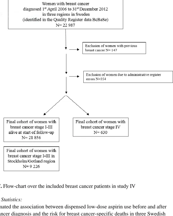
Study IV3.2.5.1 Study population:
We used a cohort study design to evaluate the association between the exposure, dispensed low-dose aspirin, and the primary outcome, risk for breast cancer-specific death. The
BcBaSe-cohort was used to identify all women with a first breast cancer during the period 1st of April 2005 - 31st December 2012. This cohort was further linked to several other Swedish registers including the Prescribed Drug Register, the NPR, the Cause of Death Register and the LISA Register including information on highest achieved educational level (≤9 years, 10-12 years, >10-12 years). Educational level was used as a proxy for socioeconomic status. Aspirin dispensings were used as a proxy for aspirin use.
Figure 7. Flow-chart over the included breast cancer patients in study IV 3.2.5.2 Statistics:
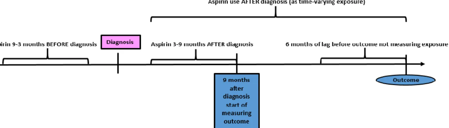
We estimated the association between dispensed low-dose aspirin use before and after breast cancer diagnosis and the risk for breast cancer-specific deaths in three Swedish regions, and time to first recurrence or metastasis in the Stockholm-Gotland region. We used a Cox proportional hazards model with HR with 95% CI as a measure of association. Low-dose aspirin use before breast cancer diagnosis was assessed during the period 9-3 months before diagnosis, dose aspirin use (yes/no), if there was ≥ 1 dispensing of low-dose aspirin or not. Low-low-dose aspirin use was also assessed during 3-9 months after breast cancer diagnosis, (yes/no), if there were ≥ 1 dispensing or not. To estimate cumulative use, low-dose aspirin use was assessed during the entire follow-up from 3 months after
diagnosis and onward as a time-varying exposure. During this classification a 180-day lag period was used in order to take into count changes in dispensing patterns immediately prior to death, since this period may entail changes in drug use due to end of life situation [135-137].
Figure 8. Flow-chart providing an overview of measurements of exposure and outcome after breast cancer diagnosis using a 180-day lag period.
We used one model with no adjustments, a second model adjusted for calendar-year of breast cancer diagnosis, age at breast cancer diagnosis, stage of breast cancer tumor, level of
education and comorbidity diagnoses before breast cancer, and a third model adjusted for the same variables as in the second model plus metformin, statin and NSAID use as well as oncological treatments for breast cancer. In analyses of low-dose aspirin use after breast cancer diagnosis, the model was additionally adjusted for low-dose aspirin use before breast cancer diagnosis. We further studied low-dose aspirin use before and after diagnosis in subgroups of women by clinical and biological breast cancer characteristics, as well as for oncological treatment and risk of breast-cancer specific death.
All the analyses in study I and III were performed the SAS software, Version 9.4 of the SAS System for Windows. Copyright © 2002-2012 SAS Institute Inc was used for all analyses. The analyses in study II and IV was made in the Stata 14 software (StataCorp. 2015. Stata Statistical Software: Release 14. College Station, TX: StataCorp LP)
4 RESULTS
4.1 STUDY I
In this Swedish cohort of 50 528 patients with breast cancer disease, median follow-up was 3.5 years and 696 (1.4%) were admitted to hospital with brain metastases during this time period. Among these 696 patients, 336 (0.7%) patients were admitted to hospital with brain metastases as their first and only distant metastasis and the other 360 (0.7%) patients were admitted to hospital with brain metastases along with other distant metastases. Brain metastases at time for breast cancer diagnosis and during follow-up were included. Admissions with other distant metastases, outside the brain, were found in 3 470 (6.9%) patients. The median time between breast cancer diagnosis and the first admission to hospital with brain metastases was 2.3 years for all the brain metastases patients, 1.8 years for the individuals with a first metastasis to the brain and 2.9 years for the individuals with brain metastases together with other metastases.
Incidence rates of brain metastases were lowest among patients diagnosed in 1999 and increased among patients diagnosed during the later time periods under study. When
compared with the time period 1998–2000, patients diagnosed with a breast cancer during the time period 2004–2006 were at a 44% increased risk (HR 1.44, 95% CI 1.13, 1.85) of being admitted to hospital due to brain metastases.
The risk was more pronounced for breast cancer patients having brain metastases together with other distant metastases (HR 1.80, 95% CI 1.23, 2.63) than for the individuals with a first metastasis to the brain (HR 1.21, 95% CI 0.87, 1.68) in 2004–2006, compared with 1998–2000. The relative risk for admissions to hospital with other distant metastases were not significantly increased in 2001-2003, 1.02 (95% CI 0.95, 1.11), and borderline increased in 2004-2006, 1.11 (95% CI 1.00, 1.24), compared with 1998–2000. When estimating the risk of brain metastases by time of follow-up, the increased incidence was primarily observed the last 1.5 years of follow-up after breast cancer diagnosis 2004–2006 (HR 2.07, 95% CI 1.45, 2.94) than during the first 1.5 years (HR 1.08, 95% CI 1.76, 1.54), compared with 1998– 2000.
Figure 9. Breast cancer patients 1998-2006 in Sweden and the cumulative incidence of brain metastases
4.2 STUDY II
In the cohort of 30 996 patients, the median age at primary breast cancer diagnosis was 62 years (range 19-102 years). Demographically 13 050 (42.1%) of the patients were from Stockholm-Gotland, 12 861 (41.5%) from Uppsala-Örebro and 5 085 (16.4%) from the North region of Sweden. In the cohort 16 415 patients (54.1%) had a stage I tumor, 11 512 (38.0%) had a stage II tumor, 1 542 (5.1%) hade a stage III tumor and 860 (2.8%) had a stage IV tumor at the time of breast cancer diagnosis. Regarding the primary breast cancer tumor characteristics, 24 260 (84.5%) were ER positive and 4 457 (15.5%) were ER negative, 2 658 (13.4%) were HER2 positive and 17 107 (86.6%) were reported as HER2 negative. There were 11 231 missing HER2. The most common clinical subtype was the luminal subtype (ER+, HER2-/HER2+), noted in 16 598 (85.9%) of the patients, whereas 974 (5.0%) had a non-luminal HER2 (, HER2+) breast cancer and 1 974 (10.1%) of the women had an ER-HER2- breast cancer tumor. There were 11 450 missing subtype due to missing variables (mostly HER2). Stratified per time period there were 7 872 (25.4%) patients in the first time period 2002-2004, 10 954 (35.3%) 2005-2008 and 12 170 (39.3%) 2009-2012.
In the cohort, 789 (2.5 %) were registered with brain metastases at diagnosis and during follow-up. The median follow up was 5 years. The time to brain metastasis from primary breast cancer diagnosis was median 31 months (range 0-142 months). Number of brain
metastases per time period were 269 in period 2002-2004, 321 in 2005-2008 and 199 in the last period 2009-2012. Compared with the time period 2002-2004 the patients diagnosed with a primary breast cancer 2005-2008 had a risk estimate of 1.09 of having a brain metastasis (HR 1.09 95% CI 0.93, 1.30) and 2009-2012, patients were at 37% risk of having a brain metastases (HR 1.37 95% CI 1.12, 1.68) when adjusted for age and stage at time for primary breast cancer diagnosis
Table 5. Relative risks for brain metastases in breast cancer in Sweden 2002-2012
Years HR (95 % CI) HR1 (95 % CI)
2002-2004 1.0 1.0 2005-2008 1.09 (0.92, 1.29) 1.09 (0.93, 1.30) 2009-2012 1.19 (0.97. 1.44) 1.37 (1.12, 1.68) HER2-positive 1.0 1.0 HER2-negative 0.29 (0.24, 0.36) 0.41 (0.33, 0.51) ER-positive 1.0 1.0 ER-negative 4.93 (4.24. 5.72) 3.73 (3.20, 4.34)
1Adjusted for age and stage at the time of breast cancer diagnosis
The median age at primary diagnosis was lower among patients with brain metastases than among other patients 56 years versus 62 years. Three-hundred and twenty-four patients (1.0%) were registered with brain metastases as their first distant metastasis and the remaining 465 (1.5 %) were registered with brain metastases in parallel with or following other distant metastases due to breast cancer. Among the patients with brain metastases 138 (34.2%) had a HER2 positive and 265 (65.8%) had a HER2-negative breast cancer tumor at primary diagnosis. Regarding ER, 372 (54.1%) were positive, 315 (45.9 %) were ER-negative.
During follow up 1 667 (5.4%) of the patients were registered with lung metastases, 1 642 (5.3%) with liver metastases and 2 297 (7.4%) with bone metastases.
4.3 STUDY III
In this cohort study, the breast cancer patients with brain metastases were treated with whole brain radiotherapy (WBRT). The median age at start of treatment was 58 years. The majority of the patients (n=212, 88%) were treated with 20 Gy in 5 fractions. Median survival after WBRT was 2.9 months (interquartile range 1.1-6.6 months). Survival was shorter if the patient was over 50 years old, had a poor performance status (>2 WHO score) or had a triple negative tumor.
Figure 10. Survival after whole brain radiotherapy for brain metastases due to breast cancer
After WBRT, 57 (24%) patients could not be discharged from the hospital. Among the individuals that were in hospital before WBRT, 45 (47%) died in the hospital without coming home again and 12 (8%) patients could be discharged from the hospital to come home again. Among patients with performance status score WHO 0-1 before WBRT, 124 (97%) returned to home after treatment and if the WHO score was 2, 46 (65%) patients returned to home, and if the WHO score was 3-4, 14 (34%) returned home. These associations could not be
explained by age.
Table 6. Number and proportions of patients coming home again after WBRT at Karolinska University hospital 1999-2012
Ever at home after WBRT Level of care before WBRT Yes No Total Home 133 (92 %) 12 (8%) 145 (100%) Hospital 51 (53%) 45 (47%) 96 (100%) Total 184 (76%) 57 (24%) 241 (100%) 4.4 STUDY IV
This cohort of breast cancer patients consisted of 21 414 patients diagnosed in stage I-III. The median follow-up time in the cohort was 3.8 years (range 0.75-7.75) and median age at primary breast cancer diagnosis was 63 years old.
Before breast cancer diagnosis (9-3 months before) 2 660 (12.4 %) patients were treated with low-dose aspirin and 2 813 (13.1%) after diagnosis (3-9 months after). Low-dose aspirin users were older at breast cancer diagnosis (median age 75 years), and more often diagnosed with breast cancer stage II-III than stage I tumors compared with non-users. When the entire follow-up period was considered, 4 091 of the women (19.1 %) used low-dose aspirin. Regarding breast cancer characteristics at the time of breast cancer diagnosis, 12 546 (58.6%), were diagnosed with a stage I breast cancer, 7 879 (36.8 %) had stage II and 989 (4.6%) had a stage III breast cancer. The most common subtype of breast cancer was the luminal subtype (ER+, HER2-/HER2+), recorded in 15 529 women (72.5%), 857 women (4%) had non-luminal HER2 (ER-, HER2+) and 1 739 patients (8.1%) an ER-HER2- breast cancer tumor.
Among all patients, we found no associations between low-dose aspirin use 9 to 3 months before breast cancer diagnosis and risk of breast cancer death in the adjusted models (HR 0.93, 95% CI 0.77, 1.12). The aspirin dose (≤75 or >75 mg/day) did not change the null association. In breast cancer subgroups by clinical and biological characteristics, reduced risks of breast cancer death were however noticed in women with ER positive breast cancer tumors (HR 0.74, 95% CI 0.57, 0.97) and among the patients who were intended for adjuvant treatment with endocrine therapy (HR 0.75, 95% CI 0.59, 0.96).
Low-dose aspirin use during the period 3 to 9 months after breast cancer diagnosis did not affect risk of breast cancer specific deaths in the fully adjusted model, including adjustment for pre-diagnostic aspirin use (HR 1.00, 95% CI 0.74, 1.37).
Dose and duration of low-dose aspirin use after diagnosis were in general not associated with breast cancer-specific deaths. However, in one subgroup of women treated with low-dose aspirin >75 mg per day during the entire follow-up, we observed an increased risk of breast cancer-specific deaths (HR 1.62, 95% CI 1.09, 2.40).
In subgroups of breast cancer patients with different clinical and tumor characteristics (stage, ER-status, HER2-status, breast cancer subtype and intended oncological treatment), low-dose aspirin use afterdiagnosis was associated with a reduced risk of breast cancer specific death for women with a stage I breast cancer tumor at diagnosis (HR 0.53, 95% CI 0.29, 0.96).
Figure 11. Survival of women with stage I-III breast cancer tumors by use of low-dose aspirin after diagnosis illustrated graphically in a Kaplan-Meier curve (to the left) and with adjusted survival curves (to the right)
In the sub cohort of women from Stockholm-Gotland, there were 9 226 women with stage I-III breast cancer, of whom 1 048 women (11.4%) used low-dose aspirin before their breast cancer diagnosis. During follow-up, 2 800 women, who were not using low-dose aspirin (34.2%) and 347 women who were using low-dose aspirin (33.1%) had a record of a first distant metastasis. Low-dose aspirin use was not associated with a reduced risk of
In another separate analysis of 621 women with stage IV disease at diagnosis, we studied low-dose aspirin use before breast cancer diagnosis and time to breast cancer death. There were no protective association for low-dose aspirin users (N=61) with stage IV disease when compared to non-users in the adjusted analyses (N= 334) (HR 0.91, 95% CI 0.67, 1.23).
5 DISCUSSION
In Sweden we have well developed national registration of diseases in registers and with often highly valid data. Due to this we have the opportunity to identify a certain population with a disease and study exposure and outcome within this cohort, using epidemiological strategies to understand associations. Patients with breast cancer are registered in several registries and therefore this disease is possible to study in large observational studies, such as cohort studies [138]. The National Breast Cancer Quality Register contains collected data on breast cancer patients, tumor characteristics, surgical treatment intended oncological
treatment and have a high validity. In 2016, the coverage was 98% [134].
In Sweden breast cancer is the most common malignant disease among women and it is also the most diagnosed malignant disease in women worldwide, affecting 1.7 million individuals in 2012 [139, 140]. When the breast cancer reaches its most advanced stage, tumor cells have the ability to spread out to form new tumors in different organs in the body, such as liver, lungs, bone, skin or brain. Breast cancer with distant metastases are rarely curable [141]. Breast cancer is also the second most common cause (after lung cancer) of brain metastases [96, 142, 143]. Among patients diagnosed with breast cancer, about 5-10% will develop brain metastases [10, 12]. Studies indicate that the risk of brain metastases is higher among patients of young age at primary breast cancer diagnosis, and if the primary tumor is triple negative (ER-negative, PR-negative and HER2-negative) or of HER2-type (ER-negative, PR-negative and HER2-postitive) [99, 100, 144].
It has been suggested in a few studies that the incidence of brain metastases in breast cancer has increased over time [84, 85, 87]. If so, this may be due to improved survival after primary breast cancer and/or that available adjuvant and palliative treatments are believed to be less efficient in treating micrometastastic disease in the central nervous system compared with in other organs. We also have refined imaging and maybe greater attention to neurological symptoms which could lead to an increase in diagnosed and registered brain metastases [145, 146].
Patients with minor brain metastases due to cancer may be treated with neuro-surgery, sometimes followed by radiotherapy or with stereotactic radiotherapy [5, 108, 147]. However, whole brain radiotherapy (WBRT) is still a common treatment for patients with poor performance status, poor prognosis, massive burden of brain metastases and
uncontrolled systemic disease.
The goal of WBRT is symptom control and if there are neurological deficits, improvement of those [109]. In some subgroups of patients, there is a risk of overtreatment, particularly among poor prognosis patients [114]. When treating with WBRT previous studied indicate that 50-80% of the patients respond to treatment and experience improvement of


![Figure 3. A HER2 amplified breast cancer cell and a normal cell. Reprinted with permission from the publisher [35]](https://thumb-us.123doks.com/thumbv2/123dok_us/456673.2553148/17.892.154.712.219.452/figure-amplified-breast-cancer-normal-reprinted-permission-publisher.webp)