Neoadjuvant chemoradiotherapy
and surgery
Slokdarmcarcinoom
Neoadjuvante chemoradiotherapie en chirurgie
Esophageal Cancer
Neoadjuvant chemoradiotherapy
and surgery
Slokdarmcarcinoom
Neoadjuvante chemoradiotherapie en chirurgie Thesis
to obtain the degree of Doctor from the Erasmus University Rotterdam by command of the rector magnificus
Prof.dr. R.C.M.E. Engels
and in accordance with the decision of the Doctorate Board, The public defense shall be held on
Wednesday 5 September 2018 at 15:30 hours by
Bo Jan Noordman born in Leiden
Promotor
Prof.dr. J.J.B. van Lanschot
Overige leden
Prof.dr. M.I. van Berge Henegouwen Prof.dr. E.W. Steyerberg
Dr. V.M.C.W. Spaander
Copromotor
CONTENTS
Introduction 11
Chapter 1
General introduction and outline of the thesis 13
PART I Implications of neoadjuvant chemoradiotherapy on surgical treatment 23
Chapter 2
Surgical approaches to remove the esophagus: open 25
Chapter 3
Chemoradiation in esophagogastric junction cancer 49
Chapter 4
Multimodality treatment for esophageal adenocarcinoma: multi-center propensity-
score matched study 69
Chapter 5
Optimal surgical approach for esophageal cancer in the era of MIE and neoadjuvant therapy 89
Chapter 6
Effect of lymphadenectomy on survival in oesophageal cancer 103
Chapter 7 109
Chaper 7A The impact of surgical approach on long-term survival in esophageal adenocarcinoma patients with or without neoadjuvant chemoradiotherapy 111 Chapter 7B Letters of correspondence 129
Chapter 8
Effect of neoadjuvant chemoradiotherapy on health-related quality of life in
esophageal or junctional cancer: results from the randomized CROSS trial 137
Chapter 9
Quality of life during and after completion of neoadjuvant chemoradiotherapy for oesophageal and junctional cancer 163
Chapter 10
Impact of neoadjuvant chemoradiotherapy on health related quality of life in long- term survivors of esophageal or junctional cancer: results from the randomized
CROSS trial 181
Chapter 11
External validation of pretreatment pathological tumour extent in patients with
neoadjuvant chemoradiotherapy plus surgery for oesophageal cancer 199
PART II Active surveillance after neoadjuvant chemoradiotherapy 219
Chapter 12
The accuracy of detecting residual disease after neoadjuvant chemoradiotherapy for esophageal cancer by endoscopy with biopsy, EUS and 18F-FDG PET: a systematic
review and meta-analysis 221
Chapter 13
Accuracy of detecting residual disease after CROSS neoadjuvant chemoradiotherapy for esophageal cancer (preSANO trial): rationale and protocol 283
Chapter 14
Detection of residual disease after neoadjuvant chemoradiotherapy for oesophageal cancer (preSANO): a prospective multicentre, diagnostic cohort study 301
Chapter 15
Endosonographic measurements of tumour thickness and tumour area to detect
residual disease after neoadjuvant chemoradiotherapy for oesophageal cancer 331
Chapter 16
Accuracy of 18F-FDG PET/CT in predicting residual disease after neoadjuvant
chemoradiotherapy for oesophageal cancer 347
Chapter 17
Active surveillance in clinically complete responders after neoadjuvant
chemoradiotherapy for esophageal or junctional cancer 371
Chapter 18
Patients’ preferences for treatment after neoadjuvant chemoradiotherapy for
Chapter 19
Neoadjuvant chemoradiotherapy plus surgery versus active surveillance for
oesophageal cancer: a stepped-wedge cluster randomised trial. 405
Chapter 20
Organ-sparing treatment in oesophageal cancer: feasible and safe? 429
Chapter 21 439
Chapter 21A Summary in English 441 Chapter 21B Samenvatting in Nederlands 447
Chapter 22 Future perspectives 455 Appendices 461 List of publications 462 PhD portfolio 469 Acknowledgements 472
Chapter
1
General introduction and outline of the thesis
Esophageal cancer is a highly lethal malignancy, as reflected by an overall 5-year survival of 17%.1 In the Netherlands, the incidence of esophageal cancer resembles the growing
trend in Western countries, with an incidence of 15/100,000 for men and 6/100,000 for women, and more than 2,600 new cases annually.2 Two main histological subtypes can
be distinguished, i.e. esophageal adenocarcinoma (AC) and esophageal squamous cell carcinoma (SCC). Globally, SCC is the most common subtype, especially in areas with high incidence such as eastern and southern Africa and eastern Asia. However, in most Western countries, the incidence of esophageal and esophogastric-junctional (EGJ) AC has surpassed that of SCC.
At the time of diagnosis, only 50% of all patients are potentially curable. Surgical re-section has long been considered the primary curative treatment modality for esophage-al and junctionesophage-al cancer. Historicesophage-ally, the Ivor-Lewis procedure has been widely applied, including a thoracotomy with limited lymphadenectomy and thoracic anastomosis.3
Ever since, two main surgical techniques have evolved. First, the extended en bloc trans-thoracic esophagectomy (TTE) was developed, with extensive two-field lymphadenec-tomy (upper abdomen and posterior mediastinum). This technique attempts to increase locoregional tumor control by enhancing the radicality of resection.4-8 It is well
estab-lished that extensive lymphadenectomy provides the benefit of more accurate staging, but its beneficial effect on survival is still unclear.9-12 Second, the limited transhiatal
esophagectomy (THE) was introduced, which focused on minimization of postoperative morbidity and mortality by preventing a formal thoracotomy. The optimal surgical approach for the treatment of patients with esophageal cancer is still topic of debate.
In the literature reported 5-year survival rates for patients treated with primary sur-gical resection range from six to 50%, but rarely exceed 35% in Western countries.13-17
To improve long-term survival, many trials investigated the added value of neoadjuvant chemo- and/or radiotherapy.18-24
In most countries, two neoadjuvant approaches have been adopted as standard of care. The first is neoadjuvant chemoradiotherapy (nCRT), now generally based on the CROSS regimen, which resulted in a 5-year overall survival benefit of 14%, compared to surgery alone.22, 23 An alternative option is perioperative or preoperative chemotherapy
using the OEO2, MAGIC or FLOT protocol, which showed an absolute risk reduction of 6%, 13% and 16% at 5-years, respectively.19, 24, 25 Except partly in Japan and China, it is
widely accepted that chemoradiotherapy is the neoadjuvant treatment of choice for patients with squamous cell carcinoma.26 For patients with adenocarcinoma the optimal
multimodality regimen is still topic of debate.27-29
The ChemoRadiotherapy for Oesophageal cancer followed by Surgery Study (CROSS) was a multicenter, randomized phase-III trial, comparing neoadjuvant chemo-radiotherapy followed by surgery with surgery alone.22 The study included and analyzed
in The Netherlands during a five-year period. Most patients (75%) had an adenocarci-noma and most tumors were located at the EGJ (24%) or in the distal esophagus (58%). Results from the CROSS-trial showed that the addition of nCRT (carboplatin, paclitaxel and 41.4 Gy of concurrent RT) to surgery significantly increases survival as compared to surgery alone in patients with potentially curable SCC or AC of the esophagus or EGJ. Neoadjuvant treatment was well tolerated, with >90% of all patients receiving full treat-ment. Therefore, neoadjuvant CRT plus surgery is now considered the therapy of first choice in the Netherlands and several other countries for potentially curable esophageal cancer (cT2-3N0-1M0 and cT1N1M0) in patients fit to undergo this treatment.
Application of nCRT prior to surgery has important implications for prognostica-tion and surgical treatment. Most of the convenprognostica-tional prognostic factors identified in the era of primary esophagectomy lose their prognostic value in patients treated with nCRT plus surgery.30 After nCRT, only the number of clinically suspected metastatic
regional lymph nodes (cN-stage) prior to treatment and the number of metastatic re-gional lymph nodes in the resection specimen (ypN-stage) are independently associated with survival after application of nCRT plus surgery. A model using these parameters to determine prognosis showed only limited prognostic strength. This emphasizes the need for new prognostic parameters. One such parameter is the pretreatment p-TNM staging. This novel staging system aims to determine the pretreatment tumor extent based on the extent of tumor fibrosis, the location of residual tumor cells and regressional chang-es of lymph nodchang-es in the rchang-esection specimen after nCRT. Previously, this staging system has been shown reproducible.31 Furthermore, it was demonstrated that especially the
number of pretreatment metastatic lymph nodes is an important and independent prognostic parameter. Patients with pre-treatment metastastic lymph nodes, which became negative for disease thanks to nCRT, have worse prognosis than patients with-out pretreatment nodal involvement.31
Neoadjuvant CRT downstages both the primary tumor and the regional lymph nodes. The first leads to an increase in the radical resection rate, whereas the latter ques-tions the necessity of extended lymphadenectomy. Some studies have shown previously that the number of resected lymph nodes has prognostic impact on survival, and proba-bly even therapeutic impact in patients after surgery alone.32 However, after nCRT the
number of resected nodes has been shown unrelated to survival.33 These data question
the necessity for maximization of lymphadenectomy after nCRT as can be performed during TTE.
Furthermore, the effects of nCRT on health-related quality of life (HRQOL) are largely unknown. Esophagectomy has a profound and lasting impact on patients’ HRQOL.34 Patients who undergo nCRT might experience a deterioration in HRQOL
effects of especially radiotherapy on heart and lungs have insufficiently been investigat-ed and might have an impact on long-term HRQOL.
In subsequent analyses of secondary endpoints in the CROSS-trial it was found that nearly a third of the patients had a pathologically complete response (pCR) in the resec-tion specimen. A pCR after nCRT was seen in 49% of patients with SCC and 23% of patients with AC. In the OEO2 and MAGIC trials this was substantially less, i.e. 4% and 5%, respectively.22, 36 This observation raises the question whether a surgical resection is
of benefit to patients in whom no vital tumor cells can be detected in the resection spec-imen. Theoretically, an organ sparing approach might be feasible since, intuitively, an esophagectomy in patients with no residual viable tumor cells in the resection specimen has probably no effect on clinical outcome. This imposes an ethical imperative to recon-sider the necessity of standard esophagectomy in patients after nCRT. An individualized approach to surgery after nCRT needs to be studied and defined; a new treatment algo-rithm in which not every patient with potentially curable esophageal cancer needs a resection after completion of nCRT to achieve long-term survival. In an active surveil-lance approach, patients will be subjected to frequent clinical investigations after nCRT. Esophagectomy will be offered only to those with a proven locoregional recurrence, in the absence of distant metastases. An active surveillance approach could have great advantages given the high postoperative morbidity and substantial mortality, and the impact of surgery on quality of life.22, 37, 38 However, an active surveillance approach
would be justified only if oncological outcome is non-inferior to standard surgery. In order to select patients for active surveillance, the disease should be re-staged after nCRT by means of meticulous clinical response evaluation (CRE). CREs need to accu-rately categorize patients as clinically complete responders (cCR) or clinically incom-plete responders. Such an active surveillance strategy is currently applied in selected patients who refuse surgery or are medically unfit for major surgery after completion of nCRT.39-42 Explorative retrospective studies in such patients show promising results,
with comparable long-term survival for active surveillance (i.e. postponed esophagec-tomy only in patients who develop a locoregional regrowth in the absence of distant metastases) vs immediate standard surgery.39-42
Outline of the thesis
This thesis consists of two parts. In part I the implications of neoadjuvant chemoradio-therapy on surgical treatment of esophageal cancer are described. Part II is focused on the feasibility of an active surveillance approach instead of standard esophagectomy in clinically complete responders after nCRT.
PART I. Implications of neoadjuvant chemoradiotherapy on surgical treatment
Since the publication of the CROSS-trial, nCRT followed by surgery is the standard of care in several countries, including the Netherlands. Overviews of nCRT and surgery are provided in chapters 3 and 4, respectively. In some other countries, perioperative chemotherapy is applied in patients with esophageal adenocarcinoma. The optimal multimodality treatment for these patients remains undetermined. In chapter 5, the CROSS nCRT-regimen is retrospectively compared with perioperative chemotherapy regimens in terms of survival, tumor down-staging and effect of lymphadenectomy on survival.
Earlier studies have shown that nCRT leads to substantial down staging of both pri-mary tumor and regional lymph nodes. This questions the need for extended lymphad-enectomy. In chapter 6-8, the role of lymphadenectomy after application of nCRT is critically appraised and further explored.
In chapter 9-11, we investigate the effect of nCRT as standard treatment on health-related quality of life early after completion of neoadjuvant treatment, in the postopera-tive phase and in the long-term.
In chapter 12, we aim to externally validate a previously introduced pre-treatment pathological staging system in the resection specimen after nCRT.
PART II. Active surveillance after neoadjuvant chemoradiotherapy
The high pathologically complete response rate after nCRT provides the rationale to explore an organ-sparing active surveillance approach after nCRT. In order to select patients who might benefit from active surveillance, the disease should be re-staged after nCRT by means of clinical response evaluation (CRE). CREs need to accurately catego-rize patients as clinically complete responders (cCR) or clinically incomplete respond-ers. In chapter 13 a systematic review and meta-analysis of the literature is described on the accuracy of detecting residual disease after neoadjuvant chemoradiotherapy. In
chapter 14, we describe the study protocol of a diagnostic trial aimed to determine which combination of diagnostic tests is adequate for CRE by determining the accuracy of detecting substantial residual disease after nCRT. The main results of this trial are described in chapter 15. Chapter 16 and 17 report the results of in depth analyses on the accuracy of detecting residual disease using endosonographic measurements and PET-CT, respectively. Active surveillance and standard esophagectomy carry specific risks and benefits. Active surveillance may avoid the risk of postoperative complications and decreased health-related quality of life (HRQOL), but patients need to undergo frequent diagnostic tests and it is unknown if survival is non-inferior compared to nCRT plus standard surgery. In chapter 18, we investigated factors that influence pa-tients’ preferences, and trade-offs that patients are willing to make in their choice
be-tween surgery and active surveillance after nCRT. This will likely improve shared deci-sion making in the future. In chapter 19, we provide an overview of the current litera-ture on active surveillance after nCRT for esophageal cancer. Based on this literalitera-ture and based on the results of chapter 14 and 15, we have designed the phase-III SANO-trial (Surgical As Needed for Oesophageal cancer), assessing the (cost)effectiveness of active surveillance after nCRT, as compared to standard surgery. The protocol of this multicenter stepped wedge randomized controlled trial is described in chapter 20. Fi-nally, in chapter 21, we illustrate the possible scenarios of an active surveillance strategy, by describing clinical outcomes of three typical patients who underwent active surveil-lance after nCRT in the Erasmus MC – University Medical Center – Rotterdam.
References
1. Jemal A, Siegel R, Ward E, Hao Y, Xu J, Thun MJ. Cancer Statistics, 2009. CA: A Cancer Journal for Clinicians. 2009;59(4):225-49.
2. Hoeymans N MJ, Schoemaker CG (red.). . Gezondheid en determinanten. Deelrapport van de Volksge-zondheid Toekomst Verkenning 2010 Van gezond naar beter. . Bilthoven: RIVM, 2010 Contract No.: RIVM-rapport nr. 270061006.
3. Lewis I. The surgical treatment of carcinoma of the oesophagus; with special reference to a new operation for growths of the middle third. Br J Surg. 1946;34:18-31.
4. Chu KM, Law SY, Fok M, Wong J. A prospective randomized comparison of transhiatal and transthorac-ic resection for lower-third esophageal carcinoma. Am J Surg. 1997;174(3):320-4.
5. Goldminc M, Maddern G, Le Prise E, Meunier B, Campion JP, Launois B. Oesophagectomy by a transhiatal approach or thoracotomy: a prospective randomized trial. Br J Surg. 1993;80(3):367-70. 6. Hulscher JB, Tijssen JG, Obertop H, van Lanschot JJ. Transthoracic versus transhiatal resection for
carcinoma of the esophagus: a meta-analysis. Ann Thorac Surg. 2001;72(1):306-13.
7. Jacobi CA, Zieren HU, Muller JM, Pichlmaier H. Surgical therapy of esophageal carcinoma: the influence of surgical approach and esophageal resection on cardiopulmonary function. Eur J Cardiothorac Surg. 1997;11(1):32-7.
8. Muller JM, Erasmi H, Stelzner M, Zieren U, Pichlmaier H. Surgical therapy of oesophageal carcinoma. Br J Surg. 1990;77(8):845-57.
9. Altorki N, Kent M, Ferrara C, Port J. Three-field lymph node dissection for squamous cell and adenocar-cinoma of the esophagus. Ann Surg. 2002;236(2):177-83.
10. Jamieson GG, Lamb PJ, Thompson SK. The role of lymphadenectomy in esophageal cancer. Ann Surg. 2009;250(2):206-9.
11. Lerut T, Nafteux P, Moons J et al. Three-field lymphadenectomy for carcinoma of the esophagus and gastroesophageal junction in 174 R0 resections: impact on staging, disease-free survival, and outcome: a plea for adaptation of TNM classification in upper-half esophageal carcinoma. Ann Surg. 2004;240(6):962-72; discussion 72-4.
12. Tong D, Law S. Extended lymphadenectomy in esophageal cancer is crucial. World J Surg. 2013;37(8):1751-6.
13. Gignoux M, Roussel A, Paillot B et al. The value of preoperative radiotherapy in esophageal cancer: Results of a study of the E.O.R.T.C. World J Surg. 1987;11(4):426-32.
14. Wang M, Gu XZ, Yin WB, Huang GJ, Wang LJ, Zhang DW. Randomized clinical trial on the combina-tion of preoperative irradiacombina-tion and surgery in the treatment of esophageal carcinoma: report on 206 pa-tients. Int J Radiat Oncol Biol Phys. 1989;16(2):325-7.
15. Nygaard K, Hagen S, Hansen HS et al. Pre-operative radiotherapy prolongs survival in operable esopha-geal carcinoma: A randomized, multicenter study of pre-operative radiotherapy and chemotherapy. The second scandinavian trial in esophageal cancer. World J Surg. 1992;16(6):1104-9.
16. Arnott SJ, Duncan W, Kerr GR et al. Low dose preoperative radiotherapy for carcinoma of the oesopha-gus: results of a randomized clinical trial. Radiother Oncol. 1992;24(2):108-13.
17. Kelsen DP, Ilson DH. Chemotherapy and combined-modality therapy for esophageal cancer. Chest. 1995;107(6 Suppl):224S-32S.
18. Cooper JS, Guo MD, Herskovic A et al. Chemoradiotherapy of locally advanced esophageal cancer: long-term follow-up of a prospective randomized trial (RTOG 85-01). Radiation Therapy Oncology Group. JAMA. 1999;281(17):1623-7.
19. Allum WH, Stenning SP, Bancewicz J, Clark PI, Langley RE. Long-term results of a randomized trial of surgery with or without preoperative chemotherapy in esophageal cancer. J Clin Oncol. 2009;27(30):5062-7.
20. Ychou M, Boige V, Pignon JP et al. Perioperative chemotherapy compared with surgery alone for resec-table gastroesophageal adenocarcinoma: an FNCLCC and FFCD multicenter phase III trial. J Clin Oncol. 2011;29(13):1715-21.
21. Sjoquist KM, Burmeister BH, Smithers BM et al. Survival after neoadjuvant chemotherapy or chemora-diotherapy for resectable oesophageal carcinoma: an updated meta-analysis. Lancet Oncol. 2011;12(7):681-92.
22. van Hagen P, Hulshof MC, van Lanschot JJ et al. Preoperative chemoradiotherapy for esophageal or junctional cancer. N Engl J Med. 2012;366(22):2074-84.
23. Shapiro J, van Lanschot JJ, Hulshof MC et al. Neoadjuvant chemoradiotherapy plus surgery versus surgery alone for oesophageal or junctional cancer (CROSS): long-term results of a randomised con-trolled trial. Lancet Oncol. 2015;16(9):1090-8.
24. Cunningham D, Allum WH, Stenning SP et al. Perioperative chemotherapy versus surgery alone for resectable gastroesophageal cancer. N Engl J Med. 2006;355(1):11-20.
25. Al-Batran SE, Hofheinz RD, Pauligk C et al. Histopathological regression after neoadjuvant docetaxel, oxaliplatin, fluorouracil, and leucovorin versus epirubicin, cisplatin, and fluorouracil or capecitabine in patients with resectable gastric or gastro-oesophageal junction adenocarcinoma (FLOT4-AIO): results from the phase 2 part of a multicentre, open-label, randomised phase 2/3 trial. Lancet Oncol. 2016;17(12):1697-708.
26. Ando N, Kato H, Igaki H et al. A randomized trial comparing postoperative adjuvant chemotherapy with cisplatin and 5-fluorouracil versus preoperative chemotherapy for localized advanced squamous cell car-cinoma of the thoracic esophagus (JCOG9907). Ann Surg Oncol. 2012;19(1):68-74.
27. Markar SR, Noordman BJ, Mackenzie H et al. Multimodality treatment for esophageal adenocaricnoma: multi-center propensity-score matched study. Ann Oncol. 2016.
28. Keegan N, Keane F, Cuffe S et al. Neo-AEGIS: A randomized clinical trial of neoadjuvant and adjuvant chemotherapy (modified MAGIC regimen) versus neoadjuvant chemoradiation (CROSS protocol) in adenocarcinoma of the esophagus and esophagogastric junction. J Clin Oncol. 2014;32(5s):(suppl; abstr TPS4145).
29. Klevebro F, Alexandersson von Dobeln G, Wang N et al. A randomized clinical trial of neoadjuvant chemotherapy versus neoadjuvant chemoradiotherapy for cancer of the oesophagus or gastro-oesophageal junction. Ann Oncol. 2016;27(4):660-7.
30. Shapiro J, van Klaveren D, Lagarde SM et al. Prediction of survival in patients with oesophageal or junc-tional cancer receiving neoadjuvant chemoradiotherapy and surgery. Br J Surg. 2016;103(8):1039-47. 31. Shapiro J, Biermann K, van Klaveren D et al. Prognostic value of pretreatment pathological tumor extent
in patients treated with neoadjuvant chemoradiotherapy plus surgery for esophageal or junctional can-cer. Ann Surg. 2017;265(2):356-62.
32. Peyre CG, Hagen JA, DeMeester SR et al. The number of lymph nodes removed predicts survival in esophageal cancer: an international study on the impact of extent of surgical resection. Ann Surg. 2008;248(4):549-56.
33. Talsma AK, Shapiro J, Looman CW et al. Lymph node retrieval during esophagectomy with and without neoadjuvant chemoradiotherapy: prognostic and therapeutic impact on survival. Ann Surg. 2014;260(5):786-93.
34. Scarpa M, Valente S, Alfieri R et al. Systematic review of health-related quality of life after esophagecto-my for esophageal cancer. World journal of gastroenterology : WJG. 2011;17(42):4660-74.
35. van Meerten E, van der Gaast A, Looman CW, Tilanus HW, Muller K, Essink-Bot ML. Quality of life during neoadjuvant treatment and after surgery for resectable esophageal carcinoma. Int J Radiat Oncol Biol Phys. 2008;71(1):160-6.
36. Smyth EC, Fassan M, Cunningham D et al. Effect of Pathologic Tumor Response and Nodal Status on Survival in the Medical Research Council Adjuvant Gastric Infusional Chemotherapy Trial. J Clin Oncol. 2016;34(23):2721-7.
37. Noordman BJ, Verdam MGE, Lagarde SM et al. Effect of neoadjuvant chemoradiotherapy on health-related quality of life in esophageal or junctional cancer: results from the randomized CROSS trial. J Clin Oncol. 2017:JCO2017737718.
38. Noordman BJ, Verdam MGE, Lagarde SM et al. Impact of neoadjuvant chemoradiotherapy on health related quality of life in long-term survivors of esophageal or junctional cancer: results from the random-ized CROSS trial. Ann Oncol. 2017.
39. Castoro C, Scarpa M, Cagol M et al. Complete clinical response after neoadjuvant chemoradiotherapy for squamous cell cancer of the thoracic oesophagus: is surgery always necessary? J Gastrointest Surg. 2013;17(8):1375-81.
40. Furlong H, Bass G, Breathnach O, O'Neill B, Leen E, Walsh TN. Targeting therapy for esophageal cancer in patients aged 70 and over. J Geriatr Oncol. 2013;4(2):107-13.
41. Taketa T, Correa AM, Suzuki A et al. Outcome of trimodality-eligible esophagogastric cancer patients who declined surgery after preoperative chemoradiation. Oncology. 2012;83(5):300-4.
42. Taketa T, Xiao L, Sudo K et al. Propensity-based matching between esophagogastric cancer patients who had surgery and who declined surgery after preoperative chemoradiation. Oncology. 2013;85(2):95-9.
PART
I
Implications of neoadjuvant
chemoradiotherapy on surgical treatment
Chapter
2
Surgical approaches to remove the esophagus:
open
B.J. Noordman1, S.M. Lagarde1, B.P.L. Wijnhoven1, J.J.B. van Lanschot1
Shackleford’s Surgery of the Alimentary Tract. Yeo (ed.). Philedelphia, USA, 2017.
Summary
Changes in the diagnosis, evaluation and pre-, per- and postoperative treatment of can-cer of the esophagus and esophagogastric junction have resulted in improved prognosis for patients with this uncommon, but deadly, disease. A tailored approach to the man-agement of these patients can now result in an overall 5-year survival of about 50%, which is a dramatic improvement compared to the dismal results reported in the (re-cent) past. Nevertheless, the optimal surgical approach remains unclear. The widely applied use of multimodality treatment (especially nCRT) questions the necessity of maximization of surgical lymph node retrieval and the introduction MIE might further decrease postoperative morbidity, with reduction of especially pulmonary complica-tions. However, the lack of high-quality evidence on these topics has led to persistence of substantial differences in treatment approach between individual institutions. These differences underline the ongoing need for well-designed clinical trials on specific topics in the field of esophageal cancer surgery.
Surgical therapy
Surgical resection remains the cornerstone of therapy for patients with resectable cancer of the esophagus in the absence of systemic metastases. Surgery, in current practice most of the times combined with neoadjuvant therapy, offers the highest likelihood of cure for patients with locoregional disease. To obtain the best results, the management of esophageal cancer should be individualized and based on a combination of factors in-cluding the physiologic status of the patient, tumor type and location, and stage of dis-ease. In this chapter, we describe the different open surgical approaches to remove the esophagus in patients with esophageal cancer. Although minimally invasive techniques are increasingly applied, the benefits of fully minimally invasive esophagectomy have not yet been proven unequivocally and an open or hybrid esophagectomy remains the standard procedure to remove the esophagus in many leading high-volume centers worldwide.1 At present, the only strong available evidence comes from preliminary
results of the French randomized MIRO trial comparing hybrid transthoracic esoph-agectomy (TTE, laparoscopic gastric mobilization and open thoracotomy) with fully open TTE. These results suggest that hybrid TTE significantly reduces postoperative complications compared to open TTE (odds ratio [OR] for postoperative morbidity 0.31, 95% CI 0.18–0.55, p=0.0001; percentage of pulmonary complications: 17.7% vs. 30.1%, p=0.037).2
Patient assessment
Esophageal cancer is a disease that occurs predominantly in the sixth and seventh dec-ades of life. Advanced age alone should not be considered a contraindication for esoph-ageal resection. Although the risk of mortality is higher in patients older than 70 years of age, this increased risk is due to the higher frequency of medical comorbidities such as heart, liver, and kidney disease in the elderly population rather than age per se.3 It is
important to note that when operative mortality is excluded, long-term survival after resection in the elderly population is similar to that observed in younger patients.4, 5 As a
result, octogenarians and nonagenarians can be considered candidates for potentially curative resection, but particular attention needs to be paid to the preoperative assess-ment of patients’ general condition.
The strong etiologic ties between (squamous cell) cancer of the esophagus and alco-hol and tobacco usage make it imperative that patients be carefully screened for the presence of cardiovascular, pulmonary, and hepatic dysfunction regardless of their age. It has been estimated that between 20% and 30% of patients with esophageal cancer will have evidence of cardiovascular disease if carefully screened.6 This evaluation should at
also include pulmonary function testing. Patients with significant impairment in the forced expiratory volume at 1 second (FEV1 < 1 L) and those with chronic obstructive
pulmonary disease are at increased risk of respiratory complications following surgery.7, 8 Cirrhosis of the liver is not uncommon in patients with esophageal cancer, particularly
those with squamous cell carcinoma. Well-compensated cirrhosis (Child classification A) alone is not a contraindication to resection of an otherwise curable esophageal can-cer, but one should be careful when considering resection in the setting of more ad-vanced stages of cirrhosis, especially in the presence of ascites. Furthermore, patients who are planned to undergo neoadjuvant chemo(radio)therapy should be screened for renal insufficiency.
Extent of resection for locoregional esophageal cancer
For several decades the optimal surgical strategy for the potentially curative treatment of patients with locoregional esophageal cancer is under debate. Historically, the Ivor-Lewis procedure has been widely applied, including a thoracotomy with limited lym-phadenectomy and thoracic anastomosis.9 Ever since, two main surgical techniques have
evolved. First, the extended en bloc TTE was developed. With extensive two-field lym-phadenectomy (upper abdomen and posterior mediastinum), this technique attempts to increase locoregional tumor control by enhancing of the radicality of resection.10-14 It is
established that extensive lymphadenectomy provides the benefit of more accurate stag-ing, but its beneficial effect on survival is still unclear.15-18 Second, the limited transhiatal
esophagectomy (THE) was introduced, which focused on minimization of postoperative morbidity and mortality by preventing a thoracotomy.
Lymphatic dissemination in esophageal cancer occurs early and is unpredictable.19
Once the tumor has penetrated the submucosal layer, up to one-half of patients will have nodal metastases.20 More than 80% of patients with invasion of the muscularis
propria will have at least one involved lymph node.21 In the presence of transmural
inva-sion, nodal involvement will be present in more than 85%, and the median number of involved nodes and the proportion of patients with more than four involved nodes in-crease (Table 1a).22 Extended lymphadenectomy as performed during TTE increases the
chance of removal of all tumor-positive lymph nodes and theoratically improves region-al tumor control and perhaps even long-term survivregion-al. However, high-quregion-ality clinicregion-al evidence on the optimal extent of lymphadenectomy is absent, especially in the present era of neoadjuvant treatment. Consequently, individual opinions and institutional pref-erences currently dominate the choice of surgical technique and extent of lymphadenec-tomy.
Technique of open en bloc transthoracic esophagectomy
En bloc TTE is performed through a right thoracotomy and a midline laparotomy. The proximal anastomosis is performed either through an extra incision made at the left side of the neck or in the chest (see “anastomosis”). When a cervical anastomosis is per-formed, the procedure starts with a thoracotomy followed by the abdominal part of the operation, whereas in case of an intrathoracic anastomosis the laparotomy is performed prior to the thoracic phase.
The thoracic dissection includes removal of the azygos vein with its associated nodes, the thoracic duct, and the paratracheal, subcarinal, paraesophageal, and para-hiatal nodes in continuity with the resected esophagus. Nodes in the aortopulmonary window are removed separately. The block of tissue removed is bounded laterally on each side by the excised mediastinal pleura, anteriorly by the pericardium and membra-nous part of the trachea, and posteriorly by the aorta and vertebral bodies.
During the thoracic phase the patient is placed in the left lateral decubitus position, with a posterolateral thoracotomy performed entering the chest through the fifth or sixth intercostal space. The inferior pulmonary ligament is divided to the level of the inferior pulmonary vein. The pleura overlying the right main bronchus is divided taking into account its membranous part. The pleura lying on both sides of the azygos arch is incised and the arch is ligated or closed with a stapling device and subsequently tran-sected. The pleura cranial to the azygos arch is incised and saved to create a pedicled “flap” to cover the subsequent intrathoracic anastomosis. The right paratracheal nodes are removed in between the trachea, superior vena cava and the azygos arch. The right vagal nerve and the bronchial artery are divided. The vagal nerve should not be dived with use of electrocautery to prevent injury to the right recurrent nerve. The pleura overlying the lateral aspect of the vertebral bodies is incised from the level of the azygos arch to the diaphragm and the intercostal veins are divided between ligatures or clips where they enter the azygos vein. A dissection plane is then created following each intact intercostal artery to reach the adventitial plane of the aorta. Dissection continues across the anterior surface of the aorta, until the left mediastinal pleura is reached. Direct branches of the thoracic aorta to the esophagus should be carefully ligated before divid-ing. One or two communicating veins to the hemiazygos need to be ligated as they pass behind the aorta. The mediastinal tissue posteriorly between the azygos vein and the aorta just above the diaphragm includes the thoracic duct, which should be identified and transected at this stage. A heavy non-resorbable ligature should be placed caudally to prevent the development of a chylothorax. The dissection can be ended at the level of both crura of the diaphragm.
The anterior portion of the dissection is performed along the previously incised infe-rior pulmonary ligament. Hereby the posteinfe-rior aspect of the pericardium is freed by
blunt and sharp dissection. The pericardium should only be removed when the tumor is adherent. Once the left mediastinal pleura is reached, the plane can be connected with the previous dissection over the aorta. Sometimes the left pleura is incised. The thoracic esophagus is then encircled with a Penrose drain for traction. The anterior dissection is then continued cephalad along the pericardium until the subcarinal nodes are encoun-tered. Careful dissection along the right main bronchus up to the carina and then distal-ly along the left main bronchus allows for removal of the entire subcarinal node basin in continuity with the resected esophagus. At this point, the anterior dissection is also transitioned to the wall of the esophagus by dividing the left vagal nerve where it crosses the left main bronchus. The esophagus is separated from the membranous part of the trachea. In case of an intrathoracic anastomosis, the esophagus is divided above the level of the azygos arch. In case of a cervical anastomosis the dissection is continued towards the root of the neck. The lymph nodes in the aortopulmonary window can be dissected after identification of the left vagal nerve. The left vagal nerve is divided between liga-tures at the level of the left main bronchus. The proximal side is carefully moved upward with use of the same ligature, thus preventing damage to the left recurrent nerve when dissecting the AP window nodes. The proximal thoracic duct is also ligated and cut at the level of the fourth vertebral body where it crosses from right to left.
The abdominal portion of the operation begins with a midline laparotomy and in-spection of the peritoneal cavity and liver. Normally segment two and three of the liver are mobilized by incising the left triangular ligament with electrocautery. The flaccid part of the lesser omentum is identified and incised in the direction of the right crus. The right gastric artery is identified and the lesser omentum is further mobilized. Then the gastrocolic omentum is divided, carefully preserving the gastroepiploic arcade. This dissection should begin distally at the level of the pylorus, continuing proximally to include division of the short gastric vessels. The short gastric vessels should be divided as close as possible to the spleen to preserve as many collateral vessels to the fundus as possible. In this fashion also an omental wrap around the future anastomosis can be created.
All of the lymph node–bearing tissue overlying the proximal border of the hepatic artery and portal vein is removed. This dissection is continued proximally along the hepatic artery to its origin from the celiac axis. The retroperitoneal tissue above the pancreas overlying the right crus of the diaphragm is dissected medially and superiorly to remain attached to the esophagectomy specimen. Attention is then turned to the greater curvature of the stomach where the gastrocolic omentum is divided. The gastric fundus is rotated to the right to continue the dissection in the retroperitoneum, remov-ing all of the node-bearremov-ing tissue above the splenic artery and overlyremov-ing the left crus of the diaphragm. The musculature of the diaphragmatic hiatus is then incised (in case of a bulky tumor) to meet the incision made in the diaphragm during the thoracic
dissec-tion. Often the diaphragmatic vein needs to be ligated. Retracting the stomach anterior-ly, ample exposure of the celiac axis can be achieved to allow for ligation of the coronary vein (= left gastric vein). After this, the upper abdominal lymphadenectomy around the celiac trunk can be completed. The left gastric artery is dived at its origin. A Kocher maneuver can be performed if needed to allow additional mobility of the stomach.
Reconstruction is preferably performed by creation of a gastric tube after resection of the gastric cardia. The gastric tube is created using a linear stapling device. The staple line should begin on the upper fundus at least 5 cm from the distal limit of the tumor and should continue to a point along the lesser curvature corresponding to the fourth or fifth branch of the right gastric artery, in case of a cervical anastomosis, where more length can be achieved by staying closer to the greater curve (consequently a narrower tube). When an intrathoracic anastomosis is performed, more of the right gastric ves-sels can be preserved and consequently a wider tube can be created. Finally, the staple line is oversewn.
Technique of transhiatal esophagectomy
The operation begins with an abdominal lymph node dissection and gastric mobiliza-tion (see “Technique of open en bloc transthoracic esophagectomy”). Next, the tendi-nous part of the esophageal hiatus is incised anteriorly or the muscular part is incised circumferentially after division of the diaphragmatic vein with ligatures. This ensures removal of any potentially involved parahiatal nodes, but it also enlarges the hiatal opening that facilitates the lower mediastinal dissection. Placement of appropriate re-tractors through the widened esophageal hiatus allows for en bloc dissection of all the fatty tissue and lymph nodes surrounding the lower thoracic esophagus under visual control as far as possible. Under normal circumstances this can be done up to the level of the inferior pulmonary veins. In order not to damage the thoracic duct, care should be taken not to dissect at the right side of the thoracic aorta. Subsequently, the gastric tube is created and the cervical esophagus is exposed (see “cervical anastomosis”). The upper thoracic esophagus is delivered into the cervical wound and it is divided in the neck. A large bore vein stripper is inserted through the cervical esophagus and brought out to the gastric remnant. After a long tape is tied to the distal part of the transected esophagus, it is bluntly stripped from the neck towards the abdomen, whilst the adhe-sions between the esophagus and surrounding structures are manually freed via the widened hiatus. In the lower mediastinum, the vagal nerve trunks that are separated from the esophagus by this maneuver can be divided below the carina with use of scis-sors. The right lateral attachments are mobilized by a similar maneuver passing the right hand anterior to the esophagus and using the thumb and index finger to bluntly dissect the right lateral attachments. The tape tied follows the inverting esophagus from the
neck to the abdomen. The esophagus is everted again and the resection specimen is sent for pathological examination. The tape is now sutured onto the top of the gastric tube (which has been created at an earlier stage: see above) . The gastric tube can be wrapped in a bowel bag or laparoscopic camera bag to facilitate atraumatic passage and can be brought up to the neck by pulling gently on the tape and pushing the gastric tube into the mediastinum. Care should be taken to avoid rotation of the gastric tube. A cervical anastomosis can subsequently be performed (see “Cervical Anastomosis”).
Reconstruction
In the far majority of patients undergoing resection for esophageal cancer, reconstruc-tion is performed using a gastric conduit, where only a single anastomosis is required.
The major disadvantages of using the stomach include the almost complete lack of peristaltic activity and the tendency for persistent reflux into the remaining cervical esophagus that is directly connected to the acid-secreting stomach. In long-term survi-vors, this ongoing reflux can result in the development of interstitial metaplasia (Bar-rett) in the cervical remnant.23 The need to preserve length may also result in more
lim-ited margins, especially for large or very distal tumors that can result in local recurrence. As a result, when there is extensive involvement of the stomach and the esophagus, the use of an antiperistaltic or isoperistaltic left colon interposition is preferred. Also, in cases where creation of a (sufficiently oxygenated) gastric tube is technically not possible (e.g. history of gastric surgery or aberrant blood supply of the stomach), reconstruction is performed using a colonic interposition.
During TTE, the surgeon can choose between an anastomosis at the cervical level or in the chest. In contrast, a THE always requires an anastomosis in the neck. Despite the increased rate of recurrent laryngeal nerve damage, leakage and possible stricture for-mation, some surgeons prefer a cervical anastomosis during TTE, because of a longer proximal tumor-free margin and a theoretically reduced morbidity in case of an anas-tomotic leak.24, 25 The latter is founded on the assumption that a leakage of a cervical
anastomosis is more likely to be confined to the neck, instead of leaking into the pleural cavity and mediastinum. However, a meta-analysis on this topic did not show differ-ences in pulmonary complications (OR 0.86, 95% CI 0.13 – 5.59, p=0.87) and tumor recurrence (OR 2.01, 95% CI 0.68 – 5.91, p=0.21), which suggests that a cervical anas-tomosis after TTE does not decrease the risk of thoracic complications compared to an intrathoracic anastomosis.26 Interestingly, in two large retrospective studies, it was
found that the risk of intrathoracic manifestations due to leakage of a cervical anasto-mosis is significantly less in patients after THE than in patients who underwent TTE. This is probably explained by the difference in mediastinal dissection and pleural resec-tion. After THE, the bilaterally intact parietal pleura may confine infections, which
pre-vents extension to the pleural cavity and mediastinum.27, 28 Notably, these studies were
performed before the introduction of neoadjuvant therapy. Studies comparing cervical with intrathoracic anastomoses in patients who underwent neoadjuvant therapy are lacking. The CROSS trial comparing neoadjuvant chemoradiotherapy plus surgery with surgery alone, in which most anastomoses were performed at cervical level, showed no significant difference in leakage rate.29 Nevertheless, preoperative radiotherapy likely
affects anastomotic healing, especially if the fundus (i.e. the future tip of the gastric tube) was located within the radiation field. Theoretically, the gastric tube can be shorter in case of an intrathoracic anastomosis, with potentially improved oxygenation of the tip and thus enhanced anastomotic healing. On the contrary, radiation damage on the in-trathoracic esophageal remnant might hamper inin-trathoracic anastomotic healing. This topic is currently subject of investigation in an ongoing Dutch randomized trial com-paring cervical with intrathoracic anastomosis after neoadjuvant chemoradiotherapy (ICAN trial, Dutch Trial Registry number: NTR4333).
Cervical anastomosis
When a cervical anastomosis is performed after transthoracic esophagectomy, dissec-tion of the proximal part of the thoracic esophagus should be performed as far as possi-ble into the base of the neck to facilitate the later dissection. Exposure of the cervical esophagus is accomplished through an oblique left neck incision placed along the ante-rior border of the sternocleidomastoid muscle. This incision should extend from the sternal notch to a point halfway to the ear lobe. The omohyoid, sternohyoid, and sterno-thyroid muscles are divided laterally and the jugular vein and carotid sheath are lateral-ized. The middle thyroid vein and inferior thyroid artery are ligated. Dissection is then continued posteriorly to the esophagus, down to the dissection plane with the preverte-bral fascia, into the thoracic inlet where the dissection plane performed during the thor-acotomy is reached. A dissection plane is then created between the esophagus and the trachea. The esophagus is encircled with a Penrose drain and the upper thoracic esoph-agus is delivered into the neck. The esophesoph-agus is divided at the level of the thoracic inlet and the specimen is removed via the abdomen after tying a tape to the esophagus. The cervical remnant should not be too long, thus preventing that the anastomosis will ulti-mately retract into the upper chest with a possibly increased risk of intra-thoracic mani-festation in case of leakage.
With use of the tape, which is tied to top of the gastric tube, the gastric pull-up can be completed. The previously created gastric tube can be wrapped in a plastic bag to facilitate atraumatic passage to the neck. Care should be taken to avoid excessive tension on the stomach or its gastroepiploic arcade during this maneuver, and to avoid twisting of the stomach. The anastomosis is performed between the remaining cervical esopha-gus and the gastric tube. We prefer to perform an end-to-end anastomosis with
single-layer running suture. Several nonabsorbable sutures should be placed to normalize the size of the hiatus to prevent visceral herniation into the thorax. A nasogastric decom-pression tube is then carefully passed as well as a nasojejunal feeding tube. Alternatively, one can choose for a percutaneous jejunal feeding tube.
Intrathoracic anastomosis
In case of an intrathoracic anastomosis, the proximal part of the esophagus is divided just above the arch of the azygos vein. With care to prevent rotation, the cardia together with the gastric tube, is delivered through the hiatus into the thoracic cavity, and the surgical specimen (i.e. esophagus and cardia) is removed. After placement of 4-8 sutures around the esophagus (PDS 3.0), a purse string prolene 1.0 is placed and after careful inflation of a 30ml balloon of a catheter the diameter of the circular stapler is estimated and the anvil is placed. Subsequently, the gastrotomy is made at the tip of the gastric tube, the circular stapling device is introduced and an end-to-side anastomosis is creat-ed using a 25 mm or 29 mm circular stapling device. The gastrotomy is closcreat-ed with a linear stapler and the linear staple line is oversewn. A naso-gastric tube is passed into the distal stomach. After completion of the anastomosis, omental tissue is wrapped around the anastomosis (omentoplasty).
Colon interposition
When a colon interposition is performed, the complete stomach is removed with the esophagectomy specimen by dividing the duodenum just distal from the pylorus. There are several alternatives to use the colon for interposition. Frequently the left colon is used in an isoperistaltic position. For this purpose the ascending and descending colon are mobilized completely. The left segment of the colon to be interposed derives its arterial supply from the ascending branch of the left colic artery and usually corre-sponds to the segment extending from the mid-transverse colon to the proximal de-scending colon. This segment is mobilized by dissecting the middle colic artery back to its origin from the superior mesenteric artery where it arises as a single trunk in most patients. After the middle colic artery and vein are have temporarily been occluded to ensure adequate collateral flow through the marginal artery, these vessels are ligated and divided.
The apex of the arc portended by the vascular pedicle is then marked with a suture and the distance from this point to the neck is measured with an umbilical tape. This tape is used to measure proximally from the first marking stitch to determine the point of transection of the proximal colon. The divided colon is then passed through the bed of the resected esophagus wrapped in a bowel bag, and a single-layer monofilament running anastomosis is performed to the remaining cervical esophagus. Traction is
gently applied to the colon from within the abdomen to eliminate redundancy and the colon is secured to the left crus of the diaphragm with a non-absorbable suture.
The colon is then divided with a linear stapler 5 to 10 cm below the point where it enters the abdomen. Care should be exercised not to leave too long of an in-traabdominal segment of colon as this will result in food retention. The mesentery should be divided immediately adjacent to the wall of the colon to avoid injury to the vascular pedicle. A single-layered anastomosis is then performed between the distally divided colon and the Roux-en-Y jejunal loop, and colon continuity is restored by a colo-colostomy.
Alternatively the left colon can be used in antiperistaltic position, which is based on a vascular pedicle of the middle colic artery and vein. In this way the interposed segment can be longer, by making use not only of the descending colon, but also (part of) the sigmoid colon.
Finally, the right colon can be used including the ileocecal valve in an isoperistaltic position and again based on the middle colic vessels. The advantage of this technique is that the ileocecal valve will act as an antifreflux mechanism at the proximal anastomosis. We routinely perform a catheter jejunostomy to provide early postoperative enteral feeding, and to avoid the need for parenteral nutrition in the event of postoperative complications such as an anastomotic leak. The jejunostomy catheter is removed when the patient is able to maintain body weight by oral feedings, usually 3 to 4 weeks postop-eratively.
Complications
Despite recent improvements in perioperative management, postoperative morbidity and mortality following esophagectomy for cancer remain significant. These are large, technically demanding operations that are often performed on patients with compro-mised cardiopulmonary function. Nutritional disturbances are also common, because of the combined effects of the cancer itself and the obstructing mass in the esophagus.
Recent audits suggest a hospital mortality rate varying from 3.5 to 9% in the West.30, 31 Complication rates varying from 17 to 74% are reported in both open and minimally
invasive esophagectomy series.32, 33 This wide range of complication rates can be
ex-plained by the variations in definitions of complications and the absence of standardiza-tion of time periods defining postoperative deaths.34, 35 Accurate comparison of
out-comes between centers to improve the quality of care requires consistency in definitions and data collection. Therefore, an international system for defining and recording post-operative complications associated with esophagectomy has been developed.36
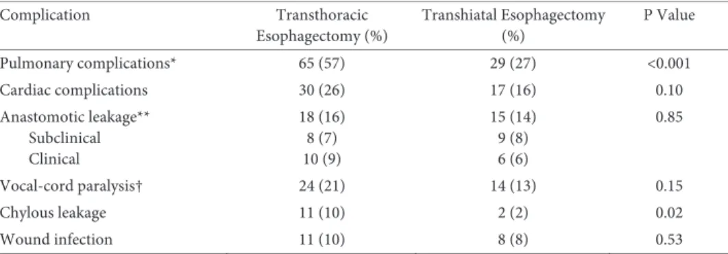
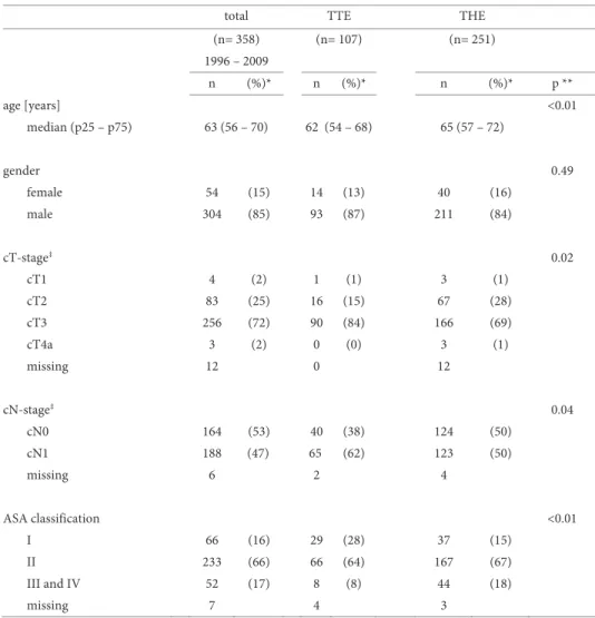
Complications occurring in a randomized trial comparing open TTE with open THE for esophageal adenocarcinoma are summarized in Table 1b.37 Pulmonary
complica-tions including pneumonia (defined as isolation of a pathogen from a sputum culture and an infiltrate on chest x-ray) and atelectasis (defined as lobar collapse on chest x-ray) are among the most common complications, occurring in 57% and 27% of patients who underwent TTE or THE, respectively. These complications can be minimized by early ambulation and careful attention to adequate pain control. Prevention of aspiration can be achieved by keeping the patient in the semi-upright position at all times, and by me-ticulous attention to maintaining a functioning nasogastric tube. When necessary, a mini-tracheostomy can provide invaluable assistance in clearing retained secretions.
Cardiac complications occur in approximately 26% and 16% of TTE and THE pa-tients, with the development of atrial fibrillation accounting for the majority of these complications. The shift of body fluids and the extensive mediastinal dissection which causes a systemic inflammatory response likely play a role in the pathogenesis. Alt-hough these are generally self-limiting, they do require cardiac monitoring and treat-ment, which can prolong the ICU stay. Atrial fibrillation can also e.g. be caused by anas-tomotic dehiscence with secondary mediastinitis or by mechanical irritation by a chest tube. For these underlying causes specific measures are needed.
Anastomotic complications occur in 10% to 30% of patients depending on the defi-nition and the type of reconstruction performed.38 Most of these leaks can be managed
with local drainage and antibiotic administration as long as the vascular supply to the reconstruction is adequate. We recommend early endoscopy in any patient who is known or suspected to have a substantial leak to exclude potentially life-threatening conduit ischemia, which can be present in as many as 14% of patients with an anasto-motic leak.39
Results
Long-term survival following esophagectomy depends on several factors including age, gender, weight loss, histological subtype, depth of tumor invasion, radicality of the re-section and the number of involved lymph nodes.29, 40, 41 The impact of surgical approach
on long-term survival remains the subject of debate.
In a retrospective analysis from nine high-volume centers on 2,303 patients (60% adenocarcinoma [AC], 40% squamous cell carcinoma [SCC]) who underwent R0
-resections, it was shown that a high total number of resected nodes is an independent prognostic factor of (favorable) survival after primary surgery. The optimal threshold for survival benefit was removal of 23 nodes, and the operation most likely to achieve this number was found to be an en bloc transthoracic resection.42 These findings are
arguments in favor of TTE over THE. In contrast, a non-randomized study by two Brit-ish high-volume centers showed similar long-term survival after THE and TTE for pa-tients with SCC (12%) or AC (88%), while hospital stay was significantly shorter after
THE.43 This advantage in short-term recovery after THE over TTE without substantially
jeopardizing oncological outcome was confirmed in a recent meta-analysis of 52 studies that included 3,389 TTE patients and 2,516 THE patients (48% SCC, 52% AC). In addi-tion to the significantly shorter hospital stay (4 days less in patients who underwent THE, 95% CI: 1–7, p<0.01), THE was associated with shorter operation time (85 minutes shorter, 95% CI 40–129, p<0.001), less pulmonary complications (17.3% vs. 21.4%, odds ratio [OR] 1.37, 95% CI 1.05–1.79, p=0.02) and lower postoperative mortal-ity (7.2% vs. 10.6%, OR 1.48, 95% CI 1.20– 1.83, p<0.001). On the other hand, anasto-motic leaks and recurrent nerve palsies occurred more frequently after THE than after TTE. Moreover, lymph node yield was higher after TTE (mean difference of eight nodes, 95% CI 1–14, p=0.02). The results of this meta-analysis should be interpreted with cau-tion, because both randomized and non-randomized studies were included. This proba-bly introduced a selection bias in favor of the THE group, because patients with more advanced tumors probably have been treated preferentially via the chest.44 On the other
hand, more frail patients may have been offered a THE because of no need for a thora-cotomy. Finally, the enhanced short-term recovery after THE could not be confirmed in a large (more than 17,000 patients), multicenter observational study that compared TTE with THE; no differences were found in morbidity and mortality. However, a preference for THE in patients with poor performance status probably resulted in selection bias in favor of patients who underwent TTE.45
Proponents of the transhiatal approach explain differences in survival by stage that have been consistently reported as being due to stage migration. This occurs when posi-tive nodes in the extended part of the dissection increase pN-stage in patients with a more favourable prognosis compared to patients with the same number of positive nodes after a limited dissection during THE. In an attempt to address this issue, Altorki
et al.. have reported outcome following en bloc TTE and transhiatal resections
per-formed in patients with T3N-positive (stage III) disease.46 In this group of patients, the
effect of stage migration was supposed to be limited because all had locally advanced tumors with lymph node involvement. They reported 4-year survival of 35% after en bloc resection, which was significantly better than the 11% survival observed after transhiatal esophagectomy. Ultimately, this debate can only be resolved by the comple-tion of a large randomized controlled trial. To date, only one such large trial (HIVEX) has been reported by Hulscher et al.37 This trial randomized 220 patients with AC of the
mid-to-distal esophagus or the gastric cardia substantially involving the esophagus be-tween THE and TTE. By avoiding a thoracotomy, artificial ventilation time (1 day after THE vs. 2 days after TTE, p<0.001) and hospital stay (15 days after THE vs. 19 days after TTE, p<0.001) were shorter and pulmonary complications were reported less frequently (27% after THE vs. 57% after TTE, p<0.001) after THE than after TTE. Nevertheless, in-hospital mortality was comparable between both groups (2% after THE and 4% after
TTE, p=0.45). Interestingly, the more extended TTE was not associated with a higher percentage of tumor-free resection margins (72% after THE vs. 71% after TTE), whereas the median number of resected lymph nodes was two times higher after TTE than after THE (median 31 vs. 16, p<0.001). This high lymph node yield did not translate into a significantly better five-year overall survival (34% after THE and 36% after TTE (p=0.71)).47 However, in a subsequent subgroup-analysis of patients with a truly
esoph-ageal (Siewert type-1) cancer, and more specifically in patients with a limited number (1–8) of positive lymph nodes, an improved long-term survival was found after TTE, (23% after THE vs. 64% TTE, p=0.02). Given the post-hoc design of this analysis, the effect of stage migration on improved survival of TTE patients cannot be excluded, because more lymph nodes were resected after TTE. Furthermore, the relevance of these results is unclear for patients with SCC (only patients with AC were included). The final conclusion of the HIVEX trial was that in patients with advanced truly esophageal can-cer (Siewert type-1) TTE is the preferred technique (especially in case of a limited num-ber of positive nodes), while THE suffices in patients with a tumor located at the EGJ (Siewert type-2) and in patients with a poor performance status (especially in case of pulmonary comorbidities), without clinically suspected nodes at or above the carina.47
The role of neoadjuvant therapy
Increasingly, the management of esophageal cancer has focused nowadays on multimo-dality therapy, with neoadjuvant chemotherapy or chemoradiotherapy being adminis-tered to nearly all patients with locally advanced disease in many centers. The concept of neoadjuvant therapy in esophageal cancer was spurred by a general disappointment in the results of primary resections, which resulted in survival of 35% or less at 5 years.37
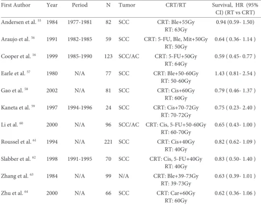
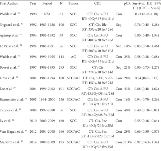
Many studies have been performed to test the additional value of preoperative neo-adjuvant therapy to surgical resection. A meta-analysis showed that both neoneo-adjuvant chemotherapy and neoadjuvant chemoradiotherapy improve long-term survival.48
Fur-thermore, this meta-analysis showed am (nonsignificant) benefit of neoadjuvant chemoradiotherapy (nCRT) over neoadjuvant chemotherapy (nCT) by comparison of the treatment arms of several trials (HR for overall mortality for nCRT vs. nCT 0.88, 95% CI 0.76-1.01, p=0.07). Unfortunately, direct comparisons are limited, especially for patients with AC.
Since the publication of this meta-analysis, the multicenter randomized CROSS trial was completed, comparing nCRT plus surgery with surgery alone in patients with esophageal or junctional cancer (both SCC and AC).29, 49 The applied regimen
(car-boplatin and paclitaxel with 41.4 Gy concurrent radiotherapy) had low toxicity com-pared to earlier trials that mostly used cisplatin and fluorouracil. Median survival
dou-bled from 24% in the surgery alone group to 49% in the nCRT group (HR 0.68, 95% CI 0.53-0.88, p=0.003), with a 5-year survival advantage of 14% (33% vs. 47%). The superi-or survival in the surgery alone arm of the CROSS trial compared to that in earlier ran-domized trials, indicates that the survival benefit can be attributed to improved survival in the multimodality arm, and is not due to poor survival in the surgery alone arm.50, 51
Based on these results, nCRT according to the CROSS regimen plus surgery is now considered standard of care in many countries.
It should be noted that the favorable results of the CROSS trial were not confirmed in a recently completed French randomized trial (Fédération Francophone de Cancérol-ogie Digestive (FFCD) 9901 trial) comparing nCRT plus surgery with surgery alone in stage I and II esophageal cancer patients. The applied neoadjuvant regimen consisted of cisplatin and fluorouracil with 45 Gy concurrent radiotherapy. No differences in 3-year overall survival rate and radical resection rate were found between both treatment arms.52
Based on the FFCD 9901 trial, the standard use of nCRT for early-stage tumors can be debated. Possibly, surgery alone suffices in this subgroup of patients. This is support-ed by the high rate of radical resections (92%) in the surgery alone arm of the French trial. However, the generalizability of the FFCD 9901 trial is questionable due to the low case volume of most participating centers, the high toxicity of the nCRT regimen with less sophisticated radiation techniques compared to the CROSS trial and a remarkably high postoperative mortality rate (11.1%). Therefore, we caution to conclude that pa-tients with early-stage esophageal cancer should not undergo nCRT. We believe that in the absence of high quality evidence on the specific effect of nCRT on early-stage tu-mors, the results from the CROSS trial (which also included stage-II-cancers) should be leading.53
The CROSS trial as well as the FFCD 9901 trial included both AC and SCC. Alt-hough nCRT also significantly improves survival in patients with AC, the maximum benefit of nCRT is observed in SCC, which is known to be more radiosensitive than AC.29, 49 Three small underpowered randomized trials comprising 119, 75 and 131
pa-tients respectively with esophageal AC did not show significant differences in survival between nCRT followed by surgery and nCT followed by surgery. Nevertheless, higher rates of pCR, R0 and ypN0 were found in the nCRT groups and two of these three trials showed a (non-significant) benefit in favor of nCRT.54-57 The optimal neoadjuvant
treatment for esophageal AC remains undetermined and is currently investigated in the randomized Neo-AEGIS (perioperative MAGIC chemotherapy vs. preoperative CROSS chemoradiotherapy, in adenocarcinoma of the esophagus and esophago-gastric junc-tion) trial which is likely to be reported in 2021.58
NCRT has a significant down staging effect on both the primary tumor and the re-gional lymph nodes. In the nCRT-arm of the CROSS trial, a substantial number of
pa-tients (29% overall, 49% SCC, 23% AC) did not have any vital tumor left in the resection specimen. This observation led to the imperative to reconsider the necessity of standard esophagectomy in all patients who undergo nCRT. Therefore, the feasibility of an active surveillance strategy in patients with a clinically complete response (cCR) after nCRT is currently being explored. In this so called SANO (i.e. Surgery As Needed in Oesophage-al cancer patients) approach, surgical resection would be offered only to patients in whom residual disease is highly suspected or proven after nCRT. Before SANO can be tested in a prospective clinical trial, we aim to determine the accuracy of clinical detec-tion of residual disease after nCRT in the present preSANO trial.59 Furthermore, the
French phase II/III randomized ESOSTRATE trial comparing standard surgery with surgery on demand in case of recurrence in patients with a clinically complete response after nCRT is currently being initiated (ClinicalTrials.gov identifier: NCT02551458).60
As outlined above, the randomized HIVEX trial comparing THE with TTE for sub-carinal AC only included patients with primary surgery. In that trial TTE did not im-prove the rate of tumor-free margins (72% after THE vs.71% after TTE), but roughly doubled the number of resected nodes (median ± standard deviation = 16 ± 9 after THE vs. 31 ± 14 after TTE, p<0.001). As discussed above, a retrospective international study has shown that after primary surgery the number of resected nodes is correlated with a favorable long-term survival.42 However, it has been reported that chemoradiotherapy
reduces lymph node yield from within the radiotherapy field.61-63 Importantly, in the
patients after primary surgery from the CROSS trial, the total number of resected nodes and the number of resected positive nodes were positively correlated. However, this positive association completely disappeared in patients who underwent nCRT. Fur-thermore, after surgery alone the total number of removed nodes was positively corre-lated with overall survival (hazard ratio (HR) per 10 additionally resected nodes, 0.76; p=0.007), which corresponds with the earlier retrospective international study.42, 64
In-terestingly, this positive correlation between the number of resected nodes and survival was absent after nCRT (HR 1.00; p=0.98). The randomized design of the CROSS trial renders differences between both treatment groups unlikely as an explanation for the (disappearance of the) association in this post-hoc analysis. These results question the necessity of maximization of surgical lymph node dissection after nCRT, both for prog-nostication and for therapeutic purposes.
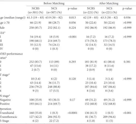
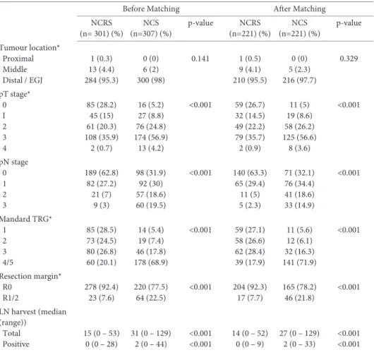
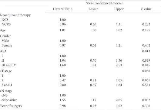
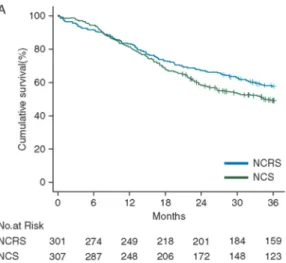
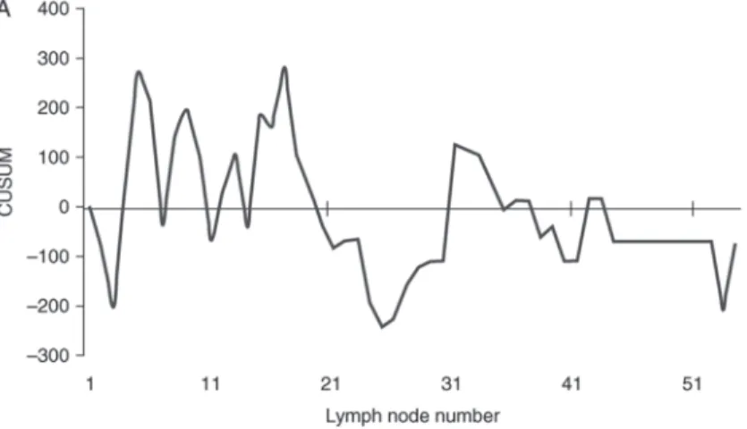
The same phenomenon was identified in a large retrospective comparison of 307 pa-tients who underwent nCRT according to CROSS plus surgery and 301 papa-tients who underwent nCT according to MAGIC followed by surgery. In the nCRT group, the association between lymph node harvest and survival was absent. However, in the nCT group, extent of lymphadenectomy seemed to be positively correlated with progression free survival. Again, these data question the necessity for maximization of surgical lymph node retrieval specifically after nCRT. However, extended lymphadenectomy
seems of importance in patients who undergo nCT followed by surgery (or surgery alone).65
These indirect arguments need confirmation in a randomized trial comparing TTE with extended lymphadenectomy and THE with limited lymphadenectomy in patients with (Siewert type-1) esophageal cancer who undergo nCRT. We believe that such trial should focus on truly esophageal cancer, and not on junctional cancer, because it al-ready has been shown that THE suffices in junctional cancer if they undergo primary surgery; let alone in patients with junctional cancer who have been treated with pre-operative nCRT.
Salvage surgery
Definitive CRT (dCRT) is frequently applied in patients with SCC of the proximal part of the esophagus (i.e. above the carina) and in patients not fit for surgery. Although organ preservation is a considerable advantage in the non-operative strategy of dCRT, this approach is associated with high rates (up to 51%) of recurrence or persistence of locoregional disease.66 In these patients, salvage esophagectomy is an option after failed
dCRT with curative intent. This selective surgery is more demanding than primary esophagectomy. Thanks to centralization of care with improvement in patient selection, in surgical technique and in perioperative management, perioperative morbidity and mortality nowadays have substantially decreased.67 Furthermore, the increased
applica-tion of nCRT has familiarized surgeons with surgical resecapplica-tion in an irradiated surgical field.
Results of salvage surgery after failed dCRT were analyzed in a non-randomized phase II trial.68 Forty-three patients were treated with induction CT (5-FU, cisplatin and
paclitaxel) followed by CRT (5-FU and cisplatin with concurrent 50.4 Gy). CT scans of the chest and abdomen, positron emission tomography (PET, optional but encourag