-3 and
-6 polyunsaturated fatty acids block
HERG
channels
Miriam Guizy, Cristina Arias, Miren David, Teresa Gonza´lez, and Carmen Valenzuela Institute of Pharmacology and Toxicology Consejo Superior de Investigaciones Cientı´ficos,Universidad Complutense de Madrid, School of Medicine, Universidad Complutense, Madrid, Spain
Submitted 27 January 2005; accepted in final form 28 June 2005
Guizy, Miriam, Cristina Arias, Miren David, Teresa Gonza´lez, and Carmen Valenzuela.-3 and-6 polyunsaturated fatty acids block
HERGchannels.Am J Physiol Cell Physiol289: C1251–C1260, 2005. First published June 29, 2005; doi:10.1152/ajpcell.00036.2005.—Dietary polyunsaturated fatty acids (PUFAs) have been reported to exhibit antiarrhythmic properties, which have been attributed to their avail-ability to modulate Na⫹, Ca2⫹, and several K⫹channels. However, their effects on humanether-a-go-go-related gene (HERG) channels are unknown. In this study we have analyzed the effects of arachi-donic acid (AA, -6) and docosahexaenoic acid (DHA, -3) on
HERGchannels stably expressed in Chinese hamster ovary cells by using the whole cell patch-clamp technique. At 10M, AA and DHA blockedHERGchannels, at the end of 5-s pulses to⫺10 mV, to a similar extent (37.7⫾2.4% vs. 50.2⫾8.1%,n⫽7–10,P⬎0.05). 5,6,11,14-Eicosatetrayenoic acid, a nonmetabolizable AA analog, induced effects similar to those of AA onHERGcurrent. Both PUFAs shifted the midpoint of activation curves of HERG channels by ⫺5.1⫾1.8 mV (n⫽10,P⬍0.05) and⫺11.2⫾1.1 mV (n⫽7,P⬍
0.01). Also, AA and DHA shifted the midpoint of inactivation curves by⫹12.0⫾3.9 mV (n⫽4;P⬍0.05) and⫹15.8⫾4.3 mV (n⫽
4;P⬍0.05), respectively. DHA and AA accelerated the deactivation kinetics and slowed the inactivation kinetics at potentials positive to ⫹40 mV. Block induced by DHA, but not that produced by AA, was higher when measured after applying a pulse to⫺120 mV (I3O). Finally, both AA and DHA induced a use-dependent inhibition of
HERGchannels. In summary, block induced by AA and DHA was time, voltage, and use dependent. The results obtained suggest that both PUFAs bind preferentially to the open state of the channel, although an interaction with inactivatedHERG channels cannot be ruled out for AA.
K⫹ channel; membrane currents; ion channels; arrhythmia; antiar-rhythmics
POLYUNSATURATED FATTY ACIDS (PUFAs) present in nature
be-long to two main classes:-6 class, mostly present in vegeta-ble oils; and-3 class, which comes mostly from fish. Both are “essential” because they are necessary for an optimal health status and cannot be synthesized de novo. In addition, mam-mals cannot convert -6 into -3 PUFAs. The dramatic in-crease in the -6/-3 ratio in the diet of the population of Western countries after the industrial revolution has contrib-uted to the rise in cardiovascular disease (7, 21). There is a growing body of evidence that dietary-3 PUFAs, especially docosahexaenoic acid (DHA), play an important role in the prevention of coronary heart disease, in decreasing the risk of sudden cardiac death, and, in particular, in preventing fatal ventricular arrhythmias (1, 5, 8, 10, 21, 37). The cardioprotec-tive effects of-3 PUFAs have been attributed to their avail-ability to modulate several ionic channels involved in the onset and maintenance of the cardiac action potential (21, 22).
PUFAs block Na⫹channels by binding to the receptor site that share local anesthetics and antiarrhythmic drugs (29, 32, 42). PUFAs also interact with other cardiac ion channels, inhibiting ultrarapid delayed-rectifier K⫹ current (IK,ur), transient
out-ward current (Ito), and Ca2⫹current (ICa) (14, 15, 22, 40) and
enhancing long-activated delayed-rectifier K⫹ current (IK1)
and slow-activated delayed-rectifier K⫹current (IKs; the latter
by their interaction with minK) (9, 24). Moreover, it has been demonstrated that membrane lipids can convert Kv A-type channels into delayed rectifiers and vice versa. Thus phospho-inositides remove N-type inactivation, whereas arachidonic acid (AA) converts Kv-delayed rectifiers into A-type rectifier channels (30). However, the effects of PUFAs on human
ether-a-go-go-related gene (HERG) channels have not been studied yet.HERGchannel activity determines the ventricular action potential duration and, therefore, the refractory period. Mutations in the gene encoding HERGchannels are involved in the genotype of congenital long QT syndrome (36). More-over, most drugs that induce acquired long QT syndrome block
HERG channels (35), and thus, HERG channels can be con-sidered a target of antiarrhythmic agents. The purpose of the present study is to analyze and compare the effects of-6 and
-3 PUFAs (AA, precursor of the prostaglandins, leukotrienes, and thromboxane cascade; and DHA) on HERG channels, whose activity determines the action potential duration (23). A preliminary report of this study has been published in abstract form (12).
METHODS AND MATERIALS
Cell culture.Stably transfected Chinese hamster ovary cells with the gene encoding HERG channels (a gift of Drs. S. Nattel, T. E. He´bert, and W. Weerapura) were cultured at 37°C in Ham’s F-12 medium supplemented with geneticine (600g/ml), penicillin-strep-tomycin (800 IU and 200 g/ml, respectively), and 10% bovine serum, in a 5% CO2atmosphere (2, 11). Cultures were passaged every 3–5 days with the use of a brief trypsin treatment. Before experimental use, the cells were removed from the dish with a rubber policeman, a procedure that left the majority of the cells intact. The cell suspension was stored at room temperature (21–23°C) and used within 12 h for all the experiments reported.
Electrophysiological recording.The intracellular pipette-filling so-lution contained (in mM) 80 K-aspartate, 50 KCl, 3 phosphocreatine, 10 KH2PO4, 3 MgATP, 10 HEPES-K, and 5 EGTA and was adjusted to pH 7.25 with KOH. The bath solution contained (in mM) 130 NaCl, 4 KCl, 1.8 CaCl2, 1 MgCl2, 10 HEPES-Na, and 10 glucose, and was adjusted to pH 7.4 with NaOH. AA, DHA, and 5,8,11,14-eicosatet-raynoic acid (ETYA) (Sigma, St. Louis, MO) were dissolved in ethanol at concentrations of 56.5, 52.5, and 10 mM, respectively. Experiments were performed to test the potential effects of ethanol
Address for reprint requests and other correspondence: C. Valenzuela, Institute of Pharmacology and Toxicology, CSIC/UCM, School of Medicine, Universidad Complutense, 28040 Madrid, Spain (e-mail: carmenva@med.ucm.es).
The costs of publication of this article were defrayed in part by the payment of page charges. The article must therefore be hereby marked “advertisement” in accordance with 18 U.S.C. Section 1734 solely to indicate this fact.
(30l/100 ml) onHERGchannels. Ethanol, at this concentration, did not modify the outward maximum current (56.3⫾10.3 vs. 52.7⫾ 11.2 pA;n⫽7,P⬎0.05) nor the maximum peak tail current (68.7⫾ 16.4 to 63.6⫾16.7 pA;n⫽7,P⬎0.05). AA and DHA were stored under argon atmosphere and maintained in sealed ampoules protected from light at ⫺40°C to prevent oxidation. HERG currents were recorded at room temperature (21–23°C) using the whole cell patch-clamp technique (13) with an Axopatch 200A patch-patch-clamp amplifier (Axon Instruments, Foster City, CA). Micropipettes were pulled from borosilicate glass capillary tubes (GD-1; Narishige, Tokyo, Japan) on a programmable horizontal puller (Sutter Instrument, San Rafael, CA) and heat polished with a microforge (Narishige). Micropipette resis-tance was 1–3 M⍀. MaximumHERG tail-current amplitudes aver-aged 717 ⫾ 123 pA, mean uncompensated access resistance was 1.5⫾0.5 M⍀, and cell capacitance 29.8⫾2.0 pF (n⫽11). Thus no significant voltage errors (⬍5 mV) were expected. HERG currents were filtered at 100 Hz and sampled at 200 Hz. Cells were held at⫺80 mV. After control data were obtained, bath perfusion was switched to fatty acid-containing solution. The effects of drug infusion were monitored with test pulses to⫺10 mV applied every 30 s until steady state was obtained. Steady-state current-voltage relationships (I-V) were obtained by averaging the current over a small window (2–5 ms) at the end of 5-s depolarizing pulses. Between⫺80 and⫺50 mV only passive linear leak was observed and least squares fit to these data were used for passive leak correction. Deactivating tail currents were recorded at⫺60 mV. The activation curves were obtained from the tail current amplitude measured at the maximum peak value. The inactivation curves were obtained from the maximum current ampli-tude measured at a test pulse at⫹30 mV applied after a two-pulse protocol that consisted of a 1-s depolarizing pulse from⫺80 mV to ⫹30 mV followed by a second pulse of 20 ms in duration to different membrane potentials between ⫺120 mV and⫹10 mV. Other pulse protocols are described in RESULTS. Command potentials and data
acquisition were generated by using the CLAMPEX program of pCLAMP 6.0.1, 9.0.1 (Axon Instruments). Data analysis was per-formed using the CLAMPfit program of pCLAMP 9.0.1, Origin 7.0.3 (Microcal Software, Northampton, MA), and other custom-made analysis programs. Deactivation was fitted to a biexponential process
y⫽C⫹A1exp共⫺t/1兲⫹A2exp共⫺t/2兲 (1) where 1 and 2 are the system time constants, A1 and A2 are the amplitudes of each component of the exponential, and C is the baseline value. Half-maximal voltages (Eh) and slope factors (s) of
activation and inactivation were determined by fitting data with a Boltzmann equationy⫽1/{1⫹exp[⫺(E⫺Eh)/s]}. The curve-fitting
procedure used a nonlinear least-squares (Gauss-Newton) algorithm; results were displayed in linear and semilogarithmic format, to-gether with the difference plot. Goodness of fit was judged by the2 criterion and by inspection for systematic nonrandom trends in the difference plot.
Statistical methods.Results are expressed as means⫾SE. Direct comparisons between mean values in control conditions and in the presence of drug for a single variable were performed by paired Student’st-test. Differences were considered significant ifP⬍0.05. Comparisons between the three groups were performed by a one-way ANOVA, with a posterior Newman-Keuls test ifP⬍0.05.
RESULTS
Free fatty acids are generally toxic to cells and are kept at low micromolar concentrations in plasma. These levels can vary greatly depending on the hormonal, metabolic, and nutri-tional state of the individual. Around 99.9% of free fatty acids are carried in plasma bound to albumin. The low plasma free fatty acid concentrations are maintained by the competition between the affinity of the albumin-binding sites and cell membrane phospholipids for free fatty acids. The range of free AA and DHA in human plasma is 5.3–13.1M and⬍2.8M, respectively (6). The choice of the concentration of DHA (10
M) is based on the reported EC50for the effects of PUFAs on
ion channels that ranges between 1 and 30M (14, 43). This concentration was also used to show antiarrhythmic properties of this fatty acid in single cell (16 –18). Following the same reasoning and for better comparisons between -3 and -6 PUFAs effects onHERGchannels, the same AA concentration was used.
Voltage-dependent block of HERG channels induced by AA and DHA. Figure 1A shows HERG currents obtained after applying 5-s pulses from a holding potential of ⫺80 to ⫹50 mV in 10-mV steps in the absence and in the presence of 10
M AA or DHA. Deactivating tail currents were measured at
⫺60 mV. Figure 1B shows the I-V relationships obtained by plotting the amplitude of the HERGcurrent measured at the end of the 5-s pulses vs. membrane potential in the absence and in the presence of AA (Fig. 1B,left) or DHA (Fig. 1B,right). Under control conditions, the I-V exhibits the characteristic bell shape that increases from⫺50 mV to⫺10 mV (⫺12.4⫾ 1.8 mV, n⫽ 19) and, due to the fast C-type inactivation of
HERGchannels, it decreased with further depolarizations (38, 39). Thus steady-state drug-induced block was measured at the end of 5-s pulses to ⫺10 mV. Blockade induced by AA (37.7⫾2.4%,n⫽ 10) was similar to that produced by DHA (50.2⫾8.1%,n⫽ 7;P⬎0.05). However, block induced by DHA was higher than that produced by AA measured at the maximum peak of the tail current recorded at ⫺60 mV after applying a 5-s depolarizing test pulse to 0 mV (47.4⫾4.2%,
n⫽ 10, vs. 58.3⫾2.7%, n⫽ 7, in the presence of AA and DHA, respectively; P ⬍ 0.05). AA and DHA shifted the midpoint of activation curves without modifying the slope factors, being the voltage shift induced by DHA higher than that produced by AA (⫺11.2⫾1.1 mV,n⫽7 vs.⫺5.1⫾1.8 mV,n⫽10;P⬍0.05) (Fig. 1C). These results,1) the higher degree of block observed in the tail currents, and 2) the negative shift of the activation curve, suggest an open-channel block mechanism.
To determine the fully activatedI-Vrelationships (Fig. 1E), a double pulse protocol was applied (Fig. 1D). A 1-s step to
⫹30 mV to activate HERG channels and a test pulse to potentials from ⫺120 to ⫹10 mV in 10-mV steps were
Fig. 1. Effects of arachidonic acid (AA) and docosahexanoic acid (DHA; 10M) on humanether-a-go-go-related gene (HERG) currents.A: current records obtained upon depolarization from a holding potential of⫺80 up to⫹50 mV in 10-mV steps and upon repolarization to 60 mV. Current records obtained in the absence and in the presence of AA (left) and DHA (right).B: current-voltage (I-V) relationships (5-s isochronal) ofHERGchannels obtained in the absence and in the presence of each fatty acid.C: activation curves ofHERGchannels obtained under control conditions and in the presence of AA or DHA. Dotted lines reflect the normalized activation curves obtained in the presence of polyunsaturated fatty acids (PUFAs) and matching control values. Open stars represent the relative current obtained at different membrane potentials. Each point represents the means⫾SE of 7–10 experiments.Eh, half-maximal voltages. *P⬍0.05
and **P⬍0.01 vs. control.D: records obtained during the application of the pulse protocol shown in thetopof the figure in the absence and in the presence of AA (left) or DHA (right).E: fully activatedI-Vrelationships ofHERGchannels obtained in the absence and in the presence of AA or DHA.
applied. The pulse to⫹30 mV was positive enough to induce full conductance ofHERGchannels but also inactivated a large number of channels (inward rectification from potentials pos-itive to⫺40 mV). Block induced by 10M AA and DHA was not voltage-dependent and when measured at⫺120 mV aver-aged 41.8 ⫾ 7.1% and 56.2 ⫾ 3.5% (n ⫽ 4; P ⬎ 0.05), respectively.
AA metabolism occurs via three principal pathways: cyclo-oxygenase, lipocyclo-oxygenase, and epoxygenase catalysis. To test whether the AA effects were due to its direct interaction with
HERGchannels or to the actions of some of its metabolites, the electrophysiological effects of its nonmetabolizable analog ETYA were analyzed (Fig. 2). ETYA (10 M) inhibited
HERGcurrent by 37.4⫾3.7% and 42.5⫾5.8%, measured at the end of 5-s pulses to⫺10 mV and at the maximum peak tail currents, respectively (n ⫽ 7; P ⬎ 0.05), similar to the inhibition produced by AA at the same concentration. More-over, as it can be observed in Fig. 2,B–D, the kinetics of block induced by ETYA was similar to that observed in the presence of AA. Therefore, these results suggest that the AA effects observed onHERGchannels are not due to AA metabolites.
Time-dependent block of HERG channels induced by AA and DHA.AA and DHA accelerated the time course of the maxi-mum outward current and decreased its amplitude (Fig. 3A). The initial increase of the current is likely due to the negative voltage shift of the activation curve induced by both PUFAs. In this figure, the control traces exponentially rise during depo-larization, whereas in the presence of both PUFAs, the current remains constant after ⬃1 s. Thus the relative current (I Fatty-Acid/IControl) vs. time of depolarization was plotted (Fig. 3A).
These ratio traces are a composite of the block, which seems to reach a maximum within the first second, which could repre-sent a mixture of tonic- and time-dependent block. The relative current decreased in the presence of both PUFAs with a similar time constant ( ⫽549.6⫾117.9 ms,n⫽8 vs. 731.6⫾109.6
ms,n⫽10, in the presence of AA and DHA, respectively,P⬍
0.05).
Time dependency of block was also apparent in the tail currents, as shown in Fig. 3B, which shows superimposed tail currents recorded under control conditions and in the presence of AA or DHA. The time course of deactivation of HERG
channels was fitted to a biexponential function (fastandslow).
AA accelerated fast(from 377.7⫾ 30.8 ms to 243.8⫾ 45.6
ms, n⫽ 8;P⬍ 0.05) andslow (from 1923.7⫾ 257.9 ms to
1599.9⫾198.1 ms,n⫽8;P⬍0.05) of deactivation without modifying the ratio of amplitudes [Afast/(Afast⫹Aslow)] (0.31⫾
0.09 vs. 0.34⫾0.06,n⫽8,P⬍0.05). DHA acceleratedfast
(from 351.6 ⫾ 41.2 to 222.3 ⫾ 24.9 ms, n⫽ 9; P ⬍ 0.05) without modifying slow (1713.7 ⫾ 264.0 vs. 1345.7 ⫾ 85.7
ms,n⫽ 9;P⬎0.05) or the ratio of amplitudes (0.51⫾0.05 vs. 0.48 ⫾0.04,n⫽9,P⬍ 0.05).
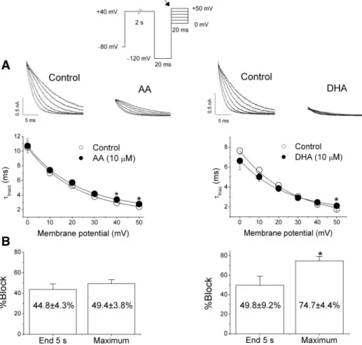
To analyze the effects of AA or DHA on the inactivation kinetics, the three-pulse protocol shown in the top of Fig. 4 was applied. Holding potential was maintained at ⫺80 mV, and after a 2-s step to⫹40 mV that fully activatesHERGchannels, a 20-ms pulse to⫺120 mV that promotes the recovery from the fast inactivation ofHERGchannels was applied. Then, 20-ms test pulses to membrane potentials between 0 and ⫹50 mV were applied. The degree of block induced by DHA when measured at the maximum peak current recorded during the application of the test pulse to 0 mV was higher than that induced by AA. Neither DHA nor AA modified the time constant of inactivation (Inac) at membrane potentials between
0 and⫹40 mV. However, at membrane potentials positive to
⫹40 mV, AA and DHA increased the Inac, which can be
explained either by a fatty acid-induced stabilization of the open state of HERG channels or a destabilization of the inactivated state. Figure 4Bshows the degree of block induced by AA and DHA at the end of 5-s pulses to 0 mV (End 5-s) and at the maximum peak current recorded during the application
Fig. 2. Effects of 5,6,11,14-eicosatetrayenoic acid (ETYA; 10 M) on HERG currents. A: current records obtained upon depolarization from a holding potential of ⫺80 mV up to ⫺10 mV and upon repolarization to⫺60 mV in the absence and in the presence of ETYA. Inset, first 5 s of the current traces shown inA.B: relative current (IETYA/IControl) vs. time of depolarization. Current exponentially decreased during depolarization. C: tail currents recorded upon repolarization to⫺60 mV after a 5-s pulse to⫺10 mV in the absence and in the presence of ETYA.D: tail current traces shown inC normal-ized to match control values.
of the test pulse to 0 mV (Maximum). DHA induced a higher block (P⬍0.05) when measured at the maximum peak current than at the end of 5-s depolarizing pulses, thus suggesting an open channel block. However, block of HERG channels in-duced by AA was similar under both experimental conditions.
Effects of AA and DHA on HERG inactivation availability.
To analyze the effects of AA and DHA on the inactivation availability, a three-pulse voltage clamp protocol was used (Fig. 5) (38, 39). Figure 5Ashows the effects of 10M AA and DHA on the voltage dependence ofHERG channels (“inacti-vation availability”). The dynamic nature of HERG currents precludes direct measurements of “steady-state” inactivation, but the data shown approximate a voltage dependence of the distribution of channels between open and inactivated states. The 1-s step to ⫹30 mV fully activates and inactivates the channels. The second step to the test potential (between⫺120 and⫹10 mV) allows some fraction of the channels to recover
from inactivation. The instantaneous current after the third step to⫹30 mV allows measurements of the fraction of channels that have recovered from inactivation during the preceding test step. Figure 5B shows a plot of the current measured at⫹30 mV as a function of voltage.HERGcurrent falls off at negative potentials because the channels also begin to deactivate during the 20-ms pulse. We corrected the current magnitude for the amount of deactivation at each voltage by fitting the deactivat-ing tail currents with an exponential function and then back-extrapolated this fit to the beginning of the hyperpolarizing pulse, which permitted us to estimate the fraction of channels deactivating during the 20-ms pulse and then increase the outward current accordingly (38). Figure 5Cshows the Boltz-mann fit of the corrected data in the absence and in the presence of the two PUFAs. AA and DHA shifted the apparent voltage dependence of channel availability toward more posi-tive potentials by ⫹12.0 ⫾ 3.9 mV (n ⫽ 4, P ⬍ 0.05) and
⫹15.8 ⫾ 4.3 mV (n ⫽ 4, P ⬍ 0.05), respectively, without modifying the slope factor.
State- and use-dependent effects of AA and DHA on HERG channels. To further analyze the interaction between AA or DHA and HERG channels, we applied the voltage protocol shown in the top of Fig. 6. From a holding potential of ⫺80 mV,HERGchannels were rapidly activated by a 5-ms step to
⫹180 mV, followed by a 200-ms step to 0 mV, before returning to ⫺60 mV. With this protocol, HERG current amplitude at 0 mV, under control conditions, was nearly constant. In the presence of AA or DHA, the amplitude of
HERGcurrent exponentially declined to a steady-state level as additional block developed during the step to 0 mV. The time course of AA- and DHA-induced HERG current decay was determined by dividing the current trace obtained in the pres-ence of the fatty acid and the control trace. This current was fitted as a monoexponential decay to calculate the time con-stant of block of the open state ofHERGchannels (B) (27.9⫾
5.6 ms,n⫽ 3, and 39.2⫾5.7 ms,n⫽5, for AA and DHA). This pulse protocol also allowed us to measure the recovery kinetics by fitting the hook of the tail current at⫺60 mV (Fig. 6A). AA or DHA, at 10 M, did not modify the recovery process (6.4⫾ 1.3 vs. 6.5⫾ 1.0 ms;n⫽4,P⬎ 0.05 in the absence and in the presence of AA and 5.9⫾0.6 vs. 4.9⫾0.5 ms; n⫽ 5, P ⬎ 0.05 in the absence and in the presence of DHA). This voltage pulse protocol was applied at 1.0 Hz and, as can be observed, AA and DHA decreased in a use-depen-dent manner the amplitude of HERGcurrent. This pattern is consistent with a preferential block of the open state of the channel. In the presence of DHA, the maximum peak current was higher than under control conditions, which can be due to the slower Inac at positive potentials (⫹180 mV).
AA and DHA HERG current inhibition during cardiac ac-tion potential.Because of the complex gating of HERG, it is difficult to predict the effects of AA and DHA on ionic currents during a cardiac action potential (AP), where the membrane voltage is continually changing with time. To assess this issue, the effects of AA and DHA were analyzed using the AP clamp technique as shown in Fig. 7. The frequency of stimulation used was 1 Hz. Under control conditions, HERG current rapidly inactivates during the upstroke. As the action potential repolarizes,HERGchannels rapidly recover from inactivation to reopen and slowly deactivate, increasing their occupancy in the open state. The current deactivates with the final phase of
Fig. 3. Time-dependent block induced by AA and DHA (10M).A, top: current traces obtained after depolarizing the cell membrane from⫺80 to⫺10 mV in the absence and in the presence of AA or DHA.Bottom, plot of the relative current (IFattyAcid/IControl) vs. time of depolarization. Current exponen-tially decreased during depolarization. B, top: tail currents recorded upon repolarization to⫺60 mV after a 5-s pulse to 0 mV in the absence and in the presence of AA or DHA. Bottom, tail current traces normalized to match control values.
repolarization. AA (10 M) decreased the maximum peak current by 43.3⫾1.6% (n⫽3). DHA increasedHERGcurrent during the upstroke and decreased it at the end of the AP to an extent similar to that of AA (48.1⫾8.3%,n⫽ 3,P⬎0.05).
DISCUSSION
In the present study, the effects of AA and DHA onHERG
channels have been studied. We found that, at relevant plasma concentrations, both PUFAs blockHERGchannels to a similar extent. Moreover, the effects of AA onHERGchannels appear to be due to this PUFA and not to its metabolites because its actions are mimicked by the nonmetabolizable analog ETYA.
Effects of AA and DHA on HERG channels.AA and DHA blockHERGchannels in a voltage-, time-, and state-dependent manner, which is consistent with an open channel block mech-anism. In fact, block induced by both PUFAs steeply increased in the range of membrane potentials that coincides with the range ofHERGchannel activation, suggesting that their bind-ing may derive a significant fraction of its voltage sensitivity through coupling to channel gating. Unfortunately, at depolar-ized voltages the open and inactivated conformations ofHERG
channels are in rapid equilibrium, making it difficult to un-equivocally identify the state(s) with which these two PUFAs interact.
Whereas AA induced a similar inhibition of the HERG
current when measured at the end of depolarizing pulses to
⫺10 mV and at the maximum tail currents, DHA inhibited this current to a higher extent when measured at the maximum tail current than at the end of depolarizing steps to ⫺10 mV.
During depolarization, HERG channels inactivate faster than they activate and thus the amplitude of the current is reduced. On repolarization, closed channels transit through the open state, resulting in tail currents with higher amplitude (38, 39). In agreement with these results, block induced by AA when measured at the maximum peak current of a test pulse to 0 mV applied after a hyperpolarizing pulse to ⫺120 mV (that pro-motes the I3O transition) was similar to that observed at the same voltage at the end of a 5-s depolarizing pulse. However, DHA-induced block, when measured at the maximum peak current of a test pulse to 0 mV applied after a hyperpolarizing pulse to⫺120 mV, was higher than that observed at the same voltage at the end of a 5-s depolarizing pulse. Block induced by AA and DHA was also time dependent, being evident after a prepulse to ⫹180 mV, suggesting a rapid drug binding to activatedHERGchannels, as previously described for cocaine and bupivacaine-type local anesthetics (11, 44). This time dependency was also evident in the deactivation process of
HERG channels that was accelerated in the presence of both AA and DHA. However, AA and DHA did not modify the onset kinetics of the inactivation process or the recovery process. The faster deactivation induced by both PUFAs, together with their lack of effect on the recovery kinetics, suggests that a very fast dissociation rate constant fromHERG
channels is consistent with an open channel block mechanism, as has been proposed for propafenone (2). Another piece of evidence of an open channel interaction between both PUFAs and HERG channels is the use-dependent inhibition of the current. Taken together, all these results suggest that both AA
Fig. 4. Effects of 10M AA or DHA onHERG inactivation kinetics.A,top: current records of test pulses obtained in the absence and in the presence of each fatty acid.Bottom, plots of the time constant of inactivation at different mem-brane potentials in the absence and in the pres-ence of each agent.B: graph showing the degree of block induced by AA or DHA measured at the end of 5-s pulses to 0 mV (End 5-s) or at the maximum peak current recorded at a test pulse to 0 mV after a test pulse to⫹40 mV during 2-s and followed by a 20-ms pulse to ⫺120 mV (Maximum) indicated by an arrow in the pulse protocol. Each point represents the means⫾SE of 7–9 experiments. *P⬍0.05 vs. control con-ditions.
and DHA preferentially bind to the open state of HERG
channels, and that DHA exhibits a higher affinity for this state of the channel.
We also observed that AA and DHA produced a positive shift in the inactivation curve. This could be explained either by 1) stabilization of the open state of HERGchannels or 2) destabilizing the inactivation process; i.e., without modifying the onset but accelerating the offset of inactivation. In both cases, the shift of the inactivation curve would be the result of the interaction between PUFAs and a closed state of HERG
channels (tonic block). This tonic block is likely to influence the apparent steady-state inactivation and perhaps the activa-tion process because both PUFAs accelerate the deactivaactiva-tion process. All of these results suggest that AA and DHA pref-erentially block the open state ofHERGchannels, but also that they interact with a closed state, thus producing changes in channel gating. Finally, the similar degree of AA-induced
inhibition of the current at the end of 5-s depolarizing pulses (when most channels are inactivated) and at the maximum tail current or at the maximum peak current after applying a⫺120 mV step cannot permit us to rule out an interaction between AA and the inactivated state ofHERGchannels. Moreover, it has been shown that AA regulates the inactivation process in other K⫹channels in such a way that introduces rapid voltage dependent inactivation into noninactivating Kv channels (30). The authors explain these results under the framework that AA closes Kv channels by inducing conformational alter-ations in the selectivity filter region (30) and propose that Kv channel inactivation is lipid dependent and that this process has a high affinity, comparable to that of KATP
channels for phosphoinositides (3). Oliver et al. (30) pro-pose that AA inserts into the cell membrane from either side, interacts with the channel protein, and, allosterically, induces a fast closure of the open Kv channel pore through
Fig. 5. Apparent voltage dependence of channel availability. The pulse protocol used to obtain each data point is shown attop.A: original traces obtained after applying such pulse protocol in the absence and in the presence of 10M AA and DHA.B: peak current observed immediately after stepping to⫹30 mV was then plotted vs. test potential in the absence and in the presence of 10M of AA (left) and DHA (right). Currents decreased at negative potentials due to deactivation during the 20-ms pulse.C: corrected data for deactivation (seeRESULTS) together with the Boltzmann fit. Each point represents the mean⫾SE of
conformational modifications in the selectivity filter. Fur-ther studies are required to discern the possible role of AA on the inactivation of HERGchannels.
Clinical implications of this study.Many studies suggest that
-3 fatty acids have beneficial effects on human health. In contrast, a diet enriched in -6 fatty acids provokes athero-sclerosis, carcinogenesis, and heart disease (20). In the present study we have demonstrated that AA and DHA blockHERG
channels. The plasma levels of these two PUFAs vary greatly depending on the hormonal, metabolic, and nutritional state of the individual. Around 99.9% of fatty acids are carried in plasma bound to albumin and the range of free AA and DHA
in human plasma is 5.3–13.1 M and⬍2.8M, respectively (6). The EC50values calculated from the blockade produced by
both PUFAs at 10 M are well within their physiological plasma range. Therefore, we can conclude that both PUFAs block HERG channels at concentrations within their physio-logical plasma levels.
It has been described that AA is able to modulate several ion currents (28). In fact, AA decreases L-typeICa(ICa,L), T-type ICa (ICa,T), Ito, INa, and Kv1.5 currents (4, 14, 40, 41) and
activatesIK1(25). AA plasma levels are known to be increased
during ischemia and reperfusion at the intracellular and extra-cellular levels (19), which, in the light of previous and present
Fig. 6. Time-dependent block of AA and DHA (10M) after a fast activation ofHERGchannels, using the pulse protocol shown at thetop.A: first record obtained in the absence and in the presence of AA or DHA.B: relative current (IFattyAcid/IControl) during the step to 0 mV together with its monoexponential fit. C: use-dependent block ofHERGchannels induced by AA and DHA obtained after applying the pulse protocol shown at top at 1.0 Hz.Inset, records obtained in the absence and in the presence of AA or DHA.
Fig. 7. Effect of 10M AA (A) and DHA (B) onHERGcurrents evoked by an action potential waveform from a guinea pig ventricular myo-cyte. The frequency of stimulation was 1 Hz. Arrow indicates 0 current level.
results, could represent a “natural” protective mechanism to prevent arrhythmias under these pathological conditions. On the other hand, it has been shown that DHA exhibits potential cardioprotective effects that have been associated with their antiarrhythmic effects (21). DHA’s antiarrhythmic effects have been attributed to its actions on the cardiac ion channels responsible for the onset and maintaining of the cardiac action potential, mostly on its inhibitory effects on Na⫹ and ICa,L
channels (22, 42), because DHA has inhibitory effects onINa,
but not onICa,L, that are higher than those of AA (41). DHA
inhibitory effects on INa and ICa theoretically should shorten
the cardiac action potential duration. Moreover, DHA enhances
IKs, which would further shorten the cardiac action potential
duration (9). However, experiments performed in rat ventric-ular myocytes demonstrate that DHA slightly prolongs the rat cardiac action potential (4, 26). In the present study we have demonstrated that DHA blocks HERG channels. Most drugs that selectively block HERG channels prolong the cardiac action potential (34). However, interactions of nonselective
HERG channel blockers with other cardiac ion channels may mitigate or exacerbate the prolongation of action potential duration (27, 31, 33). Because DHA inhibits INaand ICa and
enhances IKs (effects that would shorten the cardiac action
potential), but also inhibitsIto,IK,ur, andHERG(which should
produce a lengthening of the action potential duration), the result should be a modest effect on the time of repolarization. Besides these effects on the action potential duration, it should be stated that its inhibitory effects on Na⫹, Ca2⫹, and several
K⫹ channels (Kv4 and HERG channels) would result in a lengthening of the refractory period and a decrease of cardiac excitability, thus contributing to its antiarrhythmic effects.
To our knowledge, this is the first demonstration thatHERG
channels are modulated by AA and DHA. The-3 antiarrhyth-mic effects have been attributed to their availability to modu-late cardiac ion channels involved in the genesis and mainte-nance of the cardiac action potential (21). Our results suggest that AA and DHA blockHERGchannels mainly by binding to the open state of the channel. These PUFAs’ actions onHERG
channels have to be taken into account to explain the antiar-rhythmic effects of AA during ischemia and those previously reported for DHA in subjects consuming a diet rich in fish and fish oils.
ACKNOWLEDGMENTS
The authors thank Drs. S. Nattel, T. E. He´bert, and W. Weerapura for providing the cell line expressingHERGchannels and Guadalupe Pablo for excellent technical assistance.
GRANTS
This work was supported by Comisio´n Interministerial de Ciencia y Tecnologı´a SAF2002-02160, SAF2004-06856, and CAM GR/SAL/0854/2004 and Red Tema´tica de Investigacio´n Cooperativa FIS C03/01 grants.
REFERENCES
1. Albert CM, Campos H, Stampfer MJ, Ridker PM, Manson JE, Willett WC, and Ma J.Blood levels of long-chain n-3 fatty acids and the risk of sudden death.N Engl J Med346: 1113–1118, 2002.
2. Arias C, Gonzalez T, Moreno I, Caballero R, Delpon E, Tamargo J, and Valenzuela C. Effects of propafenone and its main metabolite, 5-hydroxypropafenone, on HERG channels.Cardiovasc Res57: 660 – 669, 2003.
3. Baukrowitz T, Schulte U, Oliver D, Herlitze S, Krauter T, Tucker SJ, Ruppersberg JP, and Fakler B.PIP2 and PIP as determinants for ATP inhibition of KATP channels.Science282: 1141–1144, 1998.
4. Bogdanov KY, Spurgeon HA, Vinogradova TM, and Lakatta EG.
Modulation of the transient outward current in adult rat ventricular myocytes by polyunsaturated fatty acids.Am J Physiol Heart Circ Physiol 274: H571–H579, 1998.
5. Burr ML, Fehily AM, Gilbert JF, Rogers S, Holliday RM, Sweetnam PM, Elwood PC, and Deadman NM.Effects of changes in fat, fish, and fibre intakes on death and myocardial reinfarction: diet and reinfarction trial (DART).Lancet2: 757–761, 1989.
6. Burtis CA, Ashwood ER, and Tietz NW.Textbook of Clinical Chemis-try. Philadelphia, PA: Saunders, 1999.
7. De Caterina R, Madonna R, Zucchi R, and La Rovere MT. Antiar-rhythmic effects of omega-3 fatty acids: from epidemiology to bedside. Am Heart J146: 420 – 430, 2003.
8. De Lorgeril M, Renaud S, Mamelle N, Salen P, Martin JL, Monjaud I, Guidollet J, Touboul P, and Delaye J. Mediterranean ␣-linolenic acid-rich diet in secondary prevention of coronary heart disease.Lancet 343: 1454 –1459, 1994.
9. Doolan GK, Panchal RG, Fonnes EL, Clarke AL, Williams DA, and Petrou S.Fatty acid augmentation of the cardiac slowly activating delayed rectifier current (IKs) is conferred by hminK.FASEB J16: 1662–1664, 2002.
10. GISSI Prevenzione Investigators. Dietary supplementation with n-3 polyunsaturated fatty acids and vitamin E after myocardial infarction: results of the GISSI-Prevenzione trial. Gruppo Italiano per lo Studio della Sopravvivenza nell’Infarto miocardico.Lancet354: 447– 455, 1999. 11. Gonzalez T, Arias C, Caballero R, Moreno I, Delpon E, Tamargo J,
and Valenzuela C.Effects of levobupivacaine, ropivacaine and bupiva-caine on HERG channels: stereoselective bupivabupiva-caine block.Br J Phar-macol137: 1269 –1279, 2002.
12. Guizy M, Arias C, Gonzalez T, Caballero R, Go´mez R, Nu´n˜ez L, Delpo´n E, Tamargo J, and Valenzuela C. Effects of fatty acids on HERGchannels.Biophys J86: 279a, 2004.
13. Hamill OP, Marty A, Neher E, Sakmann B, and Sigworth FJ. Im-proved patch clamp techniques for high-resolution current recording from cells and cell-free membrane patches.Pflu¨gers Arch391: 85–100, 1981. 14. Honore´ E, Barhanin J, Attali B, Lesage F, and Lazdunski M.External
blockade of the major cardiac delayed-rectifier K⫹channel (Kv1.5) by polyunsaturated fatty acids. Proc Natl Acad Sci USA 91: 1937–1941, 1994.
15. Jude S, Bedut S, Roger S, Pinault M, Champeroux P, White E, and Le Guennec JY.Peroxidation of docosahexaenoic acid is responsible for its effects on ITOand ISSin rat ventricular myocytes.Br J Pharmacol139: 816 – 822, 2003.
16. Kang JX and Leaf A.Effects of long-chain polyunsaturated fatty acids on the contraction of neonatal rat cardiac myocytes.Proc Natl Acad Sci USA 91: 9886 –9890, 1994.
17. Kang JX and Leaf A. Prevention and termination of -adrenergic agonist-induced arrhythmias by free polyunsaturated fatty acids in neona-tal rat cardiac myocytes.Biochem Biophys Res Commun208: 629 – 636, 1995.
18. Kang JX and Leaf A. Evidence that free polyunsaturated fatty acids modify Na⫹channels by directly binding to the channel proteins.Proc Natl Acad Sci USA93: 3542–3546, 1996.
19. Kim D and Clapham DE.Potassium channels in cardiac cells activated by arachidonic acid and phospholipids.Science244: 1174 –1176, 1989. 20. Lands WE.Biochemistry and physiology of n-3 fatty acids.FASEB J6:
2530 –2536, 1992.
21. Leaf A, Kang JX, Xiao YF, and Billman GE.Clinical prevention of sudden cardiac death by n-3 polyunsaturated fatty acids and mechanism of prevention of arrhythmias by n-3 fish oils.Circulation107: 2646 –2652, 2003.
22. Leaf A and Xiao YF.The modulation of ionic currents in excitable tissues by n-3 polyunsaturated fatty acids.J Membr Biol184: 263–271, 2001. 23. Li GR, Feng J, Yue L, Carrier M, and Nattel S.Evidence for two
components of delayed rectifier K⫹current in human ventricular myo-cytes.Circ Res78: 689 – 696, 1996.
24. Liu Y, Liu D, Heath L, Meyers DM, Krafte DS, Wagoner PK, Silvia CP, Yu W, and Curran ME.Direct activation of an inwardly rectifying potassium channel by arachidonic acid.Mol Pharmacol59: 1061–1068, 2001.
25. Liu Y, Liu D, and Krafte DS. Decrease of inward rectification as a mechanism for arachidonic acid-induced potentiation of hKir2.3. Eur Biophys J31: 497–503, 2002.
26. Macleod JC, Macknight AD, and Rodrigo GC. The electrical and mechanical response of adult guinea pig and rat ventricular myocytes to 3 polyunsaturated fatty acids. Eur J Pharmacol 356: 261–270, 1998.
27. Martin RL, McDermott JS, Salmen HJ, Palmatier J, Cox BF, and Gintant GA.The utility of hERG and repolarization assays in evaluating delayed cardiac repolarization: influence of multi-channel block.J Car-diovasc Pharmacol43: 369 –379, 2004.
28. Meves H.Modulation of ion channel by arachidonic acid.Prog Neurobiol 43: 175–186, 1994.
29. Nau C, Wang SY, Strichartz GR, and Wang GK.Point mutations at N434 in D1–S6 of 1 Na⫹ channels modulate binding affinity and stereoselectivity of local anesthetic enantiomers. Mol Pharmacol 56: 404 – 413, 1999.
30. Oliver D, Lien CC, Soom M, Baukrowitz T, Jonas P, and Fakler B.
Functional conversion between A-type and delayed rectifier K⫹channels by membrane lipids.Science304: 265–270, 2004.
31. Pearlstein R, Vaz R, and Rampe D.Understanding the structure-activity relationship of the humanether-a-go-go-related gene cardiac K⫹channel. A model for bad behavior.J Med Chem46: 2017–2022, 2003. 32. Ragsdale DS, McPhee JC, Scheuer T, and Catterall WA.Molecular
determinants of state-dependent block of Na⫹channels by local anesthet-ics.Science265: 1724 –1728, 1994.
33. Redfern WS, Carlsson L, Davis AS, Lynch WG, MacKenzie I, Palethorpe S, Siegl PK, Strang I, Sullivan AT, Wallis R, Camm AJ, and Hammond TG. Relationships between preclinical cardiac electro-physiology, clinical QT interval prolongation and torsade de pointes for a broad range of drugs: evidence for a provisional safety margin in drug development.Cardiovasc Res58: 32– 45, 2003.
34. Roden DM.Current status of class III antiarrhythmic drug therapy.Am J Cardiol72: 44B-49B, 1993.
35. Roden DM, Lazzara R, Rosen M, Schwartz PJ, Towbin J, and Vincent GM.Multiple mechanisms in the long-QT syndrome. Current knowledge,
gaps, and future directions The SADS Foundation Task Force on LQTS. Circulation94: 1996 –2012, 1996.
36. Sanguinetti MC, Jiang C, Curran ME, and Keating MT.A mechanistic link between an inherited and an acquired cardiac arrhythmia: HERG encodes the IKrpotassium channel.Cell81: 299 –307, 1995.
37. Singh RB, Niaz MA, Sharma JP, Kumar R, Rastogi V, and Moshiri M.
Randomized, double-blind, placebo-controlled trial of fish oil and mustard oil in patients with suspected acute myocardial infarction: the Indian experiment of infarct survival– 4.Cardiovasc Drugs Ther11: 485– 491, 1997. 38. Smith PL, Baukrowitz T, and Yellen G. The inward rectification
mechanism of the HERG cardiac potassium channel.Nature379: 833– 836, 1996.
39. Spector PS, Curran ME, Zou A, Keating MT, and Sanguinetti MC.
Fast inactivation causes rectification of the IKrchannel.J Gen Physiol107: 611– 619, 1996.
40. Xiao YF, Gomez AM, Morgan JP, Lederer WJ, and Leaf A. Suppres-sion of voltage-gated L-type Ca2⫹currents by polyunsaturated fatty acids in adult and neonatal rat ventricular myocytes.Proc Natl Acad Sci USA 94: 4182– 4187, 1997.
41. Xiao YF, Kang JX, Morgan JP, and Leaf A. Blocking effects of polyunsaturated fatty acids on Na⫹channels of neonatal rat ventricular myocytes.Proc Natl Acad Sci USA92: 11000 –11004, 1995.
42. Xiao YF, Ke Q, Wang SY, Auktor K, Yang Y, Wang GK, Morgan JP, and Leaf A. Single point mutations affect fatty acid block of human myocardial sodium channel alpha subunit Na⫹channels.Proc Natl Acad Sci USA98: 3606 –3611, 2001.
43. Xiao YF, Wright SN, Wang GK, Morgan JP, and Leaf A.Fatty acids suppress voltage-gated Na⫹currents in HEK293t cells transfected with the alpha-subunit of the human cardiac Na⫹channel.Proc Natl Acad Sci USA 95: 2680 –2685, 1998.
44. Zhang S, Rajamani S, Chen Y, Gong Q, Rong Y, Zhou Z, Ruoho A, and January CT. Cocaine blocks HERG, but not KvLQT1⫹minK, potassium channels.Mol Pharmacol59: 1069 –1076, 2001.