Scholarship@Western
Scholarship@Western
Electronic Thesis and Dissertation Repository
8-16-2019 10:30 AM
Assessing the structure-function relationships of the
Assessing the structure-function relationships of the
apolipoprotein(a) kringle IV sub-type 10 domain
apolipoprotein(a) kringle IV sub-type 10 domain
Matthew J. Borrelli
The University of Western Ontario
Supervisor
Koschinsky, Marlys L. Robarts Research Institute
Graduate Program in Physiology and Pharmacology
A thesis submitted in partial fulfillment of the requirements for the degree in Master of Science © Matthew J. Borrelli 2019
Follow this and additional works at: https://ir.lib.uwo.ca/etd
Part of the Biochemistry Commons, Cardiovascular Diseases Commons, Cellular and Molecular Physiology Commons, Circulatory and Respiratory Physiology Commons, Genetic Phenomena Commons, and the Molecular Biology Commons
Recommended Citation Recommended Citation
Borrelli, Matthew J., "Assessing the structure-function relationships of the apolipoprotein(a) kringle IV sub-type 10 domain" (2019). Electronic Thesis and Dissertation Repository. 6461.
https://ir.lib.uwo.ca/etd/6461
This Dissertation/Thesis is brought to you for free and open access by Scholarship@Western. It has been accepted for inclusion in Electronic Thesis and Dissertation Repository by an authorized administrator of
ii
Abstract
Elevated plasma lipoprotein(a) (Lp(a)) is the most prevalent heritable risk factor in the
development of cardiovascular disease. The apolipoprotein(a) (apo(a)) component of Lp(a) is
strongly implicated in the pathogenicity of Lp(a). It is hypothesized that the inflammatory
potential of Lp(a)/apo(a) is mediated by the lysine binding ability of the apo(a) kringle IV10
(KIV10) domain, along with its covalently bound oxidized phospholipid (oxPL). Using targeted
mutagenesis, two novel null alleles for the LPA gene that generate non-secretable apo(a)
species have been identified, resulting from amino acid substitutions in the KIV10 domain. A
potential mechanism by which KIV10 oxPL modification is enriched was identified. Finally,
RNA-Seq was utilized to demonstrate gene regulation in macrophage-like cells in response to
the lysine binding function and covalent oxPL of the KIV10 domain. It was determined that the
lysine binding ability and covalent oxPL of apo(a) KIV10 are both implicated in vascular cell
inflammation and atherosclerosis.
Keywords
Cardiovascular disease, atherosclerosis, Lipoprotein(a), apolipoprotein(a), inflammation,
iii
Lay Summary
In humans, fats and cholesterol are transported in the blood stream as components of
particles called lipoproteins. A high level of one lipoprotein variety, lipoprotein(a) (Lp(a)),
has been determined to be the single most prevalent heritable risk factor for developing
cardiovascular diseases by contributing to the build-up of plaques within the arteries, or
“atherosclerosis”. Lp(a) is very similar to the more commonly known low-density lipoprotein (LDL), which is typically considered the “bad” cholesterol (in comparison to high density lipoprotein (HDL), which is typically considered the “good” cholesterol). Lp(a) contains a
protein component called apolipoprotein(a) (apo(a)), which distinguishes Lp(a) from LDL,
and is thought to be responsible for the increased pathogenicity of Lp(a) in comparison to
LDL. Apo(a) contains many sub-sections, or “domains”, that contribute in different ways to
its characteristics. One domain, kringle IV sub-type 10 (KIV10), is thought to be particularly
important for the harmful effects of Lp(a) in the blood stream. Here, we investigated the
implications of the functions of the KIV10 domain, and the potential roles this domain may
have in the development of atherosclerosis. The KIV10 domain is able to bind lysine, which
allows apo(a) or Lp(a) to associate closely with cell surface proteins and other ligands.
Additionally, the KIV10 domain is modified covalently with an oxidized phospholipid
(oxPL). The lysine binding ability of KIV10 has been determined to have
atherosclerosis-related effects in certain vascular cell types, as have oxPLs. Together, this suggests that the
KIV10 domain may represent an important factor in mediating the harmful effects of Lp(a) in
the blood stream. We have determined that either of two independent amino acid
substitutions in KIV10 can prevent the secretion of apo(a) entirely, and that a different
substitution can change the amount of oxPL added to this domain. Beyond that, we have
determined that the lysine binding ability and covalent oxPL of the KIV10 domain are
implicated in many gene-regulatory processes that can potentially facilitate the development
iv
Co-Authorship Statement
All data presented in this thesis was collected by Matthew Borrelli. Dr. Marlys Koschinsky
and Dr. Michael Boffa contributed to the generation of appropriate experimental designs,
editing of this thesis, and general supervision.
This thesis incorporates the outcomes of joint research in collaboration with:
- Dr. E Stroes (University of Amsterdam), who identified human patients with elevated
Lp(a) and unexpectedly low oxPL, and sent our group blood samples from these
patients.
- Dr. M Junop (University of Western Ontario), with R Szabla, who performed molecular
modeling for the Met64 and Thr64 variants of the apo(a) KIV10 domain.
- David Carter (London Regional Genomics Center), who facilitated the RNA-Seq
analysis, and developed the RNA-Seq methods detailed in Section 2.10.
The use of figures in this thesis from previously published works has been approved by the
v
Acknowledgments
I would first like to sincerely thank my supervisor, Dr. Marlys Koschinsky, for welcoming me
to her research program during my undergraduate studies, and for giving me the opportunity
to pursue graduate studies under her supervision. I am grateful for the support, the knowledge,
and the numerous opportunities you have generously shared with me. Your influence has been
instrumental in my development as a trainee. I would also like to thank Dr. Michael Boffa for
all of your guidance and ever-constructive feedback, and for the laughs I have had the pleasure
of sharing with you.
Next, I would like to thank my advisory committee members, Dr. Nica Borradaile and Dr. John
Di Guglielmo. Your feedback was always incredibly helpful in the problem solving and
adaptation this work required. Thank you both for your out-of-meeting support as well; I am
grateful for everything you have done to help me succeed – you have gone above and beyond.
I would like to extend my thanks to the members of the Koschinsky-Boffa lab group. Dr. Amer
Youssef, thank you for sharing your vast knowledge with me, and for the calm and focused
presence you bring to the lab. You inspire me to work hard inside of the lab and out, to better
myself as a researcher and as a person. Julia St. John, thank you for your support over these
years and for enabling so much of the work our group does. Justin Clark, thank you for your
support, your friendship, and like you said, for the laughs. Tasnim Reza, thank you for your
unwavering kindness and selflessness.
To Dr. Corey Scipione, thank you for the efforts you made to train me, and for the patience
you showed. This project presented many challenges, but the resourcefulness and problem
solving I learned from you helped every step of the way. To Dr. Zainab Bazzi, thank you for
your continued friendship even from the other side of the country, and for always lending an
ear to listen.
And finally, those who mean the most to me: to my closest friends and family, thank you for
your constant support and encouragement. Mom, I’d like to thank you for nothing short of
everything you and Dad have ever done for me. You always put me first. Dad, though you
vi
Table of Contents
Abstract ... ii
Lay Summary ... iii
Co-Authorship Statement... iv
Acknowledgments... v
Table of Contents ... vi
List of Tables ... ix
List of Figures ... x
List of Abbreviations ... xii
Chapter 1 ... 1
1 Introduction ... 1
1.1 Lipoprotein(a) ... 1
1.1.1 Structure ... 1
1.1.2 Apolipoprotein(a) ... 6
1.1.3 Lp(a) metabolism ... 9
1.1.4 Determinants of plasma Lp(a) concentration ... 14
1.1.5 Cardiovascular disease and atherosclerosis ... 15
1.1.6 Pathophysiology of Lp(a) ... 18
1.1.7 Lp(a) as a risk factor for CVD ... 23
1.1.8 Lipoprotein(a)-lowering therapies ... 24
1.2 Aims, rationale, & hypotheses ... 28
1.2.1 Aim 1: generation of 17K apo(a) containing the KIV10 His33→Ala substitution ... 28
1.2.2 Aim 2: characterize the KIV10 mutations Met64→Thr and Arg10→Gln. 29 2 Materials and Methods ... 30
2.1 Cell culture ... 30
2.2 Generation of apo(a) expression plasmids ... 30
2.2.1 17K H33A ... 33
2.2.2 14K mutants ... 33
2.2.3 6K mutants ... 33
2.2.4 10-P mutants ... 33
2.2.5 KIV10KV mutants ... 34
vii
2.3.1 17K H33A ... 34
2.3.2 KIV10KV R10Q and KIV10KV M64T ... 34
2.4 Preparation of conditioned medium and cell lysates from stable- and transiently-transfected cells ... 35
2.4.1 Stably-expressing cell lines... 35
2.4.2 Transiently transfected cells ... 35
2.5 Purification of recombinant apo(a) ... 36
2.5.1 Purification of recombinant 17K apo(a) species ... 36
2.5.2 Purification of recombinant KIV10KV apo(a) species ... 36
2.6 Purification of plasma-derived Lp(a) ... 37
2.7 Determination of lysine binding status of recombinant apo(a) using a lysine binding assay ... 38
2.8 Determination of covalent oxPL modification of apo(a) by E06 immunoblotting 38 2.9 Detection of apo(a) by immunofluorescence microscopy ... 39
2.9.1 Sample preparation ... 39
2.9.2 Imaging of apo(a)-expressing cells ... 39
2.10 Illumina MiSeq Next Generation Sequencing: characterizing the apo(a) transcriptome in human macrophage-like cells... 40
2.11 Statistical methods for data analysis ... 41
3 Results ... 42
3.1 The amino acid residues KIV10 His33 and KIV10 Arg10 are required for processing and secretion of apo(a) ... 42
3.1.1 17K H33A is translated, but not secreted by HEK293 cells ... 42
3.1.2 Truncated apo(a) species with additional KIV10 His33 substitutions are translated, but not secreted by HEK293 cells ... 46
3.1.3 17K H33A is retained in the endoplasmic reticulum in HEK293 cells .... 50
3.1.4 6K R10Q is translated, but not secreted by HEK293 and HepG2 cells .... 53
3.1.5 14K R10Q and 14K R10A recombinant apo(a) variants are translated, but not secreted by HEK293 and HepG2 cells ... 55
3.2 Met/Thr at KIV10 position 64 modulates the degree of covalent oxPL modification, but not sLBS function ... 57
3.3 The structure and functions of apo(a) KIV10 are implicated in the induction of pro-atherogenic phenotypes in macrophage-like cells... 62
3.3.1 The KIV10 covalent oxPL has functions in enabling immune function .... 66
viii
3.3.3 Differential regulation of genes with atherogenic implications ... 71
4 Discussion ... 74
4.1 A molecular basis for novel transcript-positive apo(a) null alleles... 75
4.2 Substitutions of apo(a) KIV10 Arg10: a double-edged sword? ... 76
4.3 Implications of the requirement of KIV10 His33 for apo(a) secretion ... 78
4.4 KIV10 Thr64 and the implications of enriched covalent oxPL modification ... 79
4.5 Apo(a) LBS-facilitated gene regulation effects have atherogenic implications ... 82
4.5.1 LBS-facilitated effects in inflammation and chemotaxis: roles for CCL4L1, CCL15, CCL19, CXCL14, IL1A, IL6, IL12B, IL23A, TNFAIP2, TNFAIP2, and TNFSF15 ... 83
4.5.2 LBS-facilitated effects in cell-cell adhesion and extravasation: roles for CD302, JAML, and SELE ... 84
4.5.3 LBS-facilitated effects in extracellular matrix remodeling: roles for HAS2 and MMP13 ... 85
4.6 Study limitations and future directions ... 86
4.7 Summary and conclusions ... 87
References ... 89
ix
List of Tables
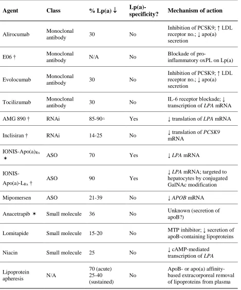
Table 1.1. Therapies that have been shown to reduce plasma Lp(a) concentrations. ... 27
Table 2.1. Primer pairs used for KIV10 sequencing and mutagenesis reactions. ... 32
Table 3.1. Selected genes differentially expressed in THP-1 macrophages in response to the
presence of the KIV10 covalent oxPL addition to apo(a). ... 67
Table 3.2. Top 15 enriched GO terms in THP-1 macrophages treated with 17K apo(a)
compared to those treated with 17K apo(a) + 200 mM -ACA. ... 69
x
List of Figures
Figure 1.1. Schematic representation of the Lp(a) particle. ... 3
Figure 1.2. Electron micrographs of LDL and Lp(a). ... 4
Figure 1.3. Schematic representation of apo(a). ... 5
Figure 1.4. Crystal structure of KIV10. ... 8
Figure 1.5. Proposed two-step model for Lp(a) formation. ... 13
Figure 1.6. Effects of Lp(a)/apo(a) on vascular cell phenotypes. ... 22
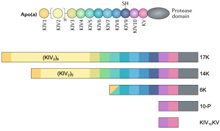
Figure 2.1. Schematic representations of recombinant apo(a) species. ... 31
Figure 3.1. HEK293 17K H33A cells exhibit abundant apo(a) content. ... 44
Figure 3.2. 17K H33A is not secreted, and is found at a reduced molecular weight in cell lysates. ... 45
Figure 3.3. 10-P apo(a) variants with substitutions to KIV10 His33 are not secreted. ... 47
Figure 3.4. 6K H33N apo(a) is not secreted by HEK293 or HepG2 cells. ... 49
Figure 3.5. 17K H33A apo(a) exhibits increased co-localization with calnexin compared with wildtype 17K. ... 52
Figure 3.6. 6K R10Q apo(a) is not secreted by HEK293 or HepG2 cells. ... 54
Figure 3.7. R10Q and R10A variants of 14K apo(a) are not secreted by HEK293 or HepG2 cells. ... 56
Figure 3.8. The KIV10KV M64T apo(a) variant binds Lysine Sepharose® in a comparable manner to the Met64 variant. ... 58
xi
Figure 3.10. Relative oxPL signal abundance determined by E06 immunoblot densitometry
of apo(a) KIV10KV Met64 and M64T variants... 61
Figure 3.11. Experimental design of functional assay used to generate RNA for RNA-Seq analysis. ... 64
Figure 3.12. t-SNE plot of biological replicates for RNA sequencing samples. ... 65
Figure 3.13. Subset of genes which are differentially regulated by apo(a) dependent on the
KIV10 sLBS and covalent oxPL, rationalized by a potential role in the pathology of
inflammation and atherosclerosis. ... 73
Figure 4.1. Molecular modeling overlay of Met64 (blue) and Thr64 (orange) variants of
xii
List of Abbreviations
17K, 14K, 6K, 10-P, KIV10KV – defined graphically in Figure 2.1
aPES – asymmetric polyethersulfone
Apo(a) – apolipoprotein(a)
ApoB-100 – apolipoproteinB-100
ASO – antisense oligonucleotide
BCA – bicinchoninic acid
CAD – coronary artery disease
CE – cholesteryl ester
CM – conditioned medium
COX-2 – cyclooxygenase 2 (prostaglandin-endoperoxidase synthase 2)
CVD – cardiovascular disease
DMEM/F12 – Dulbecco’s modified eagle medium nutrient mixture F-12
ECM – extracellular matrix
EEA1 – early endosome antigen 1
ELISA – enzyme-linked immunosorbent assay
ER – endoplasmic reticulum
ERAD – endoplasmic reticulum-assisted degradation
FBS – fetal bovine serum
FC – free cholesterol
FGF-19 – fibroblast growth factor 19
GM-CSF – granulocyte-macrophage colony-stimulating factor
GO – Gene Ontology
HBS – HEPES-buffered saline
HEK293 – human embryonic kidney 293 cells
HepG2 – hepatocellular carcinoma G2 cells
HoFH – homozygous familial hypercholesterolemia
HS2 – hyaluronan synthase 2
HUVEC – human umbilical vein endothelial cell
ICAM-1 – intercellular cell adhesion molecule 1 IFN- - interferon-
JAML – junctional adhesion molecule-like\
xiii
KIV – kringle IV
KV – kringle V
LA – lipoprotein apheresis
LAMP-1 – lysosomal-associated membrane protein 1
LASX – Leica Application Suite X
LDL – low density lipoprotein
LDLR – low density lipoprotein receptor
Lp(a) – lipoprotein(a)
LRGC – London Regional Genomics Centre
M-CSF – macrophage colony-stimulating factor
MCP-1 – monocyte chemoattractant protein 1
MEM – minimum essential medium
MMP – matrix metalloproteinase
MMP13 – matrix metallopeptidase 13
NAFLD – non-alcoholic fatty liver disease
NASH – non-alcoholic steatohepatitis
oxLDL – oxidized LDL
oxPL – oxidized phospholipid
PARC – pulmonary activation-regulated chemokine
PBS – phosphate-buffered saline
PCSK9 – proprotein convertase subtilisin/kexin type 9
PDGF – platelet-derived growth factor
PGE2 – prostaglandin E2
PIC – protease inhibitor cocktail
PL – phospholipid
PLA2 – phospholipase A2
PLG-RKT – plasminogen-receptor KT
PMA – phorbol-12-myristrate-13-acetate
PMSF – phenylmethylsulfonyl fluoride
PVDF – polyvinylidene difluoride
RhoA – Ras homolog gene family, member A
RNAi – RNA interference
xiv
RPMI-1640 – Roswell Park Memorial Institute
SDS-PAGE – sodium dodecyl sulfate polyacrylamide gel electrophoresis
sLBS – strong lysine binding site
SMC – smooth muscle cell
SNP – single nucleotide polymorphism
SREBP – sterol regulatory element-binding protein
t-SNE – t-distributed stochastic neighbour embedding
TBS – Tris-buffered saline
TFPI – tissue factor pathway inhibitor
TG – triglyceride
TGF- - transforming growth factor
TGN46 – trans-Golgi network integral membrane protein 2
THP-1 “macrophages” – THP-1 monocytes incubated with 100 mM PMA for 72 hours; macrophage-like cells
THP-1 monocytes – THP-1 human monocytic leukemia cells
TLR – toll-like receptor
TNF – tumor necrosis factor tPA – tissue plasminogen activator
UPR – unfolded protein response
VCAM-1 – vascular cell adhesion molecule 1
VLDL – very low density lipoprotein
VLDLR – very low density lipoprotein receptor
Chapter 1
1
Introduction
1.1
Lipoprotein(a)
1.1.1
Structure
Lipoprotein(a) (Lp(a)) is a unique class of lipoprotein found only in humans, apes, and Old
World Monkeys. Originally discovered in 1963 by Kåre Berg, Lp(a) is comprised of two
fundamental components: a lipoprotein moiety virtually indistinguishable from low density
lipoprotein (LDL), and a single molecule of apolipoprotein(a) (apo(a))1 (Figure 1.1). The
LDL-like lipoprotein component contains a cholesteryl ester- and triglyceride-rich core, a free
cholesterol/phospholipid monolayer outer shell, and a single copy of apolipoproteinB-100
(apoB-100); in the case of Lp(a), apo(a) is bound covalently to apoB-100 via a single disulfide
bond2. Under normal conditions, apo(a) maintains a lysine binding-dependent “closed”
conformation in association with the apoB-100 component of Lp(a), which can be inhibited by the addition of the lysine analog -aminocaproic acid (-ACA) to produce a “ball-and-chain”-type “open” conformation. This conformation can be visualized using electron microscopy to
demonstrate both the single site of covalent linkage between apo(a) and apoB-100, as well as
the “beads on a string” appearance of the kringle domains of apo(a)3 (Figure 1.2).
The presence of apo(a) imparts unique properties and characteristics to Lp(a) that differ from
those of LDL, largely owing to the presence and functions of multiple kringle domains in
apo(a) that are highly homologous to plasminogen kringle 4 (KIV)4 (Figure 1.3). Beginning
at the amino terminus, the KIV domains in apo(a) are classified into ten sub-types (KIV1
through KIV10), each differing from the others based on the amino acid sequence and, in turn,
dictating their characteristics and functions in the mature protein4. Each KIV subtype is present
in apo(a) in a single copy with the exception of KIV2, which appears in a variable number of
identical copies (from 3 to >35 in humans); the number of KIV2 domains is encoded at the
genomic level by the LPA gene, giving rise to extensive heterogeneity in apo(a) isoform size
in the human population2. Carboxyl-terminal to the KIV sequences, apo(a) contains single
copies of kringle V- (KV) and protease-like domains, each highly homologous to the
protease-like domain of apo(a) is enzymatically inactive5,6. The close homology between apo(a) and
plasminogen contributes to the disruption of the balance between thrombosis and fibrinolysis,
since apo(a) inhibits plasminogen activation in a number of contexts, resulting in prolonged
blood clot breakdown5.
Some specific functionalities relevant to the assembly and molecular pathology of Lp(a) are
attributable to the lysine binding capabilities of certain KIV domains in apo(a). Low-affinity,
or “weak” lysine binding sites (wLBS) are present in each of the KIV7 and KIV8 domains of
apo(a). The functionality of these wLBS are required for initial non-covalent interactions with
exposed lysine residues in the amino-terminal globular domain of apoB-100, thereby
facilitating the first stage of assembly of the Lp(a) holoparticle6–9. This lysine
binding-dependent non-covalent association is followed by the formation of a single covalent disulfide
bond between Cys4057 of apo(a)10, found in the KIV
9 domain, and a still-debated cysteine
residue in apoB-100. Guevara et al. demonstrated Cys3734 as the apoB-100 cysteine residue in
question using molecular modelling and fluorescent labelling of potential candidate residues11,
while Callow et al. posit that Cys4326 is the residue involved in disulfide bond formation with
apo(a) as determined using mutagenesis12.
Finally, the KIV10 domain of apo(a) possesses two characteristics that make it unique with
respect to the other KIV domains, and of most relevance to this thesis: a high-affinity, “strong”
lysine binding site (sLBS), and an amino acid residue to which an oxidized phospholipid
(oxPL) is covalently bound13,14. The covalent oxPL modification of apo(a) has been determined
empirically to absolutely require the functionality of the sLBS of KIV10 for addition. The
proposed roles for the sLBS and covalent oxPL are described in in detail Section 1.1.6.
In addition to the unique covalently-bound oxPL, Lp(a) has been shown to be the preferential
carrier of oxPL in the plasma over LDL, an important factor in the context of inducing
inflammatory phenotypes in vascular cells and subsequent development of cardiovascular
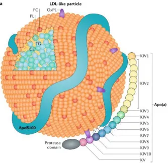
Figure 1.1. Schematic representation of the Lp(a) particle.
Lp(a) is a unique lipoprotein owing to its inclusion of apolipoprotein(a). Sharing many
characteristics with LDL, Lp(a) possesses a lipoprotein core comprised of triglycerides (TG)
and cholesteryl esters (CE), surrounded by a monolayer of phospholipids (PL) and unesterified
free cholesterol (FC), all associated with a single copy of apoB-100. In addition to its bulky
lipoprotein core, Lp(a) possesses a single copy of apo(a) covalently bound to apoB-100 by a
disulfide bridge involving the single free cysteine residue present in the apo(a) KIV9 domain.
Apo(a) contains ten kringle domains homologous to plasminogen KIV, each present in a single
copy with the exception of KIV2, which varies from 3 to >35 identical repeats corresponding
to the number of KIV2 sequences encoded in the LPA gene, and determines the overall size of
the apo(a) molecule. The specific characteristics and functions of the various kringle domains
comprising apo(a) are described in Section 1.1.2. Also shown are oxidized phospholipids
(OxPL) associated with the lipid core as well as covalently bound to KIV10. [Boffa &
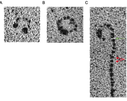
Figure 1.2. Electron micrographs of LDL and Lp(a).
LDL can be visualized using electron microscopy as (A) a tightly clustered lipoprotein particle,
exhibiting regions of high electron density corresponding to its apoB-100 component. Lp(a)
exhibits lysine-dependent interactions between apo(a) and apoB-100, similarly yielding (B) a
tightly clustered lipoprotein particle with more electron-dense regions compared with LDL due
to the presence of apo(a), or (C) as an “open” conformation with a “ball-and-chain” appearance
upon the addition of the lysine analog -ACA. In the open conformation, individual kringle
domains of apo(a) can be visualized as “beads on a string” (red arrows), and the single covalent
bond involving KIV9 near the C-terminus of apo(a) can be inferred (green arrow). [Weisel et
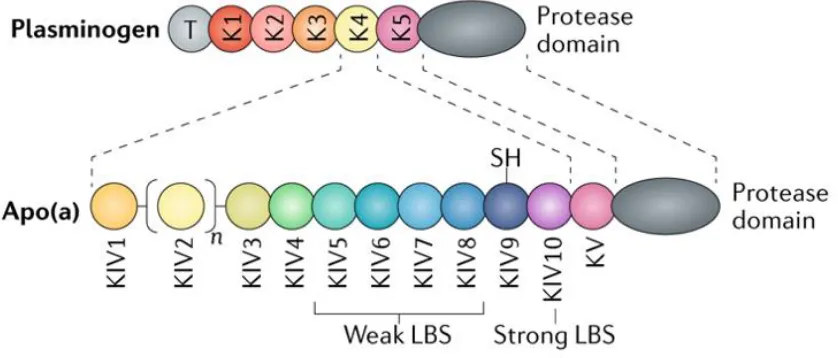
Figure 1.3. Schematic representation of apo(a).
Apo(a) strongly resembles plasminogen, sharing substantial sequence and structural
homology. Both apo(a) and plasminogen contain multiple kringle domains; the kringles that
make up apo(a) include ten unique subtypes sharing extensive sequence identity with
plasminogen K4 (KIV1-KIV10), each found in a single copy with the exception of KIV2, which
is a repeating domain ranging from 3 to >35 identical copies. In addition to its KIV domains,
apo(a) also possesses domains similar in sequence to the plasminogen K5 and protease
domains; in apo(a), the protease domain is not enzymatically active. Apo(a) lacks sequences
corresponding to the plasminogen tail, K1, K2, and K3 domains. Certain apo(a) KIV domains
possess characteristics that are important for the assembly and pathogenicity of Lp(a): the weak
lysine binding sites found in KIV7 and KIV8 are essential for initial non-covalent interaction
between apo(a) and apoB-100, the unpaired Cys4057 found in KIV9 is required for covalent
linkage to apoB-100, and KIV10 contains both a strong lysine binding site (strong LBS) as well
as a covalently-bound oxPL. Full function of the strong lysine binding site found in KIV10 is
1.1.2
Apolipoprotein(a)
Apo(a) distinguishes Lp(a) from LDL both structurally and functionally, imparting Lp(a) with
unique characteristics. Apo(a) is approximately 28% carbohydrate by weight16. As previously
stated, apo(a) is composed of repeating domains homologous to the KIV domain of
plasminogen. In fact, it has been determined that the existence of LPA in humans, apes, and
Old World Monkeys, is the result of a gene duplication of PLG, and that gene divergence of
what we now know as LPA and its parent gene PLG occurred relatively recently during primate
evolution (approximately 40 million years ago)4,17. The kringle domains found in apo(a) and
plasminogen are so named for their physical resemblance to a traditional Danish pastry of the
same name. Kringles form a characteristic tri-looped structure composed of about 80 amino
acids, wherein three invariant disulfide bonds are formed between the six invariant cysteine
residues present in each kringle (Figure 1.4). KIV9 is the only domain possessing a seventh
cysteine; nascently unpaired, this cysteine is the residue through which apo(a) links covalently
to apoB-100 in the covalent assembly of the Lp(a) holoparticle10. Between each apo(a) kringle
domain, there is an inter-kringle linker region of approximately 30 amino acids, rich in serine
and threonine residues and thus representing the major proposed sites of O-linked
glycosylation18. Each KIV domain also possesses at least one site for N-linked glycosylation19.
Kringle domains are largely considered to serve as protein-binding domains, suggesting that
the presence of apo(a) on Lp(a) allows interactions with a variety of ligands with which LDL
is unable to interact.
Due to its KIV10 lysine-binding sites and covalently bound oxPL, it has been hypothesized that
apo(a) confers pathogenic potential to Lp(a) beyond that which can be attributed to LDL. The
lysine-binding ability of apo(a) allows it to bind exposed lysine residues on a wide variety of
ligands, including fibrin and cell-surface receptors20–22. The previously mentioned zymogen,
plasminogen, also bears lysine-binding kringle domains, the binding interactions of which
facilitate its cleavage and activation to the fibrinolytic enzyme plasmin which, in turn,
promotes the dissolution of fibrin clots23,24. The presence of apo(a) on Lp(a) slows the action
of plasmin by inhibiting plasminogen cleavage in a number of contexts25,26, thereby evoking a
net anti-fibrinolytic/pro-thrombotic effect. A second mechanism by which apo(a) slows the
formation of a plasminogen-tissue plasminogen activator (tPA)-fibrin-apo(a) complex which
exhibits a reduced rate of plasmin activation compared with the same complex lacking apo(a)5.
The covalently bound oxPL of KIV10 is implicated in stimulating the production of a variety
of pathogenic stimuli, as oxidized lipids have been shown to induce inflammatory processes
in vascular cell types14,27–30. Plasma oxPL/apoB-100 ratio has a strong correlation with Lp(a)
concentration in humans, and further, oxPL/apoB-100 has been found to correlate with the
presence and extent of coronary artery disease (CAD)31,32. Elevated oxPL/apoB-100 has also
been shown to correlate with prevalence of carotid and femoral artery diseases33. The covalent
oxPL moiety of apo(a) has been shown to induce apoptosis in endoplasmic reticulum
(ER)-stressed macrophages in a CD36- and toll-like receptor 2 (TLR2)-dependent manner34, a key
step in the formation of the necrotic core in atherosclerotic lesions. Based on this information,
a strong role for apo(a), and Lp(a) by extension, is suggested in the initiation of endothelial
dysfunction, vascular cell inflammation, and necrotic core formation – key processes in the
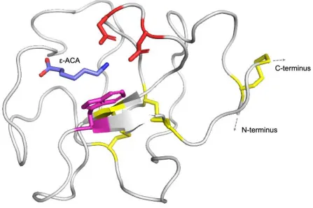
Figure 1.4. Crystal structure of KIV10.
Kringle domains are found in both plasminogen and apo(a), as well as other proteins involved
in thrombotic and fibrinolytic processes. Kringle domains possess a hallmark tri-looped
structure stabilized by three invariant disulfide linkages, shown in yellow. Kringles represent
important domains for protein-ligand interactions; KIV10 possesses a strong lysine binding site
which facilitates interactions between apo(a) and fibrin, cell surface receptors, and other
ligands. The lysine analog -ACA is shown as the bound ligand. Two aspartic acid residues
(red) and one tryptophan residue (magenta) that are crucial to the function of the sLBS are
shown. Figure generated using PyMOL version 2.3.2, pdb: 3KIV, produced by Mochalkin et
1.1.3
Lp(a) metabolism
1.1.3.1
Apo(a) secretion
Up to 90% of the variability seen in human plasma Lp(a) concentrations can be attributed to
LPA36, the gene encoding apo(a), which is found on human chromosome 6q25.3-q264. The size
of the LPA gene itself varies widely within the population, reflecting differences in the number
of KIV2-encoding sequences present. In general, an inverse correlation exists between the
isoform size encoded by a given LPA allele and the corresponding plasma Lp(a) concentration
observed7,37. It is believed that more time is required for cells to process larger apo(a) isoforms
due to the greater total number of kringles present, the complex folding required for each, and
extensive post-translational modification in the form of N- and O-linked glycosylation. This
extended processing stage results in longer periods of ER retention and subsequent
ER-associated degradation, the end result of which is reduced levels of secreted protein species
and circulating Lp(a)38–40. In light of this, it is important to note that Lp(a) levels are generally
agreed to be determined primarily at the level of what can broadly be termed “production”,
rather than uptake and catabolism of the particle from circulation. In vivo kinetic studies in
humans have ascribed differences in apo(a) production rates for different isoform sizes
(slowest production rates for largest isoform sizes), while failing to show such a relationship
for fractional catabolic rates41.
Biosynthesis of Lp(a) begins in a similar fashion to many secreted proteins: transcription of
the gene, in this case LPA, and subsequent apo(a) protein translation/ER translocation,
processing, and passage through the constitutive secretory pathway. Several
transcription-regulating elements have been identified for LPA, including an inductive interleukin (IL)-6
response element in the LPA promoter region42, a repressive DR-1 promoter element which is
bound by the bile acid-bound farnesoid X receptor (FXR)43, an Ets motif that has been shown
to mediate repression of LPA transcription by fibroblast growth factor 19 (FGF-19) via Elk-1
binding44, and several cAMP response elements thought to play a role in niacin-mediated Lp(a)
reduction45. However, the transcriptional control of LPA is not fully understood, with evidence
suggesting a gene-repressing role for estrogens based on studies in men undergoing estrogen
therapy for prostate cancer46, as well as post-menopausal women receiving estrogen
replacement47. Finally, apo(a) secretion is intrinsically regulated by intracellular processing
retention time observed in apo(a) isoforms of increasing size results in increased proteasomal
degradation and, accordingly, reduced secretion efficiency39.
With respect to apo(a) secretion, the importance of the processes underlying general protein
processing and quality control must be considered thoroughly. Classically, the process of
protein secretion involves unidirectional movement of proteins of interest through the ER, the
Golgi apparatus, secretory vesicles, and finally culminates in their extracellular release. At the
same time, quality control processes typically ensure only properly folded proteins are
secreted, targeting misfolded proteins for degradation. Most secreted proteins possess an
N-terminal signal sequence that is recognized by cytosolic targeting factors and ER-resident
translocation machinery48, which directs the translation of that protein/peptide to occur through
an ER-embedded Sec61 pore complex following association with the cytosolic ER
membrane49. The signal recognition particle, a multimeric complex comprised of both protein
and RNA components50, mediates the targeting of signal peptide-containing polypeptides to
the ER, whilst simultaneously slowing protein translation to prevent premature cytoplasmic
protein folding that would preclude ER translocation51. The N-terminal end of the nascent
polypeptide is first inserted into the ER, where a protease complex, usually signal peptidase,
cleaves the signal peptide52. Protein translation directly into the lumen of the ER then follows,
and ER-based quality control of protein folding and post-translational modification occurs.
During translation of the protein into the lumen of the ER, sequence recognition by the
oligosaccharyltransferase complex occurs, during which N-linked Glc3Man9GlcNAc2 glycan
motifs are added to Asn in the Asn-X-Thr/Ser consensus sequence53; as mentioned previously,
each kringle domain of apo(a) possesses at least one such N-glycan attachment site, making
this ER-localized stage of protein modification important in the processing of apo(a). It has
been demonstrated that the addition and trimming of N-linked glycans is important for the
subsequent folding of a great number of secreted protein species, including apo(a)39,54,55. This
folding process is facilitated by a number of catalytic isomerases (e.g. protein disulfide
isomerase), glycosidases, molecular chaperones (e.g. calnexin), and by the oxidizing
environment of the ER which favors disulfide bond formation. In the case of properly folded
and modified proteins, normal progression to the Golgi apparatus occurs by way of
COPII-coated vesicular budding56,57. However, in cases of misfolded or improperly processed
recognizes these proteins and exports them for cytosolic degradation by the proteasome58–60,
blocking their passage to the Golgi.
With respect to the processing and secretion of apo(a), three key studies in cultured baboon
hepatocytes are particularly relevant in the context of this thesis. First, a radioactive
pulse-chase study revealed that two distinct sizes of apo(a) could be isolated simultaneously from
cell lysates: both a mature, glycosylated form of apo(a) and an immature hypo-glycosylated
form of apo(a). It is crucial to note that only the mature, fully glycosylated form of the protein
was secreted by the cell61. The second study revealed that tunicamycin, an indirect inhibitor of
N-linked glycosylation, prevented the maturation and secretion of apo(a), and that
castanospermine, an inhibitor of N-linked glycan trimming, produced a similar effect39. It
should be noted that this effect was not observed previously in HEK293 cells stably expressing
apo(a)62. A third study identified variants of apo(a) that could be isolated from cell lysates but
were not secreted; it was determined using immunoprecipitation coupled with pulse-chase
analysis and endoglycosidase digests that these apo(a) species were being retained in the ER40.
Taken together, these data strongly indicate that proper folding as well as N-linked
glycosylation are both critical for the secretion of apo(a).
1.1.3.2
Lp(a) assembly
Biosynthesis of Lp(a) not only encompasses apo(a) secretion, but also the assembly of the
particle, first by initial lysine-dependent non-covalent interaction between apo(a) and
apoB-100, and followed by the covalent linkage of apo(a) to apoB-100 (Figure 1.5). Until recently,
it has been generally accepted that Lp(a) assembly occurs exclusively extracellularly, from
newly-secreted apo(a) and either newly-synthesized or circulating LDL; in both cases, the
secretion of the apoB-100-containing species is proposed to be unlinked to that of apo(a). This
model for Lp(a) assembly has come under scrutiny after an in vivo kinetic study in humans
showed that the production rate of Lp(a)-associated apoB-100 is quite different than the
production rate of apoB-100 found in other lipoprotein species, but matches very closely with
the production rate of apo(a)63, suggesting a yet uncharacterized relationship between the
secretion of apo(a) and Lp(a)-apoB-100. These findings indicate the possibility of intracellular
Lp(a) assembly, but the existence of an Lp(a)-destined apoB-100-containing lipoprotein pool
interaction, an in vitro model of Lp(a) assembly has demonstrated the extracellular activity of
an Lp(a)-specific oxidase enzyme involved in catalyzing the formation of the covalent bond
between Cys4057 of apo(a) and Cys3734 or Cys4326 of apoB-10064. However, the identity of this
enzyme and its mechanism of action have not been fully determined. When hypothesizing
about the secretion and assembly processes that comprise Lp(a) formation, we must also
consider the fact that many therapies known to reduce circulating LDL levels (e.g.
mipomersen, lomitapide, anacetrapib, and proprotein convertase subtilisin/kexin type 9
(PCSK9) inhibitors) also reduce Lp(a) levels65–70. While much is still unknown about the
specifics of Lp(a) assembly in the context of apo(a) secretion and its relationship with
apoB-100, it is known that free apo(a) is not simply secreted into the plasma prior to association with
an LDL particle; it has been demonstrated that apo(a) species with nonsense mutations
N-terminal to Cys4057 (and thereby lacking the ability to form the covalent link to apoB-100
required for Lp(a) assembly) experience rapid degradation in the plasma71. Free apo(a) is
detectable at very low levels in the plasma, but the vast majority is present in Lp(a) particles72.
1.1.3.3
Catabolism
Further complicating our incomplete understanding of Lp(a) metabolism, a robust
understanding of Lp(a) catabolism remains elusive. Although no definitive receptor for Lp(a)
has been identified, it has been established that the liver is the organ for Lp(a) catabolism,
while also representing the dominant source of apo(a) secretion. Roles in Lp(a) internalization
have been described for a number of receptors including the following: the LDL receptor
(LDLR)73,74 and LDLR family (very low density lipoprotein receptor (VLDLR), LDLR-related
protein 1, and gp330/megalin)75–77, the plasminogen receptor family (PLG-R
KT)78, and
scavenger receptor superfamily (scavenger receptor B1)79. The relative contribution of each
receptor type to the overall internalization and catabolism of Lp(a) has not been determined.
The kidney has also been identified as a minor potential contributor to Lp(a) catabolism owing
to its expression of the VLDLR and/or gp330/megalin80, but no renal Lp(a) uptake studies have
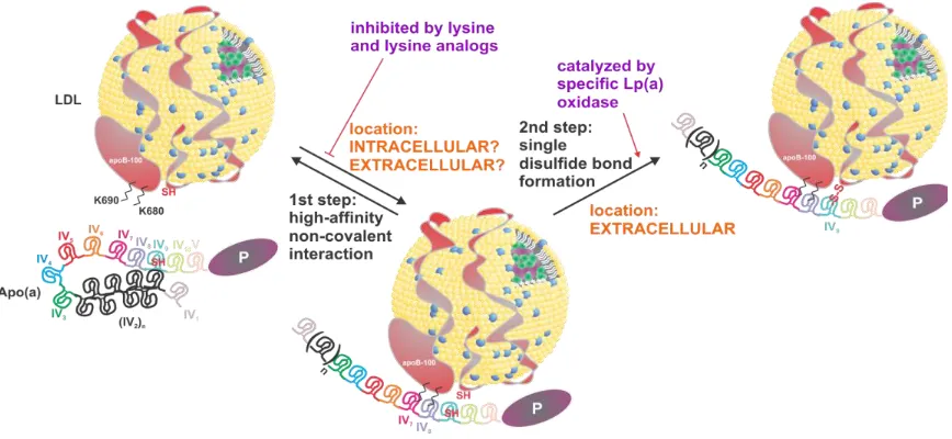
Figure 1.5. Proposed two-step model for Lp(a) formation.
The current understanding of Lp(a) formation involves a two-step mechanism. In the first step,
apo(a) associates non-covalently with free lysine residues in the N-terminal globular domain
of apoB-100 via weak lysine binding sites in the apo(a) KIV7 and KIV8 domains. The precise
location of initial non-covalent interaction between the two species is unclear; evidence has
been presented to suggest that both intracellular and extracellular association are possible.
Additionally, it has been demonstrated that the rate of Lp(a)-apoB-100 secretion closely
matches that of apo(a). Regardless of the location of this interaction, it has been shown that it
can be competitively inhibited with the addition of lysine analogs such as -ACA. The second
step of Lp(a) assembly is the formation of a single disulfide bond between Cys4057 of apo(a)
and either Cys3734 or Cys4326 (current findings indeterminate)11,12 of apoB-100. This bond
1.1.4
Determinants of plasma Lp(a) concentration
Plasma concentrations of Lp(a) vary widely within the population, from levels that are
undetectable to those in excess of 250 nanomolar. It is well established that plasma Lp(a)
concentrations are determined predominantly at the stage of production, and that there is a
general inverse relationship between concentration of Lp(a) and the size of the LPA gene
encoding apo(a). The determinants that collectively dictate the level of Lp(a) in the plasma are
LPA transcription, apo(a) secretion, assembly of Lp(a), and its catabolism.
While LPA gene size is the major genetic determinant of apo(a) production and Lp(a)
concentration, the general inverse relationship between the two only accounts for about 70%
of the observed variation in plasma Lp(a) concentrations. Approximately 22% of this variation
has been attributed to cis-acting sequences in the gene whose influences have not been fully
characterized36. There is evidence to suggest that the size of a given LPA allele affects not only
the plasma concentration of Lp(a) with the corresponding isoform, but that of the second
isoform as well81,82. Furthermore, there is evidence that single nucleotide polymorphisms
(SNPs) influence plasma Lp(a) concentrations as well: rs3798220 (protease-like domain, Ile4399→Met) and rs10455872 (KIV2, intronic) are associated with elevated Lp(a) levels and
increased incidence of CVD83.
At the genomic level, several transcription factor binding sites in the LPA promoter region
have been identified as modulators of gene transcription. As mentioned previously, these
elements include those responsive to 1) IL-6, with a 5-fold enhancement in LPA reporter gene
activity reported in HepG2 cells treated with IL-642, 2) bile acids, repressing LPA transcription
via an FXR response element43, 3) FGF-19, down-regulating transcription via Elk-1 binding
to an Ets motif44, and 4) cAMP, up-regulating expression (but depleted by niacin)45. In addition
to these factors, exogenous estrogen supplementation has been shown to reduce Lp(a)
levels46,47. However, the exact role of estrogens in this context remains elusive when one
considers that there is no significant difference between plasma Lp(a) levels in healthy men
and pre-menopausal women, despite a great discrepancy in concentrations of circulating
estrogens84. Further, it has been demonstrated that Lp(a) levels are higher in post-menopausal
women than in pre- or peri-menopausal women, indicating further differences in the handling
Interestingly, it has also been established that a given LPA allele may be associated with
considerably different concentrations of Lp(a) in the plasma, especially between individuals of
different ethnic backgrounds; multiple studies have shown that African Americans have
median plasma Lp(a) concentrations significantly exceeding that of Caucasians, and that this
discrepancy is not fully accounted for by differences in isoform size distribution81,86,87.
1.1.4.1
“Null”
LPA
alleles
Only two null alleles of the LPA gene have been identified in humans – both of which result
in the generation and secretion of apo(a) species that are unable to participate in Lp(a)
assembly. As discussed previously, it has been shown that such unbound apo(a) species are
degraded rapidly. A splice site mutation (rs41272114) results in the production of an
alternately-spliced LPA transcript, the translation of which generates an apo(a) variant that is
truncated after KIV7 and prevents both non-covalent and covalent associations with
apoB-10071. This SNP is associated with reduced plasma Lp(a) concentrations and reduced CAD
risk88. The other null allele (rs# unclear, “G4925A”89) produces a nonsense mutation in the
KIV2 region; this apo(a) species is believed to be secreted, but, once again, is unable to form
Lp(a) and subsequently results in reduced plasma Lp(a) levels90. While these two null alleles
are the only confirmed variants of this nature, recent findings suggest the discovery of novel
non-synonymous mutations in LPA that represent null alleles for apo(a), by causing the
intracellular retention and degradation of the encoded apo(a) species (unpublished data
presented at European Atherosclerosis Society 87th Congress by S McCormick, Maastricht,
The Netherlands, 2019).
1.1.5
Cardiovascular disease and atherosclerosis
Cardiovascular disease (CVD) is a broad term referring to a class of diseases characterized by
dysfunction of the heart and/or vasculature, and collectively represents the leading cause of
death in the developed world91. While CVD encompasses many specific types of pathologies,
the most common is CAD, accounting for the greatest burden of mortality and morbidity of
any CVD subtype92. CAD is a progressive disease, believed to be initiated by the exposure of
the arterial endothelial lining to some form of insult. As a result, endothelial cells undergo
structural changes that compromise the integrity of tight junctions between them, increasing
lipoproteins and leukocytes, among other blood components, within the intimal compartment
of the vessel93. The initial insult to the vessel may take the form of chronically elevated
circulating lipids (dyslipidemias), exposure to compounds found in cigarette smoke, shear
stress, hyperglycemia, obesity, hypertension, or insulin resistance94. Each of the
aforementioned insults results in activation of signaling pathways that lead to the generation
of excess reactive oxygen species (ROS) and subsequently, an environment within the
endothelial cells characterized by high levels of oxidative stress95,96. High intracellular levels
of oxidative stress activate the NF-B pathway, and in turn induce the expression of genes under the control of NF-B-response elements95,96. NF-B-inducible genes encode protein
products that serve a wide range of functions including cell surface adhesion molecules (e.g.
E- and P-selectins, intercellular adhesion molecule 1 (ICAM-1), and vascular cell adhesion
molecule 1 (VCAM-1))97–100 and pro-inflammatory cytokines (e.g. interferon- (IFN-),
IL-1/, 6, and 8, tumor necrosis factor (TNF), and monocyte chemoattractant protein 1
(MCP-1))101–106, among others.
Following initial insult and ROS-induced NF-B signaling, vascular endothelial cells undergo phenotypic changes, resulting in what can broadly be termed “endothelial dysfunction”.
Endothelial dysfunction compromises the integrity of intercellular tight junctions, allowing the
aberrant efflux of pathogenic factors such as LDL and Lp(a) from the lumen of the vessel into
the intimal (sub-endothelial) layer of the vessel wall. It should be noted that upon entering the
intimal space of the vessel wall, lipoproteins may be acted upon by enzymes or encounter
oxygen radicals, leading to their oxidative modification107; the exposure of endothelial cells to
oxidized lipoproteins promotes the adhesion and extravasation of immune cell types from the
plasma by inducing further production of cell adhesion molecules29,108,109 and secretion of
pro-inflammatory factors110. Pro-inflammatory cytokines secreted by the damaged endothelial
lining serve to further immune cell convergence and invasion at the site of injury, thereby
initiating and propagating a local inflammatory response, initiating the process of
atherosclerotic plaque formation111,112.
Following the initiation phase resultant from endothelial dysfunction, intimal invasion by
immune cells such as monocytes becomes a primary factor in the advancement of local
monocytes encounter high levels of stimulating growth factors including macrophage colony
stimulating factor (M-CSF) and granulocyte-macrophage colony stimulating factor
(GM-CSF), inducing their differentiation and maturation to macrophages113,114. In vitro, M-CSF is
constitutively expressed under basal conditions by endothelial cells and smooth muscle cells
(SMCs), as well as fibroblasts115,116. On the other hand, GM-CSF expression is inducible in
vitro by TNF and IL-1 in arterial SMCs114, and by oxidized LDL (oxLDL) in endothelial
cells and macrophages in vivo117. This suggests a mechanism by which leukocyte
chemoattratants contribute to the propagation of the chronic and non-resolving inflammatory
processes ongoing in the developing lesion. As monocyte-to-macrophage differentiation
occurs, so too does the up-regulation of TLRs and scavenger receptors in these cells, mediating
cell signaling processes that lead to inflammatory cytokine secretion and lipid uptake118,119.
Macrophages in the intimal space bind and engulf oxLDL and oxidized Lp(a) via pattern
recognition receptors including the TLRs and scavenger receptors mentioned previously, along
with CD36. Macrophage uptake of oxidized lipids leads to the formation of lipid-laden foam
cells and stimulates their secretion of pro-inflammatory factors119–122. These factors propagate
further inflammatory responses in vascular cell types while continued uptake of lipoproteins
triggers ER stress-mediated apoptotic processes in foam cells, and lead to the formation of the
necrotic core in the atherosclerotic plaque34,123. In addition to the aforementioned processes,
cytokines secreted by activated macrophages induce local migration and proliferation of
SMCs111 and contribute to downstream platelet aggregation124; stimulation of both endothelial
cell and SMC contraction, migration, and proliferation has also been observed in direct
response to apo(a) exposure in an V3 integrin- and RhoA/Rho kinase-dependent
fashion125,126. Platelet aggregation and subsequent activation, mediated by blood-borne von
Willebrand factor, promotes the secretion of platelet-derived growth factor (PDGF) and transforming growth factor (TGF-), propagating SMC migration and proliferation, as well
as synthesis and secretion of extracellular matrix (ECM) components by SMCs,
respectively127–130. The combination of SMC proliferation and migration, coupled with ECM
deposition, leads to the formation of the lesion’s fibrous cap131, while the contractile response
of SMCs to Lp(a) exposure is thought to reduce plaque stability. Overall, the summation of
these processes represents the cyclic nature of both the formation as well as the progression of
As the deposition of arterial plaques continues over the span of years to decades, blood vessel
occlusion gradually obstructs blood flow. In the case of plaque formation in the coronary
arteries, (i.e. CAD), blood flow to the myocardium itself becomes reduced and presents
symptomatically with feelings of tightness or pain in the chest, clinically termed angina132.
Over time, chronic exposure of macrophages, endothelial cells, and SMCs to inflammatory factors such as TNF and decreased nitric oxide levels promotes their secretion of collagenases
and matrix metalloproteinases (MMPs)133,134. Without intervention, the plaque continues to
grow and the stability of the fibrous cap may be compromised by MMP-mediated ECM
degradation, reducing the overall stability of the plaque and increasing its risk of rupture. If
plaque rupture occurs, exposure of blood to the components of the plaque precipitates the
formation of a thrombus which may occlude coronary artery flow completely, resulting in
ischemia of the myocardium and subsequent myocardial infarction131. Plaque rupture in other
vessels can lead to other major events such as ischemic stroke. Recalling the anti-fibrinolytic
effect of Lp(a)/apo(a) as an inhibitor of plasmin formation discussed briefly in Section 1.1.2,
it is reasonable to speculate that plasma Lp(a) concentration may be a contributing factor in
determining the prevalence and severity of cardiovascular events following plaque rupture.
1.1.6
Pathophysiology of Lp(a)
The structural duality of the Lp(a) particle, containing both an LDL-like bulky lipid moiety
and a plasminogen-like apo(a) molecule, suggests that Lp(a) may participate in both
atherogenic and anti-fibrinolytic processes in the vasculature. The potential effects of the
KIV10 sLBS and covalent oxPL on macrophages, endothelial cells, and SMCs are summarized
in Figure 1.6.
1.1.6.1
Oxidized phospholipid modification of Lp(a)
Oxidized lipoproteins are important drivers of the inflammatory processes involved in the
progression of atherosclerosis. In this context, the determination that Lp(a) is the preferential
carrier of oxPL in the plasma, compared to LDL, becomes even more relevant. In a study of
all lipoprotein-associated oxPL, it was determined that Lp(a) was found to contain
approximately 85% of the total quantified oxPL135. In fact, it has even been shown in vitro that
preferential oxPL enrichment of Lp(a), compared to LDL, increases upon oxLDL incubation,
non-covalently associated oxPL, Lp(a) is unique in that it also contains a non-covalently-bound oxPL.
Originally believed to be present within the KV domain136, it has since been determined that
the oxPL is covalently attached within the apo(a) KIV10 domain13. Interestingly, as discussed
previously, this domain also contains a sLBS. The proximity of these two unique features has
led to speculation that the sLBS, known to mediate the interaction of Lp(a) with a variety of
ligands, may also serve to bring the covalent oxPL in close proximity to these ligands and, in
doing so, facilitate the pro-inflammatory and/or oxidative effects associated with exposure to
oxPLs. The role of oxPLs in the facilitation of inflammatory processes in the context of
atherosclerosis is well understood: it has been shown that exposure of leukocytes to oxPLs
activates a variety of inflammatory responses, namely the secretion of
pro-inflammatory/chemoattractant cytokines, and macrophage apoptosis137,138. OxPLs have been
shown to induce transcription of CXCL8 and secretion of its cytokine product, IL-8, as well as
MCP-1 in endothelial cells in vitro139. Strong evidence for the role of oxPLs in the development
of atherosclerosis in vivo was published recently, showing that mice overexpressing a single
chain variable fragment transgene of the anti-oxPL E06 IgM (E06-scFv) exhibited significantly
slower progression of atherosclerosis compared with non-transgenic mice on an identical high
cholesterol diet140.
With respect to Lp(a) and apo(a), it has been shown that Lp(a)/apo(a) induce apoptosis in
ER-stressed macrophages in an oxPL-dependent manner through the TLR2-CD36 heterodimer;
removal of the covalent oxPL from apo(a) with phospholipase A2 (PLA2) destroyed its ability
to exert pro-apoptotic effects in this study34. Furthermore, it has been shown that THP-1
macrophages increase transcription of CXCL8 and secretion of IL-8 in response to apo(a)
exposure, and that these effects are greatly diminished by PLA2-mediated oxPL removal14.
Taken together, these data strongly implicate a role for Lp(a)/apo(a)-bound oxPL in the
induction and propagation of vascular inflammation, the promotion of macrophage/foam cell
apoptosis, and the overall development of atherosclerosis.
1.1.6.2
Functional significance of the lysine binding properties of
apo(a) KIV
10Apo(a) has been shown in vitro to stimulate the RhoA/Rho kinase signaling cascade in
pathway is increased stress fiber formation, endothelial cell contraction, and increased
permeability of the vessel wall. Also through the RhoA/Rho kinase pathway, apo(a) has been
demonstrated to stimulate vascular SMC migration, proliferation, and chemorepulsion125,142.
Additionally, the lysine binding activity of apo(a) has been implicated in vitro in the disruption of VE-cadherin/-catenin complexes, increased nuclear -catenin translocation, and the
subsequent stimulation of prostaglandin synthesis through cyclooxygenase 2 (COX-2)
up-regulation in human umbilical vein endothelial cells (HUVECs)110. Lastly, it has been shown
that apo(a) can inhibit tPA-mediated plasminogen cleavage on the cell surface of endothelial
cells, monocytes, and macrophages in vitro26, and within fibrin clots5,143. The cell-surface
effect was found to be related to the sLBS function of KIV10, and was diminished with the
addition of the lysine analog -ACA, strongly indicating a role for the lysine-binding properties
of apo(a) in the context of inhibiting plasmin activation.
1.1.6.3
The role of Lp(a) in thrombosis and fibrinolysis
A growing body of evidence from in vitro studies indicates pro-thrombotic/anti-fibrinolytic
roles for Lp(a) and apo(a). The striking homology between apo(a) and plasminogen coupled
with the lack of apo(a) protease activity suggests a role for apo(a) as an anti-fibrinolytic factor,
owing to its ability to act as a inhibitor of plasmin activation by preventing the binding of
plasminogen to surface receptors on endothelial cells and monocytes, and by inhibiting
tPA-mediated plasminogen cleavage on the cell surface26. Apo(a) slows plasmin formation by
inhibiting the conversion of Glu1-plasminogen to the more readily activated Lys77
-plasminogen, thereby inhibiting the feed-forward mechanism by which maximal plasmin
activation is achieved143,144. In addition to the sLBS function of KIV
10, this process also
requires sequences within the KIV5-8 and KV domains of apo(a)145,146. The apo(a) component
of Lp(a) has also been shown to participate in the formation of a
plasminogen-tPA-fibrin-apo(a) complex, which exhibits a reduced rate of plasmin activation when compared with the
same complex lacking apo(a)5. Inhibition of tPA-mediated plasmin activation by apo(a) has
also been shown to slow thrombolysis in vivo, using both transgenic apo(a) mouse and rabbit
jugular vein fibrinolysis models147,148. Beyond the inhibition of plasminogen cleavage by
apo(a), Lp(a) has also been shown to increase synthesis of plasminogen activation inhibitors
by endothelial cells and monocytes in vitro, providing another mechanism by which Lp(a) can
Finally, Lp(a) has also been implicated as a pro-thrombotic contributor due to its ability to bind
tissue factor pathway inhibitor (TFPI), suppressing the anti-coagulant effects that TFPI
normally exerts as a result upon binding to Factor Xa and the tissue factor/Factor VIIa
complex151. As TFPI normally acts as a potent inhibitor of the tissue factor-initiated
coagulation cascade, binding and inhibition of TFPI by Lp(a) supports a role for Lp(a) as a
pro-thrombotic factor in hemostasis.
Taken together, these data along with those presented in Sections 1.1.6.1 and 1.1.6.2 strongly
implicate mechanistic roles for Lp(a)/apo(a) in both inflammatory and pro-thrombotic
processes, but there remains debate as to the role of elevated Lp(a) concentrations in
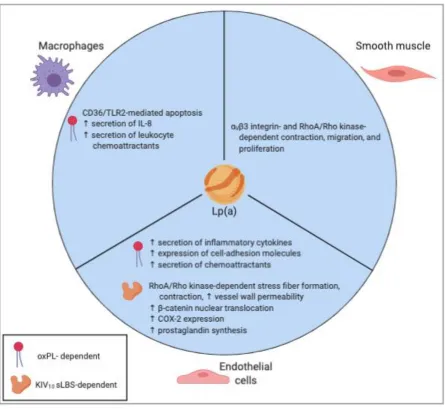
Figure 1.6. Effects of Lp(a)/apo(a) on vascular cell phenotypes.
The deleterious effects of Lp(a) and/or apo(a) on a variety of vascular cell types are well
documented. These effects importantly include increasing the secretion of IL-8 via NF-B
signaling in macrophages, increasing contraction, migration, and proliferation in smooth
muscle cells dependent on V3 integrin and RhoA/Rho kinase signaling, and increasing
prostaglandin synthesis in endothelial cells downstream of a sLBS-dependent increase in
nuclear translocation of -catenin and subsequent COX-2 up-regulation. TLR2, toll-like
receptor 2; RhoA, Ras homolog gene family, member A; COX-2,
1.1.7
Lp(a) as a risk factor for CVD
Since its discovery over half a century ago, assessing the role of Lp(a) in the development of
CVD in vivo remains a daunting challenge. The LPA gene, and therefore Lp(a) itself, exists
only in humans, apes, and Old World Monkeys. As such, the in vivo study of Lp(a) is severely
hindered by the unavailability of relevant animal models in determining the role of elevated
Lp(a) in CVD. For example, to study Lp(a) in a murine model of disease, mice must 1) be
transgenic for human apo(a), 2) produce human apoB-100 as opposed to murine apoB-48, and
3) be bred onto an atherogenic background i.e. Ldlr-/- or Apoe-/-.
Assessment of a 58,000-subject meta-analysis of 40 studies showed that individuals with <22
total KIV repeats exhibited increased CAD risk in excess of 2-fold compared to individuals
with >22 KIV153; the effect of apo(a) isoform size itself on pathogenicity has not been
definitively resolved. It is difficult to assess whether these findings are a true representation of
the effects of apo(a) isoform size rather than Lp(a) concentration, as many of the studies
involved in this meta-analysis did not control for Lp(a) concentration. While studies such as
this have reported Lp(a) as a risk factor for CVD for decades, distinguishing Lp(a) as a causal
risk factor versus a disease marker had not been possible due to the study designs utilized. A
Mendelian randomization study in 2009 confirmed the inverse relationship between LPA gene
size and Lp(a) plasma levels, and also established a causal role for such “genetically elevated”
Lp(a) concentrations in the incidence of CVD154. Since then, genome-wide association studies
have also determined LPA gene size to be a major determinant of CVD risk155,156, and other
genetic studies have associated two SNPs in the LPA gene with increased plasma Lp(a)
concentration as well as increased CVD risk83. Further genetic studies have allowed us to draw
a causal relationship between elevated plasma Lp(a) levels (>50 mg/dL) and >2-fold increased
risk for CAD, and indeed that approximately 20% of the global population falls into this risk
category157. Lp(a) has also been determined to be a causal and independent risk factor for other
CVDs such as peripheral vascular disease, ischemic stroke, and calcific aortic valve
stenosis83,154,158–161.
Despite substantial evidence implicating Lp(a) as an independent and casual risk factor for
CVD, current research is still addressing a definitive resolution to the question underlying the