Copyright © 1998, American Society for Microbiology
Evaluation of Previously Assigned Antibody Concentrations in
Pneumococcal Polysaccharide Reference Serum 89SF by the
Method of Cross-Standardization
NELYDIA CONCEPCIONANDCARL E. FRASCH*
Division of Bacterial Products, Center for Biological Evaluation and Research, Bethesda, Maryland
Received 30 June 1997/Returned for modification 13 October 1997/Accepted 18 December 1997
An enzyme-linked immunosorbent assay (ELISA) and the antibody concentrations assigned to different pneumococcal capsular polysaccharide types were used to estimate concentrations of antibody to additional pneumococcal types in reference serum 89SF and to confirm assigned antibody values. This was possible because the slopes of curves of antibody binding to all polysaccharide types evaluated (1, 3, 4, 5, 6B, 7F, 9V, 14, 18C, 19F, and 23F) were similar. The point estimates for total anti-pneumococcal antibody and immuno-globulin G (IgG) antibody determined by cross-standardization by an ELISA based on use of methylated human serum albumin (mHSA) to improve the efficiency of polysaccharide binding to the ELISA plate differed by less than 40% from those reported by Quataert et al. (Clin. Diagn. Lab. Immunol. 2:590–597, 1995) for types 1, 4, 6B, 7F, 9V, 14, 18C, and 23F. However, large differences were found between the assigned values and those obtained by our mHSA ELISA for types 3 and 19F. The mHSA ELISA and the direct polysaccharide coat ELISA may not measure antibodies to the same epitopes on polysaccharides of types 3 and 19F. The functional importance of these different antibody specificities is being investigated. We have thus confirmed the assigned IgG antibody values for most types by a different method and have extended antibody assignments to several additional types.
Streptococcus pneumoniae remains the most common
bacte-rial etiology in pediatric infections. In the United States, seven types (4, 6B, 9V, 14, 18C, 19F, and 23F) are responsible for more than 80% of pneumococcal disease in young children (18). Additional types such as 1, 5, and 7F are important causes of pneumococcal infections in other countries. Two pneumo-coccal conjugate vaccines having types 4, 6B, 9V, 14, 18C, 19F, and 23F are presently in phase III efficacy trials for prevention of otitis media or invasive disease (4). Because pneumococcal conjugate vaccines prepared by different manufacturers using differing conjugation chemistries (1) are in clinical trials, direct comparison of antibody responses to these different vaccines will assist in identifying the better conjugation methodologies. Reported studies of the immune responses of infants to two different pneumococcal conjugate vaccines showed large dif-ferences in the geometric mean responses at 7 months of age (7, 15). The question is to what extent can this difference be attributed to differences in assay methods. Development of pneumococcal polysaccharide-protein conjugate vaccines for prevention of invasive disease and otitis media in young chil-dren has necessitated standardization of assay methods for estimation of pneumococcal polysaccharide (PS) antibodies. Use of a standardized antibody assay method, including use of the internationally recognized 89SF pneumococcal reference serum, will support present and future clinical trials to bring needed pneumococcal conjugate vaccines to the market.
It is important to obtain comparable pneumococcal antibody measurements in different laboratories, because this will assist in determining minimal antibody levels associated with protec-tion. In the case of Haemophilus influenzae type b,
concentra-tions of 1mg of anti-PS antibody per ml measured a few weeks after immunization correlated with long-term protection against H. influenzae type b disease (12). Studies by Landes-man and SchiffLandes-man using a radioimmunoassay suggest that antipneumococcal PS levels of 2 mg/ml are protective (9). However, the radioimmunoassay antibody estimates are al-most certainly somewhat high because of interference by an-ti-C PS antibodies, since all pneumococcal PSs are variably contaminated with the C PS, although the degree of interfer-ence in the radioimmunoassay may be less than that expected (11, 17). We do not yet know the amount of anti-PS antibody required for protection against invasive pneumococcal disease, and this should be one of the goals of the ongoing conjugate vaccine efficacy trials. Such estimates will help facilitate addi-tion of new pneumococcal types to an approved conjugate vaccine.
Antibodies to pneumococcal PSs have, until recent years, been measured by radioimmunoassaying (9). More recently, Koskela developed a more-specific pneumococcal PS enzyme-linked immunosorbent assay (ELISA) (8). In this ELISA, the individual type PSs are adsorbed directly to the plates and C PS antibodies are inhibited in each test serum by preadsorption. The Koskela assay was further refined by Quataert et al. (14). In the present communication, we describe an alternative ELISA method to estimate antipneumococcal PS antibodies based on use of methylated human serum albumin (mHSA) to facilitate better attachment of the pneumococcal PSs to the ELISA plates (2).
The present studies were conducted to confirm the values for total and immunoglobulin G (IgG) antibody assigned to lot 89S (89SF) by Quataert et al. for 11 pneumococcal polysac-charide types (14) and to investigate whether assigned anti-body concentrations in the reference serum for one type could be used to assign antibody values to additional types.
The studies reported here show that cross-standardization is a useful means to confirm antibody assignments and to provide
* Corresponding author. Mailing address: Division of Bacterial Products, Center for Biologics Evaluation and Research, 1401 Rock-ville Pike, Mailstop HFM-428, RockRock-ville, MD 20853. Phone: (301) 496-1920. Fax: (301) 402-2776. E-mail: [email protected].
199
on August 17, 2020 by guest
http://cvi.asm.org/
antibody assignments for additional types, provided the slopes of antibody-binding curves for the different types are similar.
MATERIALS AND METHODS
Reference serum 89SF and mHSA.Serum 89S was derived from a plasma pool from 17 donors who had received the licensed 23-valent pneumococcal PS vaccine. The plasma samples for the pool were obtained from George Siber, Massachusetts Public Health Biologic Laboratories, Boston, Mass. (16). The plasma pool was defibrinated and freeze-dried in 3.0-ml volumes. The serum contained 0.1% sodium azide. The present serum, 89SF, was distributed by the Food and Drug Administration and was obtained from serum pool 89S (14). Studies were done by our laboratory and by Madore (Wyeth Lederle Vaccines and Pediatrics, Rochester, N.Y.), and both showed that when 89S and 89SF were run side by side, they gave essentially identical results (10).
mHSA was used to improve the efficiency of attachment of the different pneumococcal PSs to Immulon 1 plates. To prepare mHSA, HSA was treated with methanolic HCl, replacing carboxyl groups with methyl groups and thus creating a positively charged but hydrophobic protein (2).
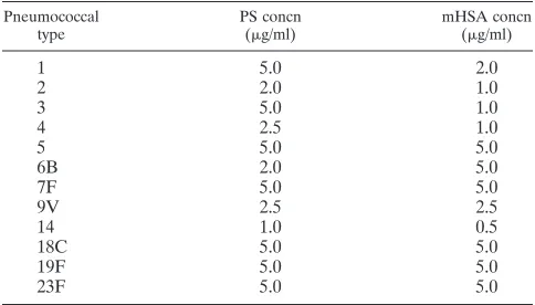
Comix mHSA ELISA method and cross-standardization.Pneumococcal PSs were obtained from the American Type Culture Collection, Rockville, Md. The optimal concentrations of PS and mHSA in mixture were determined to give the most specific binding for each Pn type (Fig. 1). The ranges were from 1 to 5mg/ml for PS and 0.5 to 5.0mg/ml of mHSA. The coating buffer was 103 phosphate-buffered saline (pH 7.4; Digene Diagnostics, Inc., Beltsville, Md.) diluted in pyrogen-free sterile water (Biofluids, Inc., Rockville, Md.). The optimal mHSA and PS concentrations for each polysaccharide type are shown in Table 1. Four different Pn types could be coated in one plate (Immulon 1 plates; Dynatech Laboratories, Chantilly, Va.). The reference serum 89SF was serially diluted
through eight dilutions beginning with 1:200 in serum/conjugate buffer contain-ing 1mg of purified C PS (State Serum Institute, Copenhagen, Denmark) per ml to inhibit C PS antibodies. Each dilution was added to PS-mHSA-coated plates in triplicate wells. The plates were developed by the addition of anti-total human immunoglobulin or anti-IgG alkaline phosphatase conjugate (Sigma Chemical Co., St. Louis, Mo.), followed by the addition of 1 mg of nitrophenol phosphate (Sigma 104 tablets) per ml. Absorbance values after 20 to 40 min were read on a Ceres 900 BioTek reader and interpolated to the value that would have occurred at 100 min, so that absorbance values from different assays could be easily compared. Dilution curves for 89SF against each of the PSs were plotted as log of optical densities versus serum dilution values.
Cross-standardization was done by the mHSA comix method. Three rows across and eight wells down on Immulon 1 plates were coated with each of the different pneumococcal type PSs. The 89SF reference serum was diluted through eight dilutions beginning at 1:200, and each dilution was added in triplicate to the coated plates. The plates were developed by the addition of anti-total immuno-globulin or anti-IgG alkaline phosphatase conjugate, followed by the addition of 1 mg of nitrophenol phosphate per ml. They were read on a Ceres 900 BioTek reader. Dilution curves for 89SF against each of the PSs were plotted as log of optical densities versus serum dilution values.
Calculation of antibody concentrations.A weighted Log-Logit computer pro-gram was used to calculate the total and IgG concentrations for each of the different PSs with the values assigned by Quataert et al. for each of the types (5). This computer program was shown to yield the same values as a standardized curve-fitting, four-parameter logistic method of calculating ELISA values devel-oped at the Centers for Disease Control in Atlanta, Ga. (6, 13). Each Pn type that has an assigned value was used as the standard to calculate the values for other types.
RESULTS AND DISCUSSION
Our studies were initiated to ascertain whether the assign-ment of antibody to one pneumococcal type could be used as a reference to estimate concentrations of antibody in the 89SF reference serum to other types, since serum 89SF was obtained from adults immunized with the licensed 23-valent pneumo-coccal PS vaccine and antibody assignments have been made for only 11 of these types (14). We chose to use the mHSA method of antigen attachment to the solid phase, because much less PS was needed for optimal PS binding compared to the direct-attachment method (8), and because we found less well-to-well variability in absorbance values between repli-cates.
The initial step in cross-standardization was to determine whether the dilution curves for antibodies against most of the types had similar slopes. The 89SF reference serum was diluted through eight twofold dilutions, starting at 1:200, and each dilution was reacted individually in triplicate against 14
differ-FIG. 1. Optimization of ELISA plate coating conditions by checkerboard titration of PS and mHSA concentrations for pneumococcal types 19F and 23F. Concentrations of 0, 1.0, 2.5, and 5.0mg of mHSA per ml comixed with each of four different PS concentrations were used. Serum 89SF was used at a dilution of 1:400. Absorbance values were read 20 to 40 min after addition of the substrate and were normalized to the values that would have occurred at 100 min.
TABLE 1. Optimal PS and mHSA concentrations for coating of Immulon I ELISA plates by the comix method Pneumococcal
type PS concn(mg/ml) mHSA concn(mg/ml)
1 5.0 2.0
2 2.0 1.0
3 5.0 1.0
4 2.5 1.0
5 5.0 5.0
6B 2.0 5.0
7F 5.0 5.0
9V 2.5 2.5
14 1.0 0.5
18C 5.0 5.0
19F 5.0 5.0
23F 5.0 5.0
200 CONCEPCION AND FRASCH CLIN. DIAGN. LAB. IMMUNOL.
on August 17, 2020 by guest
http://cvi.asm.org/
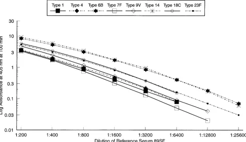
ent pneumococcal PS types by using the optimal mHSA-to-PS ratios (Table 1). The slopes of optical density versus serum dilution were essentially parallel for all serotypes, and repre-sentative types are shown in Fig. 2. The correlation coefficients for individual dilution curves with 89SF were high (r.0.98). Koskela recommended using C PS to preadsorb each serum before assaying for type-specific antibodies (8). The data shown in Fig. 3 indicate that addition of a uniform concentra-tion of 1mg of C PS per ml to the serum dilution buffer blocked binding of C PS antibodies as effectively as the previously recommended preadsorption method and support the use of a uniform 1-mg/ml concentration of inhibitor through all serum dilutions.
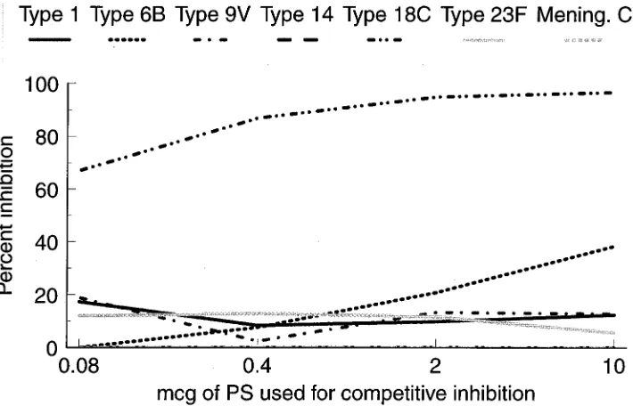
The specificity of binding of anti-PS antibodies by the comix method was shown by competitive inhibition with soluble PS inhibitors. Representative data for type 18C are shown in Fig. 4. By the comix method, all types except type 3 (see below) showed similar type specificities.
The similarity of slopes for binding of antibody (IgG and total antibody) to each of the pneumococcal PSs tested meant that the assigned antibody value for one type could be used to estimate antibody concentrations specific for each of the other types (14). For example, the assigned IgG antibody concentra-tion of 6.3mg/ml for type 1 was used to generate a Log-Logit reference curve for serum 89SF versus the type 1 PS. The ELISA absorbance values obtained for binding of IgG to each of the other 13 pneumococcal type PSs were then measured in relation to the type 1 reference curve to obtain estimates of the respective concentrations (in micrograms per milliliter) of an-tibody in the 89SF serum against each of these PSs (see row labeled type 1 in Table 2). In this example, using type 1 as the assigned reference, we obtained values of 5.8, 14.8, and 22.9mg
of IgG antibody per ml for types 4, 6B, and 14 compared to assigned values, respectively, of 4.1, 16.9, and 27.8mg/ml (Ta-ble 3). The results of a previous collaborative study indicated that an intralaboratory coefficient of variation of 620% is reasonable (6). Therefore, based on an uncertainty of620% for the assigned and standardization values, the cross-standardization estimates for types 4, 6B, and 14 were not different from the assigned values.
FIG. 2. Comparability of slopes for IgG antibodies with those for PS types used for cross-standardization. Reference serum 89SF was diluted beginning with 1:200 for each of the eight polysaccharides and used in the mHSA comix method.
FIG. 3. Comparison of two methods for blocking of C PS antibodies. Refer-ence serum 89SF was used against type 19F PS without C PS absorption (solid line), preadsorbed with 50mg of Danish C PS per ml (dotted line), without preabsorption but with 1mg of C PS per ml added to the serum dilution buffer (dashed line), or both with preadsorption and with C PS added to the serum dilution buffer (dashed and dotted line).
on August 17, 2020 by guest
http://cvi.asm.org/
The assigned values for each of the PS types were used in turn as the reference standard to estimate concentrations of antibody in serum 89SF to each of the heterologous types as described above for type 1. However, when the assigned IgG antibody concentration for type 3 was used as the reference to recalculate the standard curve, markedly lower antibody esti-mates were obtained for each of the other 13 types compared to their assigned values (Tables 2 and 3). For example, when the standard regression line was generated with the assigned type 3 value of 2.4mg of antibody per ml, the amounts of IgG antibody in 89SF against types 6B and 14 were estimated to be 4.4 and 6.9 mg/ml, respectively, compared to their assigned values of 16.9 and 27.8mg/ml. The amount of type 3 antibody estimated in this study by cross-standardization by the comix mHSA method for plate coating was 7.7mg/ml rather than the
2.4mg/ml assigned by using the method of direct PS attach-ment to the plate.
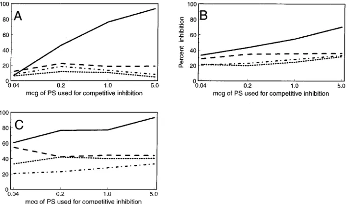
The specificity of binding of antibody to type 3 by the comix method for attachment of PS to the plate was investigated and found to be unsatisfactory due to lack of antibody specificity (Fig. 5). One microgram of the type 3 PS inhibited antibody binding by only 55%, compared to 25 to 36% for heterologous type PSs (Fig. 5B). When the type 3 PS was coated directly onto the plate as described by others (8, 14), the homologous absorption with 1 mg/ml was 77%, compared to 40 to 44% inhibition by heterologous PS types (Fig. 5C). In contrast, when the ELISA plates were precoated with mHSA and then coated with the type 3 PS, 1mg of type 3 PS inhibited by 76%, compared to less than 20% inhibition by heterologous types (Fig. 5A). When cross-standardization was repeated with type
FIG. 4. Specificity of the mHSA comix method for measurement of type 18C antibodies in reference serum 89SF.
TABLE 2. Estimation of concentrations of antibody to individual pneumococcal types in reference serum lot 89SF by cross-standardization by the mHSA comix methoda
Type used as reference standard
Concn (mg/ml) of IgG antibody in serum 89SF against the following type PSs:
1 3 4 5 6B 7F 9V 14 18C 19F 23F
1 7.6 5.8 3.5 14.8 5.3 8.3 22.9 6.8 6.7 10.6
3 2.0 1.7 1.1 4.4 1.7 2.6 6.9 2.0 2.1 3.5
4 4.7 5.6 2.6 10.0 4.1 6.1 16.7 4.8 4.9 8.8
5 11.0 13.0 10.1 23.8 9.8 13.8 40.0 11.3 11.7 17.9
6B 8.0 9.7 7.2 4.4 6.9 10.5 28.4 8.2 8.5 13.9
7F 6.2 7.8 5.4 3.5 13.2 8.9 20.9 6.2 6.4 10.6
9V 5.6 7.2 4.9 3.2 12.0 4.7 18.9 5.7 5.8 9.6
14 8.2 10.4 7.2 6.3 17.7 6.9 10.3 8.3 8.5 14.1
18C 5.1 6.6 4.3 3.0 10.9 4.1 7.7 16.5 5.2 8.4
19F 7.3 11.9 7.7 5.1 22.7 8.4 13.6 35.4 11.0 18.8
23F 5.3 7.0 4.5 3.1 10.8 4.3 8.1 17.3 5.4 5.5
Meanb 6.2 7.7 5.5 3.7 12.8 5.2 8.5 20.2 6.5 6.4 10.8
aData are for one of six assays to estimate IgG antibodies. Similar assays were done to estimate levels of total antibody to each type. Reference antibody values to
a given pneumococcal type were used.
bArithmetic means of antibody estimates obtained with individual types with their previously assigned IgG antibody concentrations (14) to generate the reference
curve. Estimates obtained with types 3, 5, and 19F as standards were excluded from the mean calculations.
202 CONCEPCION AND FRASCH CLIN. DIAGN. LAB. IMMUNOL.
on August 17, 2020 by guest
http://cvi.asm.org/
3 attached to the plate by the precoat method, the mean value for IgG anti-type 3 was 4.0 mg/ml rather than 7.7mg/ml, as shown in Table 2, but was still higher than the assigned IgG value of 2.4mg/ml (Table 3).
The mean cross-standardization values were calculated by using the means of antibody values obtained individually with types 1, 4, 6B, 7F, 9V, 14, 18C, and 23F as reference standards (Table 2). This was done because when type 3, 5, or 19F was used as the cross-standardization reference antibody, the val-ues obtained for the different types were quite different from those obtained with any of the other type PSs. Interestingly, types 5 and 19F in the mHSA comix method gave greater than
90% homologous inhibition, with almost no inhibition by het-erologous types.
The cross-standardization means for the different types were compared to the assigned IgG values (Table 3). Quataert et al. (14) found a range of assay variation for individual types in their laboratory of between 6 and 26%, with a mean variation of616%. In the mHSA ELISA, we obtained a mean coeffi-cient of variation of 14.7% for 9 different types and 13 different assays for each type. Therefore, we used 620% of the point estimates for the interlaboratory comparison of our means with assigned values. Only type 19F was outside this range for both total and IgG antibodies. Types 5 and 3 were outside the range for total antibody and for IgG, respectively. The cross-standardization procedure was repeated on six different occa-sions, and the values for all such assays for all types were within a coefficient of variation of620% of the mHSA values shown in Table 3. We conclude that the antibody assignments for the 89SF reference serum should be accepted as previously re-ported (14), except for those for types 3 and 19F, for which additional studies are needed.
The cross-standardization method was used to estimate con-centrations of IgG antibody to additional pneumococcal types for which no antibody assignments have been made (Table 4). The mean IgG antibody estimates for types 2, 9N, and 19A were 5.6, 7.9, and 11.3mg/ml, respectively.
Interestingly, the native type 3 and 19F PSs attach very poorly to the Immulon 1 plates without mHSA (see Fig. 1 for 19F). Therefore, it is possible that only a small subpopulation of type 3 or 19F PS molecules attach without mHSA and that epitopes are expressed somewhat differently depending on whether the PS is adsorbed to the plate directly or via mHSA. We have preliminary data in the case of type 19F to suggest that the mHSA and direct-binding ELISA methods preferen-tially measure antibodies to different epitopes. The mechanism of association with mHSA is primarily hydrophobic, because most of the carboxyl groups on the serum albumin are con-verted to hydrophobic methyl groups, and as reported earlier,
FIG. 5. Effect of PS coating conditions on the specificity of IgG antibodies to the type 3 pneumococcal PS in reference serum 89SF. The serum was used at a dilution of 1:200 and was examined for type specificity following attachment of type 3 PS to the plate by one of the following three methods: precoating of the ELISA plate with 5mg of mHSA per ml for 6 h and then addition of 5mg of PS per ml (A), comixing of 1mg of mHSA per ml with 5mg of PS per ml (B), and direct coating of 5mg of PS per ml without mHSA (C). In each method, the antibodies were competitively inhibited by type 3 PS (solid line), type 1 PS (dotted line), type 2 PS (dashed and dotted line), or type 4 PS (dashed line).
TABLE 3. Comparison of antibody values assigned to the 89SF reference serum with those assigned by cross-standardization by
mHSA ELISA
Pneumococcal type
Total antibody concn
(mg/ml) IgG specific-antibodyconcn (mg/ml)
Assigneda mHSAb Assigneda mHSAb
1 10.7 8.4 6.3 6.2
3 7.9 7.0 2.4 4.0c
4 7.0 8.4 4.1 5.5
5 10.0 4.1c 5.8 3.7
6B 24.3 22.0 16.9 12.8
7F 7.3 6.1 5.2 5.2
9V 10.2 8.0 6.9 8.5
14 37.0 43.2 27.8 20.2
18C 6.7 8.5 4.5 6.5
19F 18.8 8.4c 13.0 6.4c
23F 11.9 9.2 8.1 10.8
aValues assigned to 89SF serum by Quataert et al. (14) and included in the
circular sent to other laboratories with serum 89SF.
bValues were obtained by comix mHSA and cross-standardization (see Table
2 for IgG values), except for type 3, for which the mHSA precoat method was used.
cFor this value, the limits of a620% interval do not overlap for both the
assigned and the mHSA estimates.
on August 17, 2020 by guest
http://cvi.asm.org/
treatment of either meningococcal or H. influenzae type b PS with phospholipase abrogated the ability of these PSs to attach to the Immulon 1 plate through interaction with mHSA (3).
In conclusion, we have shown that a cross-standardization procedure can be used to confirm assigned antibody trations in the 89SF reference serum and to estimate concen-trations of antibody to heterologous pneumococcal PS types. This procedure is applicable to standardization of antibodies to other PS antigens.
REFERENCES
1. Anonymous. 1995. Overcoming scientific and technical barriers: pneumococ-cal vaccines, p. 8–12. In G. W. Pearson (ed.), The children’s vaccine initia-tive: continuing activities. National Academy Press, Washington, D.C. 2. Arakere, G., and C. E. Frasch. 1991. Specificity of antibodies to
O-acetyl-positive and O-acetyl-negative group C meningococcal polysaccharides in sera from vaccinees and carriers. Infect. Immun. 59:4349–4356.
3. Arakere, G., A. L. Lee, and C. E. Frasch. 1994. Involvement of phospholipid end groups of group C Neisseria meningitidis and Haemophilus influenzae type b polysaccharides in association with isolated outer membranes and in immunoassays. J. Bacteriol. 176:691–695.
4. Division of Microbiology and Infectious Diseases. 1995. The Jordan report-accelerated development of vaccines 1995, p. 18–21. National Institute of Allergy and Infectious Diseases, National Institutes of Health, Bethesda, Md.
5. Frasch, C. E., J. M. Zahradnik, L. Y. Wang, L. F. Mocca, and C.-M. Tsai. 1988. Antibody response of adults to an aluminum hydroxide-adsorbed Neis-seria meningitidis serotype 2b protein-group B polysaccharide vaccine. J. In-fect. Dis. 158:710–718.
6. Gheesling, L. L., G. M. Carlone, L. B. Pais, P. F. Holder, S. E. Maslanka,
B. D. Plikaytis, M. Achtman, P. Densen, C. E. Frasch, H. Ka¨yhty, J. P. Mays, L. Nencioni, C. Peeters, D. C. Phipps, J. T. Poolman, E. Rosenqvist, G. R. Siber, B. Thiesen, J. Tai, C. M. Thompson, P. P. Vella, and J. D. Wenger.
1994. Multicenter comparison of Neisseria meningitidis serogroup C anti-capsular polysaccharide antibody levels measured by a standardized enzyme-linked immunosorbent assay. J. Clin. Microbiol. 32:1475–1482.
7. Kayhty, H., P.-R. Ronneberg, and J. Eskola. 1993. Tetravalent pneumococcal capsular polysaccharide—meningococcal outer membrane protein conjugate vaccine is immunogenic in early infancy, abstr. 175, p. 151. In Abstracts of the 33rd International Conference on Antimicrobial Agents and Chemotherapy. American Society for Microbiology, Washington, D.C.
8. Koskela, M. 1997. Serum antibodies to pneumococcal C polysaccharide in children: response to acute pneumococcal otitis media or to vaccination. Pediatr. Infect. Dis. J. 6:519–526.
9. Landesman, S. H., and G. Schiffman. 1981. Assessment of the antibody response to pneumococcal vaccine in high-risk populations. Rev. Infect. Dis.
3:S184–S197.
10. Madore, D. V. Personal communication.
11. Nahm, M. H., G. R. Siber, and J. V. Olander. 1996. A modified Farr assay is more specific than ELISA for measuring antibodies to Streptococcus
pneu-moniae capsular polysaccharides. J. Infect. Dis. 173:113–118.
12. Peltola, H., H. Kayhty, M. Virtanen, and P. H. Makela. 1984. Prevention of Haemophilus influenzae type b bacteremic infections with the capsular poly-saccharide vaccine. N. Engl. J. Med. 310:1561–1566.
13. Plikaytis, B. D., S. H. Turner, L. L. Gheesling, and G. M. Carlone. 1991. Comparisons of standard curve-fitting methods to quantitate Neisseria
men-ingitidis group A polysaccharide antibody levels by enzyme-linked
immu-nosorbent assay. J. Clin. Microbiol. 29:1439–1446.
14. Quataert, S. A., C. S. Kirch, L. J. Quackenbush-Wiedl, D. C. Phipps, S.
Strohmeyer, C. O. Cimino, J. Skuse, and D. V. Madore.1995. Assignment of weight-based antibody units to a human antipneumococcal standard refer-ence serum, lot 89-S. Clin. Diagn. Lab. Immunol. 2:590–597.
15. Rennels, M. B., K. M. Edwards, H. I. Keyserling, M. M. Blatter, K. S.
Reisinger, D. A. Hogerman, A. Kimura, and F. J. Malinoski.1996. Immu-nogenicity and safety of 7-valent pneumococcal-CRM197conjugate vaccine, abstr. 1082. Pediatr. Res. 39:138A.
16. Siber, G. R., C. Thompson, G. R. Reid, J. Almeido-Hill, B. Zacher, M. Wolff,
and M. Santosham.1992. Evaluation of bacterial polysaccharide immune globulin for the treatment or prevention of Haemophilus influenzae type b and pneumococcal disease. J. Infect. Dis. 165:S129–S133.
17. Sorensen, U. B. S., J. Henrichsen, H.-C. Chen, and S. C. Szu. 1997. Covalent linkage between the capsular polysaccharide of streptococcus pneumoniae revealed by immunochemical methods. Microb. Pathog. 8:325–334. 18. Zangwill, K. M., C. M. Vadheim, A. M. Vannier, L. S. Hemenway, D. P.
Greenberg, and J. I. Ward.1996. Epidemiology of invasive pneumococcal disease in Southern California: implications for the design and conduct of a pneumococcal conjugate vaccine efficacy trial. J. Infect. Dis. 174:752–759. TABLE 4. Estimation of concentrations of antibody to individual
pneumococcal types in reference serum lot 89SF by cross-standardization by the mHSA comix method
Type used as reference
Concn (mg/ml) in serum 89SF of IgG antibody against the following type PSs with reference
antibody values:
2 9N 19A
4 4.5 7.4 10.3
6B 7.6 7.6 11.4
7F 5.7 6.6 9.1
9V 5.2 9.6 13.4
23F 4.8 8.3 12.5
Meana 5.6 7.9 11.3
aArithmetic mean of antibody estimates obtained by using individual types
with their previously assigned IgG antibody concentrations (14) to generate the reference curve.
204 CONCEPCION AND FRASCH CLIN. DIAGN. LAB. IMMUNOL.