Electronic Thesis and Dissertation Repository
4-23-2018 3:30 PM
Oncoplastic Surgery: Is It Time to Change? From Innovation to
Oncoplastic Surgery: Is It Time to Change? From Innovation to
Adoption Using Mentorship Program
Adoption Using Mentorship Program
Eman Khayat
The Univeristy of Western Ontario Supervisor
Brackstone, M.
The University of Western Ontario Co-Supervisor Cristancho, S.
The University of Western Ontario
Graduate Program in Surgery
A thesis submitted in partial fulfillment of the requirements for the degree in Master of Science © Eman Khayat 2018
Follow this and additional works at: https://ir.lib.uwo.ca/etd
Part of the Medical Education Commons, Oncology Commons, and the Surgery Commons
Recommended Citation Recommended Citation
Khayat, Eman, "Oncoplastic Surgery: Is It Time to Change? From Innovation to Adoption Using Mentorship Program" (2018). Electronic Thesis and Dissertation Repository. 5343.
https://ir.lib.uwo.ca/etd/5343
This Dissertation/Thesis is brought to you for free and open access by Scholarship@Western. It has been accepted for inclusion in Electronic Thesis and Dissertation Repository by an authorized administrator of
ii
Oncoplastic surgery is considered the standard of care for breast cancer therapy in numerous Western World countries, particularly in Europe. Despite the advancement of knowledge, Canada still lags in adoption of oncoplasty into the standard surgical practice. In our study, a mentorship program was used to introduce oncoplastic surgery to practicing breast surgeons at LHSC. The change in perception and adoption of oncoplastic surgery were evaluated using semi-structured interviews, before and after the intervention, by qualitative thematic analysis method. Mentorship program was validated as a superior method of learning new surgical techniques by practicing surgeons, demonstrating acceptance of different levels of oncoplastic surgery. Identified barriers to acceptance included surgeon satisfaction with their initial work, lack of formal training, limited availability of courses, and the limitations within the Canadian healthcare system. Mentorship program was found to be a valid, accessible method for adopting new surgical techniques and needs. As a result, oncoplastic surgery started to be adopted at LHSC, providing an example of how to facilitate the adoption to other surgical communities.
iii
While each of the co-authors listed below made important contributions to this work, I am the principal author who designed all the projects, performed all of the experimental design, data collection and analysis. This thesis was prepared by me, with the consultation and critical review by the co-authors.
iv
I dedicate this thesis to my Mom, who is not with me now to celebrate my success because of breast cancer. My Mom was the first guidance for me to go this way. She was the reason behind what I am now, and how I will be in the future. To you, Mom, I hope you are proud of me and I promise that I will work hard to help people through their journey with this disease, to be able to celebrate their successes with their family and loved ones.
I dedicate this thesis to my Dad, without whose continuous support and endless love I wouldn’t continue this way or wouldn’t be at this stage with my thesis. He was my role model and his prayers always surround me.
I dedicate this to my kids, Hassan, Abdulrahman and Sally, who didn’t mind the long hours of me working and being away. They never minded cancelling a fun plan for mom to be able to finish her work. Your continuous care and support to me is beyond explanation. Thank you for being proud of me.
I also dedicate this to Khalid, my husband, who has been supportive of me all through my career, up to this minute. Without your love and support I couldn’t be where I am now.
v
For any journey to be successful, there is always a supporter who makes the journey possible and enjoyable. My journey is full of people who are the reason that this work is being done. I was lucky enough to get the chance to do my project in such an accepting place as the University of Western Ontario. To work with such amazing people who made it a success at the end is my only goal.
First, I would like to thank my supervisors: Dr. Muriel Brackstone, who helped me with the idea and facilitated the best environment for me to move on with the project; and Dr. Sayra Cristancho, who was my resource for the qualitative methodology, and who helped me to find the real goal behind my project. I would like to thank them for their continuous support and priceless advice, despite all the hard time I gave them. I couldn’t plan or finish this thesis without their encouragement, and them standing behind me.
I would like also to thank my advisory committee, Dr. Leslie Scott and Dr. Chris Doherty for the feedback and advice that helped me progress through my Master’s.
vi
and data analysis, and Rachael Pack for her editorial assistance.
A big thank you to Relka Bihari, the unknown soldier working behind the scene to help getting all this work done, and to let me see the light. I couldn’t do it without you.
vii
ABSTRACT ... ii
CO-AUTHORSHIP ... iii
DEDICATION ... iv
ACKNOWLEDGEMENTS ... v
TABLE OF CONTENTS ... vii
LIST OF TABLES ... xii
LIST OF FIGURES ... xiii
LIST OF APPENDICES ... xiv
LIST OF ABBREVIATIONS ... xv
CHAPTER 1. INTRODUCTION ... 1
1.1 BREAST CANCER ... 2
1.1.1. Brief Historical Review of Breast Cancer Treatment ... 3
1.1.2 Anatomy of the Breast ... 7
1.1.3 Types of Breast Cancer ... 10
1.1.4 Risk Factors for Developing Breast Cancer ... 14
1.1.5 Epidemiology ... 16
1.1.6 Diagnosis of Breast Cancer ... 18
1.2 CLASSIFICATION AND STAGING OF BREAST CANCER ... 19
1.2.1 Classification ... 19
1.2.2 Staging ... 21
viii
1.3.1 Surgery... 24
1.3.2 Chemotherapy ... 25
1.3.3 Radiation Therapy ... 26
1.3.4 Hormone Manipulation Therapy ... 28
1.4 ONCOPLASTIC SURGERY ... 28
1.4.1 Indications for Oncoplastic Surgery ... 30
1.5 CLASSIFICATION OF ONCOPLASTIC SURGICAL TECHNIQUES ... 30
1.5.1 Level 1 Oncoplasty ... 32
1.5.2 Level 2 Oncoplasty ... 32
1.5.2.1 Tumours at the Upper Pole ... 35
1.5.2.2 Upper Inner Quadrant ... 35
1.5.2.3 Upper Outer Quadrant ... 35
1.5.2.4 Tumours at the Lower Pole ... 40
1.5.2.5 Lower Inner Quadrant ... 41
1.6.2.6 Lower Outer Quadrant ... 41
1.5.3 Level 3 Oncoplasty ... 44
1.6 OUTCOMES OF ONCOPLASTIC SURGERY ... 47
1.6.1 Therapeutic Outcome ... 47
1.6.2 Risks and Complications ... 48
ix
2.1 QUALITITVE RESEARCH ... 54
2.1.1 Overview ... 54
2.1.2 Types of Qualitative Designs and Research Questions ... 58
2.1.3 Types of Qualitative Data Collection Methods ... 58
2.1.3.1 Data Collection Models ... 60
2.1.4 Qualitative Interviewing ... 61
2.1.5 Thematic Analysis of Qualitative Data ... 63
2.1.5.1 Steps Undertaken to Perform Thematic Analysis ... 63
2.1.5.1.1 Familiarize Yourself with the Data ... 66
2.1.5.1.2 Coding Process ... 66
2.1.5.1.3 Search for Themes ... 66
2.1.5.1.4 Revision and Confirmation of Themes ... 67
2.1.5.1.5 Definition of Themes ... 67
2.1.5.1.6 Writing Report ... 68
2.1.6 Challenges of Qualitative Research ... 70
2.2 KNOWLEDGE TRANSLATION ... 70
2.2.1 Diffusion of Innovations ... 71
2.2.1.1 Process of the Adoption of Innovation ... 72
2.2.1.2 Failed Diffusion ... 73
2.3 STUDY SETTINGS ... 74
x
2.4.3 Post-Intervention Interview ... 82
2.5 DATA ANALYSIS ... 82
CHAPTER 3. RESULTS ... 84
3.1 PRE-INTERVENTION INTERVIEWS ... 85
3.1.1 Knowledge of Oncoplastic Surgery ... 88
3.2 INTERVENTION: MENTORSHIP PROGRAM ... 92
3.3 POST-INTERVENTION ... 94
3.3.1 Effect ... 97
3.4 WORKING WITH THE PLASTICS TEAM ... 100
3.5 BARRIERS FOR ADOPTION OF ONCOPLASTIC SURGERY ... 102
3.5.1 Outcome Satisfaction ... 102
3.5.2 Canadian Healthcare System ... 106
3.5.3 Courses ... 108
3.7 FUTURE OF ONCOPLASTIC SURGERY ... 110
CHAPTER 4. DISCUSSION ... 113
4.1 SUMMARY OF THE OUTCOME OF MENTORSHIP ... 115
4.2 DIFFUSION OF INNOVATION ... 118
4.2.1 Relative Advantage ... 119
4.2.2 Compatibility ... 119
4.2.3 Complexity ... 120
xi
4.4 SOCIAL SYSTEM ... 122
4.5 BARRIERS TO OVERCOME ... 125
4.6 STUDY LIMITATIONS ... 128
4.7 FUTURE DIRECTIONS ... 129
REFERENCES ... 131
APPENDICES ... 138
APPENDIX I. COPY OF REB APPROVAL ... 139
APPENDIX II. COPY OF THE LETTER OF INFORMATION ... 140
APPENDIX III. PERMISSIONS TO USE COPYRIGHTED MATERIAL .. 145
III1. British Journal of Surgery ... 145
III2. Journal of Breast Cancer ... 146
APPENDIX IV. THEMES IDENTIFIED DURING THEMATIC ANALYSIS OF QUALITATIVE DATA ... 147
APPENDIX V. CODES GENERATED DURING THEMATIC ANALYSIS OF QUALITATIVE DATA ... 150
xii
Table Page
2.1 Comparison of qualitative and quantitative studies ... 56
2.2 Research questions and their qualitative design ... 59
2.3 Advantages of thematic analysis ... 64
2.4 Steps in thematic analysis ... 65
2.5 Fifteen-point checklist for thematic analysis ... 69
2.6 Pre-intervention interview guide ... 76
xiii
Figure Description Page
1.1 Anatomy of the breast ... 9
1.2 Level 1 oncoplastic surgery ... 33
1.3 Overview of level 2 oncoplastic surgical reconstruction techniques ... 34
1.4 Round block mastopexy technique in level 2 oncoplastic surgery ... 36
1.5 Inferior pedicle mammoplasty in level 2 oncoplastic surgery ... 37
1.6 Batwing mastopexy in level 2 oncoplastic surgery ... 38
1.7 Lateral mammoplasty (‘tennis racket’) in level 2 oncoplastic surgery ... 39
1.8 V mammoplasty technique in level 2 oncoplastic surgery ... 42
1.9 J mammoplasty technique in level 2 oncoplastic surgery ... 43
1.10 Wise pattern reduction in level 2 oncoplastic surgery ... 45
xiv
Appendix I. Research Ethics Board Approval ... 139 Appendix II. Letter of Information ... 140 Appendix III. Permissions to Use Copyrighted Materials ... 145 Appendix IV. Themes Identified During Thematic Analysis
of Qualitative Data ... 147 Appendix V. Codes Generated During Thematic Analysis
xv
AC-T, adriamycin-cyclophosphamide followed by taxane ALH, atypical lobular hyperplasia
BCS, breast conserving surgery BRCA, breast-related cancer CT, computerized tomography DCIS, ductal carcinoma in situ ER, estrogen receptor
FEC-D, fluorouracil-epirubicin-cyclophosphamide followed by docetaxel FISH, fluorescent in situ hybridization
HER2, human epidermal growth factor erb2 IDC, invasive ductal carcinoma
ILC, invasive lobular carcinoma IMF, inframammary fold
KT, knowledge translation LCIS, lobular carcinoma in situ
LHSC, London Health Sciences Centre LN, lobular neoplasia
MRI, magnetic resonance imaging NAC, nipple-areola complex
OPS, oncoplastic surgical techniques
xvi QIRC, quality initiative in rectal cancer RT, radiotherapy
SJHC, St. Joseph’s Healthcare Centre SLN, sentinel lymph node
CHAPTER 1
CHAPTER 1: GENERAL INTRODUCTION AND LITERATURE REVIEW
1.1 BREAST CANCER
Breast cancer is the most common non-cutaneous cancer diagnosis for
women in Canada, with approximately 26,300 Canadian women and 230
Canadian men diagnosed in 2017, and almost 5,000 women and 43 men dying
of the disease (CanadianBreastCancerFoundation 2017). One out of every eight
women will develop breast cancer in their lifetime (Canadian Breast Cancer
Foundation 2017).
Cancer development is a complex process that is thought to occur as a
result of an interaction between an environmental factor(s) and a genetically
susceptible host (Fearon 1997, Tomasetti and Vogelstein 2015). Cell division is a
physiological process that occurs in most tissue types in the body. In order to
maintain tissue and organ integrity, the highest degree of regulation of cell
division must be occur to achieve the proper balance between proliferation and
programmed cell death (typically occurring in the form of apoptosis). Any
imbalance in this process, by mutation of the genes responsible for the control of
either of these processes, can lead to cancer. Cancer cells, therefore, behave as
cells that have lost the control over their cell replication and tissue growth. As a
result, they gain the ability to invade into surrounding tissues and spread to other
areas of the body, ultimately interfering with organ function. This can lead to
The primary cause of any cancer is thought to be irreparable DNA damage.
While normal damage to DNA is common (e.g. errors in replication, exposure to
damaging ionizing radiation), cells contain inherent repair machinery that is
designed to detect and subsequently fix mutations. If, however, there is some
form of deficiency in the DNA repair mechanism, more and more DNA damage
accumulates, thus increasing the risk of cancer. There are two main types of
genes that are responsible for regulation of cancerous cell growth and
differentiation: oncogenes – genes normally responsible for regulation of cellular
growth that have become mutated, resulting in constitutive activation (such that
protein products are present in inappropriately high numbers, or altered proteins
that now exhibit new tumour-promoting properties), and tumour suppressor
genes – mutated genes that normally inhibit cell division or survival of cancer
cells, but in their absence, the cells suffer a loss of function, which can lead to
the development of cancer (Fearon 1997, Tomasetti and Vogelstein 2015).
1.1.1 Brief Historical Review of Breast Cancer Treatment
Breast cancer is an extremely old disease, which has been recorded in
texts since the ancient times. Breast cancer had been described in the writings of
that era more than any other form of cancer (Sakorafas and Safioleas 2009).
The first account of breast cancer comes from the Edwin Smith papyrus of
the ancient Egyptians, written more than three thousand years ago (about 1600
BC). The papyrus reported five cases in which a ‘fire drill’ was used to treat
pre-Christian era, however, surgery was rarely used as a treatment option, since it
was believed that only a divine intervention from God could cure the disease
(Sakorafas and Safioleas 2009).
The first detailed description of breast cancer originated from Hippocrates
(460-377 BC). Hippocrates differentiated it from a benign tumour and, based on
its appearance of a “crab with a center and extending legs”, named it ‘carcinoma’
(Ekmektzoglou, Xanthos et al. 2009, Sakorafas and Safioleas 2009). He claimed
that surgical removal was good for ulcerating tumours, but not for hidden or silent
ones, as surgery in those cases would only lead to the patient dying sooner. In
line with the beliefs of his era, Hippocrates attributed the development of a
cancer to an increase in the level of ‘black bile’ in the body, believing that it
happened more often in older women, due to the cessation of the menstrual
cycle. Thus, he introduced the concept of breast cancer as being a systemic
disease (Ekmektzoglou, Xanthos et al. 2009, Sakorafas and Safioleas 2009).
Several centuries later, Galen (131-203 AD) further expanded upon
Hippocrates’ theory that breast cancer occurs as a result of black bile
accumulation. Galen recommended that breast cancer be treated with purging
techniques followed by surgical removal. He was the first to discuss disease
margins, and how they could be damaged by cauterization (Cotlar, Dubose et al.
2003).
It wasn’t until the 18th century that early stage of breast cancer began to be considered a localized disease, with surgery offered as an effective treatment.
breast tumour, in any cases where the disease had progressed beyond the
breast, the lymph nodes should also be resected, with the thought that this would
reduce the likelihood of the disease progressing further to other areas of the
body (Sakorafas and Safioleas 2010).
The first proper description of a surgical mastectomy came from
Jean-Louis Petit (1674-1750), a Fellow of the Royal Society of London, and the
founding director of the Académie Français de Chirurgie (Sakorafas and
Safioleas 2010). Petit described the details of the ablative cancer surgery which
included the removal of the breast, removal of any palpable axillary lymph node
and excision of the pectoralis fascia and muscle as required, to fully remove all of
the disease. Although not clear how this might have been helpful, Petit used to
leave most of the skin and the nipple intact, with the notion that it could aid with
hemostasis, as long as the tissue was not affected by the disease process.
It was Charles Hewitt Moore (1821-1870) of London who reported that a
non-enlarged lymph node could still carry the disease, and described that the
cancer recurrence always occurred in the skin (not the node). He advised that a
complete axillary dissection should be carried out in breast cancer patients, and
that as much skin as possible should also be removed (Cotlar, Dubose et al.
2003).
William Stewart Halsted (1852-1922) also described the surgical treatment
of breast cancer as involving removal of the breast, axillary lymph nodes, and as
much skin as possible, including the pectoralis fascia and muscle. He termed it a
axillary lymph nodes, pectoralis fascia and the muscle (or at least a part of it),
through a tear-drop incision. Halsted would leave the wound open, to heal by
secondary intention (Ekmektzoglou, Xanthos et al. 2009). In his 1894 publication,
Halsted described 50 cases of breast cancer treated by the radical mastectomy
approach at Johns Hopkins University, demonstrating that this resulted in a
breast cancer recurrence rate of 6% – significantly better than other approaches
that had been previously reported (i.e. 50-80% recurrence rate) (Sakorafas and
Safioleas 2010).
The advent of radiation therapy brought a big change in the therapeutic
approach to breast cancer. George Edward Pfahler (1874-1957) introduced
routine post-operative radiation to improve the 5-year survival in stage II breast
cancer (Ekmektzoglou, Xanthos et al. 2009). Robert McWhirter further
transformed clinical care by reporting that a simple mastectomy (breast and skin
only) coupled with post-operative regional radiation would yield 5-year survival
rate of 62%, results similar to those achieved by radical mastectomy (Cotlar,
Dubose et al. 2003, Ekmektzoglou, Xanthos et al. 2009). David Patey
(1899-1977) standardize the modified radical mastectomy (removal of breast and
overlying skin with axillary lymph nodes), preserving the pectoralis major muscle
unless it was also involved (Ekmektzoglou, Xanthos et al. 2009).
The next advancement in breast cancer therapy came from George Crile,
Jr., of the Cleveland Clinic. Crile was an early proponent of breast conservation.
In his 1971 publication, he reported on 57 patients with operable stage I/II breast
or post-operative radiation. He found that the 67% 5-year survival was almost
identical to that of over 300 patients that had been treated with simple or
modified mastectomy (with or without radiation). Based on his findings, a
randomized control trial was designed to compare the outcomes of mastectomy
versus the new breast conserving surgery. The results demonstrated an
important difference between stage I and stage II outcomes: while the 10-year
survival rates were similar between the two surgical approaches, the prognosis
was worse for patients who had stage II breast cancer (Crile 1971, Ekmektzoglou,
Xanthos et al. 2009).
This, as well as many subsequent trials that followed and supported the
findings (many conducted by the National Surgical Adjuvant Breast and Bowel
Project (NSABP)), led to breast-conserving surgery being recommended as the
standard of care the treatment of early stage I/II breast cancer by the National
Cancer Institute (Ekmektzoglou, Xanthos et al. 2009).
1.1.2 Anatomy of the Breast
The breast is a paired structure located on the anterior thoracic wall,
overlaying the pectoral region. Breasts are present in both males and females,
although they are more developed in females following puberty. In females, the
breast is composed of mammary glands (the key structures involved in the
production of milk for lactation) surrounded by a connective and structural
Mammary glands are thought to be modified sweat glands, as they consist
of a series of ducts and secretory lobules. Each lobule is made up of many
alveoli draining into a single lactiferous duct. The ducts then progressively meet
and drain into 12-20 main ducts behind the areolar complex that then converge
and drain out the nipple (Figure 1.1).
Connective tissue is made up of fibrous and fatty components. It functions
as a support structure, surrounding the mammary glands and ducts. The fibrous
stroma condenses to form suspensory ligaments (responsible for the fixation of
the breast to the dermis and underlying pectoral fascia, and separation of the
secretory lobules). Pectoral fascia lies at the base of the breast, acting as an
attachment point to the suspensory ligaments. A layer of loose connective tissue,
the retromammary space (often used in reconstructive plastic surgery), is found
between the breast and pectoral fascia (Gray 2000).
The blood supply to the breast is provided medially by the internal thoracic
artery (an arterial branch of the subclavian artery), while the lateral part receives
blood supply from the lateral thoracic and thoracoacromial branches (which, in
turn, are branches of the axillary artery), lateral mammary branches (originating
from the posterior intercostal arteries), and mammary branch of the anterior
intercostal artery (Gray 2000). The venous supply corresponds with the arteries,
draining into the axillary and internal thoracic veins. Innervation to the breast is
via the anterior and lateral cutaneous branches of the 4th to 6th intercostal
Figure 1.1 Anatomy of the breast.
Adapted from Wikimedia Commons 2017.
There are three groups of lymph nodes that serve as the lymphatic
drainage of the breast: axillary nodes, retrosternal nodes and variable internal
mammary nodes. Lymphatic drainage of the breast is of great clinical
importance, as it plays a significant role in the breast cancer metastasis and
staging.
1.1.3 Types of Breast Cancer
Breast cancer is a general term that encompasses several types of
neoplasm arising from breast tissue. The most common one is adenocarcinoma,
a term for all cancers originating in glandular tissues; this cancers is felt to
originate from the epithelial cells lining the milk ducts (termed ‘ductal carcinoma’)
or the terminal duct lobular units (termed ‘lobular carcinoma’). Over 80% of
breast adenocarcinomas are derived from the epithelial cells lining the ducts
specifically, thus often referred to as mammary ductal carcinoma. Ductal carcinoma in situ (DCIS) is proliferation of cancer cells within the duct itself but without invasion through the myoepithelial and basement membrane lining of the
ducts (considered Stage 0 breast cancer). Invasive ductal carcinoma (IDC) is composed of cancer cells that have invaded through the myoepithelial lining of
the ducts into the surrounding stromal tissue of the breast. Although DCIS is
believed to be a non-obligate precursor of IDC, approximately 40% of DCIS will
progress to IDC if left untreated, evidenced by DCIS and IDC having very similar
2014). The drivers of invasion, or epithelial-to-mesenchymal transition, remain
unknown.
Classic type lobular carcinoma in situ (LCIS) is a marker of increasing risk of developing breast cancer in the future (ductal or lobular) in either breast
(Weigelt, Geyer et al. 2010), although a more aggressive from of LCIS
(pleomorphic LCIS) is considered a non-invasive lobular carcinoma of the breast
and is treated the same as DCIS (excision and adjuvant radiation) (Flanagan,
Rendi et al. 2015). LCIS and its lesser form termed Atypical Lobular Hyperplasia
(ALH) are relatively uncommon, and are usually an incidental finding in a core
biopsy that had been indicated for another finding on mammogram. These
lobular neoplasias are defined by the World Health Organization as “a spectrum
of atypical epithelial lesions originating in the terminal duct-lobular unit and
characterized by a proliferation of generally small, non-cohesive cells, with or
without pagetoid involvement of the terminal ducts” (Harris, Lippman et al. 2014).
In other words, lobular neoplasms (‘in situ’ or invasive) are characterized by
neoplastic cells originating in the terminal ductal units, whereas ductal neoplasms
(‘in situ’ or invasive) are characterized by neoplastic cells originating in the main
breast ducts. They differ histologically as lobular neoplastic cells do not express
e-cadherin, whereas ductal neoplastic cells do, and this can be tested by the
pathologist using immunohistochemistry staining.
Invasive lobular cancer (ILC), arising from the lobules of the breast, is the second most common type of breast pathology. Approximately 10% of all breast
through breast tissue and metastasize to other body parts. ILC is usually a
multifocal disease and is more commonly bilateral than any other type of breast
cancer (Harris, Lippman et al. 2014). The clinical and radiological presentations
of ILC are subtle: while LCIS and ILC may present as a palpable mass, the most
common presentation is a thickening or induration, or what the patient may
describe as a ‘shrinking of the breast’. ILC is often mammographically occult (not
visible on screening mammogram), and thus presents as a contour distortion of
the breast (contracture or elevation of the affected breast compared with the
other) rather than a palpable discrete mass.
Medullary cancer is one of the rare breast tumours, accounting for about 3-5% of all breast cancer. It affects mainly middle-aged women. Although it does
have an aggressive appearance in the breast primary, it can behave in a less
aggressive way in terms of propensity for distant metastases (Harris, Lippman et
al. 2014). Patients usually present with a palpable mass and possible axillary
lymphadenopathy. Treatment consists of surgery, followed by adjuvant radiation,
as these tumours tend to be less chemosensitive.
Papillary cancer accounts for about 1-2% of breast cancer cases, and is usually found in older women, who typically present with axillary
lymphadenopathy, similar to that of medullary cancer.
Tubular cancer is one of the least aggressive types of breast cancer, and it doesn’t usually metastasise outside the breast. It used to account for less than
4% of the cases, but with the advancement of screening programs, tubular
decades of life, and it is rarely seen in men. The majority of patients have an
abnormal finding on mammogram, with the absence of any clinically palpable
findings. The mammogram finding tends to be hard to distinguish from IDC, due
to the speculated margins (Harris, Lippman et al. 2014). The treatment regime for
tubular cancer is similar to that for IDC.
Mucinous (or colloid) cancer, as the name suggests, this a tumour surrounded by mucus, secreted by the cancer cells. It is considered to be a less
aggressive type. It is unusual for it to spread to the lymph node. Studies have
demonstrated that less than 5% of invasive cancer would have some mucinous
component, with the pure mucinous cancer representing less than half of these
(Harris, Lippman et al. 2014). Mucinous cancer is usually diagnosed in patients in
their seventies and eighties. The patients usually present with a palpable mass.
While the usual treatment is surgical, a controversy exists about the role of
radiation therapy, considering the benign behaviour of this type of cancer (Harris,
Lippman et al. 2014).
Cribriform cancer is a subtype that presents as an invasive carcinoma of low-grade, accounting for about 5% of breast cancer cases. The type is well
differentiated, with features similar to those of tubular cancer. Following surgical
treatment, cribriform cancer carries a good prognosis (Harris, Lippman et al.
2014).
Finally, there are other, less common types of breast cancer. These
include micropapillary carcinoma, metaplastic carcinoma, carcinoma with
differentiation, secretory carcinoma, as well as other miscellaneous rare invasive
breast cancers. The primary management of all breast cancer subtypes is
surgical excision (Harris, Lippman et al. 2014).
1.1.4 Risk Factors for Developing Breast Cancer
Many risk factors are associated with the development of breast cancer.
These include gender (females are more prone than males), age (the risk of
developing breast cancer increases with age, particularly after menopause),
reproductive history (risk increases with higher number of ovulatory cycles and
nulliparity), lactation (lactational changes related to breast feeding reduces risk of
developing breast cancer), exposure history (ionizing radiation exposure, alcohol
intake and hormone replacement therapy all increase the risk of having breast
cancer), height and weight (taller women and those of higher BMI have a higher
chance of developing breast cancer), family history and Breast Related Cancer
(BRCA-1/BRCA-2) gene mutation (these significantly increase the chance of
breast cancer) (Duncan, Reeves et al. 1998, Anand, Kunnumakkara et al. 2008).
A number of inherited tumour suppressor gene mutations can lead to
breast cancer, particularly those within the BRCA1 and BRCA2 genes. BRCA
gene mutations significantly increase the risk of breast cancer development, from
a 1 in 8 risk for an average woman, to 65-80% lifetime risk for those who are
BRCA gene mutation carriers (Antoniou, Pharoah et al. 2003).
Hormones appear to have an important influence over the development,
progesterone. These are steroidal sex hormones produced by the ovaries in
premenopausal women. In postmenopausal women, both hormones are derived
from the conversion of androgens to estrogen by aromatase in the adrenal
glands, and (to a lesser degree) in peripheral tissues such as adipose tissue
(Ryan 1982). Progesterone is derived from pregnenolone, a precursor originating
from cholesterol (Ryan 1982). Circulating estrogen promotes the upregulation of
progesterone receptors, particularly in breast tissue (Ryan 1982). Both estrogen
and progesterone play a role in the female sexual development, maintenance of
sex characteristics and fertility.
Two different types of estrogen receptors (ER) have been described:
alpha (α) and beta (β) (ERα and ERβ, respectively). Various tissues express ER
(breast, ovaries and the endometrium express ERα, while the kidneys, brain,
lungs and several other organs express ERβ). The role of ERβ in carcinogenesis
remains controversial, whereas a clear link between ERα protein and breast
cancer has been established (Rizza, Barone et al. 2014). Most breast cancers (at
least 80%) are ER positive and/or PR positive (Ryan 1982).
A third cell surface receptor is called Her2-neu (transmembrane protein
from the class of epidermal growth factor receptors) (Hammond, Hayes et al.
2010). It is present in most tissue types, but can be over-expressed in a number
of cancers, including breast. It is associated with higher grade and more
aggressive breast cancers and conveys a worse prognosis.
Her2-neu-overexpressing tumours are treated with chemotherapy in addition to a targeted
prognosis associated with Her2-neu overexpression, as it significantly reduces
recurrence and is associated with an improved survival (Rao, Shetty et al. 2013,
Blanchette, Desautels et al. 2018).
A number of tumour and patient factors determine the risk of recurrence or
death from breast cancer (Cianfrocca and Goldstein 2004): tumour stage,
menopausal status (worse prognosis with pre-menopausal status), tumour grade
(worse prognosis with higher grade), and tumour phenotype
(ER+/PR+/Her2-neu- are most favourable, followed by ER and/or PR+/Her2+, followed by
ER-/PR-/Her2+ and finally ER-/PR-/Her2- or ‘triple negative’, which carries the worst
prognosis for survival (Diab, Clark et al. 1999).
1.1.5 Epidemiology
The total number of diagnosed breast cancer cases progressively
increased in the 1990s-2000s, but started to decrease after that. This spike is
most likely due to the improvement in and standardized use of screening
mammograms, as those assist with the early diagnosis of breast cancer
(Canadian Cancer Society, 2017).
Incidence, defined as the number of new cases diagnosed in a population
over a specified period of time, provides an understanding of the risk of
developing breast cancer to the general population. Prevalence is the number of
people living with breast cancer within a population at any give time point.
Mortality, the number of people that are likely to die from breast cancer in a
impact that breast cancer has on society, based on the number of lives lost to the
disease.
In 2017, it was estimated that 25% of all cancer diagnoses in women were
those of breast cancer, which makes breast cancer the most common
non-cutaneous malignancy diagnosis for Canadian women. This represented about
26,300 females in the Canadian population (CanadianBreastCancerFoundation
2017). The total number of women diagnosed appears to be increasing, most
likely due to the total population increase (cbcf.org). The incidence of breast
cancer is known to increase with age, since 83% of breast cancer cases
diagnosed are in women over the age of 50. Data available from 2009 suggests
that more than 157,000 Canadian women and over 1,000 Canadian men
diagnosed with breast cancer since 1999 were still living (cbcf.org), meaning that
survivorship issues are becoming more and more important (long-term form
therapy, improved surgical scars and appearance, emotional sequelae from the
diagnosis and treatment, etc.).
The last estimated 5-year net survival is about 87% for women, and 79%
for men (cancer.ca). Approximately 5,000 women, representing 13% of all cancer
deaths, are anticipated to die this year of metastatic cancer (cancer.ca).
Breast cancer in young women is known to behave more aggressively,
leading to a faster progression and a higher cancer-related death. The incidence
of young onset breast cancer in 2016 was estimated to be around 4,495
(cbcf.org). Male breast cancer typically has a delayed diagnosis, resulting in a
rate overall. The incidence of Canadian male breast cancer in 2016 was around
230 (cbcf.org).
1.1.6 Diagnosis of Breast Cancer
Patients usually present with cancer in one of two ways: a palpable breast
mass or change in breast appearance, or an abnormality such as a mass or
microcalcifications seen on screening mammogram. The diagnostic approach
begins with the appropriate medical imaging for any suspicious finding, with
mammogram as the first-line gold standard in breast imaging (May L 2014).
Ultrasound is used to interrogate any area in question on mammogram, as well
to investigate any palpable concern. Other modalities, including breast magnetic
resonance imaging (MRI) or contrast-enhanced mammography, are added as
needed, in order to obtain definitive information regarding the breast tissue and
whether any area is deemed suspicious and worthy of tissue biopsy for diagnosis.
Such suspicious results on mammogram or other imaging modality are further
assessed by an image-guided core needle biopsy. If the clinical finding persists
but the mammogram and ultrasound are negative, a surgical consultation is
obtained to determine whether this is an abnormal finding requiring an excisional
biopsy procedure, or whether further imaging (such as magnetic resonance
imaging (MRI) if not already done) might be warranted for the few cancers which
present as mammogram and/or ultrasound occult (May L 2014).
Once a biopsy is done using image guidance, the specimen is processed
microscopic examination, with hematoxylin and eosin staining. The pathologist
determines the tissue of origin of the cancer, whether the cancer is in situ or invasive, and if invasive, its histologic type and grade. Immunohistochemical
staining is done to determine whether the cancer cells are ER and/or PR positive
and whether the cells are HER2 overexpressing (Hammond MEH 2010). If the
tumour is HER2 equivocal by immunohistochemistry, testing for the HER2 gene
may be performed by fluorescent in situ hybridization (FISH), to determine whether or not the HER2 gene is amplified.
Distant staging investigations (searching for distant metastases using
imaging tests) are not recommended for early breast cancers; however, as the
risk of distant metastases rises, then staging investigations are recommended
prior to any systemic therapies. These standardly include a computerized
tomography (CT) scan of the chest, abdomen and pelvis, and a full body bone
scan. Imaging of the brain is not indicated in the absence of symptoms, as the
yield for detecting metastases is otherwise low (May L 2014).
1.2 CLASSIFICATION AND STAGING OF BREAST CANCER 1.2.1 Classification
Breast cancers can be classified and substratified using a number of
clinically relevant features. The purpose of classification is to select the best
therapy and treatment algorithms, as well as to prognosticate. The major
stage of the tumour, and the molecular (ER/PR/HER2) subtype (a surrogate for
gene expression profile classification).
Histopathological classification involves the differentiation between in situ
and invasive breast cancers, as well as their histologic type and grade.
Histological features roughly stratify the invasive cancers as either no special
type (infiltrating ductal) or special type (medullary, mucinous, lobular, tubular,
cribriform), although there are also other more rare forms (e.g. metaplastic,
apocrine, adenosquamous, etc.) (Bloom and Richardson 1957). Tumours
showing mixed ‘no special type’ and ‘special type’ features usually behave and
are classed as a ‘no special type’ tumour of the same histologic type and grade.
Grading focuses on the differentiation of the breast cancer cells compared
to that of the normal breast cells. As the cell division becomes uncontrolled,
nuclei become less uniform and cell arrangement more disorganized. The grade
of an invasive carcinoma is assessed using the Scarff-Bloom-Richardson grading
system, which involves three criteria: tubule formation (the percentage of tumour
made up of tubular structures (1 point for >75% tubules; 2 points for 10-75%
tubules and 3 points for <10% tubules)); nuclear pleomorphism (the degree of
change in the shape and size of the cells’ nuclei (1 point for small and uniform
nuclei; 2 points for medium to large nuclei but they remain consistent in shape);
and 3 points for large and varied nuclei)); and mitotic count (number of cells
under microscope that are actively dividing (1 point for slow mitotic rate; 2 points
for medium mitotic rate and 3 points for rapid mitotic rate)) (Elston and Ellis 1991).
collapsed into a score for grade out of three (grade 1=1-5 points; grade 2=6-7;
grade 3=8-9 points). These could also be described as well differentiated
(low-grade), moderately differentiated (intermediate-grade) and poorly differentiated
(high-grade) as the cells progressively lose the features and arrangement of
normal breast cells. The poorer the differentiation is (or the higher the grade), the
worse the prognosis for the patient.
1.2.2 Staging
Staging of breast cancer is based on the extent to which the cancer has
spread away from the primary site of origin. There are two different staging
approaches used in breast cancer: the Roman numeral staging system, and the
tumour, lymph nodes, metastasis (TNM) staging system, developed by the
American Joint Committee on Cancer (AJCC) (Edge, Byrd et al. 2009). The most
clinically utilized staging is TNM (Edge, Byrd et al. 2009). Although
lymphovascular space invasion does not change the stage of cancer, it is usually
associated with a more aggressive phenotype, where the cells have infiltrated
into veins or lymphatic channels in the area where the tumour is located.
According to the NSABP-B04, patients with negative nodes have a better 10-year
survival in comparison to patients diagnosed with node positive invasive breast
cancer (Harris, Lippman et al. 2014). Therefore, having a precise staging for
each patient is critical in guiding treatment decisions and providing accurate
1.2.2.1 Roman Numeral Staging System
Roman numeral staging involves assigning a number to describe the
progression of cancer. The following stages are recognized:
Stage 0: carcinoma in situ.
Stage I: T1 tumours that are lymph node negative.
Stage II: tumours up to T2 in size, with up to N1 nodal metastases, or T3
in size but no nodal metastases. This is the most common stage at diagnosis for
breast cancer. Distant staging is indicated from this stage forward.
Stage III: This stage is considered locally advanced. These cancers are all
lymph node positive (N1-N3) or invading surrounding structures (T4).
Stage IV: cancer has metastasized to other organs or throughout the body.
1.2.2.2 TNM Staging System
Tumour: tumour classification (TX, T0, Tis, T1, T2, T3 or T4) depends on
the cancer site. TX refers to an inability to assess that site; T0 means that no
primary cancer was found; Tis refers to ductal in situ carcinoma, lobular in situ carcinoma or Paget’s disease of the nipple; T1 represents tumours up to 2cm in
size; T2 represents tumours more than 2cm but less than 5cm; T3 represents
tumours 5cm or greater; T4 represents tumours invading surrounding structures
including chest wall, skin, both or infiltrating dermal lymphatics resulting in a
clinical diagnosis of inflammatory breast cancer.
Lymph Node: lymph node involvement with cancer (NX, N0, N1, N2 or N3)
axillary lymph nodes, the infra or supraclavicular lymph nodes, or the internal
mammary lymph nodes are affected. NX designation means the lymph nodes
have not been assessed; N0 signifies no lymph node metastases; N1 means 1-3
lymph nodes are involved; N2 means 4-9 nodes are involved and N3 means 10
or more nodes are involved. Clinically, all nodal basins are examined and any
biopsy proven nodes are classed based on their location: N1 means palpable but
mobile axillary nodes; N2 represents matted nodes in the axilla or infraclavicular
or internal mammary nodes; N3 represents nodes found in the supraclavicular
nodal basin.
Metastases: The clinically relevant classification for distant metastases for
breast cancer are M0 and M1, which refers to distant detectable metastases or
absence thereof. The most likely areas for breast cancer cells to harbour
clinically visible or relevant metastases are bone, lung, liver and brain.
1.3 THERAPEUTIC APPROACH TO BREAST CANCER
Treatment of breast cancer is usually multimodal, and requires the
involvement of many specialties. General approaches to breast cancer treatment
include surgery, chemotherapy, radiation therapy and hormonal manipulation
1.3.1 Surgery
Surgery is one of the primary lines of therapy for breast cancer. The
purpose is to remove all of the cancerous tissue (tumour), plus some of the
normal breast tissue all around the tumour to constitute its margins. The extent of
surgery is dictated by the staging and the type of tumour, and may include
lumpectomy (removal of the lump) or mastectomy (removal of the whole breast).
Most early breast cancers (stage I and II) consist of small primary breast cancers
easily resectable by lumpectomy – ‘breast conserving surgery’ – whereas stage
III advanced cancers tend to occupy a larger portion of the breast and, therefore,
require a mastectomy for successful removal of the entire involved area.
Standard practice requires the surgeon to establish margins clear of cancer,
indicating that the cancer has been completely excised. If the removed tissue
does not have clear margins, further operations to remove more tissue may be
necessary. Therefore, in an effort to minimize the risk of margin positivity while
reducing the amount of normal breast tissue that needs to be resected,
particularly in the later stages, neoadjuvant chemotherapy (systemic cytotoxic
chemotherapy prior to surgical excision rather than afterwards) may be used to
downsize the primary tumour to render operable breast cancers amenable to
breast conserving surgery (Wolmark, Wang et al. 2001).
For larger breast neoplasms that remain extensive despite neoadjuvant
chemotherapy, or which consist of separate tumours distributed throughout
different quadrants of the breast, a mastectomy remains the standard of care.
resecting overlying skin and nipple-areolar complex and achieving primary skin
closure over the chest.
During the operation, the lymph nodes in the axilla must be sampled or
removed entirely, in order to stage the patient for regional metastases (N stage).
Until the early 2000s, the standard of care for staging the axilla involved
resection of all axillary lymph nodes in the level I and II zones, resulting in
reduced arm mobility, dysesthaesias of the upper arm and a 10-20% risk of
permanent lymphoedema of the upper extremity. More recently, the technique of
sentinel lymph node (SLN) dissection has become popular, as it requires the
removal of far fewer lymph nodes (i.e. fewer side effects) (George, Quan et al.
2009, Brackstone, Fletcher et al. 2015). SLN mapping can spare 65-70% of
patients from having a complete lymph node dissection, for what could turn out to
be a negative nodal basin, but is indicated for early breast cancers felt clinically
to be lymph node negative.
Patients with Stage IV breast cancer are deemed incurable and, therefore,
goals of care are shifted to extension of quality of life. As a result, there is great
debate whether the patient should undergo surgery to remove the primary cancer
if it has already metastasized, especially if the primary tumour appears to be well
managed by the systemic therapies being given to control the distant disease.
1.3.2 Chemotherapy
Systemic chemotherapy can be delivered in two main regimens:
chemotherapeutic agents may be used in combination. Determining the
appropriate regimen depends on the character of the tumour (i.e. its hormonal
status), lymph node status, and the age/health of the patient. Many regimens
have been clinically evaluated and found to be efficacious in clinical trials, but for
the majority of breast cancers, the regimens typically used contain an
anthracycline and a taxane, as these have demonstrated superior survival to
regimens not containing these classes of drugs (Brackstone, Fletcher et al. 2015).
The most common regimens used to treat breast cancer include AC-T
(anthracycline and cyclophosphamide IV, q3 weekly x 4 or dose-dense as q2
weekly x 4) followed by taxane (paclitaxel or docetaxel, either q3 weekly x 4 or
q-weekly x 9-12) and FEC-D (5-fluorouracil, epirubicin and cyclophosphamide IV
q3 weekly x 3) followed by a taxane (docetaxel IV q3 weekly x 3 cycles, or
paclitaxel IV qweekly x 12 cycles).
1.3.3 Radiation Therapy
Radiation therapy (RT) is used to reduce the risk of locoregional
recurrence, and is almost always delivered in the adjuvant setting to the surgical
field. It is the standard of care for in situ or invasive disease in patients treated by
breast conservation, for reducing by more than 50% the risk of local recurrence
in the breast following lumpectomy, or following mastectomy for lymph node
positive breast cancers (Dayes, Rumble et al. 2015).
RT involves the delivery of high-energy X-rays that target the tumour, or
radiotherapy (linear accelerator), or brachytherapy (radiation source is placed
directly at the treatment site). Given that all tissue types are susceptible to
radiation damage, the dose of radiation must be strong enough to be cytotoxic to
proliferating cancer cells, but tolerable by the surrounding normal cells. Therefore,
the radiation delivery is planned using a CT scan where radiation oncologists and
physicists calculate dosage to deliver even radiation to the area in question while
constraining doses to critical structures. Acute and late radiotherapy sequelae
are then minimized by delivering the treatment over many fractions at a low dose
per fraction (typically 2-3.4Gy/fraction each day).
Despite the benefits of RT in lowering the rate of recurrence, RT carries a
lot of negative effects on the cosmetic outcome (Whelan, Pignol et al. 2010). Due
to RT-induced various degree of fibrosis, a distortion of breast shape can occur,
dramatically worsening the cosmetic result of breast conservation. In addition,
the retraction of the lumpectomy scar towards chest wall is very common
following the absorption of the seroma in the cavity following the use of standard
techniques of lumpectomy, particularly when breast tissue mobilization and
contouring techniques are not used.
Other complications of radiation therapy include (but are not limited to) fat
necrosis and breast fibrosis, radiation pneumonitis and lung fibrosis,
1.3.4 Hormone Manipulation Therapy
Systemic hormonal manipulation therapy is recommended for all ER
and/or PR positive breast cancers where there is a significant risk of distant
relapse, balanced against the toxicity profile of these agents for each individual
patient. Current recommendations support the use of a selective estrogen
receptor modulator for 10 years in premenopausal patients, and an aromatase
inhibitor for 5 years (Brackstone, Fletcher et al. 2015). Thus, the choice of
hormone manipulation therapy depends on the menopausal status of the patient,
as well as the response of the patient to treatment.
Selective estrogen receptor modulator is usually employed as a first-line
hormonal manipulation therapy in the premenopausal women. An example of this
type of medication is Tamoxifen, administered once a day for 5-10 years.
Aromatase inhibitor is employed as a first-line hormonal manipulation
therapy in post-menopausal women, in patients who failed the first-line
Tamoxifen, or instead of surgery for elderly patients with non-operable/metastatic
breast cancer. For example, Letrozole is administered once a day for 5 years.
1.4 ONCOPLASTIC SURGERY
Oncoplastic surgery is a form of breast cancer surgery that combines the
techniques for lumpectomy with those of plastic surgery, to achieve defect
closure (Urban, Lima et al. 2011, Rassu, Serventi et al. 2013, Santos, Urban et
plastic and reconstructive surgery, it allows for the maximum preservation of the
appearance of the breast, all without compromising therapeutic outcome.
In the past, the primary goal of any breast cancer surgery was a
satisfactory oncological outcome; cosmesis was not regarded as important.
However, studies have shown that many patients perceive their scar as a very
negative outcome after several years of survivorship. Up to 30% of women
surveyed have reported being unhappy or unsatisfied with their cosmetic result,
significantly worsening their quality of life (Veronesi, Banfi et al. 1990, Driul,
Bernardi et al. 2013).
Self-esteem, confidence, social interactions, sexual and emotional
relations all affect a woman’s self-image. Therefore, it was identified as very
important to find a reasonable alternative to breast cancer surgery that would not
only be oncologically safe, but also improve the self-image of the patients after
the surgery. This gave rise to the field of oncoplastic surgery: a multi-disciplinary
approach between general and plastic surgeons that offers a breast conserving
surgery (BCS) techniques in conjunction with chemotherapy and radiation
therapy, optimizing the rate of breast conservation with much improved cosmetic
outcomes and patient satisfaction.
The history of oncoplastic surgery is difficult to trace, as it has not been
extensively documented. Although the term ‘oncoplastic surgery’ was originally
proposed by Werner Audrescht in Germany in the 1990s (Audretsch, Rezai et al.
1998), several sources indicate that the combination of BCS and plastic surgery
Lima et al. 2011). Nowadays, the oncoplastic surgery has expanded well beyond
the almost-exclusive use by the European surgeons. Currently it is employed as
a standard practice for breast cancer surgery in Europe and some places in the
United States, North America, Australia, and is just beginning in Canada.
1.4.1 Indications for Oncoplastic Surgery
Oncoplastic surgery offers a solution for patients who would not be good
candidates for BCS for a variety of reasons, including the presence of large
lesions, tumours that are not chemo-sensitive (e.g. ILC, DCIS), size and location
of the tumour, particularly those in the upper inner quadrant or around the 6
o’clock position, and multifocal/multicentric tumours (Cil and Cordeiro 2016).
Oncoplastic surgery offers many advantages, particularly in the adjuvant
radiotherapy setting: less exposure of the breast tissue to radiation, hence fewer
complications attributable to radiation (Munhoz, Montag et al. 2013). For example,
in their clinical series, Gray et al (1991) found that patients with larger breast and
more fatty tissue are more prone to radiotherapy complications, particularly
retraction, symmetry issues and necrosis (Gray, McCormick et al. 1991).
1.5 CLASSIFICATION OF ONCOPLASTIC SURGICAL TECHNIQUES
Several methods can be used to classify oncoplastic surgical techniques
in order to facilitate a better understanding of the variety of procedures that fall
approaches used to manage breast defects created by lumpectomy surgery:
volume displacement and volume replacement techniques. Volume displacement
techniques combine resection of the tumour followed by breast tissue
rearrangements with mammoplasty, breast reduction and reshaping cosmetic
techniques, all done as part of the same operation. On the other hand, volume
replacement procedures combine resection with immediate reconstruction using
loco-regional flaps with tissues outside the breast. While volume displacement
methods are good for a D or larger-cup-sized breast, small to medium-sized
breast derives the maximum benefits from volume replacement techniques
(Noguchi, Yokoi-Noguchi et al. 2016).
Volume displacement oncoplastic surgery can be further divided into Level
1 and Level 2, according to the quantity of breast tissue to be removed, the
quality of the breast tissue and the tumour location (Clough, Kaufman et al. 2010).
Level 1 oncoplastic techniques are used for defect closure and tissue
undermining when the anticipated volume loss in a glandular breast is less than
20%, without the need for skin excision or mammoplasty. Level 2 includes
techniques that are more complex: they are used in fatty breasts, in instances
where the anticipated resection of the breast volume is 20-50%, and excision of
the excess skin is required to reshape the breast. The mastery of level 2
oncoplastic techniques requires specialized training in various mammoplasty
1.5.1 Level 1 Oncoplasty
The techniques, originally described in detail by Clough et al (Clough,
Kaufman et al. 2010) include general standard steps that can be adapted or
modified according to necessity in each individual case (Figure 1.2). The main
aim of the procedure is to close the defect of lumpectomy in such way as to
eliminate the formation of seroma and avoid resultant contour deformity.
The surgical procedure begins with a skin incision, followed by
undermining of the skin and nipple areolar complex to obtain easy mobilization of
the breast gland itself. Lumpectomy then proceeds as planned. Once the
excision is completed, the breast gland is mobilized off the pectoralis fascia (dual
gland mobilization from skin and chest wall) to close the defect. At this stage,
recentralization of the nipple is performed using de-epithelization, i.e. the removal
of the skin epidermal layer to plicate the dermis and recentralize the nipple over
the breast mound. No other skin removal is required, thus the existing dermal
layer with the dermal plexus and nerves provides the blood supply to the
mobilized skin of the breast and the nipple.
1.5.2 Level 2 Oncoplasty
The techniques of mammoplasty utilized in level 2 oncoplastic surgery
vary (Figure 1.3), depending on the region to which the tumour is localized and in
A B C
Figure 1.2 Level 1 oncoplastic surgery. The fibroglandular tissue is advanced over the pectoralis muscle to separate deeper tissues
from the overlying skin. (A) lumpectomy, (B) apositioning of
margins, (C) defect closure.
Adapted from Oncoplastic Breast Surgery
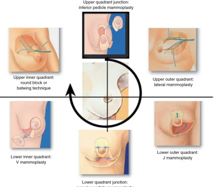
Figure 1.3 Overview of level 2 oncoplastic surgical reconstruction
techniques. A quadrant-per-quadrant approach to the choice of surgical technique depends on the area of tumour localization.
1.5.2.1 Tumours at the Upper Pole (at 12 O’Clock Position)
For tumours in the upper pole of the breast, i.e. those localized in the
superior aspect of the breast from 11-1 o’clock based on the lock orientation, two
most appropriate techniques to be used are the ‘round block’ (Benelli 1990)
(Figure 1.4) and the inferior pedicle mammoplasty (Robbins 1977) (Figure 1.5).
Inferior pedicle mammoplasty involves the use of existing breast tissue and its
blood supply from the lower pole of the breast, to fill in the generated defect in
the superior pole, as one would do in a cosmetic ‘breast reduction’ or ‘breast lift’.
1.5.2.2 Upper Inner Quadrant
Tumours in the upper inner quadrant are those localized to 9-11 o’clock
position of the left breast or 1-3 o’clock position of the right breast. The best
approach to these is to use the batwing (Figure 1.6) and the round block (Figure
1.3) procedures (Anderson, Masetti et al. 2005, Clough, Ihrai et al. 2012).
1.5.2.3 Upper Outer Quadrant
For the tumours localized to the upper outer quadrant, i.e. at 1-3 o’clock in
the left breast and 9-11 o’clock in the right breast, lateral or raquet mammoplasty
is the most suitable technique (Ballester, Berry et al. 2009) (Figure 1.7). The
procedure consists of the removal of a lateral wedge of breast tissue, including
the tumour and the overlying skin. Briefly, an incision is made that extends
laterally from the edge of the nipple areola complex (NAC), as necessary. The
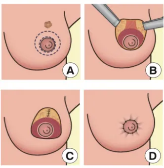
Figure 1.4 Round block mastopexy technique in level 2 oncoplastic
surgery. (A) Preoperative design with two circular skin markings, (B) lumpectomy and de-epithelization, (C) undermining and
approximation of nearby breast tissue, (D) postoperative periareolar
scar.
Figure 1.5 Inferior pedicle mammoplasty in level 2 oncoplastic surgery. The existing breast tissue and its blood supply from the lower pole
of the breast is used to fill in the generated defect
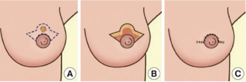
Figure 1.6 Batwing mastopexy in level 2 oncoplastic surgery. Batwing mastopexy. (A) Preoperative design with batwing form, (B)
lumpectomy, (C) pulling up the inferior breast tissue.
Figure 1.7 Lateral mammoplasty (‘tennis racket’) in level 2 oncoplastic surgery. (A) Preoperative design with racket form, (B) lumpectomy
and de-epithelization, (C) filling and nipple-areolar complex
reposition.
then carried out at the level of pectoralis major muscle. The remaining lateral and
central glandular tissue is mobilized while keeping the skin attached to the
glands; this is then used to fill the defect while preserving a good blood supply to
the tissue. Complete detachment of NAC from the underlying tissue assists with
mobilization of the central glandular tissue for volume replacement at the
lumpectomy defect (Clough, Ihrai et al. 2012). Once the defect is closed,
re-centralization of the nipple is carried out by de-epithelization of the crescent of
skin medial to NAC.
1.5.2.4 Tumours at the Lower Pole (at 6 O’Clock Position)
For the tumours localized to the lower pole of the breast, i.e. those at 5-7
o’clock position, superior pedicle mammoplasty offers the most appropriate
approach. The technique employs an inverted T-shaped skin incision. The
procedure begins by marking the superior pedicle and the tumour, followed by
de-epithelization of the pedicle. An incision is made at the infra-mammary fold
(IMF), and a wide dissection at the level of pectoralis major is carried out.
Ensuring a wide clinical margin around the tumour, the lower pole tissue and
some of the central tissue are all removed en bloc. Lateral and medial tissues are
re-approximated, and sutured to the IMF. During the final stage of the procedure,
the nipple is recentralized over the new breast mound (Clough, Ihrai et al. 2012).
This technique is also commonly used for ‘breast reduction’ or ‘breast lift’
procedures, where up to 60% of the breast volume can be resected, providing an
rounder and more aesthetically pleasing breast mound, that is usually
cosmetically better after surgery than before.
1.5.2.5 Lower Inner Quadrant
For tumours of the lower inner quadrant, i.e. those localized to 7-9 o’clock
position in the left breast and 3-5 o’clock position in the right breast, V
mammoplasty (Figure 1.8) is the best option (Clough, Ihrai et al. 2012). The
technique involves making a V-shaped incision over the tumour, with the apex
pointing towards the margin of the areola, carrying out the dissection to the level
of pectoralis major. Following tumour excision, another incision is made at the
level of the IMF, extending to the anterior axillary line. Breast tissue is dissected
at the level of pectoralis major, and the entire inferolateral breast tissue is
mobilized medially to fill the defect, suturing it to the medial breast tissue.
Following this, the skin around the NAC is de-epithelized and the NAC is then
positioned over the superomedial pedicle.
Although more appropriate for the lower pole tumours, superior pedicle
with inverted T-shape incision is another technique that may be, in some cases,
useful on tumours of the lower inner quadrant (Clough, Ihrai et al. 2012).
1.5.2.6 Lower Outer Quadrant
Tumours of the lower outer quadrant are those localized to 3-5 o’clock
position in the left breast and 7-9 o’clock position in the right breast. For these, J
Figure 1.8 V mammoplasty technique in level 2 oncoplastic surgery. (A) V-shaped incision, (B) lumpectomy, (C) inferolateral tissue
mobilization, (D) scar.
Adapted from Clough et at (2010).
A B
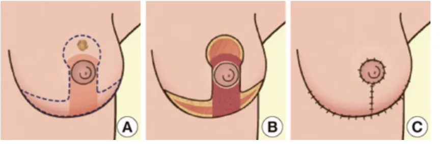
Figure 1.9 J mammoplasty technique in level 2 oncoplastic surgery. (A) J-shaped, oblique incisions from both sides of NAC, (B) lumpectomy,
(C) lateral, medial and central glandular tissue are pulled together
to achieve defect closure.
Adapted from Clough et at (2010).