O R I G I N A L R E S E A R C H
The lncRNA HOXA11-AS regulates Rab3D
expression by sponging miR-125a-5p promoting
metastasis of osteosarcoma
This article was published in the following Dove Press journal: Cancer Management and Research
Kun Cao1
Yueyang Fang1
Hao Wang1
Zheng Jiang1
Li Guo2
Yong Hu1
1Department of Orthopaedics, The First
Hospital Of Anhui Medical University, Hefei, People’s Republic of China;
2Department of Orthopaedics,
The Second Hospital Of Shanxi Medical University, Taiyuan, People’s Republic of China
Objective:Many studies have shown that long non-coding RNAs (lncRNAs) are closely
related to various cancers. This study aims to explore the roles of lncRNA HOXA11-AS in the development and progression of osteosarcoma (OS).
Methods:The expression levels of HOXA11-AS and miR-125a-5p in tumor tissues and the
adjacent tissues were detected by RT-PCR method. The proliferation, migration and invasion of MG-63 and KHOS cells were determined.
Results:It was found that HOXA11-AS expression levels in OS tissues and OS cell lines were
higher than those in OS adjacent tissues and normal human osteoblast cell lines. The higher expression level of HOXA11-AS was positively correlated with more severe clinical stage, distant metastasis and poor prognosis of OS. Inhibition of HOXA11-AS expression could reduce metas-tasis and invasion of OS cell lines. In addition, HOXA11-AS was found to be an endogenous inhibitor of miR-125a-5p, it down regulated the expression level of miR-125a-5p, and this process could promote the expression of Rab3D, the target gene of miR-125a-5p.
Conclusion: Our study elucidated the role of a new HOXA11-AS/miR-125a-5p/Rab3D
regulatory pathway in promoting OS metastasis.
Keywords: osteosarcoma (OS), long non-coding RNAs (lncRNAs), HOXA11-AS,
miR-125a-5p, Rab3D
Introduction
Osteosarcoma (OS) is the most common primary malignant tumor in childhood,
and it is also the second major pediatric tumor that causes death.1 Half of the
patients will eventually have lung metastases, although there are many treatments,
such as chemotherapy, radiotherapy and surgical treatment.2Thefive-year survival
rate of patients with OS is not more than 30%, and lung metastasis is the main
cause of death.3Therefore, it is particularly important to study the pathogenesis of
OS andfind new diagnostic markers and therapeutic targets.
Long non-coding RNAs (IncRNA) are a class of non-coding RNA with a length larger than 200 bases, and they participate in X chromosome inactivation, splicing,
epigenetic control and gene transcription regulation.4Recent studies have shown that
IncRNA can be used as competing endogenous RNAs (ceRNA) or molecular sponges to
regulate the expression of microRNAs, little is known about its other functions.5,6The
interaction between ceRNA and miRNA represents a new form of gene regulation, which
plays a role in a variety of pathophysiological processes including tumorigenesis.7
Homeobox A11 antisense (HOXA11-AS), a newly identified lncRNA, locates on the
Correspondence: Yong Hu
Department of Orthopaedics, The First Hospital of Anhui Medical University, Hefei, Anhui 230022, People’s Republic of China
Tel +86 551 629 23607 Email [email protected]
Cancer Management and Research
Dovepress
open access to scientific and medical research
Open Access Full Text Article
Cancer Management and Research downloaded from https://www.dovepress.com/ by 118.70.13.36 on 20-Aug-2020
HOXA gene cluster.8It has been reported to be up-regulated in a wide variety of carcinomas and is generally associated with
poor prognosis.9–13
miR-125a-5p is an anti-oncogene, which can inhibit the proliferation and invasion of liver cancer, gastric cancer, breast cancer, lung cancer, glioma and melanoma. It has been
con-firmed that SIRT7, PI3K, E2F3, ERBB2, TSTA3, HDAC4,
HDAC5, EGFR, Gab2 and Lin28B are target genes of
miR-125a-5p.14–24However, the role of miR-125a-5p in OS has not
been reported, and it needs further study.
Rab GTPases is a highly conserved intracellular trans-porter, it is the basic component and the major regulator of
vesicular transport signaling pathway.23 Rab GTPases
include Rab3A/B/C/D, Rab26, Rab27A/B and Rab37,
which control the corresponding transport process.
Abnormal expression of Rab GTPases can affect the
development and metastasis of tumor.24 Rab3D is one of
the most important members of the Rab GTPases family, and it is mainly expressed in the cells of non-nerve tissue
and regulates the transport of specific types of cells.
Abnormal expression of Rab3D occurred in a variety of tumor tissues, and increased expression of Rab3D in color-ectal cancer and esophageal squamous cell carcinoma is
associated with increased invasiveness of tumor cells.25
The expression of Rab3D is also increased in OS, which
is related to the proliferation and invasion of OS.26
miR-125a-5p acted on the 3’-UTR of Rab3D, and HOXA11-AS
has a ponging effect of Mi-125a-5p in intestinal cancer.13
However, the role of Rab3D in OS remains unclear. Therefore, we explored the roles of HOXA11-AS/miR-125a-5p/Rab3D regulatory pathway in the development and progression of OS in this study.
Materials and methods
Subjects
A total of 61 patients with OS who did not receive chemother-apy or radiotherchemother-apy but received surgical treatment directly were recruited in this study. They were from the Department of
orthopedics of the First Affiliated Hospital of Anhui Medical
University during the 2011–2017 years, and their demography
and clinical data were collected. Surgically resected tumor tissues para-tumor tissues were collected, some of them were
fixed with 4% polyoxymethylene, embedded with paraffin,
and tissue sections were prepared for immunohistochemical
staining. The others were sheared and put into RNAlater™
Stabilization Solution (Thermo Fisher Scientific, Waltham,
MA, USA) for RNA extraction. The clinical data of the patients are shown in Table 1.
This study was carried out in accordance with The Code of Ethics of the World Medical Association (Declaration of Helsinki). The informed consent was obtained from patients. The patient consent was written informed consent. These experiments were approved by the Ethics Committee of Anhui medical university.
Experimental animals
BALB/c-nu nude mice (4~6 week old, weighT 16–20 g)
were purchased from Anhui experimental animal center. They were bred in the SPF class barrier system of Anhui experimental animal center and were allowed to eat and drink freely. All animal experiments were con-ducted according to the Principles of Laboratory Animal Care (National Society for Medical Research). These experiments were approved by the Ethics Committee of Anhui medical university.
Cell culture
Human OS cell lines U2OS, MG-63 and KHOS, normal human osteoblast cell line Nhost were purchased from American Type Culture Collection (ATCC). The cells were cultured with DMEM medium containing 10% FBS
at 37°C in 5% CO2.
siRNA transfection
miR-125a-5p mimics, miR-125a-5p inhibitors and
AS-siRNAs (for the knockdown of HOXA11-AS expression) and Rab3D-siRNAs were purchased from
Table 1The primers used in this study
Gene Forward primer Reverse primer
HOXA11-AS GAGTGTTGGCCTGTCCTCAA TTGTGCCCAGTTGCCTGTAT
miR125a-5p TGCGGCTCCCTGAGACCCTTTAA
Rab3D ATCGCCAATCAGGAATCCTTTG CACAACACGTTCGTCCTCCA
U6 CTCGCTTCGGCAGCACA AACGCTTCACGAATTTGCGT
β-actin TGAGGATGTCACGGTTCCAG GTCACCTTCACCGTTCCAGT
Cancer Management and Research downloaded from https://www.dovepress.com/ by 118.70.13.36 on 20-Aug-2020
Gene Pharma (Shanghai, China). pcDNA3.1-HOXA11-AS plasmid was constructed. miRNA mimics, miRNA inhibi-tors, PADI2-siRNAs or HOXA11-AS-siRNAs
Rab3D-siRNAs (5ʹ- AGGAGATTCTGGGATTGCCAGTTCT-3ʹ;
5ʹ- CCATCTTCTCTTGCCTGGTCCTTGA-3ʹ; 5ʹ-GGATG
TCGTGAAGATGGCAAGAAGA-3ʹ) or
HOXA11-AS-siRNAs (5ʹ- TCGTCACTCGGTGTTCTCACCGAAA-3ʹ;
5ʹ- GCACGGTGACTTGATTACACTCTCT-3ʹ; 5ʹ-CGGA
AACGGCTAACAAGGAGATTTG-3ʹ) were transfected
into U2OS, MG-63, KHOS and Nhost cell lines, respec-tively. All siRNA transfections were performed using
Lipofectamine 2000 Reagent (Life Technologies,
Carlsbad, CA, USA), according to the manual protocol.
RNA extraction and qRT-PCR
Total RNA was extracted using a Trizol reagent kit (Invitrogen, Carlsbad, CA, USA) according to the
manu-facturer’s protocol. RNA concentration and purity were
detected using a Qubit Fluorometer (Thermo Fisher
Scientific, Inc.). A total of 1 μg RNA was subjected to
reverse transcription using a Prime Script Kit (Takara Bio Inc., Otsu, Japan). qPCR was performed using a SYBR
Premix Ex Taq™Kit (Takara Bio Inc.). The quantification
method used was the 2-ΔΔCT method. The thermocycling
conditions were as follows: pre-degeneration at 95°C for 10 mins, followed by 40 cycles of 95°C for 12 s and 62°C
for 40 s; U6 and β-actin genes were used as an internal
control. The primers used in this study are shown in Table 1.
Cell proliferation ability
The cell proliferation was detected with Cell Counting Kit-8 (CCK-Kit-8; Dojindo, Kumamoto, Japan) according to the
kit’s manual protocol. The cells in the logarithmic growth
phase were digested with trypsin and inoculated into
96-well plates (2×10⁴cell/well), and they were cultured at 37°
C with 5% CO2overnight. Each well was added with 10
μLCCK-8 solution before detection and incubation for 1
hr, and A450 values were detected by Epoch Microplate Spectrophotometer (Bio Tek, Winooski, VT, USA) after culture for 24 hrs, 48 hrs, 72 hrs and 96 hrs, respectively, to evaluate the proliferation of cells.
Cell migration and invasion assay
Cell migration and invasion were determined using a Transwell system (Corning, Inc., Corning, NY, USA). Cells were trypsinized and suspended with serum-free medium and cells (50,000 each well) were added to the
upper chamber of the 24-well Transwell system with or without Matrigel (BD Biosciences, San Jose, CA, USA). A 0.6 ml medium containing 10% FBS was added to the lower chamber. Cells were incubated at 37°C for 12 hrs for migration test and continued to culture at 37°C for 24 hes
for invasion test. The cells were fixed and stained with
0.1% crystal violet dye (Richard-Allan Scientific, San
Diego, CA, USA). They were washed twice with PBS. Stained cells were observed under an inverted microscope.
Xenograft mouse model
Each nude mouse was inoculated 5×10⁶cells of different
groups. Tumor volume was measured every three days (0.5× length × width2) after inoculation for 12 d. The
mice were sacrificed after transfection for 36 d, and the
tumor was removed for weight measurement.
Dual-luciferase reporter gene analysis
The luciferase reporter gene plasmid WT HOXA11- AS (luc-HOXA11-AS-WT) and point mutation of luciferase reporter gene plasmid Mut HOXA11-AS (luc-HOXA11-AS-MU) were constructed. The 293T cells were inoculated into 24-well plates and cultured overnight, and luciferase reporter plasmid, Renilla luciferase and miR-125a-5p mimic or control were transfected into 293T cells simulta-neously. The cells were split using Dual Luciferase Reporter Assay System (Promega, Madison, WI, USA) according to the manual after culture for 48 hrs. The results were detected using Panomics Luminometer (Affymetrix, Santa Clara, CA, USA) after the
lumines-cence was added. The sea reninfluorescence was used as
an internal reference.
Western blotting method
Cells in the logarithmic growth period were lysed with cell lysis solution (Sigma-Aldrich, St. Louis, MO, USA). The supernatant was collected after they were centrifuged at 4° C (1000 rpm) for 5 mins. Total proteins were extracted and protein concentration was determined using BCA. Proteins
(50 μg per lane) were separated using 12% SDS-PAGE.
Proteins were then electrotransferred to a PVDF mem-brane (Amersham Biosciences, Piscataway, NJ, USA).
The PVDF membrane was rinsed with TBS for 10–15
mins, placed in TBS/T blocking buffer containing 5% (w/v) skimmed milk powder. It was incubated at room temperature for 2 hrs following the addition of an appro-priate dilution of primary antibodies (1:1000 Rab3D,
Proteintech, Rosemont, IL, USA; 1:2000 β-actin,
Cancer Management and Research downloaded from https://www.dovepress.com/ by 118.70.13.36 on 20-Aug-2020
Proteintech, Rosemont, IL, USA). The membrane was
then rinsed with TBST three times (5–10 mins/wash) and
then incubated at room temperature for 1 hr with horse-radish peroxidase-labeled secondary antibody (1:50,000; Abcam, Cambridge, UK; diluted with TBST containing 0.05% (w/v) skimmed milk powder). The membrane was
then rinsed three times with TBST (5–10 mins/wash).
Protein bands were detected using an enhanced
chemilu-minescence kit (Perkin-Elmer Inc.) and quantified as the
ratio to β-actin. Quantification was performed using
Imagequant LAS4000 (GE Healthcare, Japan).
Immunohistochemical test
Briefly, the osteosarcoma resection tissues were
embedded with paraffin using a conventional method.
They were cut into 5-μm slices and incubated with
0.3% endogenous peroxidase blocking solution for 20 mins after dewaxing and hydrating. Then, they were incubated at room temperature for 10 mins with 3% hydrogen peroxide methanol solution and washed with PBS for 3 times (3 mins/time). Antigen retrieval was performed using citrate buffer (pH 6.0) at 121°C for 2 mins. After blocking with 5% BSA (Gibco; Thermo
Fisher Scientific, Inc), the cells were incubated with
a primary monoclonal antibody anti-Rab3D (abcam, ab128997; 1:500) overnight at 4°C. The cells were then incubated with goat anti-rabbit non-biotinylated regents (Zhongshanjinqiao, Beijing, China) according to the man-ual and mounted with epoxy resin. They were observed using Photo and Image Auto Analysis System (Image-Pro
-Plus, China). Five visual fields were randomly selected
for each slice, and the integrated optical density (IOD) was calculated by using Image J 6.0 software. The rela-tive expression levels of Rab3D in different tissues were analyzed by comparing IOD.
Statistical analysis
The data were analyzed using SPSS 16.0 software (SPSS Inc., Chicago, IL, USA). All results are
pre-sented as the mean ± SD. Student’s t test, the
Wilcoxon signed-rank test and Pearson’s chi-square
test were used to evaluate the differences among groups. The correlation between HOXA11-AS and miR-125a-5p and the expression level of miR-125a-5p
and Rab3D were analyzed by Pearson’s correlation
analysis. P<0.05 was considered to indicate
a statistically significant difference.
Results
The expression level of HOXA11-AS
increased in OS cases
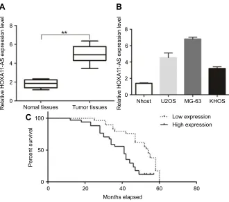
The expression levels of lncRNA HOXA11-AS in 61 OS
patients’ resected tumors and para-tumor tissues were
detected by fluorescence quantitative PCR method. The
results showed that the expression level of HOXA11-AS
in tumor tissues was significantly higher than that in
para-tumor tissues (P<0.01, Figure 1A), and its expression level
in OS cell lines was significantly higher than that in
normal human osteoblasts (P<0.01, Figure 1B). The 61
cases were divided into high-expression group and low-expression group according to the median low-expression level
of HOXA11-AS detected by fluorescence quantitative
PCR. Kaplan–Meier analysis showed that increased
expression of HOXA11-AS was associated with patients’
poor survival (P=0.002, Figure 1C), decreased expression
level of microRNA-125a-5p was associated with patients’
poor survival (P=0.002, Figure 1D) and increased Rab3D
expression was associated with patients’ poor survival
(P=0.007, Figure 1E). Analysis of the correlation between
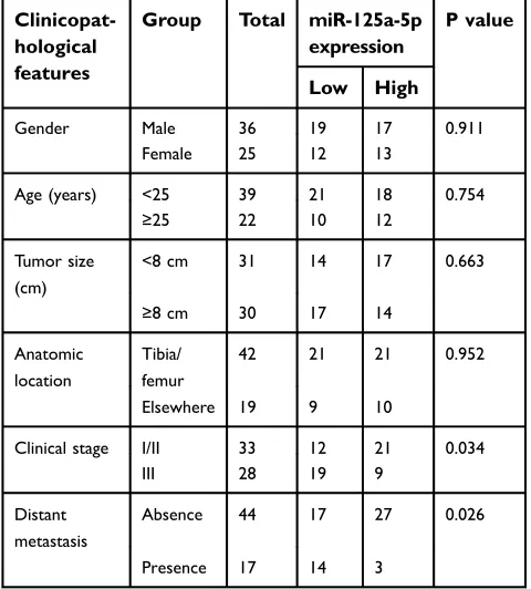
clinicopathological features and expression levels of HOXA11-AS, microRNA-125a-5p and Rab3D showed that the higher expression level of HOXA11-AS was posi-tively correlated with clinical staging, distal metastasis and
poor prognosis of OS patients (P<0.05, Tables 2–4).
HOXA11-AS promotes the proliferation
of OS
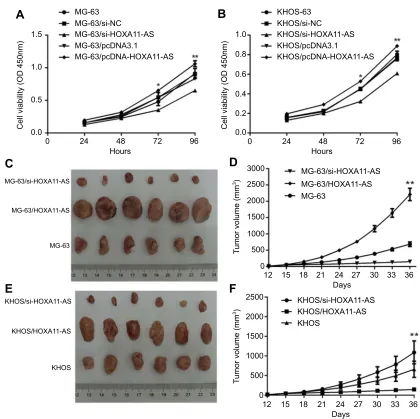
In vitro experiments showed that down-regulation of HOXA11-AS expression in MG-63 and KHOS cells could inhibit cell proliferation, while up-regulation of HOXA11-AS expression could promote cell prolifera-tion (Figure 2A and B). In vivo experiments showed
that down-regulation of HOXA11-AS expression
in MG-63 and KHOS cells could inhibit the growth of subcutaneous solid tumors in nude mice, while up-regulation of HOXA11-AS expression could promote the growth of subcutaneous solid tumors in nude mice
(Figure 2C–F).
HOXA11-AS promotes the migration and
invasion of OS
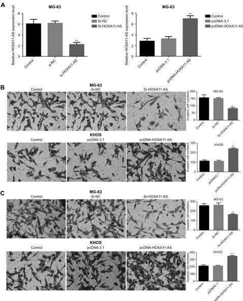
The invasion and migration ability by up-regulating and down-regulating HOXA11-AS gene in both MG-63 and KHOS cell lines were detected, respectively. The results
Cancer Management and Research downloaded from https://www.dovepress.com/ by 118.70.13.36 on 20-Aug-2020
showed that the migration and invasion ability of
MG-63 and KHOS cells decreased significantly after
HOXA11-AS siRNA inhibited HOXA11-AS expression,
the migration and invasion of MG-63, and KHOS cells
increased significantly after overexpressed HOXA11-AS
(P<0.01, Figure 3). These results suggested that
A
8
6
Relative HOXA1
1-AS expression level
Percent survival
4
2
0
100
50
0
0 20 40 60 80
Months elapsed
Nomal tissues Tumor tissues
B
C
8
6
Relative HOXA1
1-AS expression level
4
2
0
Nhost U2OS MG-63 KHOS
Low expression High expression
Figure 1RT-PCR results of HOXA11-AS expression in OS tissues and cell lines. (A) The expression level of HOXA11-AS in tumor tissues and the adjacent tissues (**P<0.01, n=61); (B) HOXA11-AS expression level in OS cell lines and normal human osteoblasts (*P<0.05); (C) The Kaplan–Meier curves by HOXA11-AS expression levels. Patients with high HOXA11-AS expression had a poor overall survival compared to those patients with low HOXA11-AS expression (log-rank test;P=0.007) (n=61).
Table 2Correlation between the expression level of HOXA11-AS and the clinicopathological features of OS
Clinicopathological features Group Total HOXA11-AS expression P-value
Low High
Gender Male 36 17 19 0.701
Female 25 13 12
Age (years) <25 39 22 17 0.245
≥25 22 10 12
Tumor size (cm) <8 cm 31 18 23 0.183
≥8 cm 30 15 16
Anatomic location Tibia/femur 44 20 22 0.615
Elsewhere 17 8 9
Clinical stage I/II 33 20 13 0.031
III 28 11 17
Distant metastasis Absence 44 30 14 0.018
Presence 17 5 12
Cancer Management and Research downloaded from https://www.dovepress.com/ by 118.70.13.36 on 20-Aug-2020
HOXA11-AS could promote the migration and invasion of OS cells.
HOXA11-AS is the molecular sponge of
miR-125a-5p
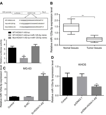
RT-PCR results showed that the expression level of
miR-125a-5p in OS tissues was significantly lower than that in
their adjacent tissues (P<0.01, Figure 4B). Luciferase
reporter gene test also confirmed that HOXA11-AS
could directly regulate the expression of miR-125a-5p
(P<0.01, Figure 4A). Inhibiting the expression of
HOXA11-AS in MG-63 and KHOS cells could signifi
-cantly increase the expression level of miR-125a-5p
(P<0.01, Figure 4C). Overexpression of HOXA11-AS
in MG-63 and KHOS cells could significantly inhibit
the expression of miR-125a-5p (P<0.01, Figure 4D).
HOXA11-AS promotes the invasion and
migration of OS through competitive
binding with miR-125a-5p
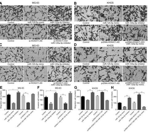
Compared with the control group, it was found that miR-125a-5p mimic could inhibit the migration and
A
C
E
B
D
F
1.5 1.0
3000
2500
2000
1500
1000
500
0
2500
2000
1500
1000
500
0
12 15 18 21 24
Days
27 30 33 36
12 15 18 21 24
Days
27 30 33 36
0.8
0.6
0.4
0.2
0.0
0 24 48 72 96
1.0
Cell viability (OD 450nm) Cell viability (OD 450nm) 0.5
0.0
0 24 48
Hours Hours
MG-63/si-HOXA11-AS
KHOS/si-HOXA11-AS
KHOS/HOXA11-AS
KHOS
MG-63/HOXA11-AS
MG-63
MG-63 MG-63/si-NC MG-63/si-HOXA11-AS MG-63/pcDNA3.1
MG-63/pcDNA-HOXA11-AS
KHOS-63 KHOS/si-NC KHOS/si-HOXA11-AS KHOS/pcDNA3.1
KHOS/pcDNA-HOXA11-AS
MG-63/si-HOXA11-AS
MG-63/HOXA11-AS MG-63
T
umor volume (mm
3)
T
umor volume (mm
3)
KHOS/si-HOXA11-AS KHOS/HOXA11-AS KHOS
72 96
Figure 2HOXA11-AS promotes the proliferation of OS. (A) Down-regulation of HOXA11-AS expression in MG-63 cells could inhibit cell proliferation, while up-regulation of HOXA11-AS expression could promote cell proliferation *P<0.05,**P<0.01. (B) Down-regulation of HOXA11-AS expression in KHOS cells could inhibit cell proliferation, while up-regulation of HOXA11-AS expression could promote cell proliferation *P<0.05,**P<0.01. (C) Down-regulation of HOXA11-AS expression in MG-63 cells could inhibit the growth of subcutaneous solid tumors in nude mice, while up-regulation of HOXA11-AS expression could promote the growth of subcutaneous solid tumors in nude mice. (D) Changes of tumor volume with time after inoculation of MG-63 cells of different groups **P<0.01. (E) Down-regulation of HOXA11-AS expression in KHOS cells could inhibit the growth of subcutaneous solid tumors in nude mice, while up-regulation of HOXA11-AS expression could promote the growth of subcutaneous solid tumors in nude mice. (F) Changes of tumor volume with time after inoculation of MG-63 cells of different groups.
Cancer Management and Research downloaded from https://www.dovepress.com/ by 118.70.13.36 on 20-Aug-2020
invasion of OS, while miR-125a-5p inhibitors could
promote the migration and invasion of OS (P<0.05,
Figure 5). When HOXA11-AS- siRNAs and
miR-125a-5p inhibitors were transfected into MG-63 cells at the same time, and the ability of HOXA11-AS-siRNAs to inhibit cell migration and invasion was counteracted. When pcDNA-HOXA11-AS and miR-125a-5p mimic were transfected into KHOS cells at the same time, it could inhibit the ability of pcDNA-HOXA11-AS to promote cell migration and invasion (Figure 5).
HOXA11-AS can regulate the expression
of its target gene Rab3D by regulating
miR-125a-5p expression
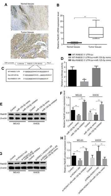
Immunohistochemical and RT-PCR results showed that the expression level of Rab3D in tumor tissues was
significantly higher than that in their adjacent tissues
(Figure 6A–C). The online software TargetScan
analy-sis showed that miR-125a-5p may be combined with 3
‘UTR of Rab3D. Luciferase reporter gene analysis
showed that miR-125a-5 could target the 3’UTR of
Rab3D (Figure 6D and E). miR-125a-5p could inhibit the expression of Rab3D (Figure 6D and E), while HOXA11-AS could promote the expression of Rab3D by inhibiting the expression of miR-125a-5p (Figure 6H and I).
Rab3D can promote the migration and
invasion of OS
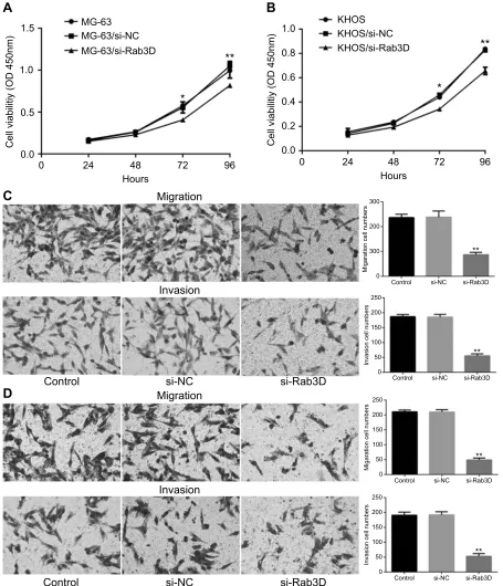
To further determine whether Rab3D is related to the proliferation, migration and invasiveness of osteosar-coma cells, we transfected Rab3D siRNA into MG-63 and KHOS cells, and it was found that the proliferation of MG-63 and KHOS decreased after down-regulation of Rab3D expression (Figure 7A and B), and the migration and invasion of MG-63 and KHOS cells
also decreased significantly after down-regulation of
Rab3D expression (Figure 7C and D). These results suggested that HOXA11-AS/miR-125a-5p/Rab3D was
involved in the invasion and metastasis of OS
(Figure 8).
Discussion
OS is common in children and young people. The incidence rate peaked around the age of 20, and it is
about 5 million cases per year.1 The current treatment
strategy is limited in the treatment of metastatic and recurrent OS, and new strategies and innovative thera-pies are urgently needed to improve the survival rate of
Table 3The correlation between the expression level of
miR-125a-5p and the clinicopathological features of OS
Clinicopat-hological features
Group Total miR-125a-5p
expression
P value
Low High
Gender Male 36 19 17 0.911
Female 25 12 13
Age (years) <25 39 21 18 0.754
≥25 22 10 12
Tumor size (cm)
<8 cm 31 14 17 0.663
≥8 cm 30 17 14
Anatomic location
Tibia/ femur
42 21 21 0.952
Elsewhere 19 9 10
Clinical stage I/II 33 12 21 0.034
III 28 19 9
Distant metastasis
Absence 44 17 27 0.026
Presence 17 14 3
Table 4The correlation between the expression level of Rab3D
and the clinicopathological features of OS
Clinicopat-hological features
Group Total Rab3D
expression
P value
Low High
Gender Male 36 18 18 0941
Female 25 13 12
Age (years) <25 39 20 19 0.938
≥25 22 11 11
Tumor size (cm)
<8 cm 31 15 16 0.617
≥8 cm 30 16 14
Anatomic location
Tibia/ femur
42 23 19 0.325
Elsewhere 19 8 11
Clinical stage I/II 33 22 11 0.031
III 28 9 19
Distant metastasis
Absence 44 29 15 0.021
Presence 17 2 15
Cancer Management and Research downloaded from https://www.dovepress.com/ by 118.70.13.36 on 20-Aug-2020
A
8
MG-63
MG-63
Control
B
C
Si-NC Si-HOXA11-AS
KHOS
Control pcDNA 3.1 pcDNA-HOXA11-AS
MG-63
Control Si-NC Si-HOXA11-AS
KHOS
Control pcDNA 3.1 pcDNA-HOXA11-AS
Control
**
**
Si-NC Si-HOXA11-AS
Relative HOXA1
1-AS expression level
6
4
2
0
Control si-NC
si-HOXA1 1-AS
8
MG-63
Control pcDNA-3.1 pcDNA-HOXA11-AS
Relative HOXA1
1-AS expression level
6
4
2
0
200 150 100 50
Invasion cell numbers
Invasion cell numbers
0
0 100 200 300
KHOS MG-63
** **
Control
Control pcDNA3.1
pcDN-HOXA1 1-AS Si-NC
Si-HOXA1 1-AS
200 300
100
Invasion cell numbers
Invasion cell numbers
0
0 100 200 300
400 KHOS MG-63
** **
Control
Control pcDNA3.1
pcDN-HOXA1 1-AS Si-NC
Si-HOXA1 1-AS
Control
pcDNA-3.1
pcDNA-HOXA1 1-AS
Figure 3HOXA11-AS could promote the migration and invasion of OS. (A) The HOXA11-AS expression level decreased significantly in MG-63 cells after HOXA11-AS siRNA was transfected into cells (**P<0.01); The HOXA11-AS expression level increased significantly in KHOS cells after pcDNA-HOXA11-AS was transfected into cells (**P<0.01). (B) The invasion ability of MG-63 cells decreased significantly after HOXA11-AS siRNA inhibited HOXA11-AS expression, the invasion of KHOS cells increased significantly after KHOS cells overexpressed HOXA11-AS (**P<0.01). (C) The migration ability of MG-63 cells decreased significantly after HOXA11-AS siRNA inhibited HOXA11-AS expression, the migration of KHOS cells increased significantly after KHOS cells overexpressed HOXA11-AS (**P<0.01).
Cancer Management and Research downloaded from https://www.dovepress.com/ by 118.70.13.36 on 20-Aug-2020
patients with OS. Understanding the pathogenesis, pro-gression and prognosis of OS at the molecular level will enable molecular targeted therapy to become a new method for the treatment of OS. In this study, we explored the role of lncRNAs in the genesis and development of OS.
lncRNA-HOXA11-AS locates on the chromosome 7p15.2; it contains 3885 nucleotides and is one of the
members of the homeobox (HOX) family.6The
HOXA11-AS expression is associated with a variety of cancers. It was up-regulated in glioma and promoted gliomagenesis
through regulating cell cycle.7 Sun et al reported that
HOXA11-AS/miR-1297/EZH2 cascade played an impor-tant role in the occurrence and development of gastric
cancer.27 HOXA11-AS expression was up-regulated in
colon cancer tissues, which was closely related to the
hepatic metastasis of colon cancer.11 Lung metastasis of
OS is one of the important causes of death, whether there is a link between HOXA11-AS and lung metastasis of OS remains unclear.
In this study, we found that Rab3D was highly expressed in OS tissues and OS cell lines, but it was relatively low in OS adjacent tissues and human osteo-blasts NHost cell line, and its expression level was
highly correlated with the expression level of
HOXA11-AS. Rab3D is one of the ras-related GTP-binding protein Rab family members. Rab GTPases are highly conservative, and they could regulate a variety
A
B
C
D
CMV promoter
Luciferase HOXA11-AS Poly A
2.5
**
2.0
1.5
1.0
Relative miR-125a-5p expression level
0.5
0.0 5
4
3
2
** 1
0
5
1.5
1.0
0.5 **
0.0 **
4
3
2
1
0
Control
Control
pcDNA3.1
pcDNA-HOXA1 1-AS
Si-NC
Si-HOXA1 1-AS
Relative luciferase activity
Relative miR-125a-5p expression level
Relative miR-125a-5p expression level
Nomal tissues Tumor tissues
WT-HOXA11-AS-luc
MG-63
KHOS WT-HOXA11-AS-luc+miR-125-5p mimic
Mut-HOXA11-AS-luc+miR-125-5p mimic
Figure 4HOXA11-AS could directly regulate the expression of miR-125-5p. (A) Construction of WT-HOXA11- AS and Mut-HOXA11-AS. (B) RT-PCR results showed that the expression level of miR-125a-5p in OS tissues was significantly lower than that in their adjacent tissues (**P<0.01). (C) Inhibiting the expression of HOXA11-AS in MG-63 cells could significantly increase the expression level of miR-125a-5p (**P<0.01). (D) Overexpression of HOXA11-AS in KHOS cells could significantly inhibit the expression of miR-125a-5p (**P<0.01).
Cancer Management and Research downloaded from https://www.dovepress.com/ by 118.70.13.36 on 20-Aug-2020
of biological processes, which include the skeleton
changes, cell movement and invasion of tumor cells.25
miR-125a-5p had tumor suppressor effect in a variety of tumors; HOXA11-AS could competitively combine
with miR-125a-5p as a molecular sponge.14,24,28,29
Therefore, we speculated that HOXA11-AS could com-petitively combine with miR-125a-5p in OS, which
leads to the increase of Rab3D expression and
promotes the metastasis and invasion of tumor cells. In this study, we found that the expression levels of HOXA11-AS, miR-125a-5p and Rab3D in OS tissues and
OS cell lines were significantly different from those in OS
adjacent tissues and human osteoblasts NHost cell line. The expression level of miR-125a-5p was negatively cor-related with that of HOXA11-AS and Rab3D. The expres-sion level of HOXA11-AS was positively correlated with
more severe clinical stage and distant metastasis. Kaplan–
Meier analysis showed that patients with high expression of HOXA11-AS had a poorer prognosis than those with low expression of HOXA11-AS. HOXA11-AS and Rab3D had a positive effect on the invasiveness of OS cell lines, while miR-125a-5p had a negative regulatory effect on the invasiveness of OS cell lines. Luciferase reporter gene test showed that HOXA11-AS could competitively combine
A
MG-63 KHOSKHOS
Control
Control si-HOXA11-AS si-HOXA11-AS+
miR-125a-5p inhibitor Control pcDNA-HOXA11-AS pcDNA-HOXA11-AS+miR-125a-5p mimic miR-125-5p mimic miR-125a-5p inhibitor Control miR-125-5p mimic miR-125a-5p inhibitor
Control pcDNA-HOXA11-AS pcDNA-HOXA11-AS+ miR-125a-5p mimic Control miR-125-5p mimic miR-125a-5p inhibitor
C
E
300 * * *
**
**
** **
** **
** 200
100
0
150 200
100
0 50
Control Control
miR-125-5p mimic
si-HOXA1 1-AS
HOXA1
1-AS+miR-125-5p mimic miR-125a-5p inhibitor
Control Control Control
miR-125-5p mimic miR-125-5p mimic
si-HOXA1 1-AS
pcDNA-HOXA1 1-AS
pcDNA-HOXA1
1-AS+miR-125-5p mimic
HOXA1
1-AS+miR-125-5p mimic
miR-125a-5p inhibitor miR-125a-5p inhibitor
Mirgration cell numbers
G
H
F
300
200
100
0
Control Control
miR-125-5p mimic pcDNA-HOXA1 1-AS
pcDNA-HOXA1
1-AS+miR-125-5p mimic miR-125a-5p inhibitor
Control
Mirgration cell numbers
300
200
100
0
Invasion cell numbers Invasion cell numbers
B
D
MG-63Control
Control
MG-63 MG-63 KHOS KHOS
si-HOXA11-AS si-HOXA11-AS+ miR-125a-5p inhibitor miR-125-5p mimic miR-125a-5p inhibitor
Figure 5HOXA11-AS promotes the invasion and migration of OS through competitive binding with miR-125a-5p. (A, E) Inhibition of miR-125-5p expression could counteract the ability of HOXA11-AS- siRNAs to inhibit cell migration (*P<0.05, **P<0.01). (B, G) Up-regulation of miR-125-5p expression could inhibit the promoting effect of HOXA11-AS overexpression on migration of KHOS cells (*P<0.05, **P<0.01). (C, F) Inhibition of miR-125-5p expression could counteract the ability of HOXA11-AS- siRNAs to inhibit cell invasion (*P<0.05, **P<0.01). (D, H) Up-regulation of miR-125-5p expression could inhibit the promoting effect of HOXA11-AS overexpression on invasion of KHOS cells (**P<0.01).
Cancer Management and Research downloaded from https://www.dovepress.com/ by 118.70.13.36 on 20-Aug-2020
A
B
D
C
E
Rab3D β-actin
Rab3D β-actin
MG-63
MG-63
MG-63
** **
** * **
**
**
*
KHOS
MG-63 KHOS
KHOS
KHOS
F
H
G
25
**
** 20
Relative Rab3D mRNA
expression level 15
10
5
5
1.5
1.0
0.5
0.0
1.5
1.0
0.5
0.0 4
3
2
Relative luciferase activity
Relative Rab3D protien expression level
Relative Rab3D protien expression level
1
0 0 Nomal tissues
Nomal tissues
WT-RAB3D 3’ UTR-luc
WT-RAB3D 3’ UTR-luc+miR-125-5p mimic Mut-RAB3D 3’ UTR-luc+miR-125-5p mimic Tumor tissues
CMV promoter
Control
Control Control pcDNA3.1-HOXA1 1-AS
pcDNA3.1-HOXA1 1-AS
Control
Control Control
si-HOXA1 1-AS
pcDNA-HOXA1 1-AS
si-HO XA1
1-AS+miR-125-5p inhibitor
pcDNA-HOXA1
1-AS+miR-125-5p mim ic Cont
rol
Control
miR-125-5p mimic
si-HOXA1 1-AS
si-HOXA1 1-AS
+miR-125-5p inhibitor +miR-125-5p mimic
miR-12 5-5p mimic
miR-125-5p mimic miR-12
5-5p inhibitor
miR-1 25-5p
inhibitor
miR-125-5p mimic miR-125-5p inhibitor miR-125-5p inhibit
or Luciferase RAB3D 3’ UTR (86-92) Poly A
Tumor tissues
Figure 6HOXA11-AS could up-regulate the expression of its target gene Rab3D by down-regulating miR-125a-5p expression. (A) Immunohistochemical results showed that the expression level of Rab3D in tumor tissues was higher than that in their adjacent tissues. (B) RT-PCR results showed that the expression level of Rab3D in tumor tissues was significantly higher than that in their adjacent tissues (**P<0.01). (C) The TargetScan software analysis showed that miR-125a-5p may be combined with 3‘UTR of Rab3D. (D) Luciferase reporter gene analysis showed that miR-125a-5 could target the 3’UTR of Rab3D (**P<0.01). (E, F) miR-125a-5p could inhibit the expression of Rab3D in OS cell lines (*P<0.05, **P<0.01). (G, H) HOXA11-AS could promote the expression of Rab3D by inhibiting the expression of miR-125a-5p in OS cell lines (*P<0.05, **P<0.01).
Cancer Management and Research downloaded from https://www.dovepress.com/ by 118.70.13.36 on 20-Aug-2020
with 125a-5p and inhibit the regulation effect of miR-125a-5p on Rab3D mRNA. These results suggested that HOXA11-AS could competitively inhibit the binding of miR-125a-5p to Rab3D mRNA and result in a high level of Rab3D expression in OS, which may be one of the
mechanisms of high metastasis of OS. Therefore,
HOXA11-AS may be a potential therapeutic target in
OS. Designing antisense oligonucleotides targeting
AS and reducing the expression of HOXA11-AS may provide a new therapeutic strategy for OS.
A
C
D
1.0
0.5 1.5
0.0
MG-63 KHOS
KHOS/si-NC KHOS/si-Rab3D
Cell viabilitiy (OD 450nm)
Cell viabilitiy (OD 450nm)
MG-63/si-NC
MG-63/si-Rab3D 0.8
0.6
0.4 1.0
0.0 0.2
0 24 48
Hours
Migration
Hours
Migration Invasion
Invasion
si-Rab3D
si-Rab3D
si-Rab3D
si-Rab3D
si-Rab3D
Migaration cell numbers
Migaration cell numbers
Invasion cell numbers
Invasion cell numbers
si-Rab3D Control
Control
Control
Control
Control
Control
si-NC
si-NC
si-NC
si-NC
si-NC
si-NC
72 96 0
200
150
100
50 250
0
200
150
100
50 250
0
200
150
100
50 250
0 200
300 300
0
24 48 72
*
**
** **
*
96
B
**
**
**
Figure 7Rab3D can promote the migration and invasion of OS. (A) The proliferation of MG-63 cells decreased after down-regulation of Rab3D expression *P<0.05,**P<0.01. (B) The proliferation of KHOS cells decreased after down-regulation of Rab3D expression *P<0.05,**P<0.01. (C) The migration and invasion of MG-63 cells were inhibited after down-regulation of Rab3D expression (**P<0.01). (D) The migration and invasion of KHOS cells were inhibited after down-regulation of Rab3D expression (**P<0.01).
Cancer Management and Research downloaded from https://www.dovepress.com/ by 118.70.13.36 on 20-Aug-2020
Conclusions
In a word, in this study, we found that HOXA11-AS could promote the expression of Rab3D by combining with miR-125a-5p competitively as its ceRNA. The HOXA11-AS /miR-125a-5p/Rab3D regulatory network was involved in the metastasis of OS. HOXA11-AS may be an important molecular marker and therapeutic target for OS prognosis.
Disclosure
The authors declare that the research was conducted in the
absence of any commercial or financial relationships that
could be construed as potential conflicts of interest.
References
1. Ottaviani G, Jaffe N. The epidemiology of osteosarcoma. Cancer Treat Res. 2009;152:3–13. doi:10.1007/978-1-4419-0284-9_1 2. Meazza C, Scanagatta P. Metastatic osteosarcoma: a challenging
multidisciplinary treatment. Expert Rev Anticancer Ther. 2016;16 (5):543–545. doi:10.1586/14737140.2016.1168697
3. Anderson ME. Update on survival in osteosarcoma. Orthop Clin North Am. 2016;47(1):283–292. doi:10.1016/j.ocl.2015.08.022 4. Wang J, Samuels DC, Zhao S, Xiang Y, Zhao YY, Guo Y. Current
research on non-coding ribonucleic acid (RNA). Genes (Basel). 2017;8(12):366. doi:10.3390/genes8120366
5. Tay Y, Rinn J, PandolfiPP. The multilayered complexity of ceRNA crosstalk and competition. Nature. 2014;505:344–352. doi:10.1038/ nature12986
6. Cesana M, Daley GQ. Deciphering the rules of ceRNA networks.
Proc Natl Acad Sci U S A. 2013;110(18):7112–7113. doi:10.1073/ pnas.1305322110
7. Karreth FA, PandolfiPP. ceRNA cross-talk in cancer: when ce-bling rivalries go awry. Cancer Discov. 2013;3(10):1113–1121. doi:10.1158/2159-8290.CD-13-0202
8. Nunes FD, de Almeida FC, Tucci R, de Sousa SC. Homeobox genes: a molecular link between development and cancer.Pesqui Odontol Bras. 2003;17(1):94–98.
9. Wang Q, Zhang J, Liu Y, et al. A novel cell cycle-associated lncRNA, HOXA11-AS, is transcribed from the 5-prime end of the HOXA transcript and is a biomarker of progression in glioma.Cancer Lett. 2016;373:251–259. doi:10.1016/j.canlet.2016.01.039
10. Chen J, Fu Z, Ji C, et al. Systematic gene microarray analysis of the lncRNA expression profiles in human uterine cervix carcinoma.Biomed Pharmacother. 2015;72:83–90. doi:10.1016/j.biopha.2015.04.010
11. Richards EJ, Permuth-Wey J, Li Y, et al. A functional variant in HOXA11-AS, a novel long noncoding RNA, inhibits the oncogenic phenotype of epithelial ovarian cancer.Oncotarget. 2015;6:34745–34757.
12. Liang J, Lv J, Liu Z. Identification of stage-specific biomarkers in lung adenocarcinoma based on RNA-seq data. Tumour Biol. 2015;36:6391–6399. doi:10.1007/s13277-015-3327-0
13. Chen D, Sun Q, Zhang L, et al. The lncRNA HOXA11-AS func-tions as a competing endogenous RNA to regulate PADI2 expres-sion by sponging miR-125a-5p in liver metastasis of colorectal cancer. Oncotarget. 2017;8(41):70642–70652. doi:10.18632/ oncotarget.19956
14. Zhang Z, Zhang S, Ma P, et al. Lin28B promotes melanoma growth by mediating a microRNA regulatory circuit. Carcinogenesis. 2015;36:937–945. doi:10.1093/carcin/bgv085
15. Sun L, Zhang B, Liu Y, Shi L, Li H, Lu S. MiR125a-5p acting as a novel Gab2 suppressor inhibits invasion of glioma.Mol Carcinog. 2016;55:40–51. doi:10.1002/mc.22256
16. Wang G, Mao W, Zheng S, Ye J. Epidermal growth factor receptor-regulated miR-125a-5p—a metastatic inhibitor of lung cancer.FEBS J. 2009;276:5571–5578. doi:10.1111/j.1742-4658.2009.07238.x 17. Sun Y, Liu X, Zhang Q, et al. Oncogenic potential of TSTA3 in breast
cancer and its regulation by the tumor suppressors miR-125a-5p and miR-125b.Tumour Biol. 2016;37:4963–4972. doi:10.1007/s13277-015-4178-4
18. Jiang L, Huang Q, Zhang S, et al. Hsa-miR-125a-3p and hsa-miR-125a-5p are downregulated in non-small cell lung cancer and have inverse effects on invasion and migration of lung cancer cells.BMC Cancer. 2010;10:318. doi:10.1186/1471-2407-10-663
19. Tang H, Li RP, Liang P, Zhou YL, Wang GW. miR-125a inhibits the migration and invasion of liver cancer cells via suppression of the PI3K/AKT/mTOR signaling pathway.Oncol Lett. 2015;10:681–686. doi:10.3892/ol.2015.3264
20. Xu Y, Huang Z, Liu Y. Reduced miR-125a-5p expression is asso-ciated with gastric carcinogenesis through the targeting of E2F3.Mol Med Rep. 2014;10:2601–2608. doi:10.3892/mmr.2014.2567
21. Kim JK, Noh JH, Jung KH, et al. Sirtuin7 oncogenic potential in human hepatocellular carcinoma and its regulation by the tumor suppressors MiR-125a-5p and MiR-125b. Hepatology. 2013;57:1055–1067. doi:10.1002/hep.26101
22. Nishida N, Mimori K, Fabbri M, et al. MicroRNA-125a-5p is an independent prognostic factor in gastric cancer and inhibits the pro-liferation of human gastric cancer cells in combination with trastuzumab. Clin Cancer Res. 2011;17:2725–2733. doi:10.1158/
1078-0432.CCR-10-2132
23. Hsieh TH, Hsu CY, Tsai CF, et al. miR-125a-5p is a prognostic biomarker that targets HDAC4 to suppress breast tumorigenesis.
Oncotarget. 2015;6:494–509. doi:10.18632/oncotarget.2674
24. Hsieh TH, Hsu CY, Tsai CF, et al. HDAC inhibitors target HDAC5, upregulate microRNA-125a-5p, and induce apoptosis in breast cancer cells.Mol Ther. 2015;23:656–666. doi:10.1038/mt.2014.247 HOXA11-AS
miR-125-5p
Rab3D
ProliferationPromoting tumor progression
Migration
Invasion
Figure 8Schematic diagram of HOXA11-AS/miR-125a-5p/Rab3D signal axis regulating OS invasion.
Cancer Management and Research downloaded from https://www.dovepress.com/ by 118.70.13.36 on 20-Aug-2020
25. Stenmark H. Rab GTPases as coordinators of vesicle traffic.Nat Rev Mol Cell Biol. 2009;10(8):513–525. doi:10.1038/nrm2728
26. Jiashi W, Chuang Q, Zhenjun Z, Guangbin W, Bin L, Ming H. MicroRNA-506-3p inhibits osteosarcoma cell proliferation and metastasis by suppressing RAB3D expression.Aging (Albany NY). 2018;10:1294–1305. doi:10.18632/aging.101468
27. Sun M, Nie F, Wang Y, et al. LncRNA HOXA11-AS promotes proliferation and invasion of gastric cancer by scaffolding the chro-matin modification factors PRC2, LSD1, and DNMT1.Cancer Res. 2016;76(21):6299–6310. doi:10.1158/0008-5472.CAN-16-0356
28. Goldenring JR. A central role for vesicle trafficking in epithelial neoplasia: intracellular highways to carcinogenesis. Nat Rev Cancer. 2013;13(11):813–820. doi:10.1038/nrc3601
29. Luo Y, Ye GY, Qin SL, et al. High expression of Rab3D predicts poor prognosis and associates with tumor progression in colorectal cancer.
Int J Biochem Cell Biol. 2016;75:53–62. doi:10.1016/j.biocel. 2016.03.017
Cancer Management and Research
Dovepress
Publish your work in this journal
Cancer Management and Research is an international, peer-reviewed open access journal focusing on cancer research and the optimal use of preventative and integrated treatment interventions to achieve improved outcomes, enhanced survival and quality of life for the cancer patient.
The manuscript management system is completely online and includes a very quick and fair peer-review system, which is all easy to use. Visit http://www.dovepress.com/testimonials.php to read real quotes from published authors.
Submit your manuscript here:https://www.dovepress.com/cancer-management-and-research-journal
Cancer Management and Research downloaded from https://www.dovepress.com/ by 118.70.13.36 on 20-Aug-2020