4(27): 4591-4606, 2014
SCIENCEDOMAINinternational www.sciencedomain.org
Modified Leishman Stain: The Mystery Unfolds
K. A. Fasakin
1*, G. R. A. Okogun
2, C. T. Omisakin
1, A. A. Adeyemi
3and A. J. Esan
11Department of Haematology, Federal Medical Centre, P.M.B 201, Ido Ekiti, Nigeria. 2Department of Medical Laboratory Science, Ambrose Alli University, P.M.B 114, Ekpoma,
Nigeria.
3Department of Chemical Pathology, Obafemi Awolowo University Teaching Hospitals
Complex, P.M.B 5538, Ile-Ife, Nigeria.
Authors’ contributions
This work was carried out in collaboration between all authors. Author KAF designed the study, performed the statistical analysis, wrote the protocol, and wrote the first draft of the manuscript. Author GRAO was also involved in study design and assessment of the literature. Authors CTO and AAA managed the analyses of the study. Authors KAF and AJE managed the literature searches. All authors read and approved the final manuscript.
Received 13thApril 2014 Accepted 21stMay 2014
Published 19thJune 2014
ABSTRACT
Background: Current Leishman staining technique for staining thin blood films for differential leukocyte count is too time consuming to meet emergency needs in hospitalized patients with infectious and other deadly diseases. This study aimed at discovering optimal phenol: Leishman powder ratio appropriate for modified Leishman stain and finding an optimized staining reaction and facilitating rapid cellular analysis of blood without alteration in quantity and quality.
Methodology: Leishman stain was modified using phenol crystals and liquefied phenol. Various ratios of phenol and Leishman powder were experimented in absolute methanol. Fixing and staining times of staining process were manipulated to develop new staining procedures that gave optimal staining reaction on thin blood films prepared within two hours of receipt. Results were presented as photomicrographs of stained slides.
Results: 30mg and 50mg of phenol crystals or 30µL and 50µL of liquefied phenol were required to give 1:5 and 1:3 phenol: Leishman powder ratios respectively. Two modified Leishman staining techniques were developed. The first fixed thin blood films for 25 seconds and stained for 50 seconds while the second technique fixed slides for 1 minute
Original Research Article RRRreResearch……..
and stained for 3 minutes. Photomicrographs of thin blood films showed excellent staining results that compared well with the conventional technique.
Conclusion: Unlike the conventional method which requires a total of 10-12 minutes, to complete the staining process, modified Leishman staining techniques require only 75.0 seconds and 4.0 minutes! Batches of blood films can be stained within a short time thus facilitating rapid diagnosis and treatment of patients.
Keywords: Phenol; accentuating agent; Romanowsky; azure b; modified Leishman stain; photomicrograph; thin film; staining time.
ABBREVIATIONS
WBC: White blood cell; RBC: Red blood cell; DMSO: Dimethyl sulphoxide; Polymorphonuclear cells: Cells having two or more nucleus separated by mitotic strands. While the term generally describes neutrophils, eosinophils and basophils it is sometimes used to refer to neutrophils; Mononuclear: Having one nucleus; pH: This is a measure of hydrogen ion concentration; a measure of the acidity or alkalinity of a solution. Aqueous solutions at 25ºC with a pH less than seven are acidic, while those with a pH greater than seven are basic or alkaline. A pH level of 7.0 at 25ºC is defined as 'neutral' because the concentration of H3O+ equals the concentration of OH− in pure water; mg: milligram; µL:
Microlitre
1. INTRODUCTION
The need for rapid testing and diagnosis of diseases has posed great challenges to us. Leishman stain which is widely used in diagnostic haematology laboratory has not enjoyed scientific touch for some decades as other Romanowsky dyes except in the work of Woronzoff-dashkoff where it constituted one of the several components that made up the complex dye [1]. Besides, to the best of our knowledge there is lack of literature on the modification of Leishman stain using accentuating agents. This study introduces accentuating agent phenol as the ‘vehicle of innovation’, manipulation of phenol: Leishman powder ratios as the ‘steering wheel’ and development of modified Leishman staining techniques as the ‘fuel’ to rapid testing and diagnosis of diseases by Leishman staining methods.
Thus, it ensures consistent results from batch to batch. The stability appeared to vary and is dependent on the concentration of the dyes, the molarity of the buffer solutions and the presence of dimethyl sulphoxide (DMSO) as a stabilizer. ‘Although most staining solutions including the routine May Grünwald-Giemsa stain showed marked loss of staining capacity soon after preparation’, it was possible to obtain an azure B–eosin Y mixture with very satisfactory staining properties which did not decrease during 8 h after its preparation, said Bins and his colleagues. Leishman stain and Wright stain have similar staining results. The latter is widely used in North America. Newer modifications of Romanowsky dyes which give superior staining of thin and thick smears have been developed. These are Romanowsky-Giemsa dyes, namely, May-Grünwald-Giemsa (MGG), Jenner-Giemsa and Wright-Giemsa. Modifications differ in the ratios of dye components and manufacturing methods used [10-15]. They require preparations of buffers at different pH to ensure proper staining else staining becomes inadequate. More complex procedures are also involved which require strict monitoring. Alone, Giemsa stain is inadequate for staining red cells, platelets and leukocyte cytoplasm and too cumbersome to meet emergency needs although batches of films can be stained [16]. In the recent times, Begaumi and Shetty described Leishman-Giemsa cocktail as a new and potentially useful cytological technique comparable to Papaniculaou staining for oral cancer diagnosis but has not been experimented for staining blood cells in peripheral blood films [17]. According to the researchers, its application requires further study. Marthur and his colleagues recently attempted using a scalable system to classify white blood cells after staining with Leishman stain for differential leukocyte count [18]. The goal of differential leukocyte count is to provide valuable clinical information in health and disease [19]. Differential leukocyte count involves enumeration of polymorphonuclear cells (neutrophils, eosinophils and basophils) and mononuclear cells (monocytes and lymphocytes) in peripheral blood film by manual microscopic approach. One hundred leukocytes must be counted and quantitative and qualitative abnormalities observed in any of the leukocytes help in clinical diagnosis. The most commonly used method is the battlement technique. Generally, it is performed as part of complete blood count analysis which involves quantitative and qualitative evaluation of red blood cells (RBC), white blood cells (WBC) and platelets (Plt) in a whole blood.
2. ACCENTUATING AGENTS
These are chemical substances that are used to improve the quality of staining of tissue (fluid tissue inclusive). Without them staining can take place but it will be of low quality. They act by changing the pH of the staining solution with overall effect of increasing the rate of staining uptake by the tissue [21].
2.1 Phenol: The Modifier
The main active ingredient responsible for modification in this study is phenol. Phenol also known as phenyl alcohol is a polar organic compound with an aromatic ring and hydroxyl functional group. It has a chemical formula C6H5OH [22]. In the crystal form it appears as a
glittering white solid but in liquefied form as colorless liquid (with aromatic smell) except at high temperature when it turns pinkish following oxidation. It has pKa of approximately 10 (9∙95) and it is more acidic than its alcoholic counterpart (cyclohexanol) with pKa of 16 but less acidic than alkanoic acids. Several articles have helped to explain the increased acidity of phenol over its alcoholic counterparts [23-26].
2.2 Aim of the Study
This study aimed at discovering optimal phenol: Leishman powder ratio appropriate for modified Leishman stain and finding an optimized staining reaction and facilitating rapid cellular analysis of blood without alteration in quantity and quality thus facilitating timely generation of results which enhance early diagnosis.
3. METHODOLOGY
3.1 Study Location
This study was carried out at the haematology department of Federal Medical Centre, Ido Ekiti. Extra thin blood films were made from blood samples of patients who came for routine full blood count analysis following an informed consent as part of the ethical guidelines.
3.2 Preparation of Blood Film for Examination
The manual wedge method described by Jean Safer was used in blood film preparation [5].
3.3 Components of Modified Leishman Stain
Component ingredients of modified Leishman stain are Leishman powder, absolute methanol, phenol crystals or liquefied phenol
3.4 Determination of Phenol: Leishman Powder Ratio/100ml of Leishman
Staining Solution Using Phenol Crystals
The preparation was done at room temperature (18-25ºC) and the stain was kept away from direct sunlight. The protocol is shown in Table 1.
Table 1. Protocol of Preparing Modified Leishman Staining Solution with Phenol Crystals
Slides 1 2 3 4 5
Phenol (mg) 30.0 37.5 50.0 75.0 150.0
Leishman powder (mg) 150.0 150.0 150.0 150.0 150.0
Phenol: Leishman powder Ratio 1:5 1:4 1:3 1:2 1:1
Volume of absolute methanol (ml) 100.0 100.0 100.0 100.0 100.0
Assessment of thin blood films after staining with modified Leishman stain
Distinctly
Stained FairlyStained DistinctlyStained PoorlyStained BadlyStained
Slides 1 and 3 became the reference slides. 1:5 and 1:3 became the reference phenol: Leishman powder ratios with optimal staining performance
3.5 Determination of Phenol: Leishman Powder Ratio/100ml of Leishman
Staining Solution Using Liquefied Phenol
We dissolved 30µL, 37∙5µL, 50µL, 75µL and 150µL each of liquefied phenol in 100ml of absolute methanol. The resultant phenol-methanol mixture was used to dissolve 150mg of commercially-prepared Leishman powder already weighed into different brown screw-capped bottles to give phenol: Leishman powder ratios 1:5, 1:4, 1:3, 1:2 and 1:1 respectively in 100ml of staining solution. The preparation was done at room temperature (18-25ᴼC) and the stain was kept away from direct sunlight. The protocol is shown in Table 2. The alternative method followed the conventional method of Leishman stain preparation [16]. Following the Leishman stain preparation, the phenols were added to the staining solution to give phenol: Leishman powder ratios 1:5, 1:4, 1:3, 1:2 and 1:1 respectively in 100ml of staining solution.
Table 2.Protocol of preparing modified leishman staining solution with liquid phenol
Slides 1 2 3 4 5
Phenol (µL) 30.0 37.5 50.0 75.0 150.0
Leishman powder (mg) 150.0 150.0 150.0 150.0 150.0
Phenol: Leishman powder Ratio 1:5 1:4 1:3 1:2 1:1
Volume of absolute methanol (ml) 100.0 100.0 100.0 100.0 100.0
Assessment of thin blood films after
staining with modified Leishman stain DistinctlyStained FairlyStained DistinctlyStained PoorlyStained BadlyStained
Slides 1 and 3 became the reference slides. 1:5 and 1:3 became the reference phenol: Leishman powder ratios with optimal staining performance
3.6 Determination of the Fixing and Staining Times of Staining Process
The fixing and staining times of staining process were determined by manipulating the experimental times thus leading to development of new staining procedures. Two categories of fixing and staining times were experimented.
3.6.1 Category 1
Seven thin blood films from enrolled patients were made and labelled 1-7. The fixing and staining times were measured in seconds. The specific times that gave the optimal staining reaction constituted the fixing and staining times of the first technique. This technique was developed for extreme emergency situation Table 3 showed the protocol.
3.6.2 Category 2
Five thin blood films from enrolled patients were made and labelled 1-5. The fixing and staining times were measured in minutes. The specific times that give the optimal staining reaction constitute the fixing and staining times of the second technique. Table 4 showed the protocol.
Table 3. Protocol showing Fixing and Staining times of Modified Leishman Staining Technique
Slides 1 2 3 4 5 6 7
Fixing time
(seconds) 5.0 7.5 10.0 15.0 20.0 25.0 30.0
Staining time
(seconds) 10.0 15.0 20.0 30.0 40.0 50.0 60.0
Staining
evaluation Poorlystained Poorlystained Poorlystained Poorlystained ModeratelyStained Goodstaining Moderatelystained Slide 6 was the reference slide. All cellular elements of the blood were satisfactorily stained
Table 4. Protocol showing Fixing and Staining times of Modified Leishman Staining Technique
Slides 1 2 3 4 5
Fixing time (minutes) 1.0 1.0 1.5 1.5 2.0
Staining time (minutes) 2.0 3.0 2.0 3.0 2.0
Staining evaluation Poorly
stained Excellentlystained Poorlystained Moderatelystained Poorlystained
Slide 2 was the reference slide. All cellular elements of the blood were excellently stained
4. RESULTS
4.1 Experimental Outcomes for the Preparation of 100ml Modified Leishman
Stain Using Liquefied Phenol
100ml of Modified Leishman stain can be prepared in two ways:
a. Weigh 150mg of Leishman powder into a brown container. Dissolve 50µL or 30 µL of phenol in 100ml of acetone-free absolute methanol depending on the desired phenol: Leishman powder ratio. The resultant methanol-phenol mixture should thereafter be used to dissolve the Leishman powder in a step-wise manner. The resulting modified Leishman staining solution must be filtered into a brown screw-capped bottle and can be used immediately or kept overnight for further ripening. b. This second approach follows the conventional method of Leishman stain
preparation [16]. Weigh 150mg of Leishman powder into a brown container and add 100ml of absolute methanol in a stepwise manner until all the stain has dissolved. Depending on the desired ratio, add 50µL or 30µL of phenol to the prepared stain.
It is worth noting that more volume can be prepared but then phenol: Leishman powder ratio must be put into consideration and the preparation must be done at room temperature.
This research work has led to the development of two modified Leishman staining techniques. The first technique stained thin blood films for overall time of 75 seconds while the second stained thin blood films for a total of 4∙0 minutes. Sörenson’s buffer or tap water of pH 6∙8 was used for dilution in such a way that the volume of buffer or tap water doubles that of stain during the staining procedure.
4.2 Experimental Outcome Describing Modified Leishman Staining
Technique 1
Blood film was made using a scrupulously clean grease-free glass slide and air-dried. The film was flooded with modified Leishman stain for exactly 25 seconds. At exactly 25 seconds Sörenson’s buffer or tap water of pH 6.8 was added in such a way that the volume of buffer or tap water doubles that of stain. It was left for 50 seconds. At exactly 50 seconds, the slide was rinsed with buffer or tap water carefully so that no stain remains on the slide. The back of the stained blood film was cleaned with dry swab to remove any residual stain at the back of the slide. The stained blood film was air-dried and made to stand upright on a slide rack. It was examined with oil immersion objective for differential leukocyte count and film appearance.
4.3 Experimental Outcome Describing Modified Leishman Staining
Technique 2
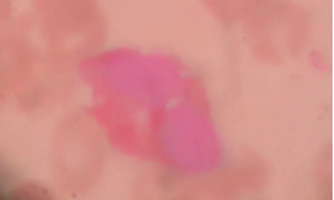
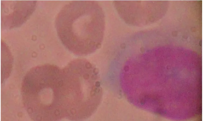
Comparative study of the first and second modified Leishman staining techniques revealed clearer morphologic pictures with the latter. Results are presented as photomicrographs of stained slides as represented by Figs. 1A and B – 3A, B and C.
Fig. 1A. Photomicrograph of Thin blood film showing a Monocyte and normocytic normochromic RBCs stained with Unmodified (Conventional) Leishman stain (x1000
magnification)
Fig. 1B. Photomicrograph of Thin blood film showing normal Lymphocyte (upper right), giant platelet (lower left) and Normocytic normochromic RBCs stained with
Fig. 2A. Photomicrograph of stained thin Blood films showing normocytic, normochromicRBCs, segmented Neutrophil and Eosinophil stained with Modified
Leishman stain, 4 minutes X 1000 magnification (Phenol-Methanol method of Preparation)
Fig. 2B. Photomicrographs of stained thin Blood films showing normocytic, Normochromic RBCs, Eosinophil stained with Modified Leishman stain, 4 minutes
Fig. 3A. Photomicrograph of Thin blood film showing Eosinophil And Normocytic normochromic RBCs stained with Modified Leishman
Stain, 75seconds (X1000 magnification)
Fig. 3C. Photomicrograph of Thin blood film showing normal Lymphocyte and Normocytic normochromic RBCs stained with Modified Leishman stain, 4 Minutes
(x1000 magnification)
4.4 The Mechanism of Action of Modified Leishman Staining Techniques
Phenol alters the pH of the modified Leishman stain thus increasing its permeability by the formed elements of the fluid tissue. This alteration in pH in turn shortened the overall time required for staining. Complete dissolution of phenol in the modified Leishman stain resulted from its high solubility and reactivity with absolute methanol. Both phenol and methanol are polar, organic compounds with terminal hydroxyl functional groups which facilitate their reactivity. Phenol turns blue litmus paper to red. Although the pH of the modified stain could not be determined due to possible impartation of colour on the sensitive membrane of the glass electrode of the pH meter, there is evidence that modified Leishman stain is slightly more acidic than its conventional counterpart. While terminal hydroxyl (-OH) functional groups of phenol and methyl alcohol aids their reactivity, the benzene ring of phenol acts as electron withdrawing group.
4.5 Principle of Modified Leishman Staining Technique
A blood film review is first performed on an appropriately prepared and stained blood film using modified Leishman staining technique. This involves a systematic microscopic scan to estimate leukocyte count and classify them into group types, identify morphological abnormalities of erythrocyte and estimate platelet number.
5. DISCUSSION
steps which make its use impractical in larger laboratories. Simeon’s modification of Boye’s and Sterenel’s method has been developed [28]. The method is only useful when methyl alcohol is unavailable to prepare Leishman stain. Its nine stepwise procedures in quick successions make it unpopular and impractical in routine staining of blood films for differential leukocyte count and peripheral blood film examination. Researchers further have sought to perform differential counts by automation but these are not without their limitations. Flow cytometry is the reference method and the most popular among them [29-31]. Cost, need for calibrations and re-calibrations of equipment, availability of reagents and consumables, trained personnel, maintenance and technicalities of these equipment and accessibility of test to patients in resource-limited setting are some of the challenges observed with automated analyzers.
aforementioned assessments regarding the staining quality of the dye on blood films on the consistency of the staining uptake by leukocytes, red blood cells and platelets during fixing stage from slide to slide, deposition of artifacts, no quantitative loss due to non-identification or mis-non-identification of blood cells, and already established appearance of red blood cells (salmon pink) leukocytes including the nuclei (purple) and cytoplasmic granules (specific for each granulocytic leukocyte), and platelets (purple). When stained thin blood films were assessed by eight different researchers and scientists, the modified staining technique demonstrated these good qualities especially when staining procedure was performed using the 4 minutes procedure (i.e. the second staining technique) more than the 75 seconds procedure. A properly stained thin film when appropriately stored can keep for 6 months to 1 year.
Many factors have been identified which interfere with staining quality. Most of the factors affect both conventional and modified Leishman stain alike. Among the factors include non-absoluteness of methyl alcohol. Absolute methanol with ≤ 1% water gave excellent staining results while methanol with ≥5% water introduces artifacts into the blood film. The degree of artifacts depends on the degree of deviation from the absoluteness of this solvent. Both refrigerated and high temperature affect the quality of staining but the effect is more on the modified Leishman stain as high temperature of close to 46ºC induces oxidation of phenol and shortens the shelf-life of the stain. Other factors that affect the quality of staining of both conventional Leishman stain and modified Leishman stain include non-performance of test according to standard operating procedures, using buffer or tap water of incorrect pH. The effect of using buffer of incorrect pH is more on the quality of staining produced by modified Leishman stain than the conventional counterpart. This includes but not limited to non-staining of the basophilic or acidophilic features of leukocytes distinctly. Our research findings did not differ from what Nguyen, Jean et al. and Dacie and his group presented on causes of faulty staining [5,16,32].
Of significant importance is our finding on the effect of using powdered latex gloves while staining. The use of powdered latex gloves creates appearance of tiny droplets on blood film as can be observed in Figs. 1A, 3A and 3C which affect absolute clarity of morphologic pictures. The more powdered the latex gloves the more the degree of appearance of tiny droplets of artefactual deposits on the blood film. Comparing the morphologic appearance of eosinophils in Figs. 2A, 2B and 3A, it is very clear that the orange-red colour which characterizes Eosinophil’s granules stands out more clearly with the second procedure than the first.
the addition of phenol in the modified method. We assume phenol’s alteration of the overall pH of modified Leishman staining solution may be responsible.
6. PRECAUTIONS
The stain should not be refrigerated or kept above room temperature for optimal staining quality. Highly purified and acetone-free absolute methanol must also be used and powdered-free gloves are encouraged. Phenol should be handled with care with limited exposure time owing to its established toxicity. The Scientist should be on necessary protective wares during preparation.
7. CONCLUSION
Modified Leishman stain and its applications will significantly affect the practice of diagnostic haematology. This inference is based on the simplicity and practicality of staining techniques and cost-effectiveness of the modified Leishman stain when compared to the ease of income generation. Besides, the stability of the stain and the understanding of the mechanism of action of this novel dye as well as opportunity to have sizeable batches of thin blood films stained lend credence to its use in diagnostic haematology laboratories. With the advent of automated slide stainer, batches of thin blood films can be stained within 75 seconds and 4 minutes in busy private and public laboratories. With the advent of these new rapid techniques in place, morbidity and mortality which resulted from late haematological findings will be drastically reduced especially in hospitalized patients with severe illness.
CONSENT
All authors declare that written informed consent was obtained from the patients (or other approved parties) for publication of this case report and accompanying images.
ETHICAL APPROVAL
This research work was performed according to ethical guidelines of Federal Medical Centre, Ido Ekiti following an informed consent to perform routine analysis. No bio-data of the patients were required for research.
COMPETING INTERESTS
Authors have declared that no competing interests exist.
REFERENCES
1. Woronzoff-Dashkoff KP. The
Erlich-Cheminsky-Grumwald-Leishman-Ructer-Wright-Giemsa-Lillie-Roe-Wilcox stain. The mystery unfolds. Clin Lab Med.1993;13:759-771. 2. Sood R. Staining of blood film: Medical Laboratory Technology, 3rdEd; 1991.
3. Marshall PN. Methylene blue-azure B-eosin Y as a substitute for May-Grumwald Giemsa stains. Micro Acta. 1977;79:153-156.
4. Wittekind D. On the nature of Romanowsky dyes and the Romanowsky-Giemsa effect.
5. Jean Safer. Preparation of blood films for examination. In: Anne S, Cheryl AL and John AK. Clinical Haematology: Principles, procedures and correlations. 2nd Ed.
Lippincott Philadelphia, New York; 1998.
6. International Committee for Standardization in Haematology. Reference method for staining blood and bone marrow films by azure B and eosin Y (Romanowsky stain). Br J of Haematol.1984;57:707-710.
7. Lapen DA. Standardized differential stain for haematology. Cytometry. 1982;2:309-315.
8. Marshall PN, Bentley SA, Lewis SM. Staining properties and stability of a standardized Romanowsky stain. J Clin Pathol. 1978;31:280-(en rule)282.
9. Bins M, Huighes W, Halie MR. Stability of azure B–eosin Y staining solutions. Br J Haematol. 1985;59:73-78.
10. Lillie RD. Factors influencing the Romanowsky staining of blood films and the role of methylene blue J Lab Clin Med. 1944;21:1181.
11. Lubrano GJ, Dean WW, Heinsohn HG et al. The analysis of some commercial dyes and stain by high performance liquid chromatography. Stain Technol 1977;52:13. 12. Marshall PN, Bentley SA, Lewis SM. Standardization of Romanowsky stains: The
relationship between stain composition and performance. Scand J Haematol. 1978;20:260.
13. Marshall PN, Galbraith WG, Navaro EF. Microspectrophotometric studies of Romanowsky stained blood cells. J Microscopy. 1981;124:197.
14. Power KT. The Romanowsky stains: A review. Am J Med Technol. 1982;48:519. 15. Woronzoff-Dashkoff KK. The Wright-Giemsa stain. Secrets revealed. Clin Lab Med.
2002;22:15-23.
16. Bain Mitchell. Lewis: Basic haematological techniques. In: Dacie and Lewis Practical Haematology, 8thEds; 1995.
17. Belgaumi UI, Shetty P. Leishman Giemsa cocktail as a new, potentially useful cytological technique comparable to Papaniculaou staining for oral cancer diagnosis. 2013;30:18-22.
18. Mathur A, Tripathi AS, Kuse M. Scalable system for classification of white blood cells from Leishman stained blood stain images. J Pathol Inform. 2013;4:15.
19. Houwen B. The differential count. Lab Haematol. 2001;7:89-100.
20. Bain BJ, Lewis SM. Preparation and staining methods for blood and bone marrow films. In: Barbara JB, Imelda B, Michael AL and Lewis SM. Dacie and Lewis Practical Haematology, Churchill Livingstone, New York. 11thEds; 2012.
21. Avwioro OG. Staining histochemistry and Tissue pathology: Principles and technique. 1stEds. 2002;137.
22. Weber Manfred, Weber Markus, Kleine-Boymann M. "Phenol". Ullmann's Encyclopedia of Industrial Chemistry; 2004.
23. IUPAC, Compendium of Chemical Terminology, 2nd ed. (the “Gold Book”); 1997. Online corrected version: (2006- ): “Alcohols”.
24. IUPAC, Compendium of Chemical Terminology, 2nd ed. (the “Gold Book”); 1997.
Online corrected version: (2006- ): “Phenols”.
25. Smith MB, March J. Advanced Organic Chemistry: Reactions, Mechanisms, and Structure (6thed.); 2007. New York: Wiley-Interscience.
26. Jim Clark. The Acidity of Phenol. Chem Guide; 2007.
27. Yvutte E, Elisabeth E, Birgit R, Hanspeter R, Niklaus W. Staining blood films with Field’s stain. Methods in Parasitology. Swiss Institute. Basel; 2005.
29. Hubi W, Wolfbauer G, Andert S, et al. Toward a new reference method for the leukocyte five-part differential. Cytometry.1997;30:72-84.
30. Fujimoto H, Sakata T, Hamaguchi Y, et al. Flow cytometric method for enumeration and classification of reactive immature granulocytes populations. Cytometry.
2000;42:371-378.
31. Faucher JL, Lacronique-Gazaille C, Frébet E, et al. ‘6 markers/5 colors’ extended white cell differential by flow cytometry. Cytometry. 2007;71:934-944.
32. Nguyen DT, Moskowitz FB, Diamond LW. Potential diagnostic pitfalls caused by blood film artifacts in prolymphocytic leukaemia. Observations in two cases. Br J Biomed Sci. 1994;51:371.
© 2014 Fasakin et al.; This is an Open Access article distributed under the terms of the Creative Commons Attribution License (http://creativecommons.org/licenses/by/3.0), which permits unrestricted use, distribution, and reproduction in any medium, provided the original work is properly cited.
Peer-review history:
The peer review history for this paper can be accessed here: