_____________________________________________________________________________________________________ ISSN: 2231-0614
SCIENCEDOMAIN international www.sciencedomain.org
An Improved Hemicorporectomy Technique
M. Burhan Janjua
1*, David C. Crafts
2,3and Frank E. Johnson
2,31
Washington University School of Medicine, St. Louis, MO, USA. 2
Saint Louis University Medical Center, St. Louis, MO, USA. 3
Veterans Affairs Medical Center, St. Louis, MO, USA.
Authors’ contributions
This work was carried out in collaboration among all authors. Author FEJ designed the study, wrote the protocol, and author MBJ wrote the first draft of the manuscript. Author MBJ managed the literature searches. Authors FEJ and DCC performed the operations while, author DCC devised the innovation in the technique. Author MBJ wrote the manuscript and author FEJ revised the final draft. All authors read and approved the final manuscript.
Article Information
DOI: 10.9734/BJMMR/2015/13742 Editor(s): (1)Salomone Di Saverio, Emergency Surgery Unit, Department of General and Transplant Surgery, S. Orsola Malpighi University Hospital, Italy. Reviewers: (1)Georgios Androutsopoulos, Department of Obstetrics and Gynecology, University of Patras, Medical School, Greece. (2)Anonymous, Neurosurgery, TUMS, Iran. Complete Peer review History:http://www.sciencedomain.org/review-history.php?iid=715&id=12&aid=6593
Received 1st September 2014 Accepted 23rd September 2014 Published 22nd October 2014
ABSTRACT
Background: The first attempted hemicorporectomy, also known as translumbar amputation (TLA), was reported in 1960. The first TLA with survival was performed in 1961. It is a lifesaving procedure initially designed for carefully selected patients with otherwise terminal cancer. The most common indications now are benign conditions such as chronic osteomyelitis of the pelvis in paraplegic patients. It is also the only procedure in which the spine is electively divided. We report our experience with four patients who had this operation, all done in two stages.
Methods: We reviewed the current literature and report techniques used in our series.
Results: We found 20 references via computer search; 14 described technical features. We describe our current technique in this report.
Conclusion: TLA can be carried out with good results. Our technique minimizes blood loss, decreases operative time, and preserves one vertebral body, compared to other techniques.
Summary: Hemicorporectomy is rarely performed. We discuss the history and rationale of the operation and describe what we consider the optimal technique, based on our series of four, with a minimal complication rate and zero mortality.
Keywords: Hemicorporectomy; translumbar amputation; division of the spine.
1. INTRODUCTION
Translumbar amputation (TLA) was first proposed by Kredel in 1950 as a salvage procedure for patients with cancer too advanced for pelvic exenteration [1]. The first attempted clinical use of the procedure in 1960 is attributed to Kennedy et al. [2]. but this patient did not survive. The TLA reported by Aust and Absolon [3] and Aust and Page [4] was the first with patient survival. Miller reported a personal series of 10 patients in 1982 [5]. Thirty-four TLA procedures had been reported in the world literature by 1990 [6]. In 2009, Janis et al., at the University of Texas Southwestern Medical Center reported a single-institution experience of nine patients. By this time, 66 TLA procedures had been reported [7]. Events have proved that the operation is conceptually sound, humane and ethically acceptable.
We found no reports describing any surgical technique in detail and enumerating the current indications for TLA. In this report we describe the operative techniques we used in our series of four patients, our results, preoperative measures to be considered, and postoperative precautions.
2. METHODS
We reviewed our series, all carried out by one senior surgeon (FEJ). All of our patients had a two-stage procedure. The PubMed/MEDLINE online database was accessed using keywords “hemicorporectomy” and “translumbar amputation.” Various relevant medical and surgical textbooks were also reviewed. We did not consider any reports dealing with a one-stage TLA procedure. In our series there were three patients with paraplegia and intractable pelvic osteomyelitis and 1 with a recurrent sacral chordoma causing ulceration, severe radicular pain and severe progressive paraparesis with debilitating infection.
3. RESULTS
We found 20 pertinent references via computer search; 14 described some technical features of this operation; [3-16] 4 dealt with [17-20] metabolic events. One was a collective review [7]. The technique of TLA has not been described in detail before, as far as we can determine.
The one-stage procedure is generally done for emergencies only because of the complications that are likely to arise when operating on ill patients under emergency circumstances [13]. It involves urinary diversion, colostomy, and division of the body wall, great vessels, and spine. We have done no one-stage procedures. The two-stage procedure is the procedure of choice for nonemergency situations.
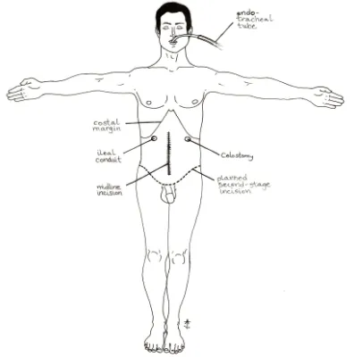
The remainder of this report concerns the second stage of an elective two-stage procedure. The anterior approach (supine position) is suitable for the first stage of a two-stage operation and is familiar to surgeons. It entails the formation of an ileal conduit and colostomy (preferably sigmoid). Other procedures deemed useful (appendectomy, cholecystectomy, etc.) are performed as needed (Fig. 1). Both stomas must be placed high enough so that the patient can see them and can eventually sit in a “bucket” prosthesis. The colon distal to the stoma is resected to the level of the rectum. After the patient recovers, stage 2 can be initiated using an anterior or posterior approach. Most reports in the literature featured an initial supine position for the second stage of a two-stage procedure [4-6,11,12]. One featured a decubitus position for a patient with a previous hemipelvectomy [8]. Our current practice is to begin with stage 2 with the patient in prone position. Our technique has not been described before.
The anterior approach for stage 2 has been considered a standard strategy. After the patient is placed in supine position, a low transverse incision is made across the pubis, retaining the inguinal ligaments (and the testicles on their pedicles). An extraperitoneal approach to the lumbar spine and great vessels is undertaken. The aorta and inferior vena cava are divided. Because it is advantageous to leave as much room as possible for the abdominal contents, the spine is divided at the lowest level compatible with fully resecting the pathology, usually L3-4, L4-5, L5-S1. Several authors describe dividing the spine by a simple discectomy. This provides very limited access to the thecal sac and posterior elements. This limits closure of the thecal sac, with unnecessary risk of CSF leak, which can cause postural headaches, impaired wound healing or meningitis. Miller [5] describes just packing the spinal canal with muscle, which may leave CSF in contact with necrosing muscle. One of his patients died at two months postoperatively of hydrocephalus, cause not specified.
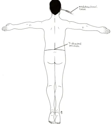
Alternatively, one can resect a vertebral body, allowing space for transverse division of thecal sac and cauda equina, with watertight closure. This costs one rostral vertebral level, takes longer, and may be accompanied by copious blood loss. The wound is then covered with sterile surgical sponges and a sterile plastic membrane dressing. The patient is turned to the prone position and a transverse incision over the pelvis is made. This connects to the anterior incision. A T-extension over spinous processes is made (Fig. 2), exposing the posterior bony elements. The spinous process and facets of the lowest remaining vertebra are also excised to complete the division of spine and to prevent a potential pressure point [14]. Remaining soft tissues of the trunk are divided and the caudal body parts are removed from the operating table. The wound is then closed in layers, dressed as required, and the patient is returned to the supine position. The patient is awakened and sent to the ICU.
We utilized this approach with vertebral corpectomy in our first two cases. At this point, one of the authors (DCC) devised a technique to minimize blood loss (500 ml. less than the initial anterior approach) that required different initial positioning for stage 2 of a two-stage TLA. We utilized this improved technique in our next two cases. This new technique features an initial prone position for the second stage of a TLA. It
allows preservation of one vertebral unit, as compared to our previous technique. A transverse incision at the level of the iliac crest is made. It crosses the midline at the level chosen as the lowest compatible with elimination of the original pathology, usually L3-4 or L4-5. A midline T extension is carried rostrally far enough to expose the spinous process and lamina of the rostral vertebra (Fig. 2).
Fig. 2. The patient is initially prone during stage 2
A transverse incision is made at the level of the iliac crests, with a T-extension to the chosen lumbar vertebra to expose the posterior bony elements. The
posterior bony elements are resected, the dura is opened transversely, the cord elements are divided, the chosen intervertebral disk is marked, and the body
wall is divided posteriorly. The wound is dressed with sterile sponges and sterile plastic membrane dressings. The patient is turned to the supine position,
the anterior body wall is divided, the aorta and vena cava are divided and the appropriate intervertebral disk is divided. The lower body parts are removed. The wound is again dressed with sterile sponges and
plastic membrane dressings. The patient is turned to the prone position and the wound is closed.
with sutures and fibrin glue. Facet joints are resected laterally and remaining bony elements are trimmed as needed to be sure there will be no pressure points. The posterior longitudinal ligament is incised across the disk space. Some disk may be removed and methylene blue can be injected so that it can be seen when the patient is turned to the supine position. Other methods such as placement of a radiopaque marker in the disc of interest and on-table x-ray also reportedly work well. The operative wound is then covered with surgical sponges and a sterile plastic membrane dressing. The patient is next turned to the supine position. The abdomen is opened with a low transverse incision, as described earlier, and the great vessels and the remaining truncal tissues are divided. The testicles can be preserved by mobilizing their vascular pedicle. The spine is divided through the marked intervebral disc with a knife. The amputated portion of the body is removed. The operative wound is then covered with surgical sponges and sterile plastic dressing. The patient is turned to the prone position again and the wound is closed in layers (Fig. 3). The patient is then awakened and sent to the ICU. This innovative technique (the initial posterior approach) reduced the operative time by 60-80min(Table 1).
The only complication in our series was minor skin necrosis in three cases. All of our patients regained good health soon after the TLA. A custom-made prosthesis enabled wheelchair use (Fig. 4). All four patients indicated that they were pleased with the results.
Fig. 3. The patient is shown after the amputation with the incision closed
Fig. 4. The patient is shown sitting in a bucket prosthesis
All artwork was created by Alea Ahmadian
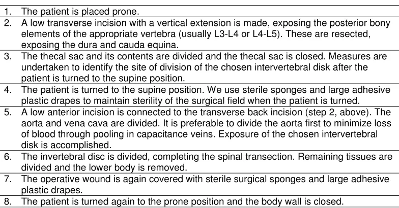
Table 1. Initial Posterior Approach for stage 2 of a 2-stage TLA
1. The patient is placed prone.
2. A low transverse incision with a vertical extension is made, exposing the posterior bony elements of the appropriate vertebra (usually L3-L4 or L4-L5). These are resected, exposing the dura and cauda equina.
3. The thecal sac and its contents are divided and the thecal sac is closed. Measures are undertaken to identify the site of division of the chosen intervertebral disk after the patient is turned to the supine position.
4. The patient is turned to the supine position. We use sterile sponges and large adhesive plastic drapes to maintain sterility of the surgical field when the patient is turned. 5. A low anterior incision is connected to the transverse back incision (step 2, above). The
aorta and vena cava are divided. It is preferable to divide the aorta first to minimize loss of blood through pooling in capacitance veins. Exposure of the chosen intervertebral disk is accomplished.
6. The invertebral disc is divided, completing the spinal transection. Remaining tissues are divided and the lower body is removed.
7. The operative wound is again covered with sterile surgical sponges and large adhesive plastic drapes.
4. DISCUSSION
TLA is rarely done. Few cases are done for cancer at present because of poor long-term results [5,6,11,12,20]. Now the dominant indication in wealthy countries is undoubtedly uncontrollable sepsis due to intractable pelvic osteomyelitis from pressure ulcers in spinal cord injury patients [2,7,10,16]
Preoperative evaluation must be extensive as candidates for TLA may be desperately ill. He or she must possess strong motivation to undergo this operation. Preoperative psychological counseling is strongly recommended [9,10,20]. Echocardiogram [11] and spirometry [7,19] are encouraged. Nutritional status should be optimized. Sepsis is treated as circumstances require [8,9,14,17].
Several cautions for the surgeon and the anesthesiologist are important. A multidisciplinary team including an experienced surgical oncologist and/or neurosurgeon is helpful [7,12]. The anesthesiologist should carefully monitor physiological parameters during administration of fluids. Anecdotal evidence implicates fluid overload as the major cause of perioperative pulmonary edema, heart failure and death [7,10,16,18]. The reason for this is that the major capacitance veins in the pelvis and legs are gone. Loss of skin surface and muscle mass can lead to impaired body temperature regulation as well [20].
Family support is important to deal with post-operative challenges [5,7,12,15,16] Males with testicles preserved on their vascular pedicles typically have poor testosterone production. Testosterone patches are typically used in such patients within two weeks of the operation.17 Males can have children if sperm has been banked. An appropriately modified automobile with hand controls can enable driving. Some patients can do sedentary work. Notably, most of our patients have given up the lower body in their body image before the TLA and their mobility is much improved postoperatively. Frequently relief from pain and/or systemic effects of infection is dramatic.
We have attempted to present pertinent considerations for those contemplating this operation. We believe our technique is the optimum technique for most patients, realizing that the physiological and anatomic details of each patient will be unique.
5. CONCLUSION
TLA can be carried out with good results. The two-stage procedure is preferred, if feasible. The initial posterior approach for stage 2 decreases operative time, minimizes blood loss, and preserves one vertebral body when compared with the initial anterior approach for stage 2.
CONSENT
Not applicable.
ETHICAL APPROVAL
Not applicable.
COMPETING INTERESTS
Authors have declared that no competing interests exist.
REFERENCES
1. Bricker EM, Modlin J. The role of pelvic evisceration in surgery. Surgery. 1951;30:76-94.
2. Kennedy CS, Miller EB, McLean DC, et al. Lumbar amputation or hemicorporectomy for advanced malignancy of the lower half of the body. Surgery. 1960;48:357-365. 3. Aust JB, Absolon KB. A successful
lumbosacral amputation, hemicorporec-tomy. Surgery. 1962;52:756-9.
4. Aust JB, Page CP. Hemicorporectomy. J Surg Oncol. 1985;30:226-230.
5. Miller TR. Traslumbar amputation (hemicorporectomy). Prog Clin Cancer. 1982;8:227-236.
6. Ferrara BE. Hemicorporectomy: a collective review. J Surg Oncol. 1990;45: 270-278.
7. Janis JE, Ahmad J, Lemmon JA, et al. A 25-year experience with hemicorporec-tomy for terminal pelvic osteomyelitis. Plast Reconstr Surg. 2009;124:1165-1176. 8. Barnett CC Jr, Ahmad J, Janis JE, et al.
Hemicorporectomy: back to front. Am J Surg. 2008;196:1000-1002.
9. Stelly TC, McNeil JW, Snypes SR, et al. Hemicorporectomy. Clin Anat. 1995;8:116-123.
11. Pearlman NW, McShane RH, Jochimsen PR, Shirazi SS. Hemicorporectomy for intractable decubitus ulcers. Arch Surg. 1976;111:1139-1143.
12. Terz JJ, Schaffner MJ, Goodkin R, et al. Translumbar amputation. Cancer. 1990;65:2668-2675.
13. Abrams J, et al. Hemicorporectomy for acute aortic occlusion: a case study. Am Surg.1992;58:509-512.
14. Norris JE, Kwon YB, Puangsuvan S, et al. Hemicorporectomy: a case report. Am Surg.1973;39:344-348.
15. Wagman LD, Terz JJ. Translumbar Amputation (hemicorporectomy): Surgical Procedures. In: Bowker JH, ed. Atlas of Limb Prosthetics: Surgical, Prosthetic, and Rehabilitation Principles 2nd ed. St. Louis: Mosby Year Book. 1992;553-561.
16. Weaver JM, Flynn MB. Hemicorporec-tomy. J Surg Oncol. 2000;73:117-124. 17. Grimby G, Stener B. Physical performance
and cardiorespiratory function after hemicorporectomy. Scand J Rehabil Med. 1973;5:124-129.
18. Lamis PA Jr, Richards AJ Jr, Weidner MG Jr. Hemicorporectomy: Hemodynamics and metabolic problems. Am Surg. 1967;33:443-448.
19. Bake B, Grimby G. Regional ventilation and gas exchange after hemicorporec-tomy. Thorax. 1974;29:366-370.
20. Shafir M, Abel M, Tasuk H, et al. Hemicorporectomy - perioperative management: a case presentation and review of literature. J Surg Oncol. 1984;26:79-82.
© 2015 Janjua et al.; This is an Open Access article distributed under the terms of the Creative Commons Attribution License
(http://creativecommons.org/licenses/by/4.0), which permits unrestricted use, distribution, and reproduction in any medium,
provided the original work is properly cited.
Peer-review history: