_____________________________________________________________________________________________________ *Corresponding author: Email: [email protected], [email protected];
www.sciencedomain.org
Prevalence of Down Syndrome in Western India:
A Cytogenetic Study
Gadhia Pankaj
1*, Kathiriya Avani
1and Vaniawala Salil
11
Molecular Cytogenetic Unit, S.N.Gene Laboratory and Research Centre President Plaza – A, Surat, India.
Authors’ contributions
This work was carried out in collaboration between all authors. Author GP designed the study, wrote the protocol, and the first draft of the manuscript. Author VS analyses of the study, performed the microscopic analysis and managed the experimental process and author KA managed the literature searches and analysis of data. All authors read and approved the final manuscript.
Article Information
DOI: 10.9734/BJMMR/2015/13648 Editor(s): (1) Paulo Ricardo Gazzola Zen, Departament of Clinical Medicine, Federal University of Health Sciences of Porto Alegre (UFCSPA), Brazil.
Reviewers: (1) Qinghua zhou, Department of Genetics, Yale University School of Medicine, New Haven, CT, USA. (2) Anonymous, Federal University of Juiz de Fora, Juiz de Fora, MG, Brazil. (3) Anonymous, Federal University of Triângulo Mineiro, Uberaba, Minas Gerais, Brazil. (4) Anonymous, Mansoura University, Egypt. Complete Peer review History: http://www.sciencedomain.org/review-history.php?iid=717&id=12&aid=6789
Received 26th August 2014 Accepted 15th October 2014 Published 5th November 2014
ABSTRACT
Aim: To study the prevalence of Down syndrome by conventional chromosome analysis and G-banded karyotyping.
Materials and Methods: A retrospective analysis was performed on the case records of2750 paediatrics patients, of which 682 cases of confirmed Down syndrome was recorded by G-banding karyotyping.
Results: Non-disjunction was the most common type of abnormality followed by Robertsonian translocation and lastly mosaic in ratio of 92.2:7.0:0.73 respectively.
Conclusion: Results suggest that advanced maternal age is classic risk factor attributed to the incidences of Down syndrome.
1. INTRODUCTION
Down syndrome (DS) is a common chromosomal anomaly associated with multiple congenital mal formations and mental retardation (MR) in humans. The incidences in India are 1 per 850-900 live births [1,2].
The extra 21 chromosome of DS individuals in 95% cases possess as free trisomy resulting from non-disjunctional error of chromosome 21 during gametogenesis. On the other hand, 2 to 4% showed a Robertsonian translocation and 1 to 3% showed mosaicism.
The clinical features are important for an early diagnosis to reduce morbidity and mortality. Apart from karyotype most characteristic features are mental retardation, congenital heart defects, facial features and in some cases developing AML at latter stage [3].
The relation of advanced maternal age to an increased risk of DS have been well documented. In addition, maternal risk other than maternal age for DS births is clearly includes genetic and other risk factors in few published studies. With regard to genetic factor, Coppode et al. [4] have studied the role of methionine synthase reductase (MTRR), G-polymorphism as maternal risk factor for birth of DS in Caucasian women. On the other hand, Shalaby [7] explained other risk factors such as consanguinity, drug and environmental toxins and reproductive functions for increase incidences of DS.
The present investigation is aimed to study the prevalence of Down syndrome in Gujarat and Western India and its possible cause.
2. MATERIALS AND METHODS
The study included a total of 682 children with DS confirmed by cytogenetic diagnosis during 10 year period (from 2004-2014). The peripheral blood samples were used for cytogenetic diagnosis. While receiving blood samples informed consent was obtained from parents.
Chromosome preparation was done from 4-5 ml of peripheral blood collected in heparin for all subjects. Giemsa-Trypsin-Giemsa (GTG) banding was performed according to method of Seabright [5]. In each case minimum 30 metaphases were examined and minimum 5 well
and they were karyotyped by IKAROSE software (Carl-Zeiss, Germany). In case of mosaicism minimum of 70 metaphases were scored.
3. RESULTS
A total of 2750 clinically suspected cases were referred to our diagnostic laboratory for cytogenetic analysis. Of which 682 cases were found confirmed DS. The rest 2068 cases were found to be normal and did not show any chromosomal anomalies. The mean maternal age of all DS calculated and it was found to be 31.3 years in free trisomy. The data analysis showed that maternal age was above 35 years in 36% cases. On the contrary, it was lowered 28.6 years in traslocation and 26.2 years in mosaic DS (Table 1).
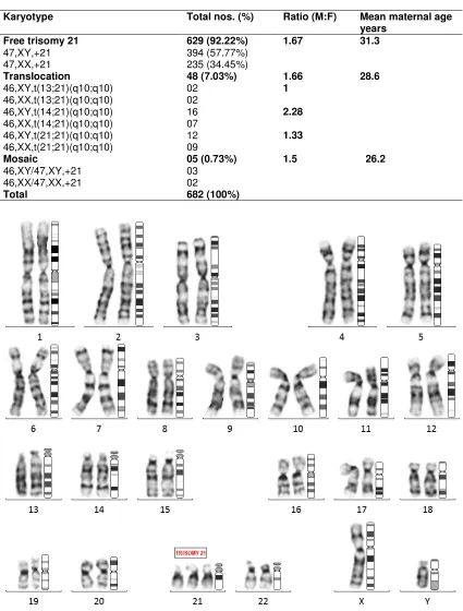
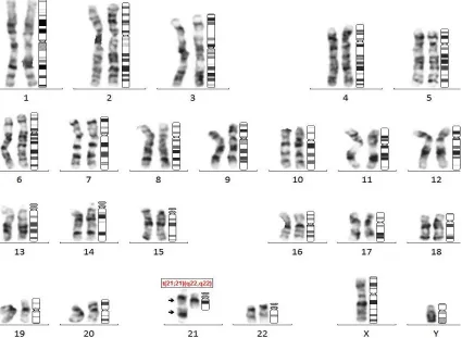
The abnormal karyotypes are listed in Table 1. Chromosomal non-disjunction was the most common type of abnormality followed by translocation and lastly mosaic 92.2, 7.0 and 0.7% respectively. Table 1 also shows a marked excess of males in non-disjunction and translocation groups as compared to mosaic DS. Fig. 1 shows G-banded karyotyped of male with 21 trisomy, while Fig. 2 depicts male DS with 46,XY,der(21;21)(q10;q10) +21.
4. DISCUSSION
The patients reported in this study on DS in Gujarat and Western India were all diagnosed postnatally. The frequency of free trisomy reported in the present study is most common seen in 92.2% of cases which is in accordance with earlier reports [1,2,11].
Cytogenetic study was carried out from March 2004 to July 2014 in which 2750 cases were referred to our laboratory for confirmation of DS, from these 682 were confirmed as DS cases. The frequency of free trisomy was 92.2% (629 cases), RS translocation was 7.0% (48 cases)
and frequency of mosaicism was 0.73% (5 cases).
Table 1. Karyotype analysis of 682 Down syndrome case
Karyotype Total nos. (%) Ratio (M:F) Mean maternal age
years Free trisomy 21
47,XY,+21 47,XX,+21
629 (92.22%)
394 (57.77%) 235 (34.45%)
1.67 31.3
Translocation
46,XY,t(13;21)(q10;q10) 46,XX,t(13;21)(q10;q10) 46,XY,t(14;21)(q10;q10) 46,XX,t(14;21)(q10;q10) 46,XY,t(21;21)(q10;q10) 46,XX,t(21;21)(q10;q10)
48 (7.03%)
02 02 16 07 12 09
1.66 1
2.28
1.33
28.6
Mosaic
46,XY/47,XY,+21 46,XX/47,XX,+21
05 (0.73%)
03 02
1.5 26.2
Total 682 (100%)
Fig. 2. Shows G-banded karyotyped of male Down syndrome with 46, XY, t(21;21)(q10;q10),+21
In this study the overall sex ratio (male/female) was 1.67:1. The excess of males appears to be universal and reported in literature from all over the world. In addition excess of males were also reported in the present study in both translocation and mosaic which are unique in nature as compared to previous studies [2].
Maternal age at birth of DS children ranged from 25 to 43 years with mean maternal age of 31.3 years. There were reports of older maternal age having DS in previous studies from different countries [9,10]. On the other hand, few studies have also reported incidences of DS to a much young maternal age [11,12].
Although several studies have been carried out on the incidence of DS, but a complete understanding of the mechanism is yet to be ascertained. In our study mean maternal age was 31.3 years in free trisomy but in translocation it was 28.6 years and the least was in mosaic 26.2 years. Similar study carried out by Sheth et al. [11] have shown mean maternal age
translocation, 25.0 years in mosaic which are not in accordance with our study, though both the studies were basically carried out in Gujarat.
With regards to other risk factors, unlike maternal age, recently Coppode et al. [4] have suggested folate polymorphism as genetic risk factor for birth of DS child in Caucasian women and Shalaby [7] reported consanguinity, drug and environmental toxins as other risk factors.
In conclusion, it is important to note that advanced maternal age in the present study could be one of the classic risk factors for higher incidences in Down syndrome. Hence, it is important to educate women at high risk occurrence (e.g. advanced maternal age) to go for screening during pregnancy. The concept of preventive genetics should be reinforced with the National policy in the form of health insurance.
5. CONCLUSION
common type of Down syndrome chromosomal abnormality in Western India.
CONSENT AND ETHICAL APPROVAL
All authors declare that informed consent was obtained from the patients for publication of this research work. We certify that we have participated sufficiently in the intellectual content, conception and design of this work or the analysis and interpretation of the data, as well as the writing of the manuscript, to take public responsibility for it and have agreed to have our name listed as a contributor.
We believe the manuscript represents valid work. Neither this manuscript nor one with substantially similar content under our authorship has been published or is being considered for publication elsewhere. We certify that all the data collected during the study is presented in this manuscript and no data from the study has been or will be published separately.
We hereby transfer, assign, or otherwise convey all copyright ownership, including any and all rights incidental thereto, exclusively to the British Journal of Medicine and Medical Research.
COMPETING INTERESTS
Authors have declared that no competing interests exist.
REFERENCES
1. Malini SS, Ramchandra NB. Influence of advanced age of maternal grandmother on Down syndrome. BMC Med. Genet. 2006;7:4.
2. Kaur A, Singh J. Chromosomal abnormalities: Genetic disease burden in India. Int. J. Hum. Genet. 2010;10(1-3):1-14.
3. Podder G, De A, Adhikari A, Halder A, Banerjee J, De M. Assessment of Down
syndrome patients in west Bengal, India. Pacific J Med Sci. 2012;10(2):28-35. 4. Coppedè F1, Bosco P, Lorenzoni V,
Denaro M, Anello G, Antonucci I, Barone C, Stuppia L, Romano C, Migliore L. The MTRR c.66A>G polymorphism and maternal risk of birth of a child with Down syndrome in Caucasian women: a case control study and a meta-analysis. Mol. Biol. Rep. 2014;41(9):5571-83.
5. Seabright M. A rapid banding technique for human chromosome. Lancet. 1971;2:971-2
6. Fisch H, Hyun G, Golden R, Hensle TW, Olsson CA, Liberson GL. The influence of paternal age on Down syndrome. J Urol. 2003;169:2275-78.
7. Shalaby HMA. A study of new potential risk factors for Down syndrome in Upper Egypt. The Egypt J. Med. Human Genetics. 2011;12:15-19.
8. Sherman SL, Freeman SB, Allen EG, Lamb NE. Risk factors for non – disjunction of trisomy 21. Cytogenet Genome Res. 2005;111(3-4):273-80. 9. Azman BZ, Ankathil R, Siti Mariam I,
Suhaida MA, Norhashimah M, Tarmizi AB, Nor Atifah MA, Kannan TP, Zilfalil BA. Cytogenetic and clinical profile of Down Syndrome in Northeast Malaysia. Singapore Med J. 2007;48:550-554. 10. Jaouad IC, Cherkaoni DS, Sbiti A, Natiq A,
Elkerch F, Sefiani A. Cytogenetic and epidemiological profiles of Down syndrome in Moroccan Population: A report of 852 cases. Singapore Med J. 2010;51:133-136.
11. Sheth F, Rao S, Desai M, Vin J, Sheth J. Cytogenatic analysis of Down syndrome in Gujarat. Indian Peditrics. 2007;44:774-777. 12. Chandra N, Cyril C, Lakshminarayana P, Nallasivam P, Ramesh A, Gopinath PM, et al. Cytogenetic evaluation of Down Syndrome: A review of 1020 referral cases. Int. J. Humn. Genet. 2010;10:87-93.
© 2015 Pankaj et al.; This is an Open Access article distributed under the terms of the Creative Commons Attribution License
(http://creativecommons.org/licenses/by/4.0), which permits unrestricted use, distribution, and reproduction in any medium,
provided the original work is properly cited.
Peer-review history: