Copyright 0 1992 by the Genetics Society of America
Oxygen Association-Dissociation
and Stability Analysis
on Mouse
Hemoglobins With Mutant
CY-and ,&Globins
Stephen
J.
D'Surney and RaymondA. Popp
Biology Division, Oak Ridge National Laboratory, and the University of Tennessee, Oak Ridge Graduate School of Biomedical Sciences, Oak Ridge, Tennessee 37831
Manuscript received March 24, 1992 Accepted for publication June 12, 1992
ABSTRACT
Oxygen association-dissociation and hemoglobin stability analysis were performed on nlouse he- moglobins with amino acid substitutions in an a-globin (a89, His to Leu) and a &globin (059, Lys to Ile). The variant a-globin, designated chain 5'" in the Hbag' haplotype, had a high oxygen affinity and was stable, The variant &globin, (p') of the Hbb" haplotype, also had an elevated oxygen affinity and in addition was moderately unstable in 19% isopropanol. Hemoglobins from the expected nine (lfbag'l Hbag2;Hbbs/Hbbs X Hba"/Hba";Hbb"'/Hbb"') FS genotypes can be grouped into five classes of P5(, values
characterized by strict additivity and dependency on mutant globin gene dosage; physiologically, bot,h globin variants gave indistinguishable effects on oxygen affinity. The hemoglobin of normal mice
(Hba"/Hba";Hbb'/Hbb') had a Pa,, = 40 mm Hg and the hemoglobin of Hbag2/Hbag2;Hbb2/Hbbr? Fs
mice had a P5,) = 25 mm Hg (human P5,, = 26 mm Hg). Peripheral blood from Hba@/Hba@;Hbbs/ Hbb', Hba"/Hbaa;Hb6"'/Hbb'' and Hbag'/Hbag';Hbb''/Hbb'' mice exhibited normal hematological values
except for a slightly higher hematocrit for Hbag'/Hbag';Hbb'/Hbb' and Hbag2/Hbag2;HbP2/Hbb2 mice,
slightly elevated red cell counts for mice of the three mutant genotypes, and significantly lower values for the mean corpuscular volume and mean corpuscular hemoglobin for Hbagz/Hbag22;Hbb'/Hbb2 mice.
number of high oxygen affinity and unstable
A
variants of human hemoglobin have beenidentified. Currently, a t least 19 high oxygen affinity a-chain variants and 50 high oxygen affinity @-chain variants have been characterized in humans (BUNN and FORGET 1986). Hemoglobin Chesapeake, a n a-
globin variant (a92, Arg to Leu) has a P ~ o of 19 mm H g (normal for humans is 26 mm Hg), and individuals carrying this variant have greatly elevated red blood cell counts (CLEGG, NAUGHTON and WEATHERALL
1966). Hemoglobin Brisbane, a @-globin variant (868, Leu to His) also has an elevated oxygen affinity and is mildly unstable (BRENNAN et al. 1981). At the mo- lecular level, most high oxygen affinity variants of
human hemoglobin can be attributed to amino acid changes in three regions: (1) the interface, (2)
the C-terminal end of the @-chain and (3) the 2,3- diphosphoglycerate and Bohr effect sites.
T h e r e a r e also a large number of unstable human hemoglobins. Nineteen unstable a-globin variants and 74 unstable @-globin variants have been characterized (BUNN a n d FORGET 1986). Hemoglobin Bristol (@67, Val to Asp) is a highly unstable hemoglobin variant that comprises 36% of the total hemoglobin in he- moglobin Bristol patients. Severe hemolytic anemia is observed in these patients and large Heinz body inclu- sions are found in the red blood cells. Hemoglobin Bristol is unstable because the amino acid substitution
Genetics 134: 545-551 (October, 1992)
weakens the heme-to-globin linkage (STEADMAN, YATFS and HUEHNS 1970). Other unstable human hemoglobin variants are caused by mutations that interfere with secondary (hemoglobin Ann Arbor: a80, Leu to Arg), tertiary (hemoglobin Baylor: P8 1 , Leu to Arg) and quaternary (hemoglobin Khartoum: @124, Pro to Arg) structures.
In mice, only one high oxygen affinity variant (Hbbd4/Hbbd4) and one unstable 0-globin variant
(Hbb"'/Hbb"') have been reported. Both variant glo- bins are products of mutant genes that were induced by treatment of parental mice with LV-ethyl-Ai-nitro- sourea (ENU). The mutant hemoglobin (01 45, Tyr to Cys) in Hbbd4/Hbbd4 mice has characteristics similar to hemoglobin Rainier in humans (PETERS et al. 1985); it has an increased oxygen affinity, reduced Bohr
effect, and decreased cooperativity. Severe polycy- themia occurs in animals homozygous for the Hbbd4/ ~ bmutation. b ~ ~
The mutant hemoglobin (P.59, Lys to Ilr) in mice of the Hbb5' haplotype (LEWIS et al. 1985) is moder- ately unstable (WAWRZYNIAK and POPP 1986), but its
oxygen binding properties have not been determined. An ENU-induced mutation at the mouse a-globin
locus in the Hbag' haplotype has also been described
( P ~ P P et al. 1983). In mice of the mutant Hba"' hap-
report describes the oxygen association-dissociation a n d stability properties of hemoglobins from parental mice of the Hbag2 a n d Hbb"' haplotypes and their
(Hbag'/Hbag';Hbb'/Hbb" X Hba"/Hba";Hbb"*/Hbb"') F2 offspring.
MATERIALS AND METHODS
Mice: Mice homozygous for two mutant hemoglobin hap-
lotypes Hbb" (Hba"/Hbaa;Hbb"/HbP2) B59, Lys to Ile, and Hbag' (Hbag'/Hbag';Hbb'/Hbb') a89, His to Leu, were initially selected for oxygen association-dissociation analysis because their hemoglobins contain amino acid substitutions in the vicinity of the heme-binding sites. The proposed P60 Val to Glu substitution for HbP2 (LEWIS et al. 1985) was incorrect, the correct substitution is 059 Lys to Ile
u.
JONES, personal communication). Control mouse strains were C57BL/6 (Hba"/Hba";Hbb'/Hbb') and DBA/2J (Hbag/Hbag;Hbbd/Hbbd).Homozygous Hba@ and Hbb" mice were mated to pro- duce doubly heterozygous (Hba"/Hbag';Hbb'/Hbb'') F, mice that were mated to produce F n progeny for ox gen associ- ation-dissociation and stability analyses. Hbag2j!bbag2;Hbb'/ Hbb', Hba"/Hbaa;Hbb'2/Hbb'2, and Hbag2/Hbag2;Hb6"'/Hbb'' F2 mice were selected for peripheral blood hematology analysis using an Ortho ELT-15 Automatic Hematology Analyser (Ortho Diagnostic Systems Inc.) that has been modified specifically for the analysis of mouse blood (POPP et al. 1986). The hemoglobin genotypes of all mice were determined by isoelectric focusing of hemolysates in a pH range of 7 to 9 (WHITNEY et al. 1979).
Oxygen association-dissociation analysis: Oxygen asso-
ciation-dissociation analysis on mouse hemoglobin was per- formed on a TCS Hemox Analyzer (FESTA and AKASURA 1979). A Clark oxygen electrode monitors the oxygen con- centration in the sample during oxygenation with air and deoxygenation with nitrogen. Analyses were made on 20- 70-pl samples of freshly drawn blood suspended in a solution of 4 ml of Hemox buffer at pH 7.4 plus 20 wl of serum albumin solution (additive A) and 10 PI of antifoaming agent (additive B) purchased from TCS Medical Products, South- ampton, Pennsylvania. After equilibration at 37" the he- moglobin was fully oxygenated by bubbling air through the blood sample and then completely deoxygenated by bub- bling with purified nitrogen. The hemoglobin was slowly oxygenated again by bubbling with air over a 20-min period while recording the oxygen association curve, which was used to read the P,,, values. Seven or more blood samples were analyzed from mice of each hemoglobin genotype. The oxygen association curves were similar to the oxygen dissociation curves, but the latter were not routinely meas- ured.
Hemoglobin stability in isopropanol: Isopropanol solu-
tions of 0 , 15, 16, 17, 18, 19, 20, 22 and 25% (v/v) were made in 0.1 M Tris-HCI buffer and the pH was adjusted to 7.4 with HCI. All isopropanol solutions were at room tem- perature prior to mixing with the hemolysates. Mice were bled from the supraorbital sinus and the blood (0.5 ml) was suspended in 30 ml of saline citrate solution (0.67% NaCl and 1.0% sodium citrate). The pelleted red cells (centri- fuged at 600 X g for 10 min) were washed twice in 0.85 % NaCl and lysed in 5 volumes of 0.1 M Tris-HCI, pH 7.4. T h e lysates were centrifuged at 10,000 X g for 30 min to remove cell membranes and the concentration of the clear hemolysate was adjusted to of 1.5 (WAWRZYNIAK and POPP 1986). In each reaction tube, 11 1 pl of hemolysate was mixed with 1.0 ml of buffered isopropanol. The he-
Segregation and assortment of Hbd' and Hbb" genes among F n
progeny of Hbd'/Hbd'; HbL'lHbb' X Hba"lHba"; HbL'Z/Hbb'2 mice
No. of mice Hemoglobin
genotypea Observed Expected
a/a;s/s 7 8.75 alg2;sls 20 17.50
g2Ig2;sls 9 8.75
a/a;sjs2 18 17.50
a/g2;s/s2 39 35.00
g2lg2;sls2 15 17.50
a/a;s2/s2 8 8.75
a/g2;s2/s2 12 17.50
g2/g2;s2/s2 12 8.75
Total = 140 'x = 4.55, P < 0.75.
a The Hba alleles are given first and the Hbb alleles are given last. a = Hba" (homozygous in Hbb" stock; g' = Hba@ (homozygous in Hbag2 stock); s = Hbb' (homozygous in Hbac2 stock); s 2 = Hbb"
(homozygous in Hbb" stock).
molysate-isopropanol mixture was incubated at 37" in a water bath for 30 min prior to reading the absorbancy of the mixture at 604 nm in a Beckman ACTA 111 spectropho- tometer. Hemolysates from each mouse were run alongside hemolysates of C57BL/6 controls. The formation of a sus- pension of precipitated protein in the diluted hemolysate- isopropanol mixture interfered with light transmittance at 604 nm and was recorded as AsO4 ,,,,, values.
RESULTS
Genetic study: Hbag2/Hbag2;Hbb"/Hbb" a n d Hba"/
Hbaa;HbbS2/HbV2 mice were mated to produce the FI a n d F2 progeny shown in Table 1. Mice of the nine expected genotypes were observed among the 140 F2
progeny in ratios expected for a dihybrid cross
(x'
= 4.55, P<
0.75). T h e largest departure from the predicted number of offspring occurred when 12doubly homozygous Hbag2/Hbag2;Hbbs2/Hbbs2 mice
were found among the F2 progeny where nine were expected.
Oxygen association-dissociation analysis: T h e av-
erage values for the P50 measurements on hemoglo-
bins from control and mutant mice are shown in Table 2. Control C57BL/6 and DBA/2J mice had P ~ o values of about 40 mm Hg. Hemoglobins from the nine
(Hbag2/Hbag2;Hbb"/Hbb" X Hbb"/Hba";Hbb"2/Hbb"2) FB
genotypes exhibited a progressive decrease in P50
values (increase in oxygen affinity) with increasing
gene dosage of mutant a-globin (Hbag2) a n d P-globin
(Hbb"') alleles; e.g., hemoglobins from mice of the
Hba"/Hbag2;Hbb"2/Hbb"2, P50 = 28 mm Hg; Hbag2/ Hbag2;Hbb5/Hbb5', P50 = 28 mm Hg; and doubly homo-
Stability of Mutant Hemoglobins
TABLE 2
401
547
Whole blood Pso values for mice of various hemoglobin genotypes
Hemoglobin Mouse stocks and ps0 valueb genotypea strains -C SEM a/a;s/s C57BL/6 41.6 t 0.2
g/g;dld DBA/2J 38.4 t 0 . 3
a/a;s/s (Hbag’ X Hbb”)F, 38.9 t 0.6
a/g2;s/s (Hbag’ X Hbb”)Fz 34.7 t 0.4
gz/g2;s/s (Hbag’ X Hbb”)F2 31.5 t 0.2
a/a;s/s’ (Hbag‘ X Hbb”)Fz 35.1 t 0.2
a/&;s/s’ (Hbag’ X Hbb”)Fz 32.3 t 0.3
g2/gz;s/s2 (Hbag’ X Hbb*’)F2 27.8 t 0.4
a/a;s’/s’ (Hbag’ X Hbb”)FZ 3 1.2
*
0.2a/g2;s’/s2 (Hbag’ X Hbbi2)Fp 28.2 t 0.4 NUMBER OF MUTANTGLOBIN GENES
23
1 2 3 4
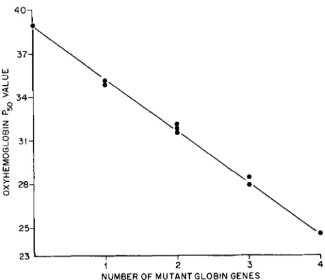
g2/g2;sZIsZ (Hba@ X HbbS2)FZ 24.5 t 0.6 FIGURE Z.-Relationship of the oxygen P50 values and the num- The Hba alleles are given first and the Hbb alleles are given ber of mutant globin genes. The PW values were determined from last. a = Hba“ (homozygous in C57BL/6 and Hbb” stocks);
2
= the oxygen association curves shown in Figure 1 and plotted againstHba@ (homozygous in Hbagz stock); s = (homozygous in the number of mutant globin genes in mice of the nine hemoglobin C57BL/6 mice and Hba@ stock); 5’ = Hbb” (homozygous in Hbb” genotypes: 0 , Hban/Hbaa;Hbp/Hbbs (control); 1, Hba”/Hba@;Hbp/
Stock); g = Hbag (homozygous in DBA/2J mice); d = Hbbd (homo- HbbS and Hba*/Hbaa;HbbS/Hbb”2; 2, H b a . @ / H b a @ ; H b ~ / H b ~ , Hbaa/
zy
‘
ous in DBA/2J mice).Seven to ten determinations for each hemoglobin genotype. Hbaa;Hbbs’/HbP2 and Hba”/Hbag’;Hb6”/Hbbs2; 3 , Hba”/Hbagz;Hbb”/
Hbb“ and HbagZ/HbagZ;Hbb”/Hbb‘2; and 4, HbagZ/Hbag’;Hbb’’/Hbb’2
(double mutant).
z 75-
s
5
m
W
0
50- 0
+
z
W V
W
a 2 5 -
0 20 40 60 80 100 120 140
p50
-
I 24.5 3 37 5
2 28.0
4 35 0 5 39.0
PARTIAL PRESSURE OFOXYGEN (m Hg)
FIGURE 1 .-Oxygen association curves and P ~ o values for hemo- globins from F2 mice of nine genotypes. Curve 1, Hbag’/Hbag’;Hbb”/ Hbb”; 2, Hbaa/Hba@;Hbb“/Hbb’2 and Hbag2/HbagZ;Hbb’/Hbb’2; 3,
Hba”/Hbag2;Hbb’/Hbb”, Hba@/Hbagz;Hbb’/Hbbs and Hba“/ Hba”;Hbb”/Hbb”’; 4, Hba”/Hbag’;Hbb’/Hbb’ and Hba”/Hba”;Hbb’/ Hbb”; and 5, controI-Hba”/Hba’;Hbb”/Hbb’.
have Pso values similar to that of normal adult human hemoglobin (P50 = 26 mm Hg).
T h e oxygen association-dissociation curves of he- moglobins from representative F2 offspring are shown
in Figure 1. T h e nine hemoglobin genotypes exhib- ited five oxygen association-dissociation curves; the quantities of the mutant forms of the a- and @-globins are correlated with the P50 values for each of these hemoglobins. Cooperativity appears to be undisturbed for the mutant hemoglobins as sigmoidicity is main-
tained for each of the oxygen association-dissociation curves. T h e additive effects of both the a-globin (Hbag2) and the @-globin (Hbb”’) mutations are de- picted in Figure 2. The left shift in hemoglobin P 5 0
values among mice of the nine F2 genotypes (Table 2)
is linearly related to the number of mutant globin genes. T h e mutant a-globin chain (5”) in Hbag’ mice and the mutant @-globin chain (p”’) in Hbb”’ mice appear to be physiologically equivalent in terms of their effect on oxygen binding. The hematological values determined from peripheral blood of normal Hba”/Hba“;Hbb”/Hbb” and mutant Hbag2/Hbag2;Hbb”/ Hbb”, Hbaa/Hbaa;Hb6’2s/Hbb’2, and Hbag2/Hbag2;Hbb”’/ Hbb”’ mice are shown in Table 3. Mild erythrocytosis (elevated red cell counts, RBC) was observed in blood from mice of the three mutant genotypes in compar- ison to that of controls. Hematocrit (HCT) values were slightly elevated in Hbag2/Hbag2;Hbb‘/Hbb‘ and doubly homozygous mutant HbagZ/Hbag2;Hbb“’/Hbbs2 mice, but were normal in Hbaa/Hba“;Hbb”2/Hbb”2 mice. In addition, the mean corpuscular volume (MCV) and mean corpuscular hemoglobin (MCH) values were significantly lower in Hbag2/Hbag2;Hbb”2/Hbbs2 mice.
Stability analysis: Clear hemolysates of blood from
Hbag2/Hbag2;Hbb”/Hbb”, Hbaa/Hbaa;Hbb’2/Hbb’2, and doubly homozygous mutant Hbag2/Hbag2;Hbb”’/Hbbr2
Hematologic data on mouse blood of C57BL/6 (Hbaa'a;Hbb"'), HbdZ1S2;Hbb'/', Hba"'";HbL'Z''z, and Hbd2182;Hbb'21'2 genotypes
Hemoglobin No. of WBC (X
genotype samples 106/ml)
Hba"'" 7 8.6" 10.2 16.3 48.2 47.0 15.9 33.8
Hbb'l' 0.5 0.1 0.3 0.6 0.2 0.2 0.4
46.3 16.0 34.5
RBC (x HGB
1 o ~ / ~ I ) (g/dl) (W) (pg/RBC) (pg/RBC) (g/dl)
HCT MCV MCH MCHC
HbaK2/KZ 8 3.1 11.2 17.9 51.9
Hbb"$
Hba"l" 7 4.6 10.6 16.1 48.3 45.6 15.2 33.5
0.6 0.2 0.3 1.1 0.7 0.3 0.9
51.1 43.0 13.8 32.2
0.2 0.1 0.3 0.6 0.3 0.3 0.8
Hbb'2/'2
HbaK2k2 7 6.2 11.9 16.8
Hbb'2/'2 0.9 0.4 0.5 1.5 0.8 0.5 1
.o
' The average for each value is listed first and the f SEM is listed below each average value.
R
0 6 -
0 4 -
~
E
c r t
s
a
0.2-
+:
8 " 8 -*
0
-8 I
0 19 ( 5 16 18 17 20 21 22
ISOPROPANOL 1%)
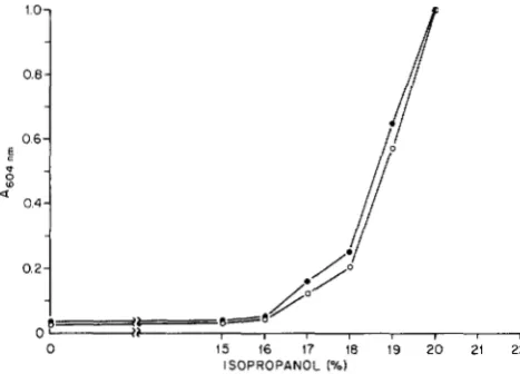
FIGURE 3.-Denaturation profile of hemoglobins from Hba@ and C57BL/6 mice in increasing concentrations of isopropanol. The increase in A,,,, is an inverse measure of the decrease in trans- mittance with increasing amounts of precipitate formed during 30- min incubation of hemolysates at 37" in isopropanol. (0) Hba@/ Hba@;Hbb'/Hbb"; (0) Hba"/Hba";Hbb'/Hbb'. Hemoglobin from
HbaK2/HbaK2;Hb6'/Hbb' mice is equally stable or only slightly less stable than C57BL/6 hemoglobin.
were found at 542 and 572 nm and minimal absorb- ance (maximal light transmittance) occurred above 600 nm (data not given), so the A604nm of the hemo- lysate-isopropanol reaction mixtures was measured after a 30-min incubation at 37" to compare the relative amounts of insoluble protein formed.
A comparison of the ACo4 nm values for Hbug' (Hbug'/
Hbug2;Hbb'/Hbb') and C57BL/6 (Hbu"/Hba";Hbb'/ Hbb") hemolysates is shown in Figure 3. For both hemolysates the denaturation profiles with increasing concentrations of isopropanol are nearly identical and both hemoglobins become completely denatured in 20% isopropanol.
In Figure 4 the denaturation profiles of hemoglobin from Hbb" (Hba"/Hba";Hbb"2/Hbb"2) and C57BL/6 mice are compared. A marked difference in the amount of precipitate between the two hemoglobin samples occurs from 16 to 20% isopropanol. Hemo-
"9
0.6
' I
:.
;//
0.2
0
0 15 16 17 18 19 20 21 22
ISOPROPANOL (%)
FIGURE 4,"Denaturation profile of hemoglobins from Hbb" and C57BL/6 mice in increasing concentrations of isopropanol. (0)
Hba"/Hba";Hbb'/Hbb' (C57BL/6); (.) Hba"/Hba";Hbb'2/Hbb'2; and
(0) Hba"/Hba";Hbb'2/Hbb'2 A604 nm values extrapolated to represent
the expected profile if the Hbb" mutant B-globin chain comprised 100% (rather than 70%) of the adult hemoglobin.
globin from Hbb"' mice forms 15-fold more precipitate in 19% isopropanol than C57BL/6 hemoglobin. Since 70% of the adult hemoglobin tetramers in Hbb"' mice contain the variant /Y2-globin chain (WAWRZYNIAK
and POPP 1985), a hypothetical extrapolation is shown
in Figure 4 for hemoglobin containing the /Y2-chain in Hbb"' mice.
T h e denaturation profile of hemoglobin from dou- bly homozygous
(Hbug2/Hbug2;Hbb"2/Hbb"2)
F:! mice is compared to C57BL/6 hemoglobin in Figure 5. The denaturation profile of the doubly mutant hemoglo- bin is nearly identical to that of Hb~"/Hba~;Hbb"/Hbb"~ hemoglobin shown in Figure 4. Since the hemoglobin of Hbug' mice is relatively stable, the instability of hemoglobin of Hbug2/Hbug2;Hbb"2/Hbb"2 mice is pri- marily due to the presence of the,&'
variant globin chain.DISCUSSION
Stability of Mutant Hemoglobins 549
ISOPROPANOL (%)
FIGURE 5,"Denaturation profile of hemoglobins from Hbag'/
Hbag';Hbb"/Hbb" and C57BL/6 mice in increasing concentrations of isopropanol. (0) Hba"/Hba';Hbb'/Hbb' (C57BL/6); (A) Hbag'f Hba6Z;Hbb'2/Hbb'2; and (A) HbagZ/Hba6Z;Hbb"2/HbbrZ Ason n m values
extrapolated to represent the expected profile if the Hbb"' and the
Hba@ (assuming that the Hbag' mutant a-globin chain is stable) mutant globin chains comprised 100% (rather than 70 and 55%, respectively) of the total adult hemoglobin.
moglobin and are based on the spectral differences between 400 and 800 nm of both oxyhemoglobin and deoxyhemoglobin. T h e oldest method made use of a tonometer to deoxygenate and oxygenate hemoglobin in a rotating V-shaped flask at 37" (BENESCH, MAC- DUFF and BENESCH 1965). T h e procedure required large amounts of blood sample and oxygenation and deoxygenation were performed manually. T h e devel- opment of automated oxygen association-dissociation analyzers permitted microliter samples of blood to be used. T h e Aminco Hem-O-Scan Analyser (NEWTON and PETERS 1983) uses as little as 2 pl of blood placed between a glass slide and a gas permeable membrane to allow oxygenation and deoxygenation of a thin film of blood where the pH and temperature are main- tained in a heated, humidified chamber flushed with a mixture of 0 2 , Nz and COP. T h e more advanced model TCS Hemox ODC Analyzer, which uses a Clark oxygen electrode, measures oxygen association-disso- ciation properties of hemoglobin in microliter samples of blood suspended in a pH 7.4 buffered physiological solution maintained at 37 O in a cuvette through which
gases are bubbled to oxygenate and deoxygenate the hemoglobin. T h e Hemox Analyzer is easy to operate and produces highly accurate oxygen association-dis- sociation curves with as little as 2 p1 of blood (FESTA and AKASURA 1979).
In adult Hbb"' (Hbba/Hbba;Hbb"'/Hb6'') mice, 70% of the adult hemoglobin contains the variant
p"'
@-globin polypeptide (WAWRZYNIAK and POPP 1985). In adult Hbag' (Hbag'/Hbag';Hbb'/Hbb") mice, 55% of the adult hemoglobin contains the variant a-globin chain 5'" (Popp et al. 1983). Both of these a- and @-globin chain variants are present at these same levels in doubly homozygous Hbag2/Hbag2;Hb6'2/Hbb'2 mice
(our unpublished results). T h e variant chains are not present in 100% of the adult hemoglobin because mice express two adult a-globin and two adult @-globin genes (LYON, BARKER and POPP 1988).
In Hbag', Hbb"', and (Hbag'/Hbag';Hbb"/Hbb' X Hbaa/Hbaa;Hbb"'/Hbb'') F2 mice, both mutant a- and @-globin chains are physiologically equivalent; that is, the oxygen affinity of hemoglobin with both globin variants is increased in proportion to the mutant gene dosage regardless of genotypic combination (Table 2, Figures 1 and 2). When both mutant globins are present in the same animal, they exert their effect on oxygen affinity in an additive manner. Although both mutant globins are physiologically similar, the pres- ence of 55% a-chain 5"' us. 70%
p2
in hemoglobins Hbag' and Hbb"' mice, respectively, implies that he- moglobin with a-chain 5"' at the molecular level has a 30% greater oxygen affinity than hemoglobin with the p'-globin. Minor differences in oxygen associa- tion-dissociation properties among normal C57BL/6 and DBA/2J mice (Table 2) may reflect a composite of globin chain variant oxygen affinities in addition to minor physiological differences such as intracellular levels of 2,3-diphosphoglycerate and pH.settling of the flocculated precipitate which allows for increased light transmittance in the supernatant at 604 nm.
T h e denaturation profile of Hbug' hemoglobin (Fig- ure 3) indicates that the mutant a-globin chain 5" is relatively stable in comparison to a-globin chain 1 in C57BL/6 mouse hemoglobin. In contrast, Figure 4 shows that the P'-globin in Hbb"' mouse hemoglobin is moderately unstable, as reported in an earlier study (WAWRZYNIAK and POPP 1986). Figure 4 also depicts an extrapolation of the denaturation profile of Hbbs2 hemoglobin expected if all of the hemoglobin con- tained the variant P2-globin chain instead of 70% of the adult hemoglobin. T h e shift in the denaturation profile of C57BL/6 hemoglobin shown in Figures 3 and 4 most likely resulted from slight variations in incubation temperature and time between completion of incubation and spectrophotometric analysis and demonstrates the need for simultaneous controls for each analysis. Some of this variation can be controlled by placing the reactions on ice immediately after incubation prior to spectrophotometric analysis. Peak precipitation of C57BL/6 hemoglobin occurred in 20 or 22% isopropanol in all experiments. Hbb" hemo- globin is moderately unstable in contrast to the highly unstable human hemoglobin variants Christchurch and Belfast (BUNN and FORGET 1986). Red cells from individuals with these hemoglobin variants contain Heinz body inclusions and the elevated hemolysis causes hemolytic anemia. In Hbug'/Hbug2;Hbb"'/Hbbs2 mice, the denaturation profile is nearly identical to that of Hbu"/Hbu";Hbb"2/Hbb"2 mice (compare Figures
4 and 5). There appears to be no interactive effect between the moderately unstable P2-globin chain and the relatively stable a-chain 5" in the a25m@2s2 hemo-
globin tetramers formed in Hbug2/Hba~';Hbb''/Hbbs2 mice. In these mice, the expected quantity of the
~
t
~
tetramer would~
~
f
l
be 0.55~
~
X~
0.70, or 38% of the total hemoglobin. T h e quantity found by isoelec- tric focusing was 36% (data not presented), which suggests that the tetrameric hemoglobin containing both the mutant a- and ,&globins is quite stable under normal physiological conditions.T h e a- and @-globin variants do not abolish the cooperativity of oxygen binding but they do alter the kinetics of transition of deoxyhemoglobin to oxyhe- moglobin (left shift in P50 values) (Figure 1). T h e tertiary structures of a- and @-globins of human and other mammalian hemoglobins are highly conserved. By homology, the a89, His to Leu substitution in a- chain 5" of Hbug' mice, though near the proximal heme-binding histidine residue, is on the hydrophilic, external surface of the a-globin molecule between two
residues involved in the a& contact in the hemoglo- bin tetramer. This region is important in regulating
allosteric cooperativity during the transition of oxy- hemoglobin to deoxyhemoglobin via the breaking and forming of salt bridges (ionic interactions). T h e ( ~ 8 9 , His to Leu substitution in H b d 2 mice may alter such interactions which can result in an increased oxygen binding (BUNN and FORGET 1986; BALDWIN 1980; PERUTZ 1980).
In Hbb"' mice, the site of the p59, Lys to Ile amino acid substitution lies near the hydrophobic heme
pocket, but does not involve any residues that associ- ate with the nonpolar porphyrin ring in the heme group. The positively charged lysine residue which projects its side chain toward the similarly charged Fe+'-ligand binding, coordination axis was replaced by a neutrally charged isoleucine moiety. The in- creased oxygen affinity in Hbbs2 hemoglobin may re- sult from reducing the dielectric constant in the vicin- ity of the iron and distal histidine region, which has a local positive charge (LAUFER, WARREN and MCIVOR 1983). A reduction of the positive charge in this area resulting from the lysine to isoleucine substitution could facilitate oxygen binding to the sixth coordina- tion position of the iron atom and increase the rate of transition of deoxyhemoglobin to oxyhemoglobin via subsequent heme-heme interaction.
T h e most plausible explanation for the instability of HbV2 hemoglobin is that the decrease in local positive charge reduces the hydrophobicity of the heme pocket in the vicinity of the distal histidine residue, and isopropanol causes a more rapid dena- turation of globin tertiary structure resulting in the formation of methemoglobin, via irreversible hemi- chrome formation followed by expulsion of the heme and precipitation of globin (BUNN and FORGET 1986). If this explanation is applicable for P'-globin, then this mouse hemoglobin variant is unique among both human and mouse hemoglobin variants described to date.
This research was sponsored jointly by National Institute of Health R01 HL-43375 and the Office of Health and Environmental Research, U.S. Department of Energy, under contract DE-AC05- 840R21400 with Martin Marietta Energy Systems, Inc.
LITERATURE CITED
BALDWIN, J., 1980 Structure and cooperativity of hemoglobin. Trends Biochem. Sci. 5 224-228.
BENFSCH, R., G. MACDUFF and R. BENESCH, 1965 Determination of oxygen equilibria with a versatile new tonometer. Anal. Biochem. 11: 81-87.
BRENNAN, S. O., R. M. WELLS, H. SMITH and R. W. CARRELL, 1981 Hemoglobin Brisbane: p-68, Leu to His. A new high oxygen affinity variant. Hemoglobin 5: 325-334.
BUNN, H . F., and B. G . FORGET (Editors), 1986 Hemoglobin: Mo- lecular, Genetic and Clinical Aspects.
w.
B. Saunders, Philadel- phia.Stability of Mutant Hemoglobins 55 1
CLECC, J. B., M. A. NAUGHTON and D. J. WEATHERALL,
1966 Abnormal human hemoglobins, separation and char- acterization of the a- and &chains by chromatography and the determination of two new variants-Hb Chesapeake and Hb J
(Bangkok). J. Mol. Biol. 1 9 91-108.
DACIE, J. V., A. J. GRIMES, A. MEISLER, L. STEINCOLD, E. H. HEMSTED, G . H. BEAVEN and J. C. WHITE, 1964 Hereditary Heinz body anemia. A report of studies on five patients with mild anemia. Br. J. Haematol. 10: 388-402.
DACIE, J. V., N. K. SHINTON, P. J. GOFFNEY, R. W. CARRELL and H. LEHMAN, 1967 Haemoglobin Hammersmith (p-42, (CD1)
Phe to Ser). Nature 216: 663-665.
FESTA, R. S., and T. AKASURA, 1979 The use of an oxygen dissociation curve analyser in transfusion therapy. Transfusion
1 9 107-113.
HUEHNS, E. R., 1970 Diseases due to abnormalities of hemoglobin LAUFER, R. S., E. WARREN and D. MCIVOR (Editors),
1983 Hemoglobin and the chemistry of respiration, pp 100- 140 in Principles of Biochemistry: Mammalian Biochemistry. McGraw-Hill, New York.
LEWIS, S. E., F. M. JOHNSON, L. C. SKOW, D. M., POPP, L. B. BARNETT and R. A. POPP, 1985 A mutation in the @-globin gene detected in the progeny of a female mouse treated with ethylnitrosourea. Proc. Natl. Acad. Sci. USA 82: 5829-5831.
LYON, M. F., J. E. BARKER and R. A. POPP, 1988 Mouse globin gene nomenclature. J. Hered. 7 9 93-95.
structure. Annu. Rev. Med. 21: 157-178.
NEWTON, M. F., and J. PETERS, 1983 Physiological variation of mouse hemoglobins. Proc. R. SOC. Lond. 2 1 8 443-453.
PERUTZ, M. F., 1980 Stereochemical mechanism of oxygen trans- port by hemoglobin. Proc. R. SOC. Lond. 2 0 8 135-162.
PETERS, J., S. J. ANDREWS, J. F. LOUTIT and J. B. CLECC, 1985
A mouse &globin mutant that is an exact model of Hemoglobin Rainier in man. Genetics 110: 709-721.
POPP, R. A., E. G . BAILIFF, L. C. SKOW, F. M. JOHNSON and S. E. LEWIS, 1983 Analysis of a mouse a- globin gene mutation induced by ethylnitrosourea. Genetics 1 0 5 157-167.
POPP, D. M., R. A. POPP, S. LOCK, R. C. MANN and R. E. HAND,
JR., 1986 Use of multiparameter analysis to quantitate he- matological damage from exposure to a chemical (ethylene oxide). J. Toxicol. Environ. Health 18: 543-565.
STEADMAN, J. H., A. YATES and E. R. HUEHNS, 1970 Idiopathic Heinz body anemia: Hb Bristol (867 (El 1) Val to Asp). Br. J.
Haematol. 1 8 435-446.
WAWRZYNIAK, C. J., and R. A. PCJPP, 1985 Use of a new mouse &globin haplotype (Hbb”) to study hemoglobin expression during development. Dev. Biol. 112: 477-484.
WAWRZYNIAK, C. J., and R. A. POPP, 1986 Detection of an unsta- ble murine hemoglobin. Biochem. Biophys. Res. Commun.
WHITNEY, J. B., G . T. COPLAND, L. C. SKOW and E. S. RUSSELL,
1979 Resolution of the duplicated hemoglobin a-chain loci by isoelectric focusing. Proc. Natl. Acad. Sci. USA 7 6 867- 871.
139: 551-556.