Copyright2001 by the Genetics Society of America
Development and Applications of a Complete Set of Rice Telotrisomics
Zhukuan Cheng,*
,†,‡Huihuang Yan,
†Hengxiu Yu,* Shuchu Tang,*
Jiming Jiang,
‡Minghong Gu* and Lihuang Zhu
†*Yangzhou University, Yangzhou 225009, People’s Republic of China,†Institute of Genetics, Chinese Academy of Sciences, Beijing 100101, People’s Republic of China and‡Department of Horticulture, University of Wisconsin, Madison, Wisconsin 53706
Manuscript received August 4, 2000 Accepted for publication September 13, 2000
ABSTRACT
We previously isolated a complete set of primary trisomics along with many other aneuploids from triploid plants derived from anindicarice variety “Zhongxian 3037.” About 30,000 progeny from these trisomic and aneuploid plants were grown each year from 1994 to 1999. The variants that differed morphologically from both the diploids and the original primary trisomics were collected for cytological identification. From these variants, a complete set of telotrisomics covering all 24 rice chromosome arms was obtained. The identities of the extra chromosomes were further confirmed by dosage analysis of the RFLP markers on extra chromosome arms. The telocentric nature of the extra chromosomes in these stocks was verified by fluorescencein situhybridization (FISH) using a rice centromeric BAC clone as a marker probe. In general, the shorter the extra chromosome arm of a telotrisomic, the stronger the resemblance it bears to the diploid; the longer the extra chromosome arm, the stronger the resemblance to the corresponding primary trisomic. We demonstrated that DNA clones can be rapidly assigned to specific chromosome arms by dosage analysis with the telotrisomics. We also showed that telotrisomics are valuable tools for chromosome microdissection and for developing chromosome-specific DNA markers.
R
ICE is the staple food of more than half of the Before producing telotrisomics for all 24 rice chromo-some arms, we first developed a complete set of primary world’s population. As a self-pollinating diploidspecies, rice has a relatively small genome,ⵑ4.3 ⫻108 trisomics of Zhongxian 3037, anindicarice variety
de-rived from a cross between IR24 and BG90-2 (Cheng
bp (Arumuganathan and Earle 1991), and can be
easily transformed and regenerated, which makes it a et al. 1996). Telotrisomics covering all 24 arms of the 12 rice chromosomes have been established from the model monocot plant for molecular biology research.
However, rice chromosomes are small and it is difficult progenies of these primary trisomics and other aneu-to consistently recognize individual chromosomes and ploids, all derived from a triploid of Zhongxian 3037. their variants in somatic cells. Today, both rice physical The telocentric nature of the extra chromosomes in mapping and molecular genomics require an efficient the telotrisomics was confirmed by fluorescencein situ
method for chromosome identification. hybridization (FISH) using a rice centromere-specific Utilization of telotrisomics is a classical method for bacterial artificial chromosome (BAC) clone, 17p22, as chromosome identification in plants. As telotrisomics a marker probe. The applications of the telotrisomics contain an extra telocentric chromosome in each cell, in marker assignment and microdissection are demon-it is often easy to distinguish the extra chromosome strated in this article.
from the rest of the chromosome complement. Since
Rhoades(1936) discovered the first telotrisomic inZea
mays, telotrisomic stocks have been developed in a num- MATERIALS AND METHODS
ber of species includingDatura stramonium(Blakeslee
Plant materials:All 12 primary trisomics and other
aneu-andAvery 1938), Nicotiana sylvestris(Goodspeedand ploids were developed from a triploid of Zhongxian 3037.
Avery 1939), Triticum monococcum (Moseman and Approximately 180,000 plants derived from the trisomics and
Smith1954),Hordeum vulgare (Tsuchiya1960), Secale other aneuploids,ⵑ30,000 annually, were evaluated in field from 1994 to 1999. Variants morphologically distinct from the cereale(KamanoiandJenkins1962),Lycopersicon
esculen-original diploid and trisomic sibs were selected for further tum(Khush andRick 1967), andOryza sativa(Singh
cytological analysis.
et al.1996a,b). However, a complete set of telotrisomics Chromosome preparation and fluorescencein situ hybridiza-covering the arms of the entire chromosome comple- tion:Young meiotic panicles of the rice variants were harvested ment has not been reported in any plant species. and fixed in 3:1 Carnoy’s solution supplemented with 0.5% FeCl3. Squashes were prepared in acetic-carmine solution
ac-cording toWu(1967). Roots of the rice variants were harvested from field-grown plants. The roots were pretreated in 0.002m
Corresponding author:Lihuang Zhu, Institute of Genetics, Chinese
8-hydroxyquinoline at 20⬚ for 2 hr to accumulate prometa-Academy of Sciences, Beijing 100101, People’s Republic of China.
E-mail: [email protected] phase cells, fixed in methanol-acetic acid (3:1), and stored at
⫺20⬚until use. Root tips were macerated in 2.5% cellulose at by Inoueet al. (1994) andChenet al. (1997), respectively. Primer pairs for STS and microsatellites on different chromo-37⬚for 1.5 hr. Squashes were made in the fixative on a glass
slide and flame dried. The chromosomes were stained with some arms were selected to amplify the PCR products from both microdissected chromosomes and control samples. 2% Giemsa solution for observation.
The procedure for FISH analysis was as described (Jiang Designation of the trisomics: The trisomic nomenclature used for tomato (Khush1973) was adopted for this article.
et al.1995) with only minor changes. A rice BAC clone, 17p22,
which produces very specific hybridization signals to each rice For example, a telotrisomic for the short arm of chromosome 1 is designated as 2n⫹·1S and that for the long arm as 2n⫹·1L. centromere was used as FISH probe (Donget al.1998). The
hybridization mixture (20l for each slide) contained 20 ng of labeled probe DNA, 50% formamide, 10% dextran sulfate,
2⫻ SSC, and 20g of sheared salmon sperm DNA. After RESULTS AND DISCUSSION
overnight incubation at 37⬚, FISH signals were detected by a
FITC-conjugated anti-biotin antibody (Vector Laboratories, Identification of the telotrisomics: Since the extra Burlingame, CA). Chromosomes were counterstained with chromosome in the trisomics,i.e., primary, secondary, propidium iodide. Images were captured with a SenSys CCD
or tertiary trisomics, could form a univalent in many of
(charge coupled device) camera (Photometrics, Tucson, AZ)
the sporocytes, misdivision of the univalent occurs at a
coupled to a Macintosh computer. Gray-scale images were
captured individually and merged using IPLab Spectrum v3.1 certain frequency. Therefore, it is possible to isolate
software. telotrisomics from the progenies of these trisomics,
es-Southern analysis for dosage effects of restriction fragment pecially from primary trisomics. length polymorphism (RFLP) markers:Genomic DNA
isola-To obtain all possible rice telotrisomics, the progenies
tion and gel blot hybridization were according toMcCouch
of different trisomic types were planted in large
popula-et al.(1988). The amount of DNA in each lane was adjusted
to equal amounts using a specific control. All the DNA from tions, up to 30,000 plants each year. The plants showing
the different aneuploids was digested withDraI. Serial volumes morphological features different from both of the origi-of digested DNA were run on a 0.8% agarose gel and stained nal trisomics and the normal diploid were collected with ethidium bromide. The gel images were captured with
for further cytological examination. As for the three
a digital camera and analyzed with molecular analysis software.
subtelocentric chromosomes with very short and/or
Consequently, nearly equal aliquots of DNA from the two
darkly stained heterochromatic short arms,i.e.,
chromo-telotrisomics, one for the extra long arm and the other for
the short arm of the same chromosome, could be run on the somes 4, 9, and 10, it is expected that the telotrisomics same agarose gel and transferred to a Hybond-N⫹membrane with these arms may resemble the diploids but differ (Amersham, Buckinghamshire, UK) for Southern analysis.
from the respective primary trisomics, while the
triso-RFLP probes were labeled with32P by random hexamer
prim-mics with the long arms of these three chromosomes
ing and hybridized to the above membranes overnight at 65⬚.
should resemble the primary trisomics but differ from
The membranes were washed sequentially in 2⫻, 1⫻, and
0.5⫻SSC with 0.1% SDS, 20 min each at 65⬚, and then exposed the diploid. Therefore, all the plants from the progenies
on X-ray film with intensifying screens at⫺70⬚for 3–7 days. of primary trisomics 4, 9, and 10, resembling either the RFLP markers designated as RG#, RZ#, and CDO# were kindly diploid or the primary trisomics, were maintained for provided by Dr. S. D. Tanksley from Cornell University (Ithaca,
further cytological identification.
NY) and clones of G#, L#, and C# were obtained from the
The somatic chromosomes at prometaphase of all the
MAFF DNA Bank at NLAR, Japan.
Chromosome microdissection and amplification: To mi- variant candidates were analyzed to confirm whether or
crodissect the extra chromosome arms from the aneuploids, not the variants were telotrisomics. The progenies of prometaphase chromosomes were prepared as follows. Briefly, the confirmed telotrisomics were planted in the follow-roots of the aneuploids were fixed in methanol-acetic acid
ing year. Because a telotrisomic might produce the
re-(5:1) for 10 min and stored in 70% ethanol until use. Squashes
verted primary trisomic in its progeny, it was convenient
were made in the fixative on a coverslip. Microdissection was
to nominate the involved chromosome according to the
performed on a Nikon inverted microscope with a Leitz
micro-manipulator. Telochromosomes were dissected through indi- morphological traits of the primary trisomics. Using this vidual microneedles with a tip of 0.5–1m. The microneedle procedure, we obtained a series of different telotrisom-with a telocentric chromosome was inserted into a 0.5-ml
Ep-ics from whose progenies a complete set of reverted
pendorf tube and the tip was broken off in the 20-l aliquot
primary trisomics was also recovered.
of 1⫻T4 ligase buffer containing 5 ng/l proteinase K. Ten
Morphological evidence showed that the selected
tel-telochromosomes were collected in a single tube reaction.
The microdissected chromosomal DNA was digested withSau otrisomics cover all 24 arms of the rice genome. To
3A, ligated toSau3A linker adaptors, and amplified by PCR further identify the extra chromosome arms, cytological according to the procedures ofChenandArmstrong(1995). investigations were conducted on the selected telotri-The positive control sample containing 10 pg of Zhongxian
somic candidates. As the centromere positions in rice
3037 genomic DNA and the negative control sample
con-molecular linkage maps have been determined by
taining no DNA were also amplified using the same protocol.
The amplified DNAs were separated in a 1.4% agarose gel Singhet al.(1996a,b) andHarushimaet al.(1998), we
and stained with ethidium bromide. used the dosage effects of the molecular markers in
Sequence-tagged site (STS) and microsatellite analyses were different chromosome arms to distinguish the extra conducted to confirm that the PCR products of microdissected
chromosomes of the telotrisomics as belonging to the
chromosomal DNA truly came from the microdissected
chro-short arm or long arm. For example, when a32P-labeled
mosome arms. The STS primer pairs and microsatellite primer
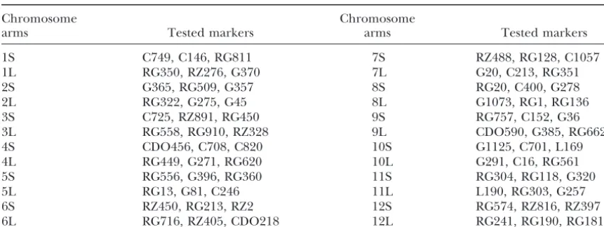
TABLE 1
The tested RFLP markers with dosage effects on the corresponding telotrisomics
Chromosome Chromosome
arms Tested markers arms Tested markers
1S C749, C146, RG811 7S RZ488, RG128, C1057
1L RG350, RZ276, G370 7L G20, C213, RG351
2S G365, RG509, G357 8S RG20, C400, G278
2L RG322, G275, G45 8L G1073, RG1, RG136
3S C725, RZ891, RG450 9S RG757, C152, G36
3L RG558, RG910, RZ328 9L CDO590, G385, RG662
4S CDO456, C708, C820 10S G1125, C701, L169
4L RG449, G271, RG620 10L G291, C16, RG561
5S RG556, G396, RG360 11S RG304, RG118, G320
5L RG13, G81, C246 11L L190, RG303, G257
6S RZ450, RG213, RZ2 12S RG574, RZ816, RZ397
6L RG716, RZ405, CDO218 12L RG241, RG190, RG181
to probe a membrane with an equal amount ofDraI- extra chromosomes in the isolated telotrisomic lines, a restricted DNA from the two telotrisomics related to rice centromeric BAC clone, 17p22, was hybridized to chromosome 1, the telotrisomic with stronger hybridiza- the prometaphase chromosomes of each aneuploid. We tion bands should be 2n⫹·1S, while the one with weaker found that the hybridization signals on the extra chro-bands should be 2n⫹·1L. Three markers on each arm mosomes were all located at one end (Figure 2). Al-were tested in this way (Table 1); thus all 24 telotrisomics though the intensities of the FISH signals varied greatly were identified by dosage analysis. Figure 1 shows the over different chromosomes, we consistently found that autoradiographs of three such examples in which G275 the signals in the extra chromosomes were always weaker on 2L, RG350 on 1L, and C749 on 1S were used to than those in their corresponding homologous chromo-identify 2n⫹·1L and 2n⫹·1S. In Figure 1A, the bands somes in the same metaphase cells, suggesting that the in all four lanes revealed by G275 are similar in intensity, telocentric chromosomes were derived from chromo-indicating that the DNA dosages corresponding to this some breaks within their centromeres. In terms of both marker in the two different rice genomes are equal. But location and intensity, the centromeric hybridization in Figure 1, B and C, dosage effects between the two signals allow us to conclude that the extra chromosomes telotrisomics are shown. The marker dosage analysis in the telotrisomics were all derived from centromere has also confirmed the identities of all 24 telotrisomics misdivisions. These results also suggest that the func-developed from a common triploid with the genetic tional rice centromeres consist of repetitive units; misdi-background of anindicarice, Zhongxian 3037. vision-derived rice centromeres, as those in maize ( Kas-To confirm the location of the centromeres on the za´sandBirchler1996, 1998), are fully functioned.
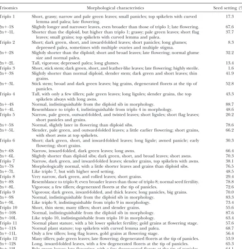
Morphological and reproductive features of the telo-trisomics:Each of the primary trisomics in Zhongxian 3037 has unique morphological features, enabling easy discrimination of all the primary trisomics from one another and from the diploid sib as well (Cheng et al.1996). Most telotrisomics have some characteristics similar to those of their respective primary trisomics. In general, however, telotrisomics with the short arms bear strong resemblance to the diploid while those with long arms bear a stronger resemblance to the corresponding primary trisomic. For the subtelocentric chromosomes 4, 9, and 10, it is difficult to morphologically distinguish the three telotrisomics of their short arms from the diploid, and it is also difficult to detect obvious
morpho-Figure1.—Dosage analysis of different RFLP markers in logical differences between the telotrisomics of their
the two telotrisomics of chromosome 1; DNA in lanes 1 and long arms and their respective primary trisomics. How-2 from How-2n⫹·1S and DNA in lanes 3 and 4 from 2n⫹·1L. (A)
ever, for the other nine metacentric or submetacentric
Probed with G275 that is mapped on 2L; (B) probed with
chromosomes, all 18 telotrisomics are morphologically
RG350 mapped on 1L; and (C) probed with C749 mapped
Figure2.—FISH analysis of probe 17p22 on prometa-phase chromosomes of dif-ferent telotrisomics. (A–X) telotrisomics with an extra chromosome arm from 1S to 12L, respectively; arrow-heads show the extra telo-centric chromosomes. All bars, 5m. (Y) 12 individual rice chromosomes and their corresponding chromosome arms presented in the telo-trisomics.
primary trisomics to varying degrees and are usually while those with long extra chromosome arms displayed lower seed set.
more vigorous than the corresponding primary
triso-mics. The morphological traits of the 24 telotrisomics Singh et al. (1996a) developed seven telotrisomics from a different rice variety, “IR36,” i.e., 2n⫹·1S, and their corresponding primary trisomics are
summa-rized in Table 2. Figure 3 shows the morphological 2n⫹·2L, 2n⫹·3L, 2n⫹·5L, 2n⫹·8S, 2n⫹·9S, and 2n⫹·10S. Most of these are quite similar to the corre-characteristics of the plants and the panicles,
respec-tively, of the diploid sib, 2n⫹·4S, 2n⫹·4L, and triplo 4. sponding telotrisomics reported here in terms of both morphology and seed set. However, these two sets of The seed-setting behavior of each primary trisomic and
telotrisomic was also investigated. In general, telotrisomics telotrisomics report differences for 2n⫹·1S and 2n⫹·10S. For 2n⫹·1S, the telotrisomic derived from have higher seed set than their respective primary
Figure2.—Continued.
IR36 is thinner in culm compared to its disomic sib polymorphism, have severe distortion segregations in the mapping populations, or include multiple and/or and therefore can be morphologically distinguished.
However, the 2n⫹·10S counterpart derived from Zhon- repetitive sequences. Still, their chromosomal mapping is of importance to genomic studies. Here, we present gxian 3037 has, rather, normal culm and cannot be
distinguished from its disomic sib by this trait. These an example of linkage assignment by dosage analysis using a complete set of rice telotrisomics. A multicopy differences may be attributed to some allelic variations
on the respective chromosome arms between the two clone, G1073, displayed three bands in a Southern analy-sis of DraI-digested Zhongxian 3037 genomic DNA. original varieties (Khushet al.1984;Chenget al.1996).
Using the telotrisomics to assign DNA clones to rice When the membranes, which contain equal amounts of
DraI-digested DNAs from the two telotrisomics with the
chromosome arms:Linkage analysis is an effective way
TABLE 2
Morphological and reproductive features of primary and telotrisomics of the rice variety Zhongxian 3037
Trisomics Morphological characteristics Seed setting (%)
Triplo 1 Short, grassy; narrow and pale green leaves; small panicles; top spikelets with curved 17.3 lemma and palea; late flowering.
2n⫹·1S Slightly longer and narrower leaves, even broader than those of triplo 1; late flowering. 67.6 2n⫹·1L Shorter than the diploid, but higher than triplo 1; grassy; pale green leaves; short flag 37.7
leaves; small grains; top spikelets with curved lemma and palea.
Triplo 2 Short; dark green, short, and inward-folded leaves; short panicles; long glumes; 8.3
depressed palea, sometimes with multiple ovaries and multiple stigma.
2n⫹·2S Slightly shorter than the diploid; short and broad leaves; late flowering; normal glume 32.2 size and normal palea.
2n⫹·2L Tall, vigorous; depressed palea; long glumes. 13.4
Triplo 3 Short, stick stem; dark green, short, and leather-like leaves; late flowering; highly sterile. 1.6 2n⫹·3S Slightly shorter than normal diploid, slender stem; dark green and short leaves; thin 41.9
grains.
2n⫹·3L Stick stem; broad and dark green leaves; big grains, degenerated florets at the tip of 52.8 panicles.
Triplo 4 Tall, with only a few tillers; pale green leaves; long ligules; slender grains, the top 43.3 spikelets always with long awns.
2n⫹·4S Normal, indistinguishable from the diploid sib in morphology. 88.7
2n⫹·4L Resemblance to triplo 4, indistinguishable from triplo 4 in morphology. 48.6
Triplo 5 Narrow, pale green, outward-folded, and twisted leaves; short ligules; short flag leaves; 20.2 short panicles and grains.
2n⫹·5S Normal, slightly later in flowering than diploid sibs. 78.6
2n⫹·5L Slender, pale green, and outward-folded leaves; a little earlier flowering; short grains, 66.2 with short awns at top spikelets.
Triplo 6 Short; dark green, short, and inward-folded leaves; long ligule; awned panicle; early 30.3 flowering; short grains.
2n⫹·6S Narrow, inward-folded, dark green leaves; long awns. 66.4
2n⫹·6L Slightly shorter than diploid sibs; dark green, short, and broad leaves; short awns. 70.3 Triplo 7 Narrow, dark green, and inward-folded leaves; slender grains, top spikelets with awns. 38.1
2n⫹·7S Morphologically normal, with a little shorter leaves and grains than diploid sibs. 76.5
2n⫹·7L Like triplo 7, but with higher seed setting. 48.5
Triplo 8 Very narrow, dark green, and rolled leaves; short grains. 29.4
2n⫹·8S Resemblance to triplo 8; even broader leaves than those of triplo 8; normal seed fertility. 78.8
2n⫹·8L Vigorous; a few tillers; degenerated florets at the tip of panicles. 72.6
Triplo 9 Vigorous; dark green, inward-folded, and thick leaves; long panicles, big grains. 70.0
2n⫹·9S Normal, indistinguishable from the diploid sib in morphology. 83.3
2n⫹·9L Like triplo 9, indistinguishable from triplo 9 in morphology. 73.4
Triplo 10 Short, thin stem; many tillers; short and slender grains. 55.6
2n⫹·10S Normal, indistinguishable from the diploid sib in morphology. 87.6
2n⫹·10L Like triplo 10, indistinguishable from triplo 10 in morphology. 63.4
Triplo 11 Normal plant stature, with a bit lower spikelet fertility; gold grains at flowering stage. 57.6
2n⫹·11S Normal plant stature; top spikelets with curved lemma and palea. 68.7
2n⫹·11L Only a few tillers; long flag leaves, gold grains at flowering stage. 56.4
Triplo 12 Many tillers; pale green leaves; late flowering; degenerated florets at the tip of panicles. 53.9 2n⫹·12S Long, inward-folded leaves, with a few degenerated florets at the tip of panicles. 65.3 2n⫹·12L Pale green leaves; late flowering, with a few degenerated florets at the tip of panicles. 64.1
bridization signal in 2n⫹·8L than that in 2n⫹·8S, and Microdissection and amplification of the two arms of chromosome 5:The techniques of chromosome micro-the two smaller fragments showed stronger signals in
2n⫹·1L than those in 2n⫹·1S (Figure 4, A and B), while dissection and microcloning represent the combination of traditional cytology with modern molecular biology. no differences in signal intensity were detected among
others. Thus, the largest fragment is located on chromo- The microdissection procedure, first performed on Dro-sophila polytene chromosomes by Scalenghe et al.
some arm 8L, and both smaller fragments are on 1L.
Using the same approach we assigned RG684, a clone (1981), has been applied to many plant species such as barley (Schondelmaier et al. 1993), wheat (Vega et
showing severe distorted segregation, to chromosome
Figure3.—Morphology of different trisomic sibs of chro-mosome 4. (A) Plants: from left to right, diploid, 2n⫹·4S, 2n⫹·4L, and 2n⫹4S·4L, respectively. (B) Panicles.
Figure 5.—Microdissection and amplification of the two
Armstrong1995), maize (Ponelieset al.1997), bean
separate chromosome arms of chromosome 5. (A)
Prometa-(Pichet al.1994), andCrepis capillaris(Jamilena et al. phase cell of 2n⫹·5S⫹·5S before chromosome
microdis-1995). However, this technique can hardly be applied section; arrows show the two extra telochromosomes. (B)
Prometaphase cell of 2n⫹·5S⫹·5S after chromosome
micro-to rice due micro-to its small chromosomes and lack of
charac-dissection; arrows point to the areas remaining after
dissec-teristics amenable to chromosome identification. The
tion. (C) Amplified DNA after the second round of PCR run
rice telotrisomics are ideal for identification of
individ-in agarose gel: 1, positive control sample; 2, negative control
ual chromosome arms, which are distinguishable from sample; 3, microdissected chromosomal DNA from 5S; 4, mi-the normal rice chromosomes. In mi-the present study, two crodissected chromosomal DNA from 5L. (D) DNA amplified
by the STS primer of the molecular marker G396; 0, the
aneuploids, 2n⫹·5S⫹·5S, derived from 2n⫹·5S, and
genomic DNA of Zhongxian 3037; 1–4, same as in C. (E) DNA
2n⫹·5L were used for microdissection of the extra arms
amplified by the microsatellite primer RM233 on 5L: 0–4,
of rice chromosome 5. The two extra chromosome arms
same as in D.
of 2n⫹·5S⫹·5S, which could be easily identified under an inverted microscope at ⫻400 magnification, were successfully microdissected from the prometaphase cells
(Figure 5C). The negative control stained very lightly, with a microneedle controlled by a Leitz
micromanipu-which may be attributed to contamination. To verify lator. Figure 5, A and B, shows the same prometaphase
whether or not the amplified DNAs came from the dis-cell before and after microdissection. Using the same
sected chromosome arms, the molecular markers on procedure, the extra chromosome arms 5L were also
5S, 5L, and other chromosomes were selected to test microdissected from the prometaphase cells in 2n⫹·5L. the DNA products of the second round of PCR. For After two rounds of PCR amplification a smear of DNA example, the STS marker RG396 on 5S and the microsa-fragments could be clearly seen in the two dissected tellite marker RM233 on 5L could be detected only in chromosomal DNA samples and in the positive control the amplified DNAs from the microdissected
chromo-some arm 5S and 5L, respectively (Figure 5, D and E). As expected, all the tested markers on the other chromosomes,e.g., RM23, RM26, RM29, RM44, RM48, RM49, RM53, RM80, RM84, RM205, and RM214, could not be detected in both of the DNA samples from the microdissected 5S and 5L. Thus we conclude that the amplified DNAs from the microdissected chromosome arms came from the extra chromosome arms, 5S and 5L. The amplified DNAs can be used to isolate the chromosome 5-specific DNA sequences for further stud-ies. Because a complete set of rice telotrisomics are available now, all 24 chromosome arms in rice can be
Figure4.—Assignment of DNA clones to rice chromosome microdissected and amplified in this manner.
arms by dosage analysis. DNA in lanes 1 and 2 from 2n⫹·8S,
DNA in lanes 3 and 4 from 2n⫹·8L, DNA in lanes 5 and 6 This work was cosupported by the Rockefeller Foundation, the Rice Functional Genomics Program of China, the Chinese National Natural from 2n⫹·1S, DNA in lanes 7 and 8 from 2n⫹·1L, DNA in
lanes 9 and 10 from 2n⫹·10L, DNA in lanes 11 and 12 from Science Foundation, and the Rice Genome Project of China. The FISH study was supported by a Consortium for Plant Biotechnology 2n⫹·10S. (A) and (B) probed with G1073, and (C) probed
correlate with physical features of rearranged centromeres in Minghong Gu at Yangzhou University, Yangzhou 225009, People’s
maize. Genetics150:1683–1692.
Republic of China.
Khush, G. S.,1973 Cytogenetics of Aneuploids.Academic Press, New York.
Khush, G. S.,andC. M. Rick,1967 Tomato telotrisomics: origin,
identification, and use in linkage mapping. Cytologia33:137–
LITERATURE CITED 148.
Khush, G. S., R. J. Singh, S. C. SueandA. L. Librojo,1984 Primary
Arumuganathan, K.,andE. D. Earle,1991 Nuclear DNA content
trisomics of rice: origin, identification, morphology, cytology and
of some important plant species. Plant Mol. Biol. Rep.9:208–218.
use in linkage mapping. Genetics107:141–163.
Blakeslee, A. F.,andA. G. Avery,1938 Fifteen-year breeding
re-McCouch, S. R., G. Kochert, Z. H. Yu, Z. W. Wang, G. S. Khush
cords of 2n⫹1 types inDatura stramonium.Pub. 501. Cooperation
et al., 1988 Molecular mapping of rice chromosome. Theor. in Research, Carnegie Institute, Washington, DC. pp. 315–351.
Appl. Genet.76:815–829.
Chen, Q. F.,andK. Armstrong,1995 Characterization of a library
Moseman, H. G.,andL. Smith,1954 Gene location by three point
from a single microdissected oat (Avena sativaL.) chromosome.
test and telocentric half chromosome fragment inTriticum
mono-Genome38:706–714.
coccum.Agron. J.46:120–124.
Chen, X., S. Temnykh, Y. Xu, Y. G. CaoandS. R. McCouch,1997
Pich, U., A. Houben, J. Fuchs, A. MeisterandI. Schubert,1994 Development of a microsatellite framework providing
genome-Utility of DNA amplified by degenerate oligonucleotide-primed
wide coverage in rice (Oryza sativaL.). Theor. Appl. Genet.95:
PCR (DOP-PCR) from the total genome and defined chromo-553–567.
somal regions of field bean. Mol. Gen. Genet.243:173–177.
Cheng, Z. K., X. Li, H. X. YuandM. H. Gu,1996 A new set of primary
Ponelies, N., N. SteinandG. Weber,1997 Microamplification of
trisomics inindicarice, its breeding and cytological investigation.
specific chromosome sequences: an improved method for
ge-Acta Genet. Sini.23:363–371.
nome analysis. Nucleic Acids Res.21:3555–3557.
Dong, F., J. T. Miller, S. A. Jackson, G. L. Wang, P. C. Ronald
Rhoades, M. M.,1936 A cytogenetical study of a chromosome frag-et al., 1998 Rice (Oryza sativa) centromere regions consist of
ment in maize. Genetics21:491–505.
complex DNA. Proc. Natl. Acad. Sci. USA95:8135–8140. Scalenghe, F., E. Turco, J. E. Edstrom, V. PirrottaandM. Melli,
Goodspeed, T. H.,andP. Avery,1939 Trisomic and other types in
1981 Microdissection and cloning of DNA from a specific region
Nicotiana sylvestris.J. Genet.38:381–458. ofDrosophila melanogasterpolytene chromosome. Chromosoma
Harushima, Y., M. Yano, A. Shomura, M. Sato, T. Shimanoet al., 82:205–216.
1998 A high-density rice genetic linkage map with 2275 markers Schondelmaier, J., R. Martin, A. Jahoor, A. Houben, A. Graner
using a single F2population. Genetics148:479–494. et al., 1993 Microdissection and microcloning of the barley
Houben, A., R. G. Kynast, U. Heim, H. Hermann, R. N. Joneset (Hordeum vulgareL.) chromosome 1SH. Theor. Appl. Genet.86:
al., 1996 Molecular cytogenetic characterisation of the terminal 629–636.
heterochromatic segment of the B-chromosome of rye (Secale Singh, K., D. S. MultaniandG. S. Khush,1996a Secondary
triso-cereale). Chromosoma105:97–103. mics and telotrisomics of rice: origin, characterization, and use
Inoue, T., H. S. Zhong, A. Miyao, I. Ashikawa, L. Monnaet al., in determining the orientation of chromosome map. Genetics
1994 Sequence-tagged sites (STS) as standard landmarkers in 143:517–529.
the rice genome. Theor. Appl. Genet.89:728–734. Singh, K., T. Ishh, A. Parco, N. Huang, D. S. Braret al., 1996b
Jamilena, M., M. Garrido-Ramos, M. R. Rejo´ n, C. R. Rejo´ nand Centromere mapping and orientation of the molecular linkage
J. S. Parker,1995 Construction of repeated sequences from map of rice (Oryza sativa L.). Proc. Natl. Acad. Sci. USA 93:
microdissected B chromosomes ofCrepis capillaris.Chromosoma 6163–6168.
104:113–120. Tsuchiya, T.,1960 Cytogenetic studies of trisomics in barley. Jpn.
Jiang, J., B. S. Gill, G. L. Wang, P. C. RonaldandD. C. Ward, J. Bot.17:177–213.
1995 Metaphase and interphase fluorescencein situhybridiza- Vega, J. M., S. Abbo, M. FeldmanandA. A. Levy,1994 Chromosome
tion mapping of the rice genome with bacterial artificial chromo- painting in plants:in situhybridization with a DNA probe from
somes. Proc. Natl. Acad. Sci. USA92:4487–4491. a specific microdissected chromosome arm of common wheat.
Kamanoi, M.,andB. C. Jenkins,1962 Trisomics in common rye, Proc. Natl. Acad. Sci. USA91:12041–12045.
Secale cerealeL. Seiken Ziho13:118–123. Wu, H. K.,1967 Note on preparing of pachytene chromosomes by
Kasza´s, E´ .,andJ. A. Birchler,1996 Misdivision analysis of centro- double mordant. Sci. Agric.15:40–44.
mere structure in maize. EMBO J.15:5246–5255.