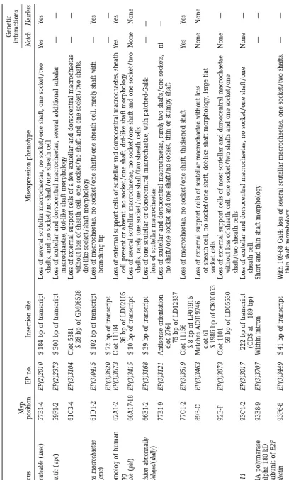
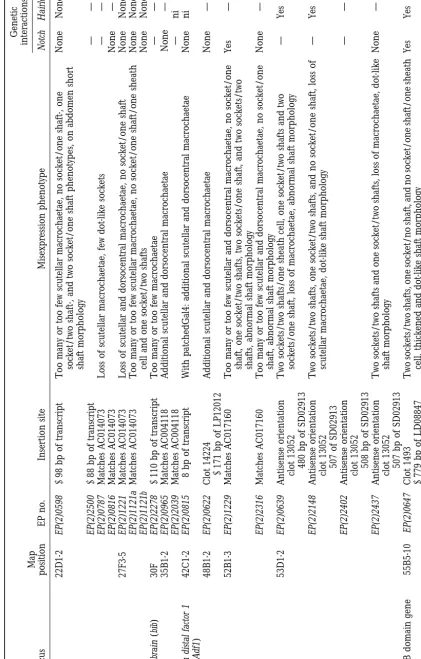
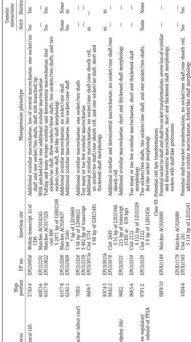
A Gain-of-Function Screen for Genes That Affect the Development of the
Drosophila Adult External Sensory Organ
Salim Abdelilah-Seyfried,* Yee-Ming Chan,* Chaoyang Zeng,
†Nicholas J. Justice,*
Susan Younger-Shepherd,* Linda E. Sharp,* Sandra Barbel,*
Sarah A. Meadows,* Lily Yeh Jan* and Yuh Nung Jan*
*Howard Hughes Medical Institute, Departments of Physiology and Biochemistry, University of California, San Francisco, California 94143-0725 and†Department of Biological Sciences, University of Wisconsin, Milwaukee, Wisconsin 53201
Manuscript received November 16, 1999 Accepted for publication February 24, 2000
ABSTRACT
The Drosophila adult external sensory organ, comprising a neuron and its support cells, is derived from a single precursor cell via several asymmetric cell divisions. To identify molecules involved in sensory organ development, we conducted a tissue-specific gain-of-function screen. We screened 2293 independent P-element lines established by P. Rørth and identified 105 lines, carrying insertions at 78 distinct loci, that produced misexpression phenotypes with changes in number, fate, or morphology of cells of the adult external sensory organ. On the basis of the gain-of-function phenotypes of both internal and external support cells, we subdivided the candidate lines into three classes. The first class (52 lines, 40 loci) exhibits partial or complete loss of adult external sensory organs. The second class (38 lines, 28 loci) is associated with increased numbers of entire adult external sensory organs or subsets of sensory organ cells. The third class (15 lines, 10 loci) results in potential cell fate transformations. Genetic and molecular character-ization of these candidate lines reveals that some loci identified in this screen correspond to genes known to function in the formation of the peripheral nervous system, such as big brain, extra macrochaetae, and numb. Also emerging from the screen are a large group of previously uncharacterized genes and several known genes that have not yet been implicated in the development of the peripheral nervous system.
T
HE development of the Drosophila adult external The transmembrane protein Notch is a receptor and its principal ligand during lateral inhibition is Delta sensory (es) organ, a mechanosensory bristle,in-volves lateral inhibition and asymmetric division, two (reviewed inArtavanis-Tsakonaset al. 1999). Within
the proneural cluster, Notch signaling is mediated mechanisms that underlie numerous developmental
processes (Posakony1994;JanandJan1995;Campos- through the transcription factor Suppressor of Hairless [Su(H)] and results in the activation of target genes at Ortega1996). First, a single sensory organ precursor
(SOP) cell is selected from a proneural cluster, a group the Enhancer of split [E(spl )] locus (Schweisguthand Posakony 1992; Fortini and Artavanis-Tsakonas of cells that are competent to become neuronal
precur-sors, via lateral inhibition. Genes within the achaete-scute 1994;BaileyandPosakony1995;Jarriaultet al. 1995;
LecourtoisandSchweisguth 1995). Hairless (H) is complex (AS-C) and the daughterless (da) gene are
re-quired to confer neuronal potential to these cells (Ghy- believed to act as an antagonist of Notch through physi-cal interaction with Su(H) (Brou et al. 1994; Banget
senandDambly-Chaudiere1989). After the SOP cell
is singled out, it divides asymmetrically to produce two al. 1995).
Both Notch-mediated cell-cell interactions and asymmet-different secondary precursor cells, IIA and IIB. IIA
gives rise to two external cells: one shaft cell (trichogen) ric segregation of the cell-intrinsic determinant Numb operate during divisions of the SOP lineage (Posakony and one socket cell (tormogen). IIB gives rise to the
internal cells: one neuron, one sheath cell, and, for 1994; Rhyu et al. 1994). During divisions of the SOP
cell and its progeny, Numb protein is unequally segre-at least one class of es organs, an additional glial cell
(HartensteinandPosakony1989;Ghoet al. 1999). gated to one of the two resulting daughter cells. In that cell, Numb inhibits the activity of N, which receives The Notch (N) signaling pathway mediates the
cell-cell interactions that occur during lateral inhibition. signals from two redundant ligands, Delta and Serrate (Rhyu et al. 1994; Frise et al. 1996; Guo et al. 1996;
Zenget al. 1998a). The pathways downstream of Notch
Corresponding author: Yuh Nung Jan, Howard Hughes Medical Insti- are different for the asymmetric divisions of IIA and
tute, Departments of Physiology and Biochemistry, Third and
Parnas-IIB cell lineages. Su(H) acts as a transducer of Notch sus Ave., University of California, San Francisco, CA 94143-0725.
E-mail: [email protected] signaling only within IIA and her daughter cells; the
downstream molecules that mediate Notch signaling in the IIB cell lineage are unknown (Wanget al. 1997). A
potential downstream target of Su(H) in IIA is tramtrack (ttk), a gene that does not appear to have a function during lateral inhibition (Guo et al. 1995, 1996).
An-other gene that affects lineage events and might be a component of the Notch signaling pathway is sanpodo (Dyeet al. 1998;SkeathandDoe1998).
The Notch signaling cascade in the SOP cell lineage
Figure 1.—Potential cell fate transformations in the IIA differs from that involved in lateral inhibition.
Addi-sublineage. (A) In wild-type, IIA divides asymmetrically to give tional components involved in N signaling during asym- rise to shaft (sh) and socket (so) cells. (B) Reduction of N metric divisions of the SOP lineage remain to be identi- signaling results in socket-to-shaft transformations. (C) Con-versely, increased N signaling (e.g., in Hairless mutants) results fied (e.g., ones that are specific for the IIB cell lineage).
in shaft-to-socket transformations. Genetic interactions were Many genes with a function in lateral inhibition or
assayed on the basis of the effects of the EP misexpression on asymmetric divisions of the adult es organ lineage, such
heterozygous mutant N or H phenotypes and vice versa. as N, Delta, numb, prospero (pros), and ttk, were initially
identified due to embryonic loss-of-function (lof) phe-notypes (Lehmannet al. 1981, 1983;Uemuraet al. 1989;
Doeet al. 1991;Vaessinet al. 1991;XiongandMontell of the es organ, i.e., the daughters of IIA. Next, we 1991; Salzberg et al. 1994). However, pleiotropy or analyzed the effect of misexpression on the sheath cell,
redundancy of gene function may hamper the identifi- a daughter of IIB. Finally, we examined the effect of cation of other genes important for the formation of reducing N or H function on the gof phenotype. These the adult es organ. One strategy to identify such genes analyses, combined with preliminary molecular charac-is to look for gain-of-function (gof) phenotypes. terizations, have led to the identification of genes pre-For this purpose, we screened 2293 independent Dro- viously shown to be important for es organ develop-sophila lines with the modular P-element-based EP (en- ment, as well as other genes that may be involved in hancer/promoter) misexpression element devised by P. this process.
Rørth (Rørth1996;Rørthet al. 1998). This
misexpres-sion element contains upstream activating sequence
MATERIALS AND METHODS (UAS) sites that are recognized by the transcriptional
activator Gal4 (Brand and Perrimon 1993). Tissue- Drosophila stocks:The collection of 2293 EP target element specific overexpression of genes that lie near the EP lines was a generous gift of P. Rørth through the Berkeley Drosophila Genome Project. For tissue-specific analysis of the element can be achieved by using a line that expresses
misexpression effects, the individual EP lines were crossed to Gal4 in specific cells. In cells that both express Gal4
sca-Gal4, a P{Gal4} line with an insertion at the scabrous locus and carry the EP element, Gal4 binds to the UAS sites
(Nakao and Campos-Ortega 1996). The sca-Gal4 line ex-and causes misexpression of the adjacent gene. presses Gal4 in SOP and surrounding cells and later in the On the basis of overexpression studies with genes pre- lineage of the es organ. To test the effects of different levels of expression, parents from initial crosses were serially trans-viously shown to be involved in adult es organ formation,
ferred and progeny from individual crosses were raised at 18, we expected certain phenotypes from such a gof screen.
25, and 29⬚during larval and pupal stages. The phenotypes Overexpression of genes such as numb, ttk, Su(H), H, at 29⬚
were generally stronger and more penetrant. All subse-and N give phenotypes opposite to the respective lof quent crosses were maintained at 29⬚.
phenotypes (Bang and Posakony1992; Lieber et al. The A101 line carries an insertion of P{lacZ,ry⫹} at the
neuralized locus (UsuiandKimura1993). It expresses nuclear 1993; Rhyu et al. 1994; Schweisguth and Posakony
-galactosidase in the SOP cell and the es organ lineage. On 1994; Guo et al. 1995; Doherty et al. 1997; Wang et
the notum, lacZ expression is strongest in the nuclei of the
al. 1997). Overexpression of N or its transducer Su(H)
two external support cells. The pros-lacZ enhancer trap line during lateral inhibition results in loss of entire es or- P{lacZ,ry⫹} expresses -galactosidase in the sheath cell. We
gans due to suppression of SOP formation. At later visualized-galactosidase expression by X-gal staining of pha-rate adults.
stages, during asymmetric division, overexpression of
Genetic interactions:To test genetic interactions with N,
these two genes produces up to four external cells, all
males from individual EP lines were crossed to waN55E11/FM6;
socket-like, due to IIB-to-IIA cell and/or shaft-to-socket
sca-Gal4/CyO females and the phenotypes of waN55E11/⫹;
sca-cell transformations (Lieberet al. 1993;Schweisguth Gal4/⫹flies carrying one copy of the EP element were com-andPosakony1994;Wanget al. 1997;Doherty et al. pared to those of FM6/⫹; sca-Gal4/⫹flies carrying one copy
of the EP element and to those of waN55E11/⫹; sca-Gal4/⫹flies
1997; Figure 1). Conversely, misexpression of H, which
without the EP element. Most lines that showed a positive antagonizes Notch signaling, results in increased
num-interaction were retested using a reciprocal crossing scheme bers of SOPs, IIA-to-IIB, and socket-to-shaft
transforma-with waN55E11/w·Y; sca-Gal4/CyO males (w·Y is a partial
duplica-tions (BangandPosakony1992). tion of the first chromosome including the N locus). Genetic In our screen, we first identified lines that produced interactions with H were tested by crossing males from individ-ual EP lines with y w ; sca-Gal4/CyO ; FRT HE21/TM3 females.
Phenotypes of y w ; sca-Gal4/⫹; FRT H /⫹ flies with one Many EP lines resulted in phenotypes with characteris-copy of the EP element were compared to those of y w ; sca- tics of more than one class. To simplify the classification, Gal4/⫹; TM3/⫹flies carrying one copy of the EP element
all EP lines with potential lineage transformation pheno-and to those of sca-Gal4/⫹; FRT HE21/⫹flies without the EP
types were grouped into class III independently of other element. For most crosses, parents were serially transferred
and progeny from individual crosses were maintained at 18, phenotypes. Similarly, among the remaining EP lines, 25, and 29⬚during larval and pupal stages. This genetic interac- those with phenotypes that include supernumerary es tion scheme allowed us to evaluate changes of the EP
misex-organs or subsets of support cells were grouped into pression phenotypes as an enhancement or suppression. In
class II independently of other phenotypes. Many lines addition, enhancement or suppression of the H mutant
phe-in all three classes also exhibited an altered morphology notype was evaluated. Since N/⫹flies lack a bristle phenotype,
only the enhancement of N haploinsufficiency could be de- of shaft or socket cells.
tected. Loss of external cells: We identified 52 lines
repre-Molecular analysis:Genomic sequences flanking the 3⬘end
senting 40 loci that produced loss of some or all of of the EP misexpression element were isolated by plasmid
the external and internal support cells. Loss of both rescue using EcoRI or SacII (Pirotta 1986). Sizes of three
independent clones for each plasmid rescue were compared external and internal support cells could arise from loss to determine the number of insertions per line. In total, there of the entire es organ. Alternatively, the support cells were 7 lines with two insertions (7/105⫽ 6.7%). Genomic
could have been transformed into neurons. Genes re-sequences adjacent to the EP element were sequenced.
sponsible for such phenotypes could interfere with lat-Flanking sequences were analyzed by searching the Berkeley
eral inhibition and function in lineage decisions, pre-Drosophila Genome Project (BDGP) and National Center for
Biotechnology Information databases. Expressed sequence vent cell cycle progression, or result in cell lethality. tags (EST) within a 3-kb distance from EP element insertion This is the largest class of EP lines and includes sites were tested for sequence similarities using “blastx”
P-element insertions into genes known to have
impor-searches. Sequenced genomic regions within a 3-kb distance
tant functions in asymmetric cell division, lateral inhibi-from EP element insertions for which no candidate transcripts
had been identified were tested using open reading frame tion, and other aspects of development. For example, finders. Only significant sequence similarities were reported misexpression of extra macrochaetae (emc) by EP(2)0415
(see Table 1). caused a loss of macro- and microchaetae (Figure 3A)
that resembles the phenotype of a dominant emc muta-tion (emcD;
Craymer1980). emc acts as a repressor that RESULTS
blocks the activity of achaete and scute gene function Using the modular misexpression system (Rørth
during sensory organ neurogenesis (Elliset al. 1990;
1996;Rørthet al. 1998), we misexpressed genes in the
Garrell and Modolell 1990; Skeath and Carroll SOP cell and its neighbors and examined the effects on
1991; Van Doren et al. 1991) and its misexpression is
the development of the adult external sensory organ.
predicted to block SOP formation. The sca-Gal4 line was chosen as driver because it is
ex-Another example is the misexpression of escargot (esg) pressed in clusters of cells surrounding the presumptive
[by EP(2)0683, EP(2)0684, EP(2)2009, EP(2)2159, and macro- and microchaetae on the notum and head
(Fig-EP(2)2408], which caused the most severe loss of es
ure 2). Expression persists in the SOP lineage. All
misex-organs observed in this screen. In EP(2)0684 and pression phenotypes described in this paper are
pro-EP(2)2009, there was an almost complete loss of es
or-duced by sca-Gal4 in conjunction with an EP insertion.
gans on the notum (Figure 3B). esg encodes a zinc finger We then examined the effects of reducing N or H
func-protein that acts as a repressor of Scute/Daughterless-tion on the gof phenotype. The enhancer trap lines
dependent transcription in vitro (Whiteleyet al. 1992; A101 and pros-lacZ were used to assist our
characteriza-Fuse et al. 1994). It also acts as negative regulator of
tion of misexpression phenotypes. A101-lacZ expresses
endoreplication in imaginal tissues (Hayashiet al. 1993;
-galactosidase strongly in the nuclei of the two external
Hayashi1996). support cells, while pros-lacZ expresses -galactosidase
We also identified several genes known to be required specifically in the sheath cell, one of the internal cells.
for correct cell cycle progression. dacapo [EP(2)2584] In total, 4.6% of the lines (105/2293) produced
phe-is a cyclin-dependent kinase inhibitor that phe-is required notypes affecting the number or fate of outer cells of
during embryogenesis for a timely exit from the cell the es organ. These phenotypes fall into three major
cycle (Laneet al. 1996;de Nooijet al. 1996).
Misexpres-classes:
sion of dacapo produced a loss of external cells of scutel-1. class I: loss of external support cells (sockets and lar and dorsocentral macrochaetae (Figure 3C). In some shafts) cases, there was a single prospero-positive cell that was 2. class II: supernumerary es organs or support cells no longer accompanied by shaft and socket cells. An-3. class III: potential cell fate transformations, with in- other gene, divisions abnormally delayed (dally), encodes creases in one cell type associated with loss of another a proteoglycan that is required for normal cell cycle cell type.
progression (Nakatoet al. 1995) and might act as
core-ceptor for Wingless (Lin andPerrimon 1999; Tsuda Tables 1 and 2 summarize the molecular, phenotypic,
(Fischer-Vize et al. 1992; Huang et al. 1995); apontic
[EP(2)2373], a gene involved in multiple processes, in-cluding head patterning (Gellonet al. 1997) and heart
morphogenesis (Suet al. 1999); Drosophila lim-domains only [EP(X)1306, EP(X)1383, and EP(X)1394], a gene
with a role in wing patterning (Milanet al. 1998; Shor-eshet al. 1998;Zenget al. 1998b), longitudinals lacking
(lola) [EP(2)0343], which is required for correct axonal projection (Giniger et al. 1994); and hnRNP 27C
[EP(2)0748], which encodes a heterogeneous nuclear RNA-associated protein. Previous studies suggest that different heterogeneous nuclear RNA-associated pro-teins may play a role in the development of the es organ (Hammondet al. 1997;zur Lageet al. 1997).
This class includes insertions at 15 previously
unchar-Figure2.—Macro- and microchaetae are arranged in
ste-acterized genes. Four of these insertions showed genetic reotyped patterns on the notum of Drosophila (for recent
interactions with N or H (see Table 2), indicating that review on es organ pattern formation, seeSimpsonet al. 1999).
(A) Four dorsocentral (dc) and four scutellar (sc) macrochae- they affect genes that are potentially in the N signaling tae decorate the adult notum. (B) sca-Gal4 expresses Gal4 (in pathway. These genes are therefore good candidates for green, driving UAS-GFP) within the four cut-expressing cells
future analyses. of the es organ (red) and surrounding cells (Blochlinger
Supernumerary es organs or support cells: Thirty-et al. 1993). On the scutellum and bThirty-etween the dorsocentral
eight lines, carrying insertions at 28 loci, caused misex-macrochaetae, sca-Gal4 is expressed not only in the developing
sensory organs but also in surrounding domains. pression phenotypes with increased numbers of internal and external cell types. We further subdivided these lines into two subclasses. One subclass of lines produced resulted in the occasional loss of scutellar or dorsocen- ectopic (i.e., spatially separate) es organs; these might tral macrochaetae. Misexpression of these genes could arise from defective lateral inhibition or ectopic pro-interfere with SOP lineage events by blocking cell cycle neural activity. The other subclass of lines exhibited super-progression (e.g., by forcing the SOP cell to exit mitosis) numerary support cells that were clustered together. This or, in the case of dally, by affecting Wingless signaling, phenotype could be due to either increased cell numbers which is involved in the patterning of es organs (Phil- within an es organ or formation of several tightly associated lipsandWhittle1993). es organs. Such phenotypes could result from defects in
A large number of P-element insertions targeted lateral inhibition or cell cycle regulation.
genes that are known to have essential functions during In this class, there are 16 previously uncharacterized development but have not previously been implicated genes (Table 1). To distinguish lines that affect lateral in sensory organ development. One line, carrying an inhibition from those that affect other functions, we insertion at the inscuteable (insc) locus [EP(2)2010], ex- tested a subset of these lines for genetic interactions hibited a loss of external structures of scutellar macro- with N and H. Eight lines representing eight indepen-chaetae without a concurrent loss of the prospero-posi- dent loci displayed significant genetic interactions (see tive sheath cell. Whether this phenotype is entirely due Table 2).
to altered expression of insc, which serves an essential Ectopic supernumerary es organs: This subclass includes function in asymmetric divisions of delaminating neuro- big brain [EP(2)2278], a gene involved in lateral inhibi-blasts and embryonic muscle progenitor cell divisions tion that encodes a channel-like transmembrane pro-(Krautet al. 1996;Carmenaet al. 1998), requires fur- tein (Rao et al. 1990). Also in this subclass are two
ther study. One potential complication is the presence genes with a known function in eye development: yan of the gene skittles, which encodes the phosphatidylinosi- [EP(2)0598 and EP(2)2500], which encodes an ETS do-tol 4-phosphate 5-kinase, in the first intron of insc. Mis- main nuclear protein that has an essential function in expression of skittles has been shown to generate ectopic photoreceptor cell development (LaiandRubin1992; es organs (Hassanet al. 1998). It is not clear whether O’Neillet al. 1994); and hedgehog [EP(3)3521], which is
misexpression of insc, skittles, or both is driven by involved in multiple developmental processes including
EP(2)2010. eye furrow progression (Heberleinet al. 1993;Maet al.
Other known developmental regulators found in this 1993). hedgehog has also been implicated in the correct screen include gliotactin [EP(2)2306], which encodes a patterning of es organs on the adult notum (Gomez-transmembrane protein that functions in peripheral Skarmeta andModolell 1996; Mulloret al. 1997). glia to establish the blood-nerve barrier (Auld et al. Another gene, split ends (spen) [EP(2)2583], resulted in
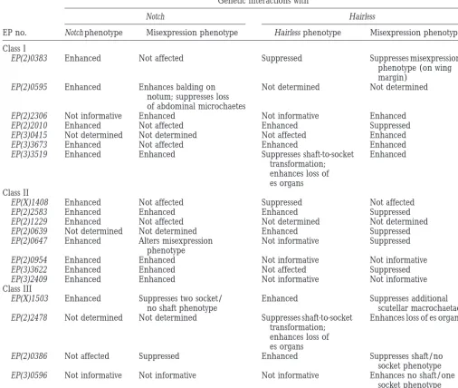
TABLE 2
Genetic interactions withNandH
Genetic interactions with
Notch Hairless
EP no. Notch phenotype Misexpression phenotype Hairless phenotype Misexpression phenotype
Class I
EP(2)0383 Enhanced Not affected Suppressed Suppresses misexpression
phenotype (on wing margin)
EP(2)0595 Enhanced Enhances balding on Not determined Not determined
notum; suppresses loss of abdominal microchaetes
EP(2)2306 Not informative Enhanced Not informative Enhanced
EP(2)2010 Enhanced Not affected Enhanced Suppressed
EP(3)0415 Not determined Not determined Not affected Enhanced
EP(3)3673 Enhanced Not affected Enhanced Enhanced
EP(3)3519 Enhanced Enhanced Suppresses shaft-to-socket Enhanced
transformation; enhances loss of es organs Class II
EP(X)1408 Enhanced Not affected Suppressed Not affected
EP(2)2583 Enhanced Enhanced Enhanced Suppressed
EP(2)1229 Enhanced Not affected Not determined Not determined
EP(2)0639 Not determined Not determined Enhanced Suppressed
EP(2)0647 Enhanced Alters misexpression Not informative Suppressed
phenotype
EP(2)0954 Enhanced Enhanced Not informative Not informative
EP(3)3622 Enhanced Enhanced Not affected Suppressed
EP(3)2409 Enhanced Enhanced Not informative Not informative
Class III
EP(X)1503 Enhanced Suppresses two socket/ Enhanced Suppresses additional
no shaft phenotype scutellar macrochaetae
EP(2)2478 Not determined Not determined Suppresses shaft-to-socket Enhances loss of es organs transformation;
enhances loss of es organs
EP(2)0386 Not affected Suppressed Enhanced Suppresses shaft/no
socket phenotype EP(3)0596 Not informative Not informative Not informative Enhances no shaft/one
socket phenotype
A total of 76 EP lines (64 loci) were tested for genetic interactions with N and H. Misexpression of the genes targeted by 19 independent lines showed interpretable genetic interactions in heterozygous backgrounds of either N or H. Genetic interactions were scored as the effect of the EP misexpression on the haploinsufficient N or H phenotype and as the effect of these mutations on the EP misexpression phenotype. For N, EP misexpression enhanced the N phenotype when socket-to-shaft transformations occurred. Conversely, for Hairless, EP misexpression enhanced the H phenotype when the number of es organs with shaft-to-socket transformations was increased (relative to the dominant Hairless phenotype), while EP misexpression suppressed the H phenotype when reduced numbers of es organs with such transformations were found. An enhancement of Hairless is also associated with the loss of es organs. None, no genetic interactions observed.
spen has multiple developmental functions including Several previously uncharacterized genes targeted by the EP element displayed genetic interactions with N correct axon formation (Kolodziej et al. 1995) and
control of correct segment identity (Wiellette et al. and H. For example, EP(3)3622 produced a misexpres-sion phenotype with additional es organs and tufts (i.e., 1999). Two insertions near nuclear fallout [EP(3)3324
and EP(3)3339] resulted in additional scutellar macro- a large number of clustered shafts; Figure 4B). The misexpression phenotype produced by EP(3)3622 is en-chaetae and in one-socket/two-shaft phenotypes. This
gene encodes a coiled-coil protein with a function in hanced by removing one copy of N and suppressed by removing one copy of H (Table 2).
cortical actin organization and cytokinesis (Rothwell
Genetic interactions with N and H were found with
EP(2)0647, an insertion at a gene that has sequence
similarities with BTB-domain-containing proteins such as Pipsqueak. Misexpression of this gene resulted in, among other phenotypes, increased numbers of support cells associated with es organs.
Potential cell fate transformations: We expected to identify P-element insertions that target genes that func-tion in the asymmetric divisions of the stereotyped es organ lineage. In total, 15 lines representing 10 loci
Figure3.—Examples of class I misexpression phenotypes.
resulted in apparent cell fate transformations. These (A) Misexpression of EP(3)0415 at the extra macrochaetae locus
lines fall into three subclasses. The first two subclasses resulted in the loss of scutellar and dorsocentral macro- and
microchaetae. (B) Several insertions targeting escargot, includ- are transformations within the IIA cell sublineage: (a) ing EP(2)0684, resulted in the loss of almost the entire popula- a socket-to-shaft cell transformation, which would result tion of macro- and microchaetae. (C) Misexpression of
in a two-shaft/no-socket phenotype (twinned pheno-EP(2)2584 at the dacapo locus resulted in the loss of external
type); and (b) a shaft-to-socket cell transformation, cells of scutellar and dorsocentral macrochaetae. The shaft
cell morphology of many macrochaetae was abnormal. The which would result in a no-shaft/two-socket phenotype. arrowhead indicates an abnormal shaft cell morphology. The third subclass is transformations from IIA to IIB, which would result in loss of external support cells (bald-ing). However, mechanisms other than transformations Supernumerary internal and external support cells
may cause these phenotypes as well (e.g., ectopic cell could arise from ectopic cell divisions caused by altered
division of one type of support cell combined with the cell cycle regulation. A previously uncharacterized gene
elimination of another type of support cell). targeted by EP(3)3559 has sequence similarities with
Potential transformations of socket cell to shaft cell: The
human regulatory subunits of protein phosphatase 2A
misexpression of numb by EP(2)2542 resulted in socket-(PP2A). Genes coding for the regulatory subunit B of
to-shaft transformations similar to the numb over-PP2A (abnormal anaphase, twins) are involved in both cell
expression phenotype (Figure 5A; Rhyu et al. 1994).
cycle progression and cell fate determination (Gomeset
The misexpression phenotype of EP(2)2542 also
in-al. 1993;Shiomiet al. 1994). EP(3)3559 shows increased
cluded the loss of external structures of macrochaetae. numbers of support cells in each es organ (Figure 4C).
This phenotype might be the result of IIA-to-IIB trans-This misexpression phenotype mimics the phenotype
formations. observed in twins, a mutation in the regulatory B subunit
Each of the two insertions [EP(X)1149 and of PP2A (Uemuraet al. 1993). Regulatory subunits that
EP(X)1179] that target the same unknown gene
pro-are under temporal or tissue-specific control in turn
duced both socket-to-shaft and reciprocal shaft-to-socket regulate the activity of PP2A. It will be of interest to test
transformations (Figure 6C). Both lines also caused a how the newly identified regulatory subunit regulates
loss of external support cells on the notum. the function of PP2A.
Potential transformations of shaft cell to socket cell: This
Three insertions at a novel locus, EP(2)0639,
subclass includes string, twine, and grapes, three genes
EP(2)2148, and EP(2)2437, produce supernumerary
sup-with a function in mitotic or meiotic cell cycle regulation port cells in the es organ (Figure 4D). The orientation
(EdgarandO’Farrell1989;Alpheyet al. 1992; Cour-of the EP elements at this locus is such that they
presum-totet al. 1992;Fogartyet al. 1994, 1997). We identified
ably generate a partial antisense transcript. Therefore,
four independent insertions at or near the string locus the phenotypes could be caused by lof or neomorphic
effects. [EP(3)1213, EP(3)3261, EP(3)3426, and EP(3)3432].
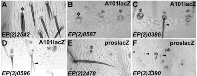
Figure5.—Examples of class III misex-pression phenotypes. (A) Misexmisex-pression of EP(2)2542 at the numb locus resulted in apparent socket-to-shaft transforma-tions. (B) EP(2)0587 at the grapes locus caused apparent shaft-to-socket transfor-mations on the abdomen. Double sock-ets are indicated by the presence of two large A101 lacZ-positive nuclear stains. (C) Misexpression of EP(2)0386 pro-duced apparent shaft-to-socket transfor-mations on the abdomen, as indicated by the presence of two large A101 lacZ-positive nuclear stains. (D) The ab-dominal misexpression phenotypes of EP(3)0596 were apparent shaft-to-socket transformations (asterisk) and branching of shaft cells (arrowhead). (E) Misexpression of EP(2)2478 resulted in apparent IIA-to-IIB or neuron-to-sheath transformations. In the absence of external support cells, two proslacZ- positive sheath cells were tightly associated (asterisk). (F) Similarly, misexpression of EP(3)3390 resulted in apparent IIA-to-IIB or neuron-to-sheath transformations. Two associated proslacZ-positive sheath cells were commonly scored in the absence of differentiated external structures (asterisk). However, abnormal cuticular structures were visible (arrowheads). Potential transformation phenotypes are indicated with an asterisk.
With the exception of EP(3)1213, which carries an inser- IIB cell fate transformations, with two or more prospero-positive cells in the absence of external support cells. tion ⵑ1.5 kb upstream of the normal transcript, the
other three insertions lie close to the transcription initia- With EP(2)2478, both macro- and microchaetae exhib-ited a loss of external support cells as well as a duplica-tion site (see Table 1). However, only EP(3)1213 resulted
in possible shaft-to-socket transformations, raising the tion of presumptive sheath cells (Figure 5E). Similarly, the misexpression caused by EP(3)3390 resulted in a loss question whether a gene other than string is affected in
this line. The misexpression by EP(3)3261 produced of external support cells of macro- and microchaetae as well as duplication of prospero-positive sheath cells increased numbers of internal and external support
cells. X-gal staining with enhancer trap lines A101 lacZ (Figure 5F). In rare cases, up to four sheath cells were present.
and prospero lacZ, which mark the external and the
sheath cells, respectively, showed an approximate dou- Defective morphology of the es organ: At least 41 lines, representing 38 loci, identified in this screen pro-bling of the cell number in many es organs (not shown).
Insertions near grapes [EP(2)0587] and twine duced aberrant morphology of either the socket or the shaft cell. The following are examples of different mor-[EP(2)0613] resulted in potential shaft-to-socket
trans-formations on the abdomen and notum, respectively phology phenotypes observed.
Misexpression driven by EP(2)2356 produced an ab-(Figure 5B). Mutations in grapes, a protein kinase with
homologies to Saccharomyces cerevisiae CHK1, have been normal shaft cell morphology. Most prominently, the shaft cell was short and branched into many distal tips shown to interfere with the DNA replication checkpoint
control of the cell cycle (Fogartyet al. 1997). In addi- (Figure 6A). Branching of the shaft cell into two distal tips was observed in several lines [i.e., in EP(3)0596, tion, embryos mutant in grapes exhibit cortical
cytoskele-tal defects during syncytial divisions (Sullivan et al. Figure 5D].
Morphologically abnormal socket cells were pro-1993). Misexpression of twine caused, in addition to
possible shaft-to-socket transformations, a four-socket duced by EP(3)3463. Among other phenotypes, the socket cells frequently were large and flattened (Figure phenotype. twine, a cdc25 homolog, has a function
dur-ing male and female meiotic divisions and participates 6B). EP(X)1149 (see also phenotype in class III) duced an abnormal socket cell morphology with a pro-in some aspects of mitotic control at the syncytial
em-bryo stage (Alphey et al. 1992; Courtot et al. 1992; truding tip similar to a short shaft (Figure 6C). We observed a massive reduction in the size of shaft EdgarandDatar1996).
The most prominent phenotype found with two other cells and morphologically abnormal socket cells with
EP(2)2317, an insertion at el F-4A (Figure 6D). Similar
lines, [EP(2)0386 and EP(2)0988], was apparent
shaft-to-socket cell transformations on the abdomen. X-gal phenotypes were seen with several other lines.
The sensitivity of cell morphology to the misexpres-staining with enhancer trap line A101 lacZ, which
pre-dominantly marks two large nuclei of the two external sion of candidate genes might yield an entry point to identify genetic components involved in differentiation cells of the es organ, confirmed the presence of two
socket cells (Figure 5C). A third line, EP(3)0596, pro- and morphogenesis. Several of the phenotypes de-scribed here resemble phenotypes caused by mutations duced a similar misexpression phenotype (Figure 5D).
Potential transformations of IIA to IIB: Two insertions at of genes that function in cytoskeletal assembly (Cant
et al. 1994;Tilneyet al. 1995, 1996).
IIA-to-DISCUSSION phenotypes in the biological context of choice. In vari-ous screens, phenotypes that affected eye development, Analyzing development of the es organ using a
gain-wing development, and follicle cell migration were ana-of-function approach: Traditionally, genetic screens
lyzed (Rørthet al. 1998).
have been based on the isolation of lof mutations. This
In this study, these 2293 randomly inserted P elements approach has been invaluable in unraveling the
mecha-were each driven by a sensory-organ-specific Gal4 driver nisms underlying many biological processes, including
and any resulting misexpression phenotypes in the es the formation of the peripheral nervous system
(Salz-organ were analyzed. Of these lines, 105 produced es berg et al. 1994; Kania et al. 1995; Go et al. 1998).
organ phenotypes. Our preliminary phenotypic and mo-However, lof screens have several limitations.
Redun-lecular analyses suggest that we have identified genes dancy between genes that have overlapping functions
that are involved in lateral inhibition, cell cycle control, might partially or completely mask gene function. In
cell fate specification, and cell differentiation. A subset such cases, it is necessary to make double or multiple
of these genes is likely to play a role in es organ forma-mutant combinations to produce a phenotype, an
ap-tion. proach that is not generally applicable during lof
One potential drawback of gof screens is that misex-screens. Moreover, early phenotypes caused by a
muta-pression of a gene may affect the development of tissues tion might prevent the detection of later phenotypes
in which that gene is not normally expressed. In some (MiklosandRubin1996). Such limitations can be
par-cases, misexpression of a gene may ectopically effect a tially circumvented by screens that are based on
analyz-signaling pathway that functions in multiple develop-ing the phenotypes of clones of mutant tissue generated
mental processes. Another concern is that phenotypes by somatic recombination (XuandRubin 1993) or by
may be artificial. For example, the phenotype caused screens for enhancers or suppressors of a particular
by misexpression of a gene at levels much higher than mutant phenotype (Simon et al. 1991). Nevertheless,
normal may interfere with development, even if that many genes might have escaped detection by lof
ap-gene does not have a function in development. proaches.
To identify those genes that normally function in es The gof screening system devised by P. Rørth
comple-organ development, it will be important to examine ments lof approaches. This system is based on the
analy-the lof phenotype, analy-the expression pattern, and genetic sis of phenotypes generated by tissue-specific
misexpres-interactions with genes known to be involved in es organ sion of genes using the UAS-Gal4 system. Any gene that
development. produces a misexpression phenotype is detectable by
The systematic misexpression screen identifies candi-the system in spite of possible functional redundancy
date genes that interfere with distinct developmental and pleiotropy of gene function (Rørth1996;Rørth
aspects of es organ formation: Among the 105 lines
et al. 1998). In addition, the tissue specificity of the
UAS-(78 loci) identified in the screen, 49 lines (37 loci) Gal4 system allows the examination of misexpression
correspond to previously characterized genes. A subset of these genes has been shown to have roles during es organ development. Some, such as emc and big brain, have a function in lateral inhibition (Skeathand Car-roll 1991; Rao et al. 1992). Several are genes with a
function in cell cycle regulation, including dacapo and
string, and thus might be required during es organ cell
division. Others, such as numb, are known to be involved in asymmetric cell division (Rhyuet al. 1994). Moreover,
a large group of genes with essential roles in other developmental processes were identified. Some of these genes, such as hedgehog and yan, have not been tested for their role in es organ development, but it is possible that they are involved in this developmental process as well. Since many of the known genes identified in this
Figure 6.—A group of 41 EP lines carry insertions near screen are likely to have normal functions in es organ genes that when misexpressed, produced an abnormal es
or-development, the concern of the potentially artificial gan morphology. Examples are as follows: (A) EP(2)2356
nature of the gof screen may be alleviated. It thus seems caused branching of shafts into multiple tips (arrowheads).
(B) Flattened and enlarged socket cells were commonly scored likely that at least a substantial subset of the new genes with EP(3)3463. (C) EP(X)1149 resulted in potential shaft- identified in our screen will turn out to be important to-socket transformations. Socket cells frequently displayed for the formation of es organs, perhaps in some of protruding shaft-like tips. (D) EP(2)2317 resulted in the severe
the less understood aspects of es organ development, reduction of shaft cells into shortened or dot-like structures.
including the following: Arrowheads indicate abnormal cell morphology. Potential
The transducers of N signaling in IIB and her daughters phology defects. These include genes that affect differ-entiation of a single cell type (e.g., shaft cell differentia-are currently not known (Wanget al. 1997). EP(2)2478
and EP(3)3390 target genes with possible functions in tion controlled by pax2; Kavaler et al. 1999) or that
affect proper regulation of cytoskeletal dynamics. We IIB and her daughters. Misexpression of those genes
was sufficient to generate potential IIA-to-IIB or neuron- found a large number of lines that, when misexpressed, resulted in aberrant morphogenesis of the socket or to-sheath transformations. One possible explanation for
this phenotype is ectopic activation of IIB-specific target shaft cell. One phenotype observed was the branching of shafts. It has been suggested that mutations causing genes (e.g., by IIB or sheath-cell-specific N-signaling
components). branched hairs are in genes that regulate the actin cy-toskeleton (TurnerandAdler1998). Consistent with
Cell cycle regulation of stereotyped lineage events: One likely
link between cell cycle regulation and asymmetric cell this prediction, mutations of genes with a function in actin bundle formation display similar branching phe-division is the cell-cycle-dependent asymmetric
localiza-tion of cell fate determinants and adaptor proteins (Hir- notypes (Cant et al. 1994; Tilney et al. 1995, 1996).
Several of the lines identified in this screen might pro-ataet al. 1995; Knoblichet al. 1995; Spanaand Doe
1995;Krautet al. 1996;Ikeshima-Kataokaet al. 1997; vide additional components involved in executing shaft cell morphology or in regulating the actin cytoskeleton Shen et al. 1997; Lu et al. 1998, 1999;Schuldt et al.
1998). Untimely cell cycle progression or defective inte- in other tissues. Less is known about the morphogenesis of socket cells. EP lines that affected predominantly gration of cell cycle with the localization of Numb
pro-tein may create a phenotype reminiscent of numb lof, socket cell morphology might provide clues to this pro-cess.
a phenotype that was observed with misexpression of
the cell cycle regulatory genes grapes and twine. Genomic considerations and perspectives: Genome sequencing by the European and Berkeley Drosophila In addition, cell cycle regulatory genes may serve
addi-tional functions that affect cell fate specification. grapes, Genome Projects (EDGP and BDGP) and the ease with which genomic sequences flanking the EP element can for example, is essential for the normal formation of
the cortical cytoskeleton during syncytial divisions (Sul- be cloned have greatly facilitated the identification of targeted genes. Of the insertion sites we sequenced, 49 livanet al. 1993). Given the importance of the cortical
cytoskeleton during asymmetric division (Broadusand (37 loci; 46.7% of all lines) matched known genes, 34 (28 loci; 32.4% of all lines) matched EST, and 22 (13 Doe 1997;Knoblich et al. 1997), genes that regulate
the dynamics of this structure may also turn out to be loci; 20.9% of all lines) matched sequenced genomic regions but still have no candidate transcripts.
essential during cell fate decisions.
Highly stereotyped division patterns occur through- Altogether, 105 lines or 4.5% of the lines tested gave rise to misexpression phenotypes.Rørthet al. (1998)
out Drosophila development (Foe 1989; Gho et al.
1999). Several cell cycle regulators, including dacapo, reported comparable frequencies of misexpression phe-notypes: 7% with ombGal4, 4% with dppGal4, 3% with are required to control the cell division patterns in the
neural lineages of the embryonic nervous system (Cui slboGal4, and 2% with sevGal4. Among the few genes
that were reported from those screens, we have isolated andDoe1995;WeigmannandLehner1995;de Nooij
et al. 1996;Laneet al. 1996;HassanandVaessin1997). escargot, hedgehog, yan, scalloped, and big brain. It will be
interesting to compare those screens to obtain an esti-It is not known at this time whether dacapo normally
functions during the development of the es organ to mate of the overlap of the genes used in those different developmental processes.
control precise cell division patterns.
Execution of morphogenesis: There are different types of In a separate database analysis, we searched for EP element insertions that target genes with a known func-genes that when misexpressed could give rise to
mor-TABLE 3
Summary of EP element insertions
Map
Locus position EP no. Insertion site
kuzbanian (kuz) 34D4 EP(2)2503 ⫺916 bp of transcript
neuralized (neur) 85D EP(3)3026 ⫹466 bp of transcript (CDS at⫹278 bp) Enhancer of split
transcript m2 (E(spl)m2) 96F9 EP(3)3635 ⫺2702 bp of transcript
Enhancer of split EP(3)3272 ⫺11 bp of transcript
transcript m7 (E(spl)m7) 96F9 EP(3)3587 ⫺646 bp of transcript
tion in neurogenesis and sensory organ development. will be to determine the biological significance of the genes identified during this screen.
Among seven EP element insertions that target six genes
(extra macrochaetae, big brain, kuzbanian, neuralized, and We are most grateful to P. Rørth for the generous gift of EP lines.
Enhancer of split transcripts m2 and m7), only two inser- We thank Todd Laverty and G. Rubin for kindly providing us with the EP lines, and the lab of J. Campos-Ortega and the Bloomington tions near two loci yielded misexpression phenotypes
Drosophila Stock Center for fly strains. Our sequence analysis was in our assay (extra macrochaetae, big brain). Five insertions
helped by the sequencing efforts of BDGP and EDGP. Thanks to B. near four loci did not cause obvious misexpression phe- Lu and S. Zhou for critical reading of the manuscript; to D. Doherty notypes (Table 3). Therefore, the misexpression screen for providing us with Figure 2; and to other current and former was not fully efficient. Similarily, there may be other members of the Jan lab for discussion, suggestions, and help. S.A.-S. was supported by a fellowship from the Deutsche Forschungsgem-unknown genes with a function in es organ development
einschaft. Y.-M. C. currently is supported by the Program in Biological that escaped detection even with an EP element inserted
Sciences Markey Grant and the Herb Boyer Fund. C.Z. is a postdoc-nearby.
toral associate, N.J.J. is a predoctoral associate, and L.Y.J. and Y.N.J. Determining the exact insertion site and orientation are investigators of the Howard Hughes Medical Institute.
of the EP element is essential to the interpretation of misexpression phenotypes. In the lines for which we identified a transcript, most of the EP transposons were
LITERATURE CITED inserted between⫺850 bp upstream and⫹800 bp
down-Alphey, L., J. Jimenez, H. White-Cooper, I. Dawson, P. Nurseet stream of the transcription start site (61/83⫽73.5%).
al., 1992 twine, a cdc25 homolog that functions in the male and
Seven lines (8.4%) were identified with insertions at female germline of Drosophila. Cell 69: 977–988.
greater distances from the transcription start site of pu- Artavanis-Tsakonas, S., M. D. RandandR. J. Lake,1999 Notch signaling: cell fate control and signal integration in development. tative target genes. In these cases it is possible that
addi-Science 284: 770–776.
tional transcripts that have not been identified might Auld, V. J., R. D. Fetter, K. BroadieandC. S. Goodman, 1995 be located closer to the EP element. One example is Gliotactin, a novel transmembrane protein on peripheral glia, is required to form the blood-nerve barrier in Drosophila. Cell 81:
EP(3)1213, which carries an insertionⵑ1.5 kb 5⬘of the
757–767.
transcriptional start site of string. The misexpression Bailey, A. M.,andJ. W. Posakony, 1995 Suppressor of hairless phenotype produced by this line was qualitatively differ- directly activates transcription of enhancer of split complex genes in response to Notch receptor activity. Genes Dev. 9: 2609–2622. ent from other EP insertions closer to the string
tran-Bang, A. G.,andJ. W. Posakony,1992 The Drosophila gene Hairless scriptional start site. Whether these differences are at- encodes a novel basic protein that controls alternative cell fates tributable to different levels of expression or are caused in adult sensory organ development. Genes Dev. 6: 1752–1769.
Bang, A. G., A. M. BaileyandJ. W. Posakony,1995 Hairless pro-by an unidentified transcript needs to be determined.
motes stable commitment to the sensory organ precursor cell Another 9 lines (10.8%) carried EP elements with an fate by negatively regulating the activity of the Notch signaling apparent antisense orientation and might generate par- pathway. Dev. Biol. 172: 479–494.
Blochlinger, K., L. Y. JanandY. N. Jan,1993 Postembryonic pat-tial antisense transcripts. How these antisense messages
terns of expression of cut, a locus regulating sensory organ identity might cause phenotypes is not clear. In addition, there in Drosophila. Development 117: 441–450.
are several lines (6/83⫽ 7.2%) that carried insertions Brand, A. H.,andN. Perrimon,1993 Targeted gene expression as a means of altering cell fates and generating dominant phenotypes. 3⬘ of the CDS, or insertions within new transcripts for
Development 118: 401–415.
which the CDS is not known. In these cases, the pheno- Broadus, J.,andC. Q. Doe,1997 Extrinsic cues, intrinsic cues and types might be caused by truncated transcripts. microfilaments regulate asymmetric protein localization in
Dro-sophila neuroblasts. Curr. Biol. 7: 827–835. The EP transposon allows only the unidirectional
Brou, C., F. Logeat, M. Lecourtois, J. Vandekerckhove, P.
Kouril-transcription of potential target genes. Therefore, skyet al., 1994 Inhibition of the DNA-binding activity of
Drosoph-ⵑ50% of the EP lines are expected to be in the correct ila suppressor of hairless and of its human homolog, KBF2/ RBP-J kappa, by direct protein-protein interaction with Drosophila orientation to drive misexpression of a sense transcript
hairless. Genes Dev. 8: 2491–2503.
[only nine of the lines that gave rise to phenotypes with Campos-Ortega, J.,1996 Numb diverts notch pathway off the
tram-sca-Gal4 (8.6%) had an inverted or antisense orienta- track. Neuron 17: 1–4.
Cant, K., B. A. Knowles, M. S. MoosekerandL. Cooley,1994 Dro-tion]. Thus, the total number of genes targeted for
sophila singed, a fascin homolog, is required for actin bundle forma-overexpression in the screen might be no more than tion during oogenesis and bristle extension. J. Cell Biol. 125: 1150. The number of targeted genes is further reduced 369–380.
Carmena, A., B. Murugasu-Oei, D. Menon, F. JimenezandW. Chia,
by multiple lines targeting the same gene (1.33
inser-1998 Inscuteable and numb mediate asymmetric muscle
progeni-tions/locus) and by insertions that lie too distantly to
tor cell divisions during Drosophila myogenesis. Genes Dev. 12: drive sufficient transcriptional activation. 304–315.
Courtot, C., C. Fankhauser, V. SimanisandC. F. Lehner,1992 The current estimate for the number of genes in
The Drosophila cdc25 homolog twine is required for meiosis. Devel-the Drosophila genome by Devel-the BDGP is around 14,000
opment 116: 405–416.
(based onMiklosandRubin1996). Therefore, the EP Craymer, L.,1980 [New mutants report.] Dros. Inf. Serv. 55: 197– 200.
collection targetsⵑ10% of the genome. In an
extrapola-Cui, X.,andC. Q. Doe,1995 The role of the cell cycle and cytokinesis tion, for a genome-wide saturation screen we would
in regulating neuroblast sublineage gene expression in the Dro-expectⱖ800 different loci orⵑ5–6% of all genes to give sophila CNS. Development 121: 3233–3243.
cyclin-dependent kinase inhibitor, Dacapo, is necessary for timely during asymmetric division: interaction of Numb and Notch. Neuron 17: 27–41.
exit from the cell cycle during Drosophila embryogenesis. Cell 87:
Hammond, L. E., D. Z. Rudner, R. KanaarandD. C. Rio,1997 Mu-1237–1247.
tations in the hrp48 gene, which encodes a Drosophila
heteroge-Doe, C. Q., Q. Chu-Lagraff, D. M. WrightandM. P. Scott,1991
neous nuclear ribonucleoprotein particle protein, cause lethality The prospero gene specifies cell fates in the Drosophila central
and developmental defects and affect P-element third-intron nervous system. Cell 65: 451–464.
splicing in vivo. Mol. Cell. Biol. 17: 7260–7267.
Doherty, D., L. Y. JanandY. N. Jan,1997 The Drosophila neurogenic
Hartenstein, V.,andJ. W. Posakony,1989 Development of adult gene big brain, which encodes a membrane-associated protein,
sensilla on the wing and notum of Drosophila melanogaster. Devel-acts cell autonomously and can act synergistically with Notch and
opment 107: 389–405. Delta. Development 124: 3881–3893.
Hassan, B.,andH. Vaessin,1997 Daughterless is required for the
Dye, C. A., J. Lee, R. C. Atkinson, R. Brewster, P. Hanet al., 1998
expression of cell cycle genes in peripheral nervous system precur-The Drosophila sanpodo gene controls sibling cell fate and encodes
sors of Drosophila embryos. Dev. Genet. 21: 117–122. a Tropomodulin homolog, an actin/tropomyosin associated
pro-Hassan, B. A., S. N. Prokopenko, S. Breuer, B. Zhang, A. Paululat
tein. Development 125: 1845–1856.
et al., 1998 skittles, a Drosophila phosphatidylinositol 4-phosphate
Edgar, B. A.,andP. H. O’Farrell,1989 Genetic control of cell
5-kinase, is required for cell viability, germline development and division patterns in the Drosophila embryo. Cell 57: 177–187.
bristle morphology, but not for neurotransmitter release.
Genet-Edgar, B. A.,andS. A. Datar,1996 Zygotic degradation of two
ics 150: 1527–1537. maternal Cdc25 mRNAs terminates Drosophila’s early cell cycle
Hayashi, S.,1996 A Cdc2 dependent checkpoint maintains diploidy program. Genes Dev. 10: 1966–1977.
in Drosophila. Development 122: 1051–1058.
Ellis, H. M., D. R. SpannandJ. W. Posakony,1990
extramacrochae-Hayashi, S., S. Hirose, T. MetcalfeandA. D. Shirras,1993 Con-tae, a negative regulator of sensory organ development in
Drosoph-trol of imaginal cell development by the escargot gene of Drosophila. ila, defines a new class of helix-loop-helix proteins. Cell 61: 27–38.
Development 118: 105–115.
Fischer-Vize, J. A., G. M. RubinandR. Lehmann,1992 The fat
Heberlein, U., T. WolffandG. M. Rubin,1993 The TGF beta facets gene is required for Drosophila eye and embryo development.
homolog dpp and the segment polarity gene hedgehog are required Development 116: 985–1000.
for propagation of a morphogenetic wave in the Drosophila retina.
Foe, V. E.,1989 Mitotic domains reveal early commitment of cells
Cell 75: 913–926. in Drosophila embryos. Development 107: 1–22.
Hirata, J., H. Nakagoshi, Y. NabeshimaandF. Matsuzaki,1995
Fogarty, P., R. F. KalpinandW. Sullivan,1994 The Drosophila
Asymmetric segregation of a homeoprotein, prospero, during cell maternal-effect mutation grapes causes a metaphase arrest at
nu-divisions in neural and endodermal development. Nature 377: clear cycle 13. Development 120: 2131–2142.
627–630.
Fogarty, P., S. D. Campbell, R. Abu-Shumays, B. De Saint Phalle,
Huang, Y., R. T. BakerandJ. A. Fischer-Vize,1995 Control of
K. R. Yuet al., 1997 The Drosophila grapes gene is related to
cell fate by a deubiquitinating enzyme encoded by the fat facets checkpoint gene chk-1 rad27 and is required for late syncytial
gene. Science 270: 1828–1831. division fidelity. Curr. Biol. 7: 418–426.
Ikeshima-Kataoka, H., J. B. Skeath, Y. Nabeshima, C. Q. Doeand
Fortini, M. E.,andS. Artavanis-Tsakonas,1994 The suppressor
F. Matsuzaki,1997 Miranda directs Prospero to a daughter of Hairless protein participates in Notch receptor signaling. Cell
cell during Drosophila asymmetric divisions. Nature 390: 625–629.
79:273–282.
Jan, L. Y.,andY. N. Jan,1995 Maggot’s hair and bug’s eye: role of
Frise, E., J. A. Knoblich, S. Younger-Shepherd, L. Y. JanandY. N.
cell interactions and intrinsic factors in cell fate specification.
Jan,1996 The Drosophila Numb protein inhibits signaling of
Neuron 14: 1–5. the Notch receptor during cell-cell interaction in sensory organ
Jarriault, S., C. Brou, F. Logeat, E. H. Schroeter, R. Kopanet lineage. Proc. Natl. Acad. Sci. USA 93: 11925–11932.
al., 1995 Signalling downstream of activated mammalian Notch.
Fuse, N., S. HiroseandS. Hayashi,1994 Diploidy of Drosophila
Nature 377: 355–358. imaginal cells is maintained by a transcriptional repressor
en-Kania, A., A. Salzberg, M. Bhat, D. D’Evelyn, Y. Heet al., 1995 coded by escargot. Genes Dev. 8: 2270–2281.
P-element mutations affecting embryonic peripheral nervous
sys-Garrell, J.,andJ. Modolell,1990 The Drosophila extramacrochaetae
tem development in Drosophila melanogaster. Genetics 139: 1663– locus, an antagonist of proneural genes that, like these genes,
1678. encodes a helix-loop-helix protein. Cell 61: 39–48.
Kavaler, J., W. Fu, H. Duan, M. NollandJ. W. Posakony,1999
Gellon, G., K. W. Harding, N. McGinnis, M. M. MartinandW. An essential role for the Drosophila Pax2 homolog in the differenti-McGinnis,1997 A genetic screen for modifiers of Deformed ho- ation of adult sensory organs. Development 126: 2261–2272. meotic function identifies novel genes required for head
develop-Knoblich, J. A., L. Y. JanandY. N. Jan,1995 Asymmetric segrega-ment. Development 124: 3321–3331. tion of Numb and Prospero during cell division. Nature 377:
Gho, M., Y. BellaicheandF. Schweisguth,1999 Revisiting the 624–627.
Drosophila microchaete lineage: a novel intrinsically asymmetric Knoblich, J. A., L. Y. JanandY. N. Jan,1997 The N terminus of
cell division generates a glia cell. Development 126: 3573–3584. the Drosophila Numb protein directs membrane association and
Ghysen, A.,andC. Dambly-Chaudiere,1989 Genesis of the Dro- actin-dependent asymmetric localization. Proc. Natl. Acad. Sci.
sophila peripheral nervous system. Trends Genet. 5: 251–255. USA 94: 13005–13010.
Giniger, E., K. Tietje, L. Y. JanandY. N. Jan,1994 lola encodes Kolodziej, P. A., L. Y. JanandY. N. Jan,1995 Mutations that affect a putative transcription factor required for axon growth and the length, fasciculation, or ventral orientation of specific sensory guidance in Drosophila. Development 120: 1385–1398. axons in the Drosophila embryo. Neuron 15: 273–286.
Go, M. J.,andS. Artavanis-Tsakonas,1998 A genetic screen for Kraut, R., W. Chia, L. Y. Jan, Y. N. JanandJ. A. Knoblich,1996 novel components of the notch signaling pathway during Drosoph- Role of inscuteable in orienting the asymmetric cell divisions in
ila bristle development. Genetics 150: 211–220. Drosophila. Nature 383: 50–55.
Gomes, R., R. E. Karess, H. Ohkura, D. M. GloverandC. E. Sunkel, Lai, Z. C.,andG. M. Rubin,1992 Negative control of photoreceptor
1993 abnormal anaphase resolution (aar): a locus required for development in Drosophila by the product of the yan gene, an
progression through mitosis in Drosophila. J. Cell Sci. 104: 583– ETS domain protein. Cell 70: 609–620.
593. Lane, M. E., K. Sauer, K. Wallace, Y. N. Jan, C. F. Lehneret al.,
Gomez-Skarmeta, J. L.,andJ. Modolell,1996 araucan and caupoli- 1996 Dacapo, a cyclin-dependent kinase inhibitor, stops cell
can provide a link between compartment subdivisions and pat- proliferation during Drosophila development. Cell 87: 1225–1235.
terning of sensory organs and veins in the Drosophila wing. Genes Lecourtois, M.,andF. Schweisguth,1995 The neurogenic sup-Dev. 10: 2935–2945. pressor of hairless DNA-binding protein mediates the
transcrip-Guo, M., E. Bier, L. Y. JanandY. N. Jan,1995 tramtrack acts down- tional activation of the enhancer of split complex genes triggered stream of numb to specify distinct daughter cell fates during by Notch signaling. Genes Dev. 9: 2598–2608.
asymmetric cell divisions in the Drosophila PNS. Neuron 14: 913– Lehmann, R., U. Dietrich, F. JimenezandJ. A. Campos-Ortega,
925. 1981 Mutations of early neurogenesis in Drosophila. Roux’s
Arch. Dev. Biol. 190: 226–229.