FOR APPLICATIONS IN THE
PERIPHERAL NERVOUS SYSTEM
Ruth Helen Branston
Department of Molecular Pathology
The W indeyer Institute of Medical Sciences
University College London
46, Cleveland Street
London W 1 P 6DB
This thesis is submitted for the degree of
Doctor of Philosophy
at University College London
ProQuest Number: U642526
All rights reserved
INFORMATION TO ALL USERS
The quality of this reproduction is dependent upon the quality of the copy submitted. In the unlikely event that the author did not send a complete manuscript and there are missing pages, these will be noted. Also, if material had to be removed,
a note will indicate the deletion.
uest.
ProQuest U642526
Published by ProQuest LLC(2015). Copyright of the Dissertation is held by the Author. All rights reserved.
This work is protected against unauthorized copying under Title 17, United States Code. Microform Edition © ProQuest LLC.
ProQuest LLC
789 East Eisenhower Parkway P.O. Box 1346
ABSTRACT
Many thanks to Rob Coffin and David Latchman for giving me the opportunity to work on this project and for the advice over the years.
Huge, huge thanks go to Dr P., who not only contributed towards and helped me achieve much of the work seen in this thesis, but who also taught me much of what little neuroscience I know today. James thanks also for the never ending supply of advice, gossip and for always being around for a long coffee. Special thanks go to Jill and Caroline who gave me advice from the beginning, both in terms of work and pleasure. Thanks for your friendship and for always being around. Many thanks also go to my fish-man Kevin, the “sweetest pea” of us all. Thank-you for your support, advice, lots of laughter, and a brilliant car service, even if I do abuse it from time to time. Big thanks to Suzanne for the copious , technical advice and for having to put up with me, and my mess, on the bench next to hers! Thanks also go to the many members of the big and little labs, plus Biovex, who have made the lab a fun place to work and the 'King and Queen’ an attractive after work venue.
I would also like to thank Scott, who has not only been a great friend over the years, but has also provided me with much stimulating conversation and advice on the preparation of this manuscript. I would also genuinely like to thank many of my non-scientist friends, especially Darren, Joanna, Sara, Kate and Susie who have supported my enthusiasm for science even when it comes in the way of other, more sociable events.
statement of Conjoint Work
Work in chapters 3 and 4 of this thesis was performed jointly with Dr Janes Palmer, Biovex, London.
Galanin Immunohistochemistry (chapter 7) was performed by Dr Ficna Holmes, University of Bristol, Bristol.
AAV Adeno-associated virus
Ad Adenovirus
ADA Adenosine deaminase
ALS Amyotrophic lateral sclerosis APS Ammonium persulphate (3-Gal P-Galactosidase
P-Geo P-Galactosidase/neomycin fusion BAG Bacterial artificial chromosome BHK Baby hamster kidney
BmEcR Bombyx mari ecdysone receptor
bp Base pair
CAT Chloramphenicol acetyltransferase , CNTF Ciliary neurotrophic factor
CFTR Cystic fibrosis transmembrane conductance regulator CMC Carboxymethylcellulose
CMV Cytomegalovirus
CNS Central nervous system CPE Cytopathic effect
Cre Cre recombinase CTL Cytotoxic T-lymphocyte
DB-EcR Drosophila/Bombyx ecdysone receptor DEPC Diethyl pyrocarbonate
□MEM Dulbecco’ modified Eagle’s media DMSO Dimethylsulphoxide
DNA Deoxyribonucleic acid DRG Dorsal root ganglia
E Early gene
EBV Epstein Barr virus
ECL Enhanced chemiluminescence
EcR Drosophilia melangaster ecdysone receptor EDTA Ethylenediaminetetra-acetic acid
E/G RE Ecdysone/gluccocorticoid response element EGFP Enhanced green fluorescent protein
EHV Equine herpes virus
EtOH Ethanol
FCS Foetal calf serum
FGFR Fibroblast growth factor receptor FGM Full growth media
g Gram
g Glycoprotein
GAL4 Yeast DNA binding domain GAP-43 Growth associated protein-43 GDNF Glial derived neurotrophic factor GFP Green fluorescent protein
GL-VP Progesterone responsive transactivator HBSS Hank's balanced saline salt
'HEBES HEBES transfection buffer HEL Human embryonic lung cells
HeLa Human cervical adenocarcinoma cell line HCF Host cell factor
hCMV Human cytomegalovirus
hEGF Human epidermal growth factor hGH Human growth hormone
HIV Human Immunodeficiency Virus HMBA Hexamethylene b/s-acetemide
HPRT Hypoxanthine-guanine phosphoribosyltransferase MRP Horse radish peroxidase
HSDNA Herring sperm DNA HSV Herpes simplex virus
HSV-TK Herpes simplex virus thymidine kinase lAA Isoamyl alcohol
ICP Infected cell protein lE Immediate early gene IRL Internal repeat long 1RS Internal repeat short
ISRTPCR In situ reverse transcription polymerase chain reaction ITR Inverted terminal repeat
Kb Kilobase
kDa Kilo Dalton
L Late gene
LacZ p-galactosidase
LAP1 Latency associated promoter 1 LAP2 Latency associated promoter 2 LATP2 Latency associated promoter P2 LAT Latency associated transcript LTE Long term element
LTR Long terminal repeat MAR Matrix attachment region 'MMTV Murine mammary tumour virus
MMLVLTR Moloney murine leukaemia virus long terminal repeat MOI Multiplicity of infection
MurA MuristeroneA
ND7 Mouse neuroblastoma DRG fusion cell line NGF Nerve growth factor
nis Nuclear localisation signal
nm Nanometre
NP40 Nonidet P40
nt Nucleotide
NT-3 Neurotrophin-3 ORF Open reading frame
P Plasmid
Pa h s p Heat shock protein minimal promoter
pA Polyadenylation signal
PAGE Polyacrylamide gel electrophoresis PBL Peripheral blood lymphocyte
PCR Polymerase chain reaction RFA Paraformaldehyde
pfu Plaque forming unit
PMSF Phenylmethylsulphonyl fluoride PNS Peripheral nervous system pi Postinoculation
PonA PonasteroneA
PR-LBD Progesterone-receptor ligand binding domain RCR Replication competent retrovirus
RNA Ribonucleic acid
rpm Revolutions per minute
RSV Rous sarcoma virus promoter RT Reverse transcription
RT Room temperature
RTPCR Reverse transcription polymerase chain reaction 'rtTA Reverse tetracycline transcriptional transactivator
RXR Retinoid-X-receptor RU 486 Mifepristone
SCID Severe combined immuondeficiency SDS Sodium dodecyl sulphate
SFM Serum free media SIN Self-inactivating virus
SIV Simian immunodeficiency virus TAE Tris-acetate EDTA buffer
TEMED N,N,N’N’-tetramethylethylenediamine Tet Tetracycline
TetR Tetracycline repressor TH Tyrosine hydroxylase
TRE Tetracycline response element Tris T ris(hydroxyl)aminomethane TRL Terminal repeat long
TRS Terminal repeat short
tTA Tetracycline controlled transcriptional transactivator TTR Transthyretin
USP Ultraspiraca! gene
Vero African green monkey cell line
VgEcR Drosophila melanogaster ecdysone receptor VgRXR Ecdysone controlled transcriptional transactivator vhs Virion host shut off protein
VP Herpes simplex virus virion protein VSVG Vesticular stomatitis virus G protein
WT Wild type
X-Gal 4-chloro, 5-bromo, 3-indolyl-p-galactosidase
P9 Microgram
Contents
Title Page
Number
CHAPTER 1: INTRODUCTION. 1-71
1.1. GENE THERAPY-AN OVERVIEW. 2
1.2. GENE THERAPY FOR MONOGENIC DISEASES. 3
1.2.1. Treatment of Monogenic Disorders with Viral Vectors. 5
1.3. GENE THERAPY FOR ACQUIRED DISORDERS. 6
1.4. GENE THERAPY VECTOR. 7
1.4.1. Non Viral Vectors. 7
1.4.2. Viral Vectors. 10
1.4.2.1. Retroviral Vectors. 10
1.4.2.2. Lentiviral Vectors. 13
1.4.2.3. Adenoviral Vectors. 14
1.4.2.4. Adeno-Associated Virus. 19
1.4.2.5. Other Viral Vectors. 20
1.4.3. Herpes Simplex Virus. 22
1.4.3.1. A Vector for Gene Therapy. 22
1.4.3.2. Biology of HSV-1. 22
1.4.3.3. Gene Expression. 24
1.4.3.4. The Immediate-Early Genes. 27 1.4.3.5. Herpes Simplex Virus Latency. 29 1.4.3.6. The Latency Associated Promoters. 30
1.4.3.7. LAT Function. 32
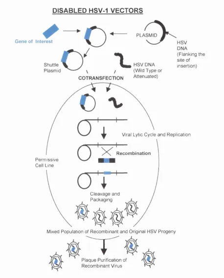
1.4.3.8. The Detection of Latent HSV. 34 1.4.4. Herpes Simplex Virus as a Vector for Gene Therapy. 36 1.4.4.1. Disabled HSV-1 Vectors. 36 1.4.4.2. Defective HSV-1 Vectors. 42 1.4.4.3. Long-Term Transgene Expression from HSV Vectors. 46 1.5. THE PERIPHERAL NERVOUS SYSTEM - A BRIEF OVERVIEW. 53
1.5.4. Dorsal Root Ganglion. 56
1.5.5. Motor Neuron Cell Bodies. 56
1.5.6. Gene Therapy in the PNS. 56
1.6. INDUCIBLE GENE EXPRESSION. 58
1.6.1. The Tetracycline Inducible System. 59
1.6.2. A Novel Tet-Repressor System. 63
1.6.3. The Ecdysone Regulated System. 65
1.6.4. The Progesterone Antagonist System. 69
1.7. THESIS AIMS. 71
CHAPTER 2: MATERIALS AND METHODS. 72-94
2.1. MOLECULAR BIOLOGY. 73
2.1.1. Laboratory Reagents. 73
2.1.2. Bacterial Strains and Growth Conditions. 73
2.1.2.1. Bacterial Strains. 73
2.1.2.2. Propagation of Bacteria. 73
2.1.2.3. Preparation of Competent XLI-Blue Cells. 74 2.1.2.4. Transformation of XL1-Blue Cells. 74
2.1.3. DNA Extraction Techniques. 74
2.1.3.1. Small Scale Plasmid DNA Extraction from 74 XL1-B cells.
2.1.3.2. Large Scale Plasmid DNA Extraction. 75 2.1.3.3. Small Scale Viral DNA Extraction. 75 2.1.3.4. Large Scale Viral DNA Extraction. 76
2.1.4. Cloning Techniques. 77
2.1.4.1. Analytical Restriction Digests. 77 2.1.4.2. Isolation of DNA Fragments. 77
2.1.4.3. Blunt-End Reactions. 77
2.1.4.4. Phenol Extraction of Digested DNA. 78
2.1.4.5. DNA Ligations. 78
2.1.5.1. Colony Transfer. 79
2.1.5.2. Hybridisation. 79
2.1.5.3. Radiolabelling DNA Probe. 80
2.1.6. Agarose Gel Electrophoresis. 81
2.1.7. Southern Blot Analysis of Viral DNA. 81
2.1.7.1. DNA Preparation. 81
2.1.7.2. DNA Transfer (Southern Blot). 81
2.1.7.3. Membrane Analysis. 82
2.2. TISSUE CULTURE. 82
2.2.1. Virus Strain. 82
2.2.2. Mammalian Cell Lines. 82
2.2.2.1. Media. 82
2.2.2 2. Baby Hamster Kidney Cells. 83
2.2.2 3. 27/12/M:4 Cells (MAM49). 83
2.2.2 4. ND7 Cells. 83
2.2.2 5. T-Rex^^^ -HeLa Cells. 83
2.2.2.G. Vero Cells. 84
2.2.3. Cell Line Storage. 84
2.2.4. Transient Transfections Assays. 84
2.2.4.1. DNA Transfections. 84
2 2.4.2. p-Galactosidase Activity Assay. 85 2.2.4.3. Detection of p-Galactosidase by X-Gal Staining. 86 2.2.5. Production of Recombinant Replication Competent HSV-1 86
Vectors.
2.2.51. Homologous Recombination Transfections. 86
2.2.5 2. Viral Titre Assay. 86
2.2.5 3. Purification of Viral Recombinants by Plaque 87 Selection.
2.2.5 4. Production of High Titre Stock of Recombinant 87 Vector.
2.2.6. Production of Recombinant Replication Incompetent HSV-1 88 Vectors.
Selection.
2 2.6.3. Growing High litre Stocks of Virus. 89 2.2.7. Protein Extraction and Analysis. 89 2.2.7.1. Standard Protein Extraction from Cultured Cells. 90 2 2.7.2. Extraction of Multiple TM Spanning or Fragile 90
Proteins from Cultured Cells.
2.2 7.3. SDS-Polyacrylamide Gel Electrophoresis 91 (SDS-PAGE).
2.2.7.4. Transfer of Proteins to Nitrocellulose Membranes 91 (Western Blot).
2.2.7.5. Immunodetection of Proteins on Western Blots. 92 2 2.7.6. Equalisation of Protein Loading in Western Blot 92
Analysis.
2.2.8. In Vivo Vector Delivery. 92
2.2.8.1. Footpad Inoculation. 93
2.2.8 2. Sciatic Nerve Inoculation. 93 2.2.8 3. Detection of p-Galactosidase Activity in Extracted 93
Dorsal Root Ganglia.
2.2.8.4. Detection of Green Fluorescent Protein in Extracted 94 Dorsal Root Ganglia.
2.2.8.5. Counterstaining of Sectionned Animal Tissue. 94
CHAPTER 3: IDENTIFICATION OF OPTIMAL REPLICATION 95-145
COMPETENT VECTORS FOR GENE DELIVERY TO THE PNS.
3.0. INTRODUCTION. 96
3.1. MATERIALS AND METHODS. 99
3.1.1. Cyclosporin Injection. 99
3.1.2. Fluororuby Injection. 99
3.1.3. In Situ Hybridisation. 99
3.1.3.1. Poly-L-lysine Coating Microscope Slides. 99
3.1.3.3. Fixation of Sections. 100 3.1.3.4. Labelling Oligonucleotide Probes. 100 3.1.3.5. Preparation of Hybridisation Buffer. 101 3.1.3.6. Hybridisation of Probe to Tissue Sections. 102 3.1.3.7. Washing the Sections after Hybridisation. 103 3.1.3.8. Emulsion Dipping In Situ Hybridised Sections. 103
3.1.3.9. Developing the Slides. 103
3.2. RESULTS. 104
3.2.1. LAP1 and LATP2. 104
3.2.2. Single Transgene Expression from a Reporter Cassette 104 in the LAT Region.
3.2.2.1. Expression Cassettes and Vector Production. 105
3.2.2.2.//7 V/Vo Analysis. 107
3.2.3. Multiple Transgene Expression from a Cassette ^ 109 in a Non-Essential Locus.
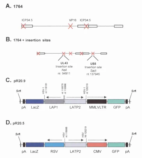
3.2.31. Non-Essential Genes UL43 and US5. 109 3.2.3 2. Expression Cassettes and Vector Production. 111 3.2.3.3. in Vitro Vector Analysis on Permissive Cells. 111 3.2.3.4. In Vivo Gene Expression is Affected by Promoter 115
Choice and Delivery Route.
3.2.4. Deletion of Vhs. 120
3.2.4.1. Expression Cassettes and Vector Production. 120 3.2.4.2. Deletion of Vhs Reduces the Efficiency of Reporter 121 Gene Expression In Vivo Following Peripheral Administration. 3.2.5. Estimation of the Efficiency of Vector Uptake by Ganglionic 125
Neurons that Project to the Footpad using Fluororuby.
3.2.51. In V/vo Analysis of Fluororuby Uptake. 125 3.2.6. Stategies to Avoid Possible Immune Responses Following 128
Vector Adminisatration.
3.2.61. immunosuppression using Cyclosporin. 128 3 2.6.2 Vector Choice for In Vivo Immunosuppression 128
Analysis.
3.2.6 3. Cyclosporin Treatment does not Affect Reporter 129 Gene Expression.
3.2.7. Detection of Latent HSV-1 in Lumbar Ganglia by In Situ 135 Hybridisation.
3.2.7.1. Vector Choice for Detection of Latent HSV. 135 3 2.7.2. Decline in Transgene Expression During Latent 138
Infection does not Appear to be a Function of Promoter Shutdown.
3.3. DISCUSSION. 142
CHAPTER 4: IDENTIFICATION OF OPTIMAL REPLICATION 146-165 INCOMPETENT VECTORS FOR GENE DELIVERY TO THE PNS.
4.0. INTRODUCTION. 147
4.1. RESULTS. 150
4.1.1. Vectors with a Deletion in the Essential Gene ICP27. 150
4.1.1.1. Vector Production. 150
4.1.1.2. In Vitro Vector Analysis on Permissive Cells. 151 4.1.1.3. In Vivo Gene Delivery with a Vector Deleted 153
for ICP27.
4.1.2. A Vector with a Further Deletion in the Essential Gene ICP4. 157
4.1.2.1. Vector Production. 157
4.1.2.2. Reporter Gene Expression In Vivo using a 158 Multiple IE Gene Deficient Replication
Incompetent HSV Vector is a Function of Vector Titre and Delivery Route.
4.1.2.3. Gene Expression from 1764 27-/4- pR19/acZ is 161 Maintained Over Time.
4.2. DISCUSSION. 163
CHAPTER 5: STUDIES TO DEVELOP INDUCIBLE 166-201 TRANSGENE EXPRESSION SYSTEMS USING REPLICATION
5.0. INTRODUCTION. 167
5.1. MATERIALS AND METHODS. 169
5.1.1. Assessment of Ligand Inducible lacZ Activity in Plasmid 169 Constructs.
5.1.2. Ex Vivo Characterisation of Ligand Inducible Expression 169 Vectors.
5.2. RESULTS. 170
5.2.1. A Single Cassette can Incorporate Multiple Elements of a 170 Ligand Inducible Expression System.
5.2.2. Expression Cassettes and Cloning Strategies. 171
5.2.2.1.Tet-On-pR20.4. 171
5.2.2 2. Ecdysone-pR20.8. 171
5.2.2.3. Mifepristone-pR20.11. 172
5.2.3. Transient Transfection Assays of Ligand Inducible , 174 Expression Cassettes.
5.2.4. Production of Tet-On, Ecdysone and Mifepristone HSV 179 Vectors.
5.2.5. In Vitro Analysis of Vectors on Permissive Cells. 179 5.2.6. The Tet-On’ Vector Shows Poor Efficacy In Vivo. 182 5.2.7. Characterisation of the Ecdysone Inducible Vector. 184 5.2.8. The Ecdysone Inducible Vector Shows Limited Efficiency 186
in Vitro.
5.2.9. Efficacy of the Ecdysone Inducible Vector Ex Vivo. 188 5.2.10. A Replication Competent Vector Causes Induction of a 191
Ligand Inducible Response Element in the Absence of Transactivator or Ligand.
5.3. DISCUSSION. 196
CHAPTER 6: STUDIES TO DEVELOP INDUCIBLE TRANSGENE 203-243
EXPRESSION SYSTEMS USING REPLICATION INCOMPETENT
VECTORS.
6.0. INTRODUCTION. 204
6.1. MATERIALS AND METHODS. 207
6.1.2. FACS Analysis of Transfected Cells. 207
6.2. RESULTS. 207
6.2.1. Ecdysone Inducible Transgene Expression. 207
6.2.1.1. Vector Design. 208
6.2.1.2. Vector Production. 208
6.2.1.3. In Vitro Characterisation of Plasmids. 214 6.2.1.4. Characterisation of Vector 1764 27-/P2-/4- A/IR/US5.218 6.2.1.5. In Vitro Characterisation of Replication Incompetent 220
Ecdysone Inducible Viral Vectors.
6.2.1.6. Ex Vivo Analysis of Replication Incompetent 224 Ecdysone Inducible Viral Vectors.
6.2.1.7. In l//Vo Analysis of Replication Incompetent 227 Ecdysone Inducible Viral Vectors.
6.2.2. Tetracycline Repressor Inducible Transgene Expression. 228
6.2.2.1. Vector Design. 228
6 2.2.2. Plasmid Construction. 230
6.2.2.3. In Vitro Characterisation of Tet-Repressor Plasmids. 233 6.2.2 4. In Vitro Characterisation of Tet-Repressor Plasmids 236
on T-Rex™ -HeLa Cells.
6.3. DISCUSSION. 239
CHAPTER 7: APPLICATIONS IN THE PNS. 244-281
7.0. INTRODUCTION. 245
7.0.1. Cre Recombinase. 245
7.0.2. Galanin. 248
7.0.3. Summary. 249
7.1. MATERIALS AND METHODS. 251
7.1.1. Cre Recombinase Methods. 251
7.1.1.1 DRG Tissue Sectioning. 251
7.1.2.1. DRG Sections. 253
7.2. RESULTS. 254
7.2.1. Vectors Expressing Cre Recombinase. 254 7.2.1.1. The ROSA26-R Cre Reporter Strain. 254 7.2.1.2. Genetic Analysis of ROSA26-R Mice. 256 7.2.2. A Replication Competent Vector Expressing Cre 256
Recombinase.
7.2.2.1. Cloning Strategy. 256
7.2 2.2. Vector Preparation and Characterisation. 259 7.2 2.3. A Replication Competent HSV-1 Viral Vector 261
Expressing Cre Recombinase Shows Efficacy
In Vivo.
7.2.2.4. In Vivo Efficiency of a Replication Competent 263 Vector Expressing Cre Recombinase is High but
Variable.
7.2.2.5. Kinetics of Cre Recombinase when Delivered by 265 a HSV-1 Vector.
7.2.3. A Replication Incompetent Vector Expressing Cre 268 Recombinase.
7.2.3.1. Cloning Strategy and Vector Preparation. 268 7.2.3.2. A Replication Incompetent HSV-1 Vector Expressing 270
Cre Recombinase Shows Efficacy In Vivo.
7.2.4. A Viral Vector Expressing the Neuropeptide Galanin. 272 7.2.4.1. Cloning Strategy and Vector Production. 272 7.2.4.2. Ex Vivo Detection of Galanin Expression from a 275
HSV-1 Vector.
7.3. DISCUSSION. 278
CHAPTER 8.0: DISCUSSION 282-293
Reference List 294
Page Number CHAPTER 1
Figure 1.1. Gene therapy protocols by disease. 4 Figure 1.2. Schematic representation of recombinant retroviral vector 12
production.
Figure 1.3. Schematic representation of the adenovirus genome. 15 Figure 1.4. Schematic representation of ‘gutless’ adenoviral vector 18
production using the Cre/lox system.
Figure 1.5. Schematic diagram of the HSV-1 genome. 24 Figure 1.6. The HSV-1 replication cycle. 26 Figure 1.7. The LAT region of the HSV-1 genome. 31 , Figure 1.8. Schematic diagram to show the generation of 37
disabled HSV vectors.
Figure 1.9. Packaging of defective HSV vectors. 45 Figure 1.10. Schematic diagram to show long-term expression cassettes
used in HSV vectors. 51-52
Figure 1.11. Schematic diagram representing the peripheral nervous 55 system at the L4 and L5 level.
Figure 1.12. Schematic representation of the tetracycline inducible 61 expression systems.
Figure 1.13. Schematic representation of the tetR-mediated transcription 63 repression switch.
Figure 1.14. Schematic diagram of two ligand inducible gene expression 68 systems.
CHAPTER 3
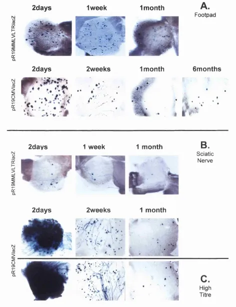
Figure 3.2. Replication competent vectors, 1764pR19MMLV/acZ and 108 1764pR19CMV/acZ tested for gene delivery to DRG during acute and latent infection.
Figure 3.3. Expression cassettes and replication competent vector 113 backbone.
Figure 3.4. Plaque phenotype of three replication competent vectors. 114 Figure 3.5. Gene delivery to peripheral ganglia following footpad 117-118
inoculation with replication competent vectors.
Figure 3.6. Gene delivery to peripheral ganglia following sciatic 119 nerve inoculation with replication competent vectors.
Figure 3.7. Expression cassettes and replication competent vector 122 backbone.
Figure 3.8. Gene delivery to peripheral ganglia following footpad or 123-124 sciatic nerve inoculation with a replication competent
vector deleted for vhs.
Figure 3.9. Ganglionic neurons and sciatic nerve after retrograde 127 axonal labelling using Fluororuby.
Figure 3.10. Effect of transient immunosuppression on the efficiency of 151 reporter gene expression from HSV vectors in mice.
Figure 3.11. Schematic diagram to show the 1764 vector backbone 133 and pR20.9 expression cassette containing blue
fluorescent protein.
Figure 3.12. Nucleotide sequence homology between EGFP and BFP. 134 Figure 3.13. Schematic diagram to show vector controls used for 137
in situ hybridisation experiments.
Figure 3.14A. In situ hybridisation to detect LAT RNA in lumbar ganglia. 140 Figure 3.14B. In situ hybridisation to detect LAT RNA in lumbar ganglia. 141
CHAPTER 4
Figure 4.1. Expression cassettes and replication incompetent 152 vector backbone.
Figure 4.2. Gene delivery to peripheral ganglia following sciatic nerve inoculation of replication incompetent vectors deleted for
for ICP27.
Figure 4.4. Expression cassette and replication incompetent vector 159 backbone.
Figure 4.5. Gene delivery to peripheral ganglia using a replication 160 incompetent vector deleted for ICP4 and ICP27.
Figure 4.6. Gene delivery to peripheral ganglia following sciatic 162 nerve inoculation with a replication incompetent vector
deleted for IC27 and ICP4.
CHAPTER 5
Figure 5.1. Vector backbone and ligand inducible expression cassettes. 173 Figure 5.2. In vitro analysis of ligand inducible expression cassettes. 176 Figure 5.3. Inducible transactivation of p-galactosidase after transient
transfection of ligand inducible expression cassettes into
BHK cells. 177-178
Figure 5.4. Plaque phenotype of replication competent ligand 181 inducible vectors on permissive BHK cells.
Figure 5.5. Ex Vivo analysis of a Tet-On’ replication competent vector. 183 Figure 5.6. Western blot detection of retinoid-X-receptor (RXR) 185
expression from a replication competent vector.
Figure 5.7. in vitro Inducible transactiviation of p-galactoidase in the 187 replication competent vector pR20.8lacZ/UL43.
Figure 5.8A. Inducible transactiviation of p-galactoidase in the replication 189 competent ecdysone regulatable vector pR20.8/acZ/UL43.
Figure 5.88. Inducible transactiviation of p-galactoidase in the 190 Replication competent ecdysone regulatable vector
pR20.8/acZ/UL43.
CHAPTER 6
Figure 6.1. Schematic diagram to show production of ecdysone 212 inducible replication incompetent vectors.
Figure 6.2. Replication incompetent ecdysone inducible vectors. 213 Figure 6.3A. Inducible transactivation of p-galactosidase after transient 216
transfection of ligand inducible expression cassettes.
Figure 6.3B. Inducible transactivation of p-galactosidase after transient 217 transfection of ligand inducible expression cassettes.
Figure 6.4. Western blot detection of replication incompetent vectors 219 expressing Retinoid-X-Receptor (RXRa).
Figure 6.5. Phenotype of replication incompetent ecdysone inducible 221 vectors on complementing cells.
Figure 6.6A. Phenotype of replication incompetent ecdysone inducible 222 vectors on non-complementing cells.
, Figure 6.6B. In vitro characterisation of ligand inducible replication 223 incompetent viral vectors.
Figure 6.7. Ex vivo characterisation of ecdysone inducible expression 225 vectors.
Figure 6.8. Schematic representation of ligand inducible expression 232 cassettes containing the tet-repressor system.
Figure 6.9. In vitro characterisation of inducible expression cassettes 235 containing the tet-repressor system
Figure 6.10. In vitro characterisation of pR20.1/27/TO/TR on a 238 T-REx™ -HeLa cell line expressing the Tet-Repressor.
CHAPTER 7
Figure 7.1. Schematic diagram of the ROSA26 locus and the 255 ROSA26 reporter construct.
Figure 7.2. PCR genotyping of ROSA26-R mice. 257 Figure 7.3. Cre recombinase expression cassettes and replication 258
competent vector backbone.
Figure 7.4. Western blot detection of Cre recombinase from HSV-1 260 vectors.
Figure 7.6. in vivo efficiency of a replication competent virus 264 expressing Cre recombinase.
Figure 7.7. Kinetics of Cre recombinase activity when delivered by a 267 HSV-1 vector.
Figure 7.8. Schematic representation of Cre recombinase and galanin 269 expression cassettes and replication incompetent vector
backbone.
Figure 7.9. In vivo efficacy of a replication incompetent vector 271 expressing Cre recombinase.
Figure 7.10. Southern blot detection of galanin genomic DNA in 274 recombinant viral vectors genomes.
Tables
CHAPTER 1
Table 1.1.
Table 1.2.
Table 1.3.
A summary of monogenic disorders and their potential target for gene therapy protocols.
Summary of some of the properties that would be possessed by an ideal gene therapy viral vector. Classification of peripheral nerve fibres.
Page
Number
53
CHAPTER 2
Table 2.1. SDS-PAGE gel composition.
Table 2.2. Western blot protein detection protocol. Table 2.3. Primary antibody details.
91 92 92
CHAPTER 5
Table 5.1. Ligand stock solutions and working concentrations used 169 in transient transfection assays and in vector purification.
Table 5.2. Ligand stock concentrations and working concentrations 170 for ex vivo vector analysis.
Table 5.3. Viral vectors used in superinfection experiments. 192
CHAPTER 6
Table 6.1. Details of transient transfection assays. Table 6.2. FACS analysis of tet-repressor constructs.
214 233
CHAPTER 1 :
Chapter 1____________________________________________________________________Introduction
CHAPTER 1:
INTRODUCTION
1.1. GENE TH ERAPY- AN OVERVIEW.
In the last few decades our increased awareness of the inherited nature of many diseases has meant that we are now able to understand the mechanisms involved in their pathologies. The recent announcement of a ‘working draft’ of the human genome sequence (Macilwain 2000), provides us with a wealth of information and perhaps the most exciting achievement in scientific terms since the work of James Watson and Francis Crick in identifying the structure of DMA (Watson and Crick 1953). In terms of gene therapy the completion of the human genome sequence will increase the potential of the field significantly. Geneticists predict that the number of genes likely to be identified, based on the number of protein coding regions, could be somewhere between 35,000 and 150,000 (Butler and Smaglik 2000). Once characterised these are likely to ' provide a wealth of potential new targets for traditional drug or gene therapy.
Gene therapy can be defined as the introduction of genetic material into cells to bring about a therapeutic effect. So far gene therapy has been limited to somatic gene therapy whereby the genetic material is inserted into diploid cells of an individual. In this case none of the acquired genetic material is passed between generations. Germline gene therapy is so far ethically unacceptable, but might be regarded as the ultimate form of gene therapy. So far somatic gene therapy can be divided into three general areas based on the route of gene administration; ex vivo, and direct or indirect in vivo (Drew and Martin,
1999). The ex vivo approach involves explant of a patient’s cells, for example stem cells or lymphocytes, followed by culture in vitro. The cells are then manipulated and transduced with the therapeutic gene and finally re- implantated back into the patient. This appears to be a relatively simple procedure and in theory should minimise problems with immunogenicity and rejection, which might otherwise be associated with the use of allogeneic cells. However, a frequent occurrence and significant limitation is the low level of cell viability that is found after re-implantation. Direct in vivo gene delivery involves direct gene transfer to the site of interest and is currently an area in which
considerable research activity is underway. Most of the commonly used vectors (see later) have been used in this approach to transfer genetic material directly into target diseased tissue, although the use of naked DNA itself has also been explored. In situ gene delivery removes any need for targeting of vectors but at present the efficiency of transduction is variable and sometimes problematic. Indirect in vivo delivery refers to the administration of genetic material by peripheral routes, for example into the blood stream, followed by targeting of the vector to the tissue requiring therapy. This approach is theoretically the most straightforward, based on the simplicity of administration, but successful targeting of therapeutic doses to the appropriate tissue is a major problem.
1.2. GENE THERAPY FOR MONOGENIC DISEASES.
Many disease states arise from chromosomal abnormalities that are either inherited or acquired. These diseases can be monogenic or polygenic. Conventional disease treatment often treats only the symptoms of the disease. ^ However to completely cure a disease, it is necessary to correct any
abnormalities which might be genetically based i.e. gene therapy would be necessary. Ideal targets for gene therapy are thus monogenic disorders, as these diseases are more easily understood and only a single gene would need to be introduced/replaced (see figure 1.1.). Currently gene therapy is aimed at gene augmentation, where-by a normal copy of a gene is introduced into the cell such that it can produce sufficient quantities of the correct gene product and can compensate for the lack of expression of the mutant host gene. Using strategies of this kind when the mutant gene has a dominant negative effect would be technically difficult. Thus disease targets have been limited to mutant genes which do not have an active negative effect such as cystic fibrosis or the severe combined imunodeficiency diseases, (X-linked SCID [SCID-X1] and adenosine deaminase deficency-SCID [ADA-SCID])(Cheng et al.
Chapter 1 Introduction
Monogenic Disorder Disease Causing Gene
Cystic fibrosis Cystic fibrosis transmembrane conductance regulator (CFTR)
Haemophilia A Blood Clotting Factor VIII B Blood Clotting Factor IX
X-linked SCID Gamma chain of IL2, IL4, IL7, IL11 and IL15 receptors
ADA SCID Adenosine Deaminase
Gauchers disease Glucocerebrosidase Duchenne muscular dystrophy Dystrophin
Familial Hypercholesterolemia Low density lipoprotein (LDL) receptor Phenylketonuria Phenylalanine hydroxylase
Huntingdons chorea Huntingtin
Table 1.1. A summary of monogenic disorders and their potential target
for gene therapy protocols. Adapted from, Concise Oxford Text book of Medicine (2000) and Samter’s Immunologic Diseases (1995).
Gene Therapy Protocols by Disease
12.9%
65.6% 0.5%
□ Monogenic Disorders □ Infectious Disease ■ Other Disease □ Gene Marking ■ Healthy volunteers ■ Cancer
1.2.1. Treatment of Monogenic Disorders with Viral Vectors.
Cystic Fibrosis is caused by a dysfunction of an epithelial cell surface chloride channel (CFTR: cystic fibrosis transmembrane conductance regulator) (Cheng
at al. 1990). Sufferers have a life expectancy of approximately 30 years, death resulting from effects in the respiratory epithelium. The respiratory epithelium provides an apparently easy and available target for gene therapy. Indeed viral and other vectors have been extensively tested in such approaches. Adenoviral vectors seem the instinctive choice for gene therapy in the lung as they have a natural tropism for airway epithelia (Shenk 1996). The first reported clinical trial using a recombinant adenovector was reported in 1993 (Zabner at al. 1993). Three CF patients showed a correction in the defective chloride channel, in a small area of the nasal epithelium, after administration of the Ad(CFTR) vector. Since then there have been a number of further published clinical trials for CF using first generation El deleted adenoviruses (Bellon at al. 1997;Boucher at al.
1994;Crystal at al. 1994). These phase one studies have looked at ' immunological and inflamatory response to the El deleted vectors, although
evidence for efficiency has been limited.
Chapter 1____________________________________________________________________Introduction
Haemophilia A and B are both X-linked inherited bleeding conditions, caused by defects in the blood clotting factors VIII and IX respectively (Martin and Drew,
1999). Untreated the conditions are lethal. Successful treatment only requires that the missing blood clotting factors are present in the plasma at about 4-5% of normal circulating levels (Scriver 1989). As the protein is secreted it may be supplied from almost any tissue, such as muscle, blood or fibroblast cells. The first reported gene therapy clinical trial for haemophilia B was in 1993 (Lu et al.
1993). Here a group in China adopted an ex vivo approach whereby a retroviral vector encoding factor IX was used to transduce autologous fibroblasts from two patients. After 3-4 administrations over a period of four months, one of the two patients showed an increase in factor IX antigen and blood clotting activity. The results of this trial provided the first real indications that human gene therapy could be effective in practice.
1.3. GENE THERAPY FOR ACQUIRED DISORDERS.
' Although monogenic inherited genetic disorders were the first targets for gene therapy, today the field is dominated by clinical trials in cancer patients (See http//:www.wiley.co.uk/genetherapy/vectors.html) (see figure 1.1). Cancer might seem an unusual choice for gene therapy strategies, the classical idea of which is that of the insertion and expression of a normal copy of the mutated gene. Cancer generally arises as the culmination of a multi-step process involving many genes. Currently there are many cancer gene therapy approaches in development (reviewed in Roth and Cristiano 1997;Vile et ai. 2000)). These include the use of recombinant vaccines for immunotherapy, the protection of bone marrow from cytotoxicity during chemotherapy (via transduction of drug resistance genes to bone marrow stem cells) and the delivery of enzymes which convert inactive prodrugs into active drugs. The genes delivered include those for cytokines and tumor suppressor genes.
Herpes simplex virus-1 (HSV-1) thymidine kinase (HSV-TK) has the ability to phosphorylate a range of nucleosides and their analogues whereas human TK cannot (French-Anderson 1998). Because of this characteristic it has been much used in suicide gene therapy for cancer patients. Currently there are phase I/ll clinical trials underway using for example, adenoviral vectors (Morris
et al. 2000;Trask et al. 2000). Gene therapy employing TK usually involves the administration of a vector containing the TK gene to the tumor cells directly or into the blood vessels supplying the tumor. In theory after systemic administration of a pro-drug such as acyclovir or gancyclovir, only cells to which the HSV-TK gene had been delivered will phosphorylate the drug to give cytotoxic acyclovir or gangcyclovir triphosphates respectively (French-Anderson 1998). These are incorporated into newly synthesised DNA resulting in cell death. This strategy relies on the selective uptake and compartmentalisation of the vector by malignant tissue. Similar strategies involving transfer of the cytosine deaminase gene, which gives sensitivity to 5-fluorocytosine, to tumor cells have also been tested using adenoviral vectors (Shirakawa et al. 1999; Ichikawa et al. 2000).
1.4. GENE THERAPY VECTORS.
To facilitate the entry of therapeutic genes into cells a delivery vehicle is ' required. This vehicle is termed a vector and vectors for gene therapy can be broadly divided into non-viral and viral systems. The optimal vector should possess a number of characteristics and these are defined in table 1.2. However, to date no single vector system possesses all of the desirable attributes. The choice of currently available vector will thus depend upon the disease being treated and the particular requirements for gene delivery.
1.4.1. Non Viral Vectors.
C hapter 1____________________________________________________________________Introduction
The vast majority of non-viral vectors are based on cationic liposome complexes (lipoplexes) (Miller AD 1999). The lipoplex consists of a postively charged lipid complex, bound to nucleic acid. The complex enters the cell by endocytosis brought about by the interaction between the positvely charged lipid and the negatively charged cell surface proteoglycans. Thus, cells deficient in proteoglycans are difficult to transfect. At this point the lipoplex in the endosome can either become trapped in a late endosome or the nucleic acid can be released into the cytoplasm. However both liposome and polysome based complexes (which are largely similar in their method of gene delivery) have some enormous problems (Li and Huang 2000;Miller 1999). The complexes tend to aggregate before endocytosis and/or are unstable in the presence of serum proteins. The uptake procedure can also take hours and once inside the cell most of the complexes probably remain within late endosome particles and are thus unable to direct gene expression. A number of methods have been employed to circumvent these problems such as the ' complexing of the liposomes with polyethylene glycol (PEG), in order to stop aggregation (Hong et al. 1997). Another potential disadvantage is that cationic lipid/DNA complexes tend to show a discrepancy between in vitro and in vivo
transfection capabilities and thus there is a need for specific tailoring for each in vivo trial and route of administration.
Properties of an Ideal
Vector
Easy production
Long term expression
Immunologically inert
' Wide host range
Cell targeting
Size capacity
The virus should be easy to manipulate and grow to high titre. Vectors must allow for easy manipulation of their genes and regulatory elements and subsequent insertion of transgenes. Production of high titres are necessary for transduction of the large number of cells often required for gene therapy.
Once delivered the vector should for many applications have the ability to express the transgene for a sustained period of time and if necessary in a regulated fashion.
The vector should be able to avoid immunological clearance, avoiding both humoral and cell mediated responses.
An Ideal vector would have a broad host range and be able to infect both dividing and non-dividing cells, although in cancer therapies it is advantageous to only infect dividing cells.
Vector targeting to specific cell types can also be highly desirable. This is particularly the case where the target cells are distributed throughout the body or are part of a heterogeneous population.
The vector should accept inserts of varying sizes. Genes delivered in gene therapy protocols vary greatly in size, insulin being 350bp (Demeterco and Levine 2001) and Dystrophin -14kb (Hartigan- O'Connor and Chamberlain 2000).
Table 1.2. Summary of some of the properties that would be possessed by
C hapter 1____________________________________________________________________Introduction
1.4.2. Viral Vectors.
The first deliberate use of a virus in disease therapy took place over one hundred years ago, when the cowpox virus was used for vaccination against small pox (Nolan and Shatzman 1998). Since then viruses and virus subunits have been extensively used in vaccination strategies against viral disease. Viruses are now also being developed as vectors for gene therapy as they have naturally evolved to enter cells in order to deliver their genetic material.
Perhaps the biggest blow to gene therapy to date came in September 1999 with the death of Jesse Gelsinger (Lehrman 1999). Jesse suffered from partial deficiency of the liver enzyme ornithine transcarbamylase (OTC), which is characterised by the build up of toxic levels of ammonia within the body. He was taking part in a phase 1 escalating dose clinical trial, using an El-deleted E2A temperature sensitive adenovirus vector (see later). Jesse was one of the two patients who were receiving the highest dose of the adenoviral vector used (3.8 ' xIO^^ virus particles). Many questions have been raised since this death and some researchers have questioned why patients suffering from partial enzyme deficiency were even involved in the trial, since a strict diet and ammonia clearing compounds can control this mild form of the disease. Since this time the public perception of gene therapy has suffered a severe setback.
1.4.2.1. Retroviral Vectors.
Retroviruses are enveloped single stranded RNA viruses with genomes of 7- 11 kb. The genome encodes three core genes termed gag, pel and env (see figure 1.2). The coding sequences are flanked by two long terminal repeats (LTRs), and a packaging signal (reviewed in Murphy 1999;Glorioso et a i 2001). Upon entry into a host cell the genome is reverse transcribed to double stranded DNA and is then subsequently integrated into the host cell genome as a provirus (Boris-Lawrie and Temin 1994). Retrovirus integration into the host cell genome thus provides the basis for long term transgene expression. However integration is not site specific and thus this can result in activation of oncogenes or inactivation of tumor suppressor genes (Donahue at al. 1992). Despite this possibility there has been little reported evidence of this phenomenon so far (see later).
Retroviruses have long been the vector in most widespread use for gene therapy protocols since they are relatively simple and easy to manipulate. Most retroviruses are relatively non-pathogenic and harmless in humans. Currently 40% (http//;www.wiley.co.uk/genetherapy/vectors.html) of gene therapy protocols utilise retroviral vectors, most commonly those derived from Moloney murine leukemia virus, MoMuLV (Vile 1997). Vector construction involves the removal of all viral genes and their replacement with the transgene of interest, flanked by the remaining cis regulatory elements (reviewed in Murphy, 1999) (see figure 1.2.). This renders the resulting virus replication incompetent but able to integrate and direct transgene expression. This allows for the insertion of up to 8kb of foreign genetic material but thus presents upper size constraints on the therapeutic gene that can be inserted (Miller 2000). Complementing cell lines have been produced that express the gag, pol and env genes in trans to allow the production of recombinant vector (Miller 1990). Separation of the
Chapter 1 Introduction
5' LTR
f
P ack agin g s e q u e n c e
R e v e r s e
C ore Transcription E n velop e P roteins in te g ra se Proteins
gag pol env
3' LTR
In e x p r e ssio n c a s s e t t e s g a g .p o l and en v are rep la ced by a therap eu tic g e n e
Packaging cell
T h erap eu tic g e n e
Retroviral e x p r e ssio n c a s s e t t e
Vector genomes
A G ag an d pol A proteins
env construct
gag/pol construct
E n v elo p e p roteins
onn
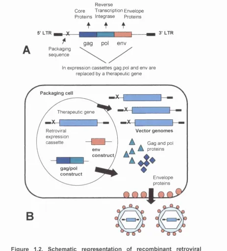
Figure 1.2. Schematic representation of recombinant retroviral vector production. A. The viral genome consists of the gag, pol and env
genes plus the packaging sequence X. B. The packaging cell line constitutively expresses the 3 viral genes, separating gag and pol from
env in order to reduce the chances of creating replication competent vectors. After transfection of a cassette, containing a transgene flanked by LTRs, into the cell, vector genomes are produced. Vector genomes are then packaged into capsids along with the gag and pol proteins and the virus buds from the cell surface. The resulting retroviral particles are then ready to infect target cells.
Retroviruses have a broad host range that is dependent on the tropism of the envelope protein (Miller 2000). One of the major drawbacks of retroviruses is their inability to infect non-dividing cells, as they require the onset of mitosis in order to enter the nucleus (Miller et al. 1990). To overcome this problem many retroviral protocols use an ex vivo gene therapy approach, as certain cells such as stem cells can be stimulated to divide in vitro (Salvatori at al. 1993 and reviewed in Verma and Somia 1997). Additionally, efforts are underway to add receptor specific ligands or alter retroviral envelope glycoproteins, (pseudotyping), in order to modify the virus tropism (Cosset and Russell 1996;Palu at al. 2000). Recently a MMLV retroviral vector pseudotyped with vesicular stomatitis virus G protein (VSV-G) was used to create a transgenic male rhesus monkey expressing GFP (Chan at al. 2001). The aim of the work was to overcome the problems in traditional gene transfer methodology used to produce transgenic primates. Here the vector, containing GFP driven by the elongation factor alpha-1 promoter (EFalpha-1P), was injected into mature ' rhesus oocytes and six hours later fertilised by direct sperm injection. Out of 224 fertilised oocytes, three live births were achieved. Of these one male contained the transgene in all tissues analysed. This was the first demonstration that a transgenic primate could be produced.
1.4.2.2. Lentiviral Vectors.
C hapter 1____________________________________________________________________Introduction
virus entry into a wide range of cells including neurons, muscle fibres and hepatocytes. Pseudotyping allowed some of the first real demonstrations of the practical use of lentiviral vectors. However, these first generation HIV-based vectors gave rise to concerns over the possibility of creating infectious HIV particles through recombination. Since then lentiviral vectors with further gene deletions have been developed (see later).
The lentiviral genome is more complex than other retroviral genomes, in addition to gag, pol and env, HIV also contains two regulatory genes, tat and
rev and four accessory genes, vif, vpr, vpu and nef (Trono 1995). The four accessory genes appear to have no obvious function in vitro, but are required for full virulence in vivo and thus do not need to be complemented in culture (Naldini 1998). Vectors have been constructed that have deletions of all four accessory genes which give effective transduction of both dividing and non dividing cells both in vivo and in vitro (Kim et ai. 1998;Zufferey et ai. 1997). ' Additionally a third generation of lentiviral vectors requiring only gag, poi, rev
and VSVG have now been produced, which do not encode rev or tat (Dull et ai.
1998). Other significant advances in lentiviral vector safety have been achieved by the generation of self-inactivating viruses (Delviks et ai. 1997;Miyoshi et ai.
1998). Miyoshi et ai used a transfer construct that had almost a complete deletion in the 3’ LTR including the TATA box and binding sites for transcription factors. This deletion is transferred to the 5’ LTR during reverse transcription, the deleted LTRs becoming integrated into transduced cells. The integrated provirus is therefore non-functional due to the loss of its c/s-acting elements in the LTRs. Inserted promoters in the transgene cassette drive the gene expression.
1.4.2.3. Adenoviral Vectors.
subgroup, Ad1, Ad2, Ad5 and Ad6 are regarded as endemic within the population (Murphy 1999). The genome is functionally divided into two major regions, termed late and early, according to their time of transcription after infection. These two regions contain overlapping transcriptional units and in total code for over 50 polypeptides (reviewed in Shenk 1996), see figure 1.3.
LI L2 L3 14 L5
— ► --- ► ► ► 1
E3
ITR — — ITR
E1A E1B
E2B E2A E4
Figure 1.3. Schematic representaion of the adenovirus genome. The black arrows show the four early coding regions, of which Eland E3 are transcribed from the upper DNA strand and E3 and E4 are transcribed from the lower strand. The blue arrows show the late coding regions, LI-5. The genome is flanked by the cis-acting inverted terminal repeats (ITRs) and the packaging sequence vp.
The tropism of Ads and their resulting vectors is extremely broad and they can infect both dividing and non-dividing cells (Benihoud et al. 1999). The primary cell targets in natural infection are epithelial cells of the respiratory tract. After infection the E1A gene is the first gene to be transcribed, and its products act to transactivate the expression of the other early and late genes (Shenk 1996). The E3 gene product is non-essential in vitro but in vivo it plays an important role in the persistance of adenovirus infection as it acts to inhibit a cytotoxic T- lymphocyte (CTL) induced lysis of the adenovirally infected cell (Beier at al.
1994). First generation Ad vectors were deleted for the El region so that theoretically the virus would be unable to replicate on non-complementing cells lines, due to its inability to transactivate other early genes. The human embryonic kidney cell line 293 was produced that constituitively expressed the El gene products in order to allow the in vitro growth of first generation Ad vectors (Graham at al. 1977). Generally these vectors also had the E3 coding
C hapter 1____________________________________________________________________Introduction
region deleted, since it is not required in vitro, in order to increase the packaging capacity of the vectors to approximately 7.5kb (Bett et al. 1993). However it was found that these vectors still produced low levels of the remaining viral genes in vivo, probably due to a cellular EIA-Iike transactivating factor (Yang at al. 1994). Thus transgene expression from these vectors, with few exceptions, was found to be transient most likely due to host immune responses to the foreign viral antigens (reviewed in Murphy 1999).
Following the limited prospects of first generation adenoviral vectors, increasingly attenutated vectors have since been developed (reviewed in Glorioso at al. 2001 and Murphy 1999). Second and third generation viral vectors were created that were deleted for the E2 (Engelhardt at al. 1994;Yang
at al. 1994) and E4 regulatory regions (Dedieu at al. 1997), in addition to El and E3, and were thus less immunogenic to the host. These vectors now tend to have a minimal 19kDa part of the E3 coding region remaining, since it is ' thought to be responsible for inhibition of CTL induced cell lysis (Beier at al.
1994).
Although these second and third generation vectors have decreased cytotoxic profiles, transgene expression generally remains short. Since then gutless adenoviral vectors have been produced that contain no viral genes (Kochanek
at al. 1996;Parks at al. 1996). Production of these gutless vectors relies on a helper-packaging virus. The helper virus contains all of the necessary Ad replication and packaging signals that act in trans on a vector containing the necessary cis acting elements and the foreign gene. The helper virus is unable to package itself but aids the production of recombinant virions that lack all the Ad genes except for the cis acting elements. Systems of this nature allow for an increased packaging capacity of the recombinant virus, up to 36kb, and theoretically should have a reduced cytotoxic profile compared to earlier Ad vectors, due to the lack of Ad proteins. However one drawback to this system is the inability to completely remove the helper virus from recombinant vector preparations (Glorioso at al. 2001). Parks at al (Parks at al. 1996) have developed a method to produce gutless adenoviral vectors using a 293/
recombinase packaging cell line that reduces the helper virus in vector preparations to very low levels (see figure 1.4.).
Chapter 1 Introduction
HELPER VIRUS
5 ’LTR 3 ’LTR
LoxP sites 5 ’ LTR
Stuffer DNA
Foreign Gene
ADENOVIRAL EXPRESSION CASSETTE
3 ’LTR
293 Packaging Cell
Cre Recombinase
5'L T R — — ^ ---1 X = 1 - — 3 'LTR AE1
HELPER VIRUS GENOME 5 ’ LTR
Stuffer D N /v
j
L
\
Foreign Gene
RECOMBINANT VECTOR GENOME
Packaged
3 ’LTR
RECOMBINANT ADENOVIRAL PARTICLES
Figure 1.4. Schematic representation of ‘gutless’ adenoviral vector production using the Cre/lox system. The Ad helper virus contains two loxP sites flanking the packaging signal 4^. The Ad expression cassette contains the transgene flanked by stuffer DNA and the adenoviral cis regulatory elements. After transfection into 293 cells expressing Cre-recombinase, the helper virus provides all the necessary gene products in trans that are necessary for replication and packaging of an Ad vector containing the appropriate regulatory sequences. Cre recombinase mediates the conditional excision of the packaging signal in the helper virus such that it cannot be incorporated into the new virion particles.Thus there is minimal contamination of recombinant Ad particles with helper virus.
1.4.2.4. Adeno-Associated Virus.
Adeno-associated virus (AAV) belongs to the parvovirus family and is a single stranded non-pathogenic DNA virus. In humans there are 6 serotypes of which most gene therapy studies focus on the best-characterised serotype AAV-2 (Murphy 1999). The non-pathogenic nature of these viruses makes them potentially powerful tools for gene therapy protocols (reviewed in Rabinowitz and Samulski 1998). AAV requires the presence of a helper virus, usually adenovirus or herpes simplex virus, in order to establish a productive infection. In the absence of helper virus AAV integrates into the host cell genome (Berns
et al. 1975). Wild type AAV preferentially integrates into a specific locus in chromosome 19, facilitated by the rep gene products (Muzyczka 1992). However AAV can also transduce cells without integration (Duan et al. 1999). AAV infects a wide variety of cell types that are both dividing and non-dividing (Samulski 1998), another benefit for gene therapy protocols.
' The AAV-2 genome is 4680bp and consists of a packaging signal and ITRs flanking two coding regions, rep and cap. The rep gene encodes the proteins involved in replication and integration and the cap gene codes for capsid proteins (reviewed in Verma and Somia 1997).
Creation of AAV vectors usually involves the replacement of the rep and cap
genes with a transgene. The loss of the rep gene means that recombinant AAV vectors loose their advantage of preferential integration into chromosome 19 and AAV vectors are thought to integrate at random sites (Muzyczka 1992). AAV vector propagation thus requires complementation of the rep and cap
genes plus additional complementation with a helper virus. To produce AAV vectors, usually the AAV plasmid, (containing the therapeutic gene, ITRs and packaging signal) is transfected with a plasmid containing the rep and cap
C hapter 1____________________________________________________________________Introduction
toxicity associated with the required rep and helper virus proteins has thus far made the generation of packaging cell lines very difficult (Verma and Somia 1997). An alternative approach made by Conway et al, (Conway et ai. 1999) used a recombinant replication defective herpes simplex virus (HSV) containing the AAV replcap genes. When used to infect a cell line containing the AAV vector genome, the HSV vector supplies both helper virus and AAV genes in a single simple step, which avoided transfection. Since transfection is hard to scale up, this potentially provides a significant advance in the area.
The small size of the AAV genome means that the vector has a limited packaging capacity of 4.7kb (Kremer and Perricaudet 1995), including ITRs and regulatory sequences, a major disadvantage of this vector system. However some groups have recently made significant advances in overcoming this problem (Nakai et ai. 2000;Sun et al. 2000;Yan et al. 2000) utilising the fact that AAV genomes usually concatermerise after transduction. Based on this ' concatermerisation a candidate gene can be split in two and each half placed separately into two AAV vectors. Upon transduction the relevant 5’ and 3’ ITR s can fuse and after mRNA processing resulting from appropriately positioned splice donor and acceptor sites, a functional full length gene is produced. Nakai
et al (Nakai et al. 2000) split an expression cassette such that one vector contained a promoter-less nuclear localising LacZ gene (nIsLacZ), and another contained the human EFalpha-1 promoter/enhancer (EFalhpa-lEP). After injection of the two vectors into the portal vein of a mouse, LacZ expression was observed at levels of about 60-70% of that obtained using a vector containing a complete nlsLacZ/EFalhpa-1 EP expression cassette. This provided evidence that AAV concatermerisation indeed occurs and that it can facilitate the fusion of split genes.
1.4.2.5. Other Viral Vectors.
Most current gene therapy protocols are concerned with the viral vector systems utilising retroviruses, lentiviruses, adenoviruses, AAV or HSV (see later). However there are now other viral systems which have demonstrated a growing potential. One such system is a hybrid vector that encompasses attractive features of more than one virus. Leiber et al (Lieber et al. 1999)
produced an adenovirus-AAV hybrid vector that aimed to couple the advantages of Ad (high titre, high infectivity and large insertional capacity) with the long-term gene delivery and integration capability seen with AAV vectors. Using Ads it had previously been shown (Steinwaerder et al. 1999) that inverted repeat sequences (IRs) inserted into an Ad vector genome mediated predictable genomic rearrangements resulting in vector genomes devoid of all viral genes. Such genomes termed DeltaAd.lR, were packaged into functional Ad particles and contained only the transgene cassette flanked by repeat sequences, packaging signal and ITR sequences. It was subsequently demonstrated that a hybrid vector, DeltaAd.AAV, could also be produced as a by-product of first generation Ad-AAV vector amplification (Lieber at al. 1999).
DeltaAd.AAV genomes contained only the transgene flanked by AAV ITRs, Ad packaging signals and Ad ITRs. These vectors could be produced at high titre and purity, were thought to integrate into the host cell genome in vitro and had a reduced cytotoxic profile compared to first generation Ad vectors. They thus ' presented a promising tool for further gene therapy protocols.
Additionally, hybrid vectors using HSV and AAV (see previous) (Conway et al.
1999) and Ad and retroviruses have also been reported (Feng et al.
1997;Zheng et al. 2000). Zheng et al produced a replication defective Ad vector containing MMLV LTRs flanking the luciferase reporter gene. They showed in vivo gene expression for up to six months and vector integration into the host cell genome of both dividing and non-diving cells.
Other viral vector systems studied include Epstein-Barr virus (EBV) (Sclimenti and Calos 1998), SV-40 (Strayer 1999) and alpha-viruses (Wahlfors et al.
C hapter 1____________________________________________________________________Introduction
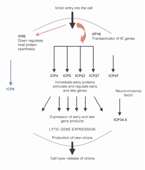
1.4.3. Herpes Simplex Virus.
1.4.3.1. A Vector for Gene Therapy.
Herpes simplex virus is a member of the alpha herpes virus family (Roizman at al. 1981). The two members of the HSV group, HSV-1 and HSV-2 are responsible for oral and genital herpes respectively. HSV-1 has been the most intensively studied to date and possesses a number of important features that make it potentially an ideal vector for gene therapy in certain tissues (reviewed in Fink at al. 1996 and Coffin and Latchman 1996). Such features include: the natural infection of both dividing and non-dividing cells; a life cycle which includes a period of latency, potentially allowing long-term transgene expression; a large genome which allows the insertion of large pieces of foreign genetic material; relatively straightforward manipulation of the genome and high titre viral growth. These properties provide a significant rationale for the development of HSV-1 vectors for gene therapy.
1.4.3.2. Biology o f HSV-1.
HSV-1 is a double stranded DNA virus with a genome of 152kb coding for over 80 polypeptides (Roizman and Sears 1996). It comprises two unique regions, short and long, each of which is flanked by inverted terminal repeats (see figure 1.5.). The unique long and unique short regions of the genome can invert relative to one another, thus yielding four possible linear isoforms during replication (Jacob at al. 1979). Approximately half of the 80 proteins encoded by HSV are essential for virus replication in vitro, but the rest are dispensable. Many of these dispensable genes are however necessary for full pathogenesis in the host (see later).
The natural route of HSV-1 infection involves uptake of the virus by epithelial cells of the skin, where-upon the virus undergoes lytic gene expression and replication (reviewed in Roizman and Sears 1996). Following lytic replication the virus infects sensory nerve terminals and is transported retrogradely to the nerve cell body. The virus can then either undergo a further round of lytic replication or the genome can remain an episomal nuclear element and enter a period of latency. Sensory neurons are the natural site of herpes latency and