1071-412X/05/$08.00⫹0 doi:10.1128/CDLI.12.8.994–1002.2005
Copyright © 2005, American Society for Microbiology. All Rights Reserved.
Cytokine Expression in Pediatric
Helicobacter pylori
Infection
Ana I. Lopes,
1* Marianne Quiding-Jarbrink,
2Ana Palha,
3Jose
´ Ruivo,
3Lurdes Monteiro,
4Mo
´nica Oleastro,
4Andrea Santos,
4and Afonso Fernandes
3Gastroenterology Unit, Paediatric Department, University Hospital Santa Maria, Lisboa, Portugal1; Department of Medical
Microbiology and Immunology, Goteborg University, Goteborg, Sweden2; Pathology Department,
University Hospital Santa Maria, Lisboa, Portugal3; and Helicobacter
Unit, Bacteriology Department, INSA, Lisboa, Portugal4
Received 31 January 2005/Returned for modification 11 April 2005/Accepted 27 May 2005
Helicobacter pyloriinfection is one of the most common gastrointestinal infections worldwide and almost invariably causes chronic gastritis in the infected host. A predominant Th1 profile has been demonstrated in
H. pylori-infected mucosa from adults, but no previous study has evaluated in situ cytokine expression in children. We therefore examined expression of proinflammatory, anti-inflammatory, and regulatory cytokines by immunohistochemistry in cryopreserved antral biopsy specimens from 10H.pylori-infected and 10 unin-fected children and correlated expression of cytokines with histology scores. Concomitant expression of interleukin-8 (IL-8), gamma interferon (IFN-␥), IL-4, transforming growth factor, and tumor necrosis factor alpha was seen in 8/10H.pylori-infected cases and in 5/10 noninfected cases; allH.pylori-infected subjects showed staining for at least two of the cytokines. The proportion of epithelial cytokine-specific staining did not differ significantly between the groups, either in surface or glandular epithelium. Furthermore, no significant differences were noticed between intraepithelial or lamina propria lymphocyte staining in the groups. There was, however, a tendency of higher numbers of IFN-␥- and IL-8-positive cells in theH.pylori-infected group. IFN-␥ and IL-8 lamina propria lymphocyte expression correlated significantly with antrum chronic inflam-mation, but there was no correlation between histology scores and epithelial cytokine expression. When the same techniques were used, the cytokine response appeared to be smaller inH.pylori-infected children than in adults, and there was no clear Th1 dominance. These results therefore suggest a different mucosal immuno-pathology in children. It remains to be determined whether the gastric immune response is downregulated in children withH.pyloriinfection and whether this is relevant to the outcome of infection.
Helicobacter pylori infection is one of the most common
gastrointestinal infections worldwide and the main cause of chronic gastritis, gastric mucosal atrophy, peptic ulcer, and some forms of gastric cancer (21, 24, 29, 30, 42). AlthoughH.
pyloriinfection almost invariably causes chronic gastritis, major
complications will develop only in a minority of infected sub-jects, predominantly in the adult host (21, 24). Epidemiological evidence ofH.pyloriacquisition during childhood (25, 44) and the rare occurrence of peptic ulcer or gastric atrophy in chil-dren (17, 18) suggests that the gastric mucosal damage result-ing from the infection might be progressive, through childhood until adulthood. During this time, bacterial determinants, the nature of the host immune responses, and exposure to poten-tial environmental factors all appear to influence outcome (20).
H.pyloriinduces a strong and complex immune response in
the gastric mucosa, both humoral and cellular (11, 19), which nevertheless fails to clear the infection and may even contrib-ute to immunopathology. The exact mechanisms by which the
H.pylori-induced immune response contributes to
gastrointes-tinal mucosal damage are still not clear.
A predominant Th1 T-cell response, associated with ele-vated levels of expression of proinflammatory cytokines gamma interferon (IFN-␥) and interleukin-2 (IL-2) and with
lower levels of expression of regulatory cytokines IL-4 and IL-10, has been clearly demonstrated inH.pylori-infected mu-cosa from adult subjects suffering from gastritis and peptic ulcers (6, 28, 32). Additionally, several studies investigating IL-8, a neutrophil chemotactic factor mainly secreted by epi-thelial cells, have shown an association between the levels of this cytokine andH.pyloriinfection (3, 10, 12, 23). At this time, it is not known whether this Th1 cell-mediated immune re-sponse is protective or whether it contributes to the pathogen-esis ofH.pylori-associated diseases.
Considering thatH.pylori-associated gastritis is usually mild in children, with low lymphocyte and neutrophil infiltration (4, 5), and if pediatric infection is viewed as an earlier stage of the
H.pylori-induced inflammatory response, a different
immuno-pathology and different patterns of cytokine expression in chil-dren could be anticipated compared to those of adults. There is, however, a paucity of information regarding local immune responses in the gastric mucosa from children, regardless ofH.
pyloriinfection status. Only a few studies have so far evaluated
the local cytokine profile in children, with somewhat conflict-ing results, but they most consistently show thatH.pylori in-fection induces production of proinflammatory cytokines and a Th1 response, similar to studies in adults (8, 9, 26, 31, 34, 41). Most of the previous pediatric studies have focused on detec-tion of cytokine mRNA in gastric biopsy specimens or on quantification of protein supernatants in gastric biopsy homog-enates or gastric juice specimens, using enzyme-linked immu-nosorbent assays (ELISAs) (8, 26, 31, 34, 41). However, these * Corresponding author. Mailing address: Gastroenterology Unit,
Paediatric Department, University Hospital Santa Maria, Avenida Professor Egas Moniz, 1600, Lisboa, Portugal. Phone: 351-919075306. Fax: 351-217588862. E-mail: anaisalopes@sapo.pt.
994
on August 17, 2020 by guest
http://cvi.asm.org/
methods do not reflect the in vivo situation as accurately as the immunohistochemistry approach does, allowing the identifica-tion, localizaidentifica-tion, and quantification of cytokine-producing cells.
To our knowledge, there are no previous pediatric studies evaluating in situ expression of different cytokines in gastric mucosa fromH.pylori-infected children by immunohistochem-istry. We therefore examined the gastric expression of proin-flammatory, anti-inproin-flammatory, and regulatory cytokines by epithelial cells, as well as by lamina propria and intraepithelial lymphocytes, inH.pylori-infected and uninfected children by immunohistochemistry. This expression was correlated with gastric mucosal inflammatory cell infiltration and withH.pylori
density of colonization and the presence of virulence factors.
MATERIALS AND METHODS
Clinical samples.Twenty children and adolescents of European origin (10 infected withH.pyloriand 10 not infected withH.pylori) referred for endoscopy with upper gastrointestinal symptoms (mostly recurrent abdominal pain), sug-gestive of organic disease and severe enough to require endoscopic evaluation, were included in the study. Informed consent from the parents and approval from the local Faculty and Hospital Ethics Committees, were obtained. The mean age ofH.pylori-positive subjects was 9.9 years (range, 7.3 to 12.6 years); the mean age ofH.pylori-negative subjects was 11.0 years (range, 5.8 to 18.8 years). Exclusion criteria were treatment with antisecretory, antimicrobial, or anti-in-flammatory medication, for the 3 months preceding the endoscopy. Subjects with peptic ulcer or severe organic disease were also excluded.
Sampling of mucosal biopsy specimens, specimen collection, and evaluation.
Upper endoscopy was performed under general anesthesia. Endoscopically, a mild to moderate erythema was present in all cases, and antral nodularity was evident in 13/20 cases (9 of these wereH.pylori-positive cases). Biopsy specimens were systematically taken from the duodenum (one or two), gastric antrum (four), and gastric body (one). One of the four antral biopsy specimens was immediately snap-frozen in isopentane previously cooled in liquid nitrogen, subsequently embedded in OCT compound (Tissue-Tek; Miles, Inc., Elkhart, Ind.), and processed for immunohistochemistry as described below. One antral biopsy specimen was fixed in 4.5% buffered formalin and embedded in paraffin;
2-m sections were stained with hematoxylin and eosin. A modified Giemsa stain was used forH.pyloriidentification, and gastritis was evaluated according to the updated Sydney system (16) by an experienced histopathologist who was unaware of the patient’sH.pyloristatus or clinical condition. Accordingly, the chronic inflammation score (mononuclear cell [MNC] infiltration), activity score (poly-morphonuclear cell infiltration), andH.pyloridensity score were determined separately and graded from 0 to 3 (for none, mild, moderate, and severe, re-spectively). Duodenal inflammation was evaluated in similarly treated biopsy specimens from all cases according to Whitehead criteria (46). The two addi-tional antral biopsy specimens were used for urease test (in-house test) and culture, respectively. The antral biopsy specimens for culture were put into sterile saline solution and processed within 3 h, according to a protocol previ-ously described (35). Briefly, biopsy specimens were ground with a tissue ho-mogenizer (Ultra Turax; Labo Moderne, France) and inoculated onto a selective medium (bioMe´rieux) and a nonselective medium, Mueller-Hinton agar (Oxoid, United Kingdom) supplemented with 10% horse blood (Probiolo´gica, Portugal). Plates were incubated at 37°C in a microaerobic atmosphere obtained with a gas-generating system (CampyGen CN 35; Oxoid) for up to 14 days of incuba-tion. Identification ofH.pyloriwas performed according to conventional tests: colony and gram stain morphology, catalase, oxidase, and hydrolysis of urea.
Diagnosis ofH.pyloriinfection.H.pyloristatus was assessed according to conventional biopsy-based criteria. Allocation toH.pylori-positive orH.pylori -negative group, was based on positivity of urease, histology, and culture, or on negativity of all three tests, respectively.
Serology.Serum samples were obtained for determination of anti-H.pylori -specific immunoglobulin G (IgG) antibodies by an ELISA (Roche) in 14/20 patients and by Western blotting (Helicoblot 2.0; Genelabs Diagnostics) in 10/20 patients (Table 1). ELISA results were in accordance withH.pyloristatus in four/fiveH.pylori-positive cases and in seven/nineH.pylori-negative cases. He-licoblot 2.0 results were positive in five/fiveH.pylori-positive cases and negative in three/fiveH.pylori-negative cases. Two currentlyH.pylori-negative cases (by culture, histology, and urease), had positive ELISA and Helicoblot 2.0 serology results, suggesting previousH.pyloriinfection.
Genotyping of isolated strains.Analysis ofvacAandcagAgenotypes was performed in allH.pylori-positive cases using PCR, according to a protocol previously described (39).cagpathogenicity island (PAI) status was also evalu-ated in 9/10 available strains by PCR using specific primers for thecagEgene and
cagPAI empty site (1, 47). The functional status of theoipAgene was determined as previously described (47).
TABLE 1. Clinical data for the 20H. pylori-infected and noninfected patients
Patient Age (yr) ELISA
serology
Helicoblot 2.0 serology
Genotype
vacA cagA cagE cagPAI oipstatus
H. pylori-infected patients
1 12.6 ⫹ ⫹ s2m2 ⫺ NP NP NP
2 10.3 NPa ⫹ s2m2 ⫺ ⫺ Absent Off
3 11.8 ⫹ ⫹ s2m2 ⫺ ⫹ Mixed Off
4 8.1 ⫹ ⫹ s2m2 ⫺ ⫺ Absent Off
5 11.0 ⫹ ⫹ s2m2 ⫺ ⫺ Absent Off
6 10.7 NP NP s2m2 ⫺ ⫺ Absent Off
7 10.4 NP NP s2m2 ⫺ ⫺ Absent Off
8 7.3 ⫺ NP s2m2 ⫺ ⫺ Absent Off
9 9.3 NP NP s2m2 ⫺ ⫺ Absent Off
10 7.3 NP NP s2m2 ⫺ ⫺ Absent Off
Noninfected patients
1 5.8 ⫹ ⫹
2 9.6 ⫺ ⫺
3 11.5 ⫹ ⫹
4 16.7 ⫺ ⫺
5 12.0 ⫺ ⫺
6 9.7 ⫺ NP
7 10.7 ⫺ NP
8 7.7 ⫺ NP
9 7.6 NP NP
10 18.8 ⫺ NP
aNP, not performed.
on August 17, 2020 by guest
http://cvi.asm.org/
Cytokine-specific MAbs.The cytokine-specific monoclonal antibodies (MAbs) used, all mouse anti-human antibodies, were anti-IL-4 (8F12; ImmunoKontact, Bioggio, Switzerland), anti-IL-8 (NAP 11; Bender, MedSystem, Vienna, Aus-tria), anti-tumor necrosis factor alpha (anti-TNF-␣) (MAb 1; Pharmingen, San Diego, California), anti-IFN-␥(1-D1K; MABTECH AB, Nacka, Sweden), and anti-transforming growth factor(anti-TGF-) (Genzyme Diagnostics, Cam-bridge, Mass.). All MAbs were of the IgG1 isotype. The MAbs were used at 5 g/ml, except for anti-TGF-, which was used at 10g/ml. Bovine serum albu-min (Sigma, St. Louis, MO.) was applied prior to the primary antibody to block unspecific staining. The specificities of the MAbs were ascertained by preabsorp-tion with recombinant cytokines. An isotype-matched mouse IgG1 antibody (Dako, Denmark) was used as a negative control in each experiment.
Immunohistochemistry.Cytokine expression was assessed in cryopreserved (OCT) antral/antral-body transition biopsy specimens as previously described (32). Briefly, 8-m-thick sections were mounted on glass slides (Superfrost/Plus; Menzel-Glaser, Braunschweig, Germany); after overnight adhesion at room tem-perature, the sections were fixed with 4% paraformaldehyde in phosphate-buff-ered saline containing 0.1% saponin, washed, and permeabilized with 0.1% saponin (Sigma, St. Louis, MO.) in phosphate-buffered saline. Endogenous per-oxidase activity was blocked with 1% H2O2and 0.02% NaN3. The tissue sections
were subsequently incubated with the cytokine-specific MAbs at 4°C overnight. The detection and amplification were performed using Envision horseradish peroxidase system (Dako, Glostrup, Denmark). A chromogen (diaminobenzi-dine) was finally applied according to the manufacturer’s instructions. The sec-tions were then washed with distilled water, counterstained with Harry’s hema-toxylin, dehydrated, and mounted with Entellan (Merck). Entire tissue sections were examined using an Olympus IMT2 microscope, excluding lymphoid folli-cles, since their random distribution in the tissue specimen may otherwise gen-erate less consistent results. However, very few cytokine-containing cells were observed within the follicles. Positive lymphocytes in epithelium or lamina pro-pria were manually enumerated in a section with a magnification of⫻300. The total section area was calculated by computer analysis (Metamorph 4.5 r6 soft-ware; Universal Imaging Corporation) with a magnification of⫻10. Lymphocyte staining in epithelium or lamina propria was expressed as the number of positive cells per square millimeter of mucosa. Only cells with a distinct intracellular (cytoplasmic) staining were included. The epithelial cytokine-stained area was determined by computer analysis, and the results were expressed as a percentage of stained epithelial area to total epithelial area in each section. Surface epithe-lial area (including foveola, neck, and pit) was evaluated separately from gland epithelium. All evaluations were performed by the same observer, who was unaware of the patient’sH.pyloristatus and histology findings. The mean tissue section area was 1.15 mm2
, ranging from 0.50 to 2.4 mm2
. One section per biopsy sample was analyzed for each cytokine, as the use of a single tissue section to represent immunostaining of an entire biopsy sample, had previously been val-idated in a reference study (32). The same study valval-idated the representativity of expression of cytokines IL-4, TNF-␣, and IFN-␥in each antral biopsy specimen, whereas the expression of TGF-and IL-8 differed substantially in biopsy spec-imens from different antral regions in each patient. Therefore, the consistency of TGF-and IL-8 staining in three different antral biopsy specimens per patient was assessed in threeH.pylori-positive cases and in threeH.pylori-negative cases at the start of the present study. The mean values for variation between biopsy specimens when the three biopsy specimens from the same subject were com-pared were 21.6% (range, 15 to 26%) for epithelial expression of IL-8 and 13.6% (range, 0 to 20%) for epithelial expression of TGF-. The mean values for variation for lamina propria lymphocyte staining were 3.1% (range, 0 to 19%) for IL-8 and 4.5% (range, 0 to 13%) for TGF-. In the majority of cases, no lamina propria lymphocyte staining could be detected, and in these instances, the sec-tions from the three biopsy samples were all negative. As the numbers of cyto-kine-producing cells and the proportion of positively stained epithelium in this material showed much less variation than the reference study, only one biopsy specimen per subject was subsequently evaluated.
Statistics.The nonparametric Mann-Whitney U test and Fisher’s exact test were used for statistical evaluation of comparisons between the two groups (H.
pyloripositive and negative) and for numerical and categorical variables, respec-tively.Pvalues ofⱕ0.05 were considered statistically significant. Spearman’s rank correlation coefficients were calculated to evaluate correlations between vari-ables. Statistical analysis was performed using SAS V8.2.
RESULTS
Histopathological evaluation. Most H. pylori-positive pa-tients showed slight to moderate chronic gastritis. Antrum and
corpus inflammation scores were higher in H. pylori-positive cases (median antrum score, 2.0 [range, 1 to 2]; median corpus score, 1.0 [range, 1 to 2]) compared toH.pylori-negative cases (median antrum score, 1.0 [range, 1]; median corpus score, 1.0 [range, 0 to 1]), with a statistically significant difference in antrum inflammation (P⫽0.007). Similarly,H.pylori-positive cases showed higher degrees of activity (median antrum activ-ity score, 1.0 [range 0 to 2]; median corpus score, 0.0 [range 0 to 1]) thanH.pylori-negative cases (median antrum activity, 0 [range, 0]; median corpus activity, 0 [range, 0]). Differences in antrum activity were statistically significant (P ⬍ 0.001). A positive correlation was found between chronic inflammation and activity scores in the antrum (r⫽0.705 andP⫽0.001), but not in the corpus, which was on the border of being significant (P⫽ 0.051). InH.pylori-positive cases, the medianH. pylori
density scores were 2.0 in the antrum (range, 2.0 to 3.0) and 1.0 in the corpus (range, 1.0 to 2.0). No positive correlation was found betweenH.pyloridensity scores and chronic inflamma-tion or activity scores in either the antrum or corpus. Lym-phoid follicles were present in 4/10H.pylori-positive cases and in 1/10H.pylori-negative cases (antrum or corpus). In oneH.
pylori-positive case, hyperplastic and regenerative features of
superficial epithelium were observed in the antrum. Duodenal histology showed a slight unspecific inflammation in 9/10H.
pylori-positive cases and in 9/10H. pylori-negative cases and
was normal in the remaining cases. As duodenal histology was uniform in most cases and no significant differences were found in corpus scores betweenH.pylori-positive and -negative cases, only antrum gastritis scores were considered for subse-quent correlation analysis between cytokine expression and histology scores.
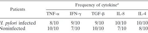
Frequency of immunostaining for different cytokines. The intracellular cytokine staining was predominantly localized to the cytoplasm of the cells. The cytokines studied could be detected in most of theH.pylori-infected and uninfected pa-tients (Table 2). Concomitant expression of all five cytokines was seen in 8 of the 10H.pylori-positive cases and in 5 of the
10H. pylori-negative cases. However, all the uninfected
sub-jects showed staining for at least two of the cytokines studied, in particular, TNF-␣, TGF-, and IL-4.
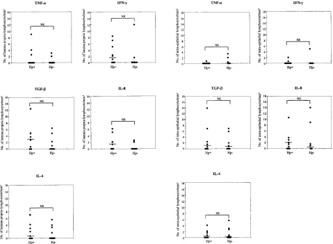
Cytokine staining of lamina propria and intraepithelial lym-phocytes in antral mucosa.When lymphocyte staining (epithe-lium and lamina propria) was compared between theH.pylori -positive and -negative groups, no significant differences were noticed, although there was a tendency towards higher fre-quencies of cytokine-producing cells in the lamina propria of
H.pylori-infected patients, especially with regard to IFN-␥and
IL-8 (Fig. 1, left panels). The intraepithelial lymphocytes, on the other hand, had a similar pattern of cytokine production in TABLE 2. Frequencies of five cytokine positively stained biopsy
specimens
Patients
Frequency of cytokinea
TNF-␣ IFN-␥ TGF- IL-8 IL-4
H. pyloriinfected 8/10 9/10 9/10 10/10 10/10
Noninfected 10/10 7/10 10/10 7/10 8/10
aNumber of biopsy samples with positively stained epithelium and/or lympho-cytes per total number of individuals studied.
on August 17, 2020 by guest
http://cvi.asm.org/
both patient groups (Fig. 1, right panels). The largest numbers of positively stained lymphocytes located intraepithelially and in the lamina propria were observed for TGF- and IFN-␥. The positive lymphocytes in the lamina propria were usually observed surrounding the glands or beneath the surface epi-thelium, particularly TGF--containing cells. A significant cor-relation was found between intraepithelial lymphocyte staining and lamina propria lymphocyte staining for TGF-(r⫽0.478 andP⫽0.033) and for IFN-␥(r⫽0.614 andP⫽0.004).
The expression of IFN-␥and IL-8 in lamina propria lym-phocytes correlated significantly with antrum chronic inflam-mation (r⫽0.539 andP⫽0.014 for IFN-␥andr⫽0.446 and
P⫽0.048), but there was no further correlation between the degree of active or chronic antrum inflammation and the stain-ing of the epithelium or lamina propria. Furthermore, theH.
pyloriantrum density score (in theH.pylori-positive group) did
not correlate with lymphocyte cytokine expression.
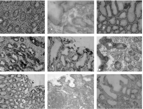
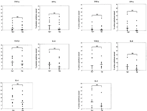
Cytokine staining of gastric epithelial cells.In addition to the cytokine staining of lymphocytes, substantial cytokine staining was localized to gastric epithelial cells in the superfi-cial epithelium and antral glands and was seen in H. pylori -positive and -negative cases (Fig. 2 and 3). The cytokine
stain-ing of epithelial cells showed a large variability between subjects for all the cytokines studied. Positive epithelial stain-ing was especially seen for TGF-, IL-8, and IL-4 and was localized to both superficial epithelial cells and to antral glands. However, the proportion of the epithelial cytokine-specific staining did not differ significantly betweenH. pylori -positive andH. pylori-negative cases for any of the cytokines studied, even though IL-8, IFN-␥, and IL-4 staining was slightly higher in the glandular epithelium ofH.pylori-positive patients (Fig. 3, right panels). Epithelial staining for IL-8 was always seen in specimens from the infected subjects but was seen in only 6/10 uninfected subjects, in whom the staining intensity was also weaker (Fig. 2). IL-4 was always expressed in
H.pylori-positive cases, where larger surface staining was
ob-served than inH.pylori-negative cases, but without significant differences. IFN-␥staining was usually detected inH. pylori -positive cases (9/10), but it was detected in only some H.
pylori-negative cases (gland, 5/10; surface epithelium, 4/10).
The proportions of epithelial staining of the anti-inflammatory cytokine TGF- did not differ between theH. pylori-positive
andH. pylori-negative subjects. This cytokine was always
de-tected inH.pylori-negative subjects and in 9/10H.pylori -pos-FIG. 1. Comparison of the cytokine staining of biopsy specimens fromH.pylori-infected (Hp⫹) and uninfected (Hp-) subjects. The numbers of cytokine-specific stained lamina propria/mm2of tissue (left panels) and intraepithelial lymphocytes/mm2of tissue (right panels) are given. Each
circle represents a biopsy specimen from one individual. Bars represent median values. NS, not significant (Mann-Whitney test).
on August 17, 2020 by guest
http://cvi.asm.org/
itive cases. TNF-␣staining was also similarly found inH.pylori -positive (5/10) andH.pylori-negative cases (5/10).
The frequency of lamina propria lymphocyte staining corre-lated significantly with epithelial staining of the epithelial sur-face and glands, respectively, for TGF-(r ⫽0.564 andP⫽
0.010;r ⫽0.509 andP⫽ 0.022), IFN-␥(r ⫽0.638 andP⫽
0.003;r⫽ 0.794 andP⬍0.001), and TNF-␣(r ⫽0.584 and
P⫽0.007;r⫽0.588 andP⫽ ⬍0.006). On the other hand, no correlation was found between antrum histology scores (in-flammation and activity) or the colonization density in theH.
pylori-positive group and epithelial cytokine expression.
Cytokine expression in subgroups ofH. pylori-negative in-dividuals.Since 2/9 cases in the H. pylori-negative group, as determined by biopsy specimen-based criteria, had serological evidence of a previousH.pyloriinfection (specific anti-H.pylori
ELISA), a subsequent statistical analysis was performed after exclusion of these twoH. pylori-negative cases (10H. pylori
-positive versus 7H.pylori-negative cases). However, very sim-ilar results (both for lymphocyte and epithelial cell staining) were found in this subset of individuals, compared to the pre-vious study sample, concerning all the cytokines studied (data not presented).
Genotyping ofH.pyloristrains.Strains werecagAnegative and harbored thevacAs2 (type II genotype). TheoipAgene was not functional in all of the cases studied. The results obtained for thecagEandcag PAI empty site corroborate the results for the absence of the cag PAI, except in one case, with a mixed infection ofcagPAI-positive and -nega-tive strains.
DISCUSSION
In the present study and to our knowledge for the first time in a pediatric population, we have evaluated in situ expression FIG. 2. Microphotographs showing immunohistochemical detection of cytokines in cryopreserved antral tissue specimens fromH. pylori -infected and un-infected subjects. (A1) Biopsy specimen from anH.pylori-infected subject showing IFN-␥staining of MNCs in the lamina propria. Original magnification,⫻300. (A and B) Epithelial IFN-␥-specific staining ofH.pylori-infected and uninfected subjects, respectively. Original magnification,⫻300. (C and D) Epithelial IL-8-specific staining ofH.pylori-infected and uninfected subjects, respectively. Original magnification,
⫻300. (E and F) Epithelial TGF--specific staining ofH.pylori-infected and uninfected subjects, respectively. Original magnification,⫻300. (G and H) Isotype controls fromH.pylori-infected (G) and uninfected (H) subjects. Original magnification,⫻300.
on August 17, 2020 by guest
http://cvi.asm.org/
of several cytokines in gastric mucosa. Children may be re-garded as an interesting natural model for the study ofH.pylori
infection, not only because they are not usually submitted to gastric mucosal noxa, such as alcohol, tobacco, and anti-inflam-matory medication, but also because of marked differences from adults regarding clinical course and gastric mucosal his-topathology. In particular, children have less intense MNC and polymorphonuclear cell infiltration (4, 5, 20). Considering the shorter duration ofH.pyloriinfection in children, gastric mu-cosal changes may represent an earlier stage of the immunoin-flammatory response compared to the adult host, and so a different immunopathology might be anticipated. In the present study, the expression of the cytokines studied in both epithelial cells and lymphocytes from lamina propria and epi-thelium did not differ significantly betweenH. pylori-positive and -negative cases, and wide variation in each group was observed.
Surprisingly, cytokine expression did not correlate with an-trum inflammation scores. Globally, these results do not totally agree with similar adult studies inH.pylori-associated gastritis
that mostly show a predominant Th1 profile with increased levels of IFN-␥, but not IL-4 and IL-5, inH. pylori-infected gastric mucosa (6, 10, 28, 32, 38, 48). Furthermore, D’Elios et al. (15) have shown that T-cell clones generated from antral biopsy specimens of H. pylori-infected peptic ulcer patients produce IFN-␥and IL-12 but usually not IL-4 or IL-5 in re-sponse to H. pylori antigen stimulation. However, recently, Holck et al. (27) found increased numbers not only of cells producing IL-8 (surface epithelium) and IFN-␥(lamina pro-pria MNCs) but also of cells producing IL-10 (lamina propro-pria MNCs) in infected adult patients, compared to uninfected subjects, which may counteract the inflammatory effect of the Th1 response.
The absence of differences between the groups may be at least partially related to the fact thatH.pylori-negative cases (status according to culture and histology) had some degree of gastritis. Other studies have also demonstrated mild chronic gastritis in a large proportion of uninfected children (9, 34). Even in the absence of clinical evidence of any chronic gastro-intestinal disease, another etiology for gastritis (including re-FIG. 3. Epithelial cytokine staining inH.pylori-infected (Hp⫹) and uninfected (Hp-) subjects. Staining of both superficial epithelium (left panels) and epithelial cells from glands (right panels) were expressed as a percentage of the total surface and glandular epithelial area in the section, respectively. Each circle represents a biopsy specimen from one individual. Bars represent median values. NS, not significant (Mann-Whitney test).
on August 17, 2020 by guest
http://cvi.asm.org/
cent viral infections, common in children) cannot be ruled out. Obviously, only symptomatic children requiring endoscopy were included, as a study design including completely healthy children would be ethically unacceptable, and in the former group, patients with normal mucosa are relatively rare. More-over, although the possibility of a missed pastH.pylori infec-tion cannot be totally excluded and it might have accounted for some degree of inflammation in the two/nineH.pylori-negative cases with positive serology, subsequent cytokine analysis did not show differences after their exclusion. It is not known how long it would take for the changes in mucosal cytokine levels to normalize after a gastric infection (whether or not due to
H. pylori). If cytokine levels remain high for a long time,
our findings of cytokine expression in some of theH. pylori -negative cases could be due to a previous, unrecognized H.
pyloriinfection (spontaneous eradication) or to another
infec-tion.
Although the potential contribution for cytokine expression of strain-related virulence factors in theH.pylorigroup could not be determined in our study, as the strains were of the type II genotype (nonfunctionaloipA), the absence ofcagA-positive strains with a functionaloipAmight be one explanation for the low proinflammatory response compared to those reported for adults (40, 48). The less pathogenic type II genotype is the most frequently found genotype in Portuguese children with nonulcerative gastritis and has been reported in other similar pediatric populations (2, 14, 39).
Previous studies on cytokine responses inH.pylori-infected children have yielded somewhat conflicting results (8, 9, 26, 31, 34, 41). However, most studies report increased levels of IL-8 and IFN-␥, as in adults, where elevated levels of IL-8 and Th1 responses are hallmarks ofH.pylori-induced gastritis. IL-8 is mainly secreted by epithelial cells and is a strong neutrophil attractant, and it is the most extensively investigated cytokine
inH.pylori-associated disease (3, 23, 32, 38, 48). However, IL-8
was also detected in the epithelium of normal gastric mucosa in some studies (10, 32). It is interesting, though, that the differ-ence in IL-8 production, as measured by immunohistochemis-try, is much higher inH.pylori-infected and uninfected adults (8) than in children (9; this study). A similar phenomenon is seen when IFN-␥levels in adults and children were compared. Both immunohistochemistry (32; this study) and analysis of secreted cytokines (8) demonstrate lower levels of IFN-␥ in children and less pronounced differences in infected and un-infected children than in adults.
Perhaps the most unexpected result of the present study was the presence of IL-4 in both H. pylori-infected and uninfected cases, which is in contrast to previous data from adult populations (6, 28, 32). In animal systems, Th2 cell responses comprising IL-4 and IL-5 have been associated with humoral responses and a reduction in bacterial load (36). The fact that children display IL-4 responses in the stomach, regardless ofH. pyloristatus, and lower levels of IFN-␥may indicate that children are more prone to mount-ing a gastric Th0 or Th2 response than adults. The lower gastritis scores in children may also be a reflection of such a skewed Th1/Th2 balance, which may result in a lower risk for developing ulcer disease.
Finally, TGF-was the sole cytokine observed uniformly in all individuals. This suggests a role as a constitutive
homeo-static cytokine also in children, in agreement with previous findings of this cytokine in normal fundic mucosa of adults (37). The inflammatory effects induced by the proinflammatory cytokines might be counteracted by IL-4 but also by other anti-inflammatory cytokines, such as TGF-, locally produced by immune cells or by gastric epithelial cells. However, TGF- production inH.pyloriinfection has not yet been extensively studied (32, 37, 43). This type of response could be relevant in trying to neutralize the Th1 response and stimulate Th2 and humoral responses, as it as been clearly demonstrated that children withH.pyloriinfection mount a humoral response, at both the systemic and local levels (7, 13, 45).
It remains to be determined whether the mucosal inflamma-tory response in the pediatric population is downregulated in response to different infectious agents and whether this is re-lated to the earlier stage of H. pylori infection or to age-dependent immunological maturation. Another interesting possibility deserving evaluation is that pediatricH.pylori infec-tion could lead to a higher activity of regulatory T cells and thereby suppress proinflammatory responses. This could ex-plain the lower degree of gastritis and higherH.pylori coloni-zation rate and may lead to the persistence of infection in children until adulthood. Indeed, the presence of circulating specific regulatory T cells in human H. pylori infection has recently been demonstrated (33). Later in the course of the infection, the persistence of H. pylori at the surface of the gastric mucosa may eventually favor a predominant Th1-type gastritis, as reported for adults.
Our data are in agreement with previous immunohistochem-istry studies showing a substantial contribution of the epithe-lium to cytokine expression. These data suggest a substantial contribution of the epithelium not only to proinflammatory and regulatory responses but also to anti-inflammatory re-sponses, similar to evidence in the adult host. The positive correlation observed between epithelial cell expression and the number of stained lymphocytes (lamina propria and/or epithe-lial) for some of the cytokines studied could suggest that at least for some cytokines, such as IFN-␥, produced only by immune cells, the epithelial staining might represent receptor binding of locally produced cytokines, rather than epithelial production. However, IFN-␥was also found in biopsy samples without detectable lymphocyte staining, suggesting the possible involvement of immune cells secreting small amounts of cyto-kine or positioned at distinct sites in the mucosa. Experiments on gastric epithelial cell lines suggest that IFN-␥may induce class II HLA expression on the epithelium, leading to in-creasedH.pyloriattachment, and possible epithelial cell apo-ptosis (22).
In conclusion, the present study indicates similar levels of gastric mucosal cytokine expression inH. pylori-positive and -negative pediatric cases, a finding that is in stark contrast to the situation in the adult host. Additionally, we have observed a considerable contribution of gastric epithelium to the antral cytokine response, similar to previous adult studies, suggesting a relevant role of the epithelium in the immunopathogenesis and outcome of infection. Further pediatric studies are war-ranted to characterize local immunopathology and cytokine responses in children.
on August 17, 2020 by guest
http://cvi.asm.org/
ACKNOWLEDGMENTS
We thank Rui Victorino, Laborato´rio de Imunologia, Instituto de Medicina Molecular, Faculdade de Medicina de Lisboa, for invaluable criticism and scientific supervision.
This study was financially supported by a grant from Comissa˜o de Fomento da Investigac¸a˜o em Cuidados de Sau´de, Ministe´rio da Sau´de, Portugal.
REFERENCES
1.Akopyants, N. S., S. W. Clifton, D. Kersulyte, J. E. Crabtree, B. E. Youree, C. A. Reece, N. O. Bukanov, E. S. Drazek, B. A. Roe, and D. E. Berg.1998. Analyses of thecagpathogenicity island ofHelicobacter pylori. Mol. Micro-biol.28:37–53.
2.Alarcon, T., D. Domingo, M. J. Martinez, and M. Lopez-Brea.1999.cagA
gene andvacAalleles in SpanishHelicobacter pyloriclinical isolates from patients at different ages. FEMS Immunol. Med. Microbiol.24:215–219. 3.Ando, T., K. Kusugami, M. Ohsuga, M. Shinoda, M. Sakakibara, H. Saito,
A. Fukatsu, S. Ichiyama, and M. Ohta.1996. Interleukin-8 activity correlates with histological severity inHelicobacter pylori-associated antral gastritis. Am. J. Gastroenterol.91:1150–1156.
4.Ashorn, M., T. Ruuska, R. Karikoski, J. Valipakka, and M. Maki.1994. Gastric mucosal densities inHelicobacter pylori-positive and -negative dys-peptic children and healthy controls. J. Pediatr. Gastroenterol. Nutr.18:146– 151.
5.Ashorn, M.1995. What are the specific features ofHelicobacter pylori gas-tritis in children? Ann. Med.27:617–620.
6.Bamford, K., X. Fan, S. E. Crowe, J. F. Leary, W. K. Gourley, G. K. Luthra, E. G. Brooks, D. Y. Graham, V. E. Reyes, and P. B. Ernst.1998. Lymphocytes in the human gastric mucosa duringHelicobacter pylorihave a T helper cell 1 phenotype. Gastroenterology114:482–492.
7.Blanchard, T. G., J. G. Nedrud, and S. H. Czinn.1999. Local and systemic antibody responses in humans with Helicobacter pyloriinfection. Can. J. Gastroenterol.13:591–594.
8.Bontems, P., F. Robert, A. Van Gossum, S. Cadranel, and F. Mascart.2003.
Helicobacter pylorimodulation of gastric and duodenal mucosal T cell cyto-kine secretions in children compared to adults. Helicobacter8:216–226. 9.Camorlinga-Ponce, M., F. Aviles-Jimenez, L. Cabrera, R. Hernandez-Pando,
O. Munoz, J. Soza, and J. Torres.2003. Intensity of inflammation, density of colonization and interleukin-8 response in the gastric mucosa of children infected withHelicobacter pylori. Helicobacter8:554–560.
10.Crabtree, J. E., J. I. Wyatt, L. K. Trejdosiewicz, P. Peichl, P. H. Nichols, N. Ramsay, J. N. Primrose, and I. J. Lindley.1994. Interleukin-8 expression in
Helicobacter pyloriinfected, normal, and neoplastic gastroduodenal mucosa. J. Clin. Pathol.47:61–66.
11.Crabtree, J. E.1996. Gastric mucosal inflammatory responses toHelicobacter pylori. Aliment. Pharmacol. Ther.10(Suppl. 1):29–37.
12.Crowe, S. E., L. Alvarez, M. Dytoc, R. H. Hunt, M. Muller, P. Sherman, J. Patel, Y. Jin, and P. B. Ernst.1995. Expression of interleukin-8 and CD54 by human gastric epithelium afterHelicobacter pyloriinfection in vitro. Gastro-enterology108:65–74.
13.Czinn, S. J., H. S. Carr, and W. T. Speck.1998. Diagnosis of gastritis caused byHelicobacter pyloriin children by means of ELISA. J. Infect. Dis.178:
460–465.
14.de Gusma˜o, V. R., E. Nogueira Mendes, D. M. De Magalha˜es Queiroz, G. Aguiar Rocha, A. M. Camargos Rocha, A. A. Ramadan Ashour, and A. S. Teles Carvalho.2000.vacAgenotypes inHelicobacter pyloristrains isolated from children with and without duodenal ulcer in Brazil. J. Clin. Microbiol.
38:2853–2857.
15.D’Elios, M. M., M. Manghetti, F. Almerigogna, A. Amedei, F. Costa, D. Burroni, C. T. Baldari, S. Romagnani, J. L. Telford, and G. Del Prete.1997. Different cytokine profile and antigen-specificity repertoire inHelicobacter pylori-specific T cell clones from the antrum of chronic gastritis patients with or without peptic ulcer. Eur. J. Immunol.27:1751–1755.
16.Dixon, M. F., R. M. Genta, J. H. Yardley, H. John, P. Correa, and the Participants in the International Workshop on the Histopathology of Gas-tritis.1996. Classification and grading of gastritis: the updated Sydney Sys-tem. Am. J. Surg. Pathol.20:1161–1181.
17.Drumm, B.1993.Helicobacter pyloriin the pediatric patient. Gastroenterol. Clin. N. Am.22:169–182.
18.Drumm, B., A. S. Day, B. Gold, F. Gottrand, S. Kato, E. Kawakami, A. Madrazo, J. Snyder, and J. Thomas.2004.Helicobacter pyloriand peptic ulcer: Working Group Report of the Second World Congress of Pediatric Gastroenterology, Hepatology and Nutrition. J. Pediatr. Gastroenterol. Nutr.39:S626–S631.
19.Ernst, P. B., S. E. Crowe, and V. E. Reyes.1997. How doesHelicobacter pylori
cause mucosal damage? The inflammatory response. Gastroenterology113:
S35–S42, S50.
20.Ernst, P. B., and B. D. Gold.1999.Helicobacter pyloriin childhood: new insights into the immunopathogenesis of gastric disease and implications for managing infection in children. J. Pediatr. Gastroenterol. Nutr.28:462–473.
21.Ernst, P. B., and B. D. Gold.2000. The disease spectrum ofHelicobacter pylori: the immunopathogenesis of gastroduodenal ulcer and gastric cancer. Annu. Rev. Microbiol.54:615–640.
22.Fan, X., S. E. Crowe, S. Behar, H. Gunasena, G. Ye, H. Haeberle, N. Van Houten, W. Gourely, P. B. Ernst, and V. E. Reyes.1998. The effect of class II major histocompatibility complex expression on adherence ofHelicobacter pyloriand induction of apoptosis in gastric epithelial cells: a mechanism for T helper cell type 1-mediated damage. J. Exp. Med.187:1659–1669. 23.Fan, X. G., A. Chua, X. J. Fan, and P. W. Keeling.1995. Increased gastric
production of interleukin-8 and tumor necrosis factor in patients with Hel-icobacter pyloriinfection. J. Clin. Pathol.48:133–136.
24.Farthing, M. J.1998.Helicobacter pyloriinfection: an overview. Br. Med. Bull.54:1-6.
25.Go, M. F.2002. Natural history and epidemiology ofHelicobacter pylori
infection. Aliment. Pharmacol. Ther.16(Suppl. 1):3–15.
26.Guiraldes, E., I. Duarte, A. Pena, A. Godoy, M. N. Espinosa, R. Bravo, F. Larrain, M. Schultz, and P. Harris.2001. Proinflammatory cytokine expres-sion in gastric tissue from children withHelicobacter pylori-associated gas-tritis. J. Pediatr. Gastroenterol. Nutr.33:127–132.
27.Holck, S., A. Norgaard, M. Bennedsen, H. Permin, S. Norn, and L. P. Andersen.2003. Gastric mucosal cytokine responses inHelicobacter pylori -infected patients with gastritis and peptic ulcers. Association with inflamma-tory parameters and bacteria load. FEMS Immunol. Med. Microbiol.36:
175–180.
28.Kartunnen, R., T. Kartunnen, H. P. Ekre, and T. T. MacDonald.1995. Interferon gamma and interleukin-4 secreting cells in the gastric antrum in
Helicobacter pyloripositive and negative gastritis. Gut36:341–345. 29.Kuipers, E. J., J. C. Thijs, and H. P. Festen.1995. The prevalence of
Helicobacter pyloriin peptic ulcer disease. Aliment. Pharmacol. Ther.9:59– 69.
30.Kuipers, E. J.1998. Relationship betweenHelicobacter pylori, atrophic gas-tritis and gastric cancer. Aliment. Pharmacol. Ther.12:25–36.
31.Kutukuler, N., S. Aydogdu, D. Goksen, S. Caglayan, and R. V. Yagcyi.1997. Increased mucosal inflammatory cytokines in children withHelicobacter py-lori-associated gastritis. Acta Paediatr.86:928–931.
32.Lindholm, C., M. Quiding-Jarbrink, H. Lonroth, A. Hamlet, and A. M. Svennerholm.1998. Local cytokine response inHelicobacter pylori-infected subjects. Infect. Immun.66:5964–5971.
33.Lundgren, A., E. Stro¨mberg, A. Sjo¨ling, C. Lindholm, K. Enarsson, A. Edebo, E. Johnsson, E. Suri-Payer, P. Larsson, A. Rudin, A. M. Svenner-holm, and B. S. Lundin.2005. MucosalFOXP3-expressing CD4⫹CD25high
regulatory T cells inHelicobacter pylori-infected patients. Infect. Immun.
73:523–531.
34.Luzza, F., T. Parrelo, and L. Sebkova.2001. Expression of proinflammatory and TH1 but not TH2 cytokines is enhanced in gastric mucosa of Helico-bacter pyloriinfected children. Dig. Liver Dis.33:14–20.
35.Me´graud, F., N. Lehn, T. Lind, E. Bayerdo¨rffer, C. O’Morain, R. Spiller, P. Unge, S. V. van Zanten, M. Wrangstadh, and C. F. Burman.1999. Antimi-crobial susceptibility testing ofHelicobacter pyloriin large multicenter trial: the MACH 2 study. Antimicrob. Agents Chemother.43:2747–2752. 36.Mohammadi, M., S. Czinn, R. Redline, and J. Nedrud.1996.Helicobacter
-specific cell-mediated immune responses display a predominant Th1 pheno-type and promote a delayed-pheno-type hypersensitivity response in the stomachs of mice. J. Immunol.156:4729–4738.
37.Naef, M., T. Ishiwata, M. Friess, M. W. Buchler, L. I. Gold, and M. Kore.
1997. Differential localization of transforming growth factor-isoforms in human gastric mucosa and overexpression in gastric carcinoma. Int. J. Can-cer71:131–137.
38.Noach, L. A., N. B. Bosma, J. Jansen, F. J. Hoek, S. J. van Daventer, and G. N. Tytgat.1994. Mucosal tumor necrosis factor-alpha, interleukin-1 beta, and interleukin-8 production in patients withHelicobacter pyloriinfection. Scand. J. Gastroenterol.29:425–429.
39.Oleastro, M., M. Gerhard, A. I. Lopes, P. Ramalho, J. Cabral, A. Sousa Guerreiro, and L. Monteiro.2003.Helicobacter pylorivirulence genotypes in Portuguese children and adults with gastroduodenal pathology. Eur. J. Clin. Microbiol. Infect. Dis.22:85–91.
40.Peek, R. M., G. G. Miller, K. T. Tham, G. I. Perez-Perez, X. Zhao, J. C. Atherton, and M. J. Blaser.1995. Heightened inflammatory response and cytokine expression in vivo to cagA⫹ Helicobacterstrains. Lab. Investig.
73:760–770.
41.Shimizu, T., H. Haruna, Y. Ohtsuka, K. Kaneko, R. Gupta, and Y. Ya-mashiro.2004. Cytokines in the gastric mucosa of children withHelicobacter pyloriinfection. Acta Paediatr.93:322–326.
42.Sipponen, P., H. Hyvarinen, K. Seppala, and M. J. Blaser.1998. Pathogen-esis of the transformation from gastritis to malignancy. Aliment. Pharmacol. Ther.12:61–67.
43.Strober, W., B. Kesall, I. Fuss, T. Marth, B. Ludviksson, R. Ehrhardt, and M. Neurath.1997. Reciprocal IFN-␥and TGF-responses regulate the occurrence of mucosal inflammation. Immunol. Today18:61–64. 44.Thomas, J. E., A. Dale, M. Harding, W. A. Coward, T. J. Cole, and L. T.
Weaver.1999.Helicobacter pyloricolonization in early life. Pediatr. Res.
45:218–223.
on August 17, 2020 by guest
http://cvi.asm.org/
45.Westblom, T. U., L. M. Lagging, B. R. Midkiff, and S. J. Czinn.1993. Differences in antigenic recognition between adult and pediatric patients infected withHelicobacter pylori: analysis using Western blot technique. Acta Gastroenterol. Belg.56:84.
46.Whitehead, R.1982. Morphological aspects of duodenitis. Scand. J. Gastro-enterol. Suppl.79:80–83.
47.Yamaoka, Y., S. Kikuchi, H. M. el-Zimaity, O. Gutierrez, M. S. Osato, and
D. Y. Graham.2002. Importance ofHelicobacter pylori oipAin clinical pre-sentation, gastric inflammation, and mucosal interleukin 8 production. Gas-troenterology123:414–424.
48.Yamaoka, Y., T. Kodama, M. Kita, J. Imanishi, K. Kashima, and D. Y. Graham.1999. Relation between clinical presentation,Helicobacter pylori
density, interleukin 1and 8 production, andcagAstatus. Gut45:804– 811.