ABSTRACT
SAN MARTIN DIAZ, VIVIANA EDITA. Effects of 1-α-hydroxycholecalciferol and Other Vitamin D Analogs on Live Performance, Bone Development, Meat Yield and Quality, and Mineral Digestibility on Broilers. (Under the direction of Dr. Edgar O. Oviedo-Rondón).
Total Ca levels in broiler diets can be under or overestimated. The reasons for this can go from simple failures on the mixing to the current tendency to considering lower dietary Ca. Vitamin D plays an important role in intestinal Ca absorption that leads to adequate blood levels of Ca and P for an adequate bone development and health. An analog molecule of VitD3,1-alpha–
hydroxycholecalciferol (1α(OH)D3), has been demonstrated to fulfill these functions. However, 1α(OH)D3 its effect under different Ca levels has not been evaluated. To address this question, two
experiments were conducted. Male broiler chicks were reared until 14d of age using 5 different Ca levels between 0.80 and 1.40% (Chapter 2) or until 35 d using 4 Ca levels between 0.54 and 1.20% (Chapter 3). In starter diets, an improvement in FCR was detected in diets with 0.80% Ca when
1α(OH)D3 is supplemented. Also, this supplementation affected bone development inducing lower
bones relative asymmetries, increased mineral digestibility when no deficient Ca diets were used. In the grower phase study, parameters related to bone development and meat quality were assessed. No effects of 1α(OH)D3 were detected on these parameters. However, an effect of dietary Ca level
was detected on valgus, meat color and spaghetti muscle myopathy incidence. Similarly, regression analyses indicated a quadratic effect of Ca on carcass and breast meat yield, with the higher yields detected at 0.76% Ca inclusion. Bone development, tibia strength and thickness and ash tibia ash content. 1α(OH)D3 increased bone ash content as well. A third study was conducted
to compare three VitD3 metabolites: 1α(OH)D3, 25(OH)D3 and 1,25(OH)2D3 (Chapter 4). These
metabolites were added on top of a basal diet containing 4,000 IU/kg of VitD3 as part of the mineral
with no metabolite addition. This treatment also obtained higher values when compared to the chickens fed with 25(OH)D3 and 1,25(OH)2D3 diets. It was also detected that chickens fed the
1,25(OH)2D3 had the worst FCR compared to the other treatments. Chickens fed 1α(OH)D3 also
obtained lower bone length relative asymmetry in comparison to other treatments. Furthermore, in meat quality, less cook loss was obtained by the non-supplemented and 1α(OH)D3 treatments. In
relation with myopathies, the highest and lower presentation of wooden breast were obtained with 25(OH)D3 and 1α(OH)D3, respectively. Therefore, it can be concluded that adequate dietary Ca
levels have considerable effects on live performance, bone health and meat quality. Based on these results, addition of 1α(OH)D3 has positive effects on improving mineral digestibility and bone
development. Likewise, the potency of each VitD3 metabolite varies differently according to the
Effects of 1-α-hydroxycholecalciferol and Other Vitamin D Analogs on Live Performance, Bone Development, Meat Yield and Quality, and Mineral Digestibility on Broilers.
by
Viviana Edita San Martin Diaz
A thesis submitted to the Graduate Faculty of North Carolina State University
in partial fulfillment of the requirements for the degree of
Master of Science
Poultry Science
Raleigh, North Carolina 2018
APPROVED BY:
_______________________________ _______________________________ Dr. Edgar O. Oviedo Rondón Dr. Peter Ferket
Committee Chair Committee Co-chair
ii DEDICATION
To my beloved parents, Iris y Pedro, for their support, love and for being the best example to follow in my life.
To my brothers, Susana y Pedro, for trusting and always being there for me.
iii BIOGRAPHY
Viviana San Martin Diaz, Peruvian Veterinarian that obtained her degree in the School of Veterinary Medicine from Universidad Nacional Mayor de San Marcos. After it, she obtained a Master’s in Animal Health from the School of Veterinary Medicine at Universidad Nacional Mayor de San Marcos.
Since 2007, she participated in various research projects as Entrenamiento de Veterinarios en enfermedades zoonóticas en países en desarrollo, porcine and avian influenza research project in Peru, Training in infectious Diseases in Perú, A demonstration Project to eliminate cysticercosis in Peru. She has experience as Graduate Teaching Assistant in undergrad courses at mathematics and Poultry Diseases courses. Furthermore, she participated as part of DVM and Master’s degree
jury in Peru.
After obtaining her Master’s degree in Peru, she followed a two year residence program for Specialist in Avian Pathology and Medicine at the Avian Pathology Laboratory - School of Veterinary Medicine Universidad Nacional Mayor de San Marcos.
Following her residency period, she worked at Ilender Pharmaceutical Corporation, multinational company responsible for the processing, distribution and marketing of nutritional and health products for animal production, especially the poultry industry. In this company she performed as Technical Service Coordinator having as one of her principal duties the development of experimental trials and field evaluations in coordination with Research and Development team.
iv ACKNOWLEDGMENTS
I need to start giving thanks to Dr. Edgar Oviedo, my advisor, for allow me to be part of his lab and work by his side. The whole experience working at NCSU and especially at his lab is invaluable as well as all the knowledge that he shared during these years. Also, I want to acknowledge my committee members Dr, Peter Ferket, Dr. Jesse Grimes, and Dr. Consuelo Arellano, for giving to this thesis part of their valuable time, for their recommendations and their guidance to obtain a final quality thesis. My gratitude to each one of them.
I would like to give thanks to the members of the Prestage Department of Poultry Science too. Thanks to all the persons that from Scott Hall, the Chicken Education Unit and/or the Feed Mill facility contributed in the success this research.
v TABLE OF CONTENTS
LIST OF TABLES………viii
LIST OF FIGURES………..…...…xi
APPENDIX………..xiii
Chapter I: Literature Review………1
Introduction………..1
Vitamin D and its metabolism………..3
Vitamin D3 analogs………..5
Calcium and Phosphorus absorption………..……….….7
Ca:P - Bone development/mineralization and strength………..10
Mineral retention and digestibility……….…12
References………..………14
Chapter II: Effect of 1-alpha-hydroxy-vitamin-D3 on Performance, Bone Development and Mineral Retention in Starter Diets Containing Different Calcium Levels for Ross-708 Broilers………..………21
Abstract………..21
Introduction………..………..…23
Materials and Methods………...…26
Treatments and Chicken Husbandry………..26
Diets………...……26
Live Performance………...………27
Bone development and mineralization………...…………27
vi
Statistical Analysis……….……28
Results ………..………..28
Discussion……….……….…30
Conclusion……….…35
References……….……….…36
Chapter III: Effect of 1-alpha-hydroxy-vitamin-D3 on Performance, Bone Development, Meat Yield and Quality and Mineral Digestibility in Grower Diets Containing Different Calcium Levels for Ross-708 Broilers………...……56
Abstract………..56
Introduction………58
Materials and Methods………...59
Dietary treatments and chicken husbandry………59
Broiler live performance………60
Leg health evaluation……….60
Processing, cook loss and bone collection………..60
Leg asymmetry, bone strength and tibia ash………...61
Ca and P Digestibility ………62
Statistical analyses...………...………...63
Results………63
Discussion………..………66
Conclusion……….69
vii Chapter IV: Effect of VitD Metabolite Sources on Broiler Performance, Carcass Yield,
Meat Quality, Leg Health and Bone Development……..………...90
Abstract……….….90
Introduction………92
Materials and Methods………...93
Treatments and chicken husbandry………93
Broiler live performance………94
Leg health evaluation……….94
Processing, carcass and cut up yield, and meat quality………...………94
Bone development and mineralization………...96
Leg asymmetry, bone strength and tibia ash………...96
Statistical analyses……….97
Results ………97
Discussion………..98
Conclusion………...101
References………102
SUMMARY………..….………..………117
viii LIST OF TABLES
CHAPTER II
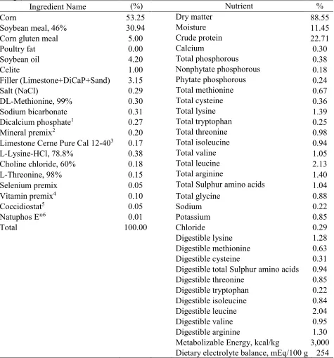
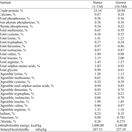
Table 2.1. Ingredient composition and calculated nutrient content of starter basal diet
(1-16 days of age) for Ross-708 broilers………...……41
Table 2.2. Effect of supplementation of 1α(OH)D3 on top of VitD3 with varying levels of
calcium in starter diets for Ross 708 male broilers .from 0-7d….……….….…...42 Table 2.3. Effect of supplementation of 1α(OH)D3 on basal diets containing VitD3 and
different Ca levels during the starter phase for Ross 708 male broilers on
performance from 0 to 14d………..………..…43
Table 2.4. Equations for effects of supplementation of 1α(OH)D3 on a basal diet containing
VitD3 and different Ca levels during the starter phase on live performance
for Ross 708 male broilers from 0 to 7d and 0-14d………..….……44
Table 2.5. Effect of supplementation of 1α(OH)D3 on basal diets containing VitD3 and
different Ca levels during the starter phase for Ross 708 male broilers on tibia
ash and bone relative length and weight at 16d……….……45
Table 2.6. Effect of dietary Ca levels and 1α(OH)D3 supplementation on ileal digestibility
of Ross 708 male broilers at 16 d of age……….46
Table 2.7. Equations for effects of supplementation of 1α(OH)D3 on a basal diet containing
VitD3 and different Ca levels during the starter phase on ileal digestibility of
Ross 708 male broilers at 16 d of age…...……….………..47
Table 2.8. Effect of supplementation of 1α(OH)D3 on basal diets containing VitD3 and
Different Ca levels during the starter phase for Ross 708 male broilers on
mineral retention at 14 days………...50
Table 2.9. Effect of dietary Ca levels and 1α(OH)D3 supplementation on Ca and
P ileal digestibility vs. Retention of Ross 708 male broilers at 16 d of age…………...53 Table 2.10. Equations for effects of supplementation of 1α(OH)D3 on a basal diet containing
VitD3 and different Ca levels during the starter phase on mineral retention and
mineral digestibility vs. retention for Ross 708 male broilers from 0 to 14 d………….55 CHAPTER III
Table 3.1. Ingredient composition of starter diet and grower basal diets for Ross-708
male broilers………...…...76
Table 3.2 Nutritional content of basal starter and grower diets for Ross-708
ix
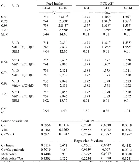
Table 3.3. Effect of 1α(OH)D3 on grower broiler diets (17-35d) with different dietary
calcium levels on BW and BWG for male broilers Ross-708………78
Table 3.4. Effect of 1α(OH)D3 on grower broiler diets (17-35d) with different dietary
calcium levels on Feed intake and FCR for male broilers Ross-708………....…..79
Table 3.5. Effect of 1α(OH)D3 on grower broiler diets with different dietary calcium levels
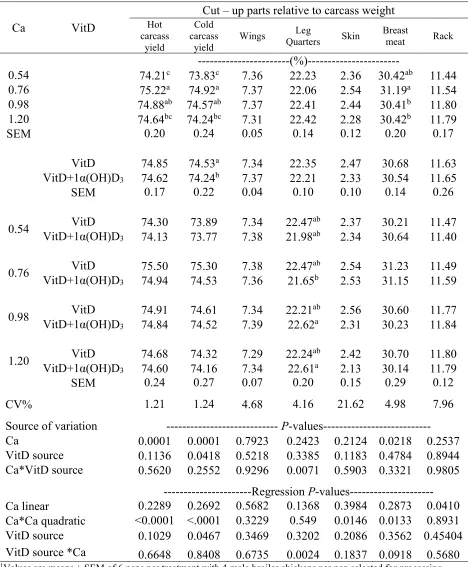
on cut-up parts yields of Ross-708 males at 34d ………80 Table 3.6. Effect of 1α(OH)D3 on grower broiler diets with different dietary calcium levels
on meat quality and color parameters of Ross-708 males at 34 d …...81
Table 3.7. Regression equation of effect of dietary Ca levels and 1α(OH)D3 supplementation
on performance, yields and meat quality parameters and breast meat color of
Ross 708 male broilers at 35 days of age...82
Table 3.8. Effect of 1α(OH)D3 on broiler grower diets with different dietary calcium
levels on wooden breast sensorial score, white stripping and spaghetti muscle of Ross-708 males at 34d………...……….…...83
Table 3.9. Effect of 1α(OH)D3 on broiler grower diets with different dietary calcium levels
on the incidence (0-1) of leg health issues or footpad dermatitis average score (0-9) of Ross-708 males at 34 d……….……….84 Table 3.10. Effect of dietary calcium level and 1α(OH)D3 on tibia strength and
mineralization of Ross 708 male broiler at 35 d of age………..……85
Table 3.11. Regression equations for effect of supplementation of 1α(OH)D3 on top of VitD
and dietary Ca levels for Ross 708 male broilers on bone strength, mineralization and on ileal digestibility at 35d……….…….……86 Table 3.12. Effect of dietary calcium level and 1α(OH)D3 on leg relative weight and
relative asymmetry of Ross 708 male broiler at 35d of age………...….87
Table 3.13. Effect of dietary Ca levels and 1α(OH)D3 supplementation on ileal digestibility
of Ross 708 male broilers at 35 d of age………....……….88 CHAPTER IV
Table 4.1. Ingredient composition of starter, grower and finisher basal diets for
Ross 708 male broilers.……….………...……...108
x of variation of male Ross 708 broilers, and total mortality………..……....110 Table 4.5. Effect of VitD sources on feed intake and feed conversion ratio (FCR) of
Ross 708 male broilers………....111 Table 4.6. Effect of VitD sources on incidence (0-1) of leg health issues, and foot pad
Dermatitis average score (0-9)1 of Ross-708 male broilers at 42 d……….111 Table 4.7. Effect of VitD sources on tibia strength of Ross 708 male broilers at 42 d.………...112 Table 4.8. Effect of VitD sources on bone ash, relative weight and relative asymmetry in diets
for Ross 708 male broilers at 42 days of age………...112 Table 4.9. Effect of VitD source on carcass and cut-up part yields of Ross-708 male
broilers at 42d………..113
Table 4.10. Effect of VitD sources on carcass and cut-up parts weights of Ross-708 male
broilers at 42 d………...……..114
Table 4.11. Effect of VitD sources on breast meat quality and color parameters of Ross-
708 male broilers at 42d………...… ………...115 Table 4.12. Effect of VitD sources on breast meat quality and on wooden breast score
xi LIST OF FIGURES
CHAPTER I
Figure 1.1 Metabolic pathway in chickens of dietary Vitamin D (cholecalciferol) and
hormonal activation...4 Figure 1.2. Hormonal regulation of Ca plasma levels to homeostasis………...8
CHAPTER II
Figure 2.1. Linear effect of calcium level with and without 1α(OH)D3 on dry matter
digestibility from broiler males at 16 d of age………….………….……….…….48
Figure 2.2. Quadratic effect of calcium level with 1α(OH)D3 and linear effect without
1α(OH)D3 on calcium digestibility from broiler males at 16 d of age.…..………..….48
Figure 2.3. Effect of calcium level with and without 1α(OH)D3 on phosphorous digestibility
from broiler males at 16 d of age. ………….……….……….………...…..49
Figure 2.4. Quadratic effect of calcium levels with and without 1α(OH)D3 on calcium
retention from 0-14 days in male broilers………….……….………...51
Figure 2.5. Quadratic effect of calcium levels with and without 1α(OH)D3 on phosphorous
retention from 0-14 days in male broilers………...…….………..51
Figure 2.6. Quadratic effect of calcium levels with and without 1α(OH)D3 on manganese
retention from 0-14 days in broiler males……….……….…….………..52
Figure 2.7. Quadratic effect of calcium level with and without 1α(OH)D3 on zinc retention
from 0-14 days in broiler males. ………….……….……….……….………...…52
Figure 2.8. Quadratic effect of calcium level with and without 1α(OH)D3 on Ca digestibility
vs. retention percentage in diets with and without 1α(OH)D3 from broiler males
at 16d of age………….……….……….……….……….……….……….…………...54
Figure 2.9. Quadratic effect of calcium level on P digestibility vs retention percentage in
xii CHAPTER III
Figure 3.1 Effect of calcium level with and without 1α(OH)D3 on Calcium digestibility
from broiler males at 35 d of age……….………...89
Figure 3.2 Effect of calcium level with and without 1α(OH)D3 on phosphorous digestibility
from broiler males at 35 d of age………89 APPENDIX
1 CHAPTER I
LITERATURE REVIEW
INTRODUCTION
Vitamin D, a fat-soluble vitamin also known as calciferol, is a nutrient, necessary for an adequate intestinal calcium (Ca) absorption and transportation, which guarantee bone health and strength. It is also important for various physiological processes. Phosphorous absorption and homeostasis with Ca, as well as micro mineral regulation (P, Mn, I, Cu, Zn), and even immunity of the bird can be affected by blood VitD levels (Biehl et al., 1995; Vazquez et al., 2018).
VitD can be synthesized in the skin by sun exposure and ultraviolet B radiation. Therefore, for many decades, since broiler rearing conditions made sunlight access limited, it is impossible or limited that the skin of the animal receive the necessary radiation to synthetize adequate VitD levels. For this reason, the consideration of VitD in poultry feed formulation is necessary, especially because of the rapid bone and body growth rate present in broilers.
Different VitD precursors have been study in the past decades (Haussler et al., 1973; Mitchell and Edwards, 1995; Han et al., 2009; Bachmann et al., 2013). The interest in searching for alternative molecules to provide cholecalciferol in the diets is due to the different biopotency and faster metabolic activity that can allow to obtain better broiler live performance. How they affect broiler performance and mineral metabolism under different conditions is a topic of truly importance (Biehl et al., 1995; Rao et al., 2009; Han et al., 2016).
There are three metabolites of VitD currently used in poultry and animal nutrition. The 25-hydroxycholecalciferol (25(OH)D3), 1,25-dihydroxycholecalciferol (1,25(OH)2D3) and
1-alpha-hydrocxicholecalciferol (1α(OH)D3) are metabolites that can be found as commercial products.
2 stressors, anti-nutritional factors and toxic compounds like mycotoxins and heavy metals, can affect liver and kidney function (Devegowda, 2009; Bryden, 2012; Murugesan et al., 2015) or the Ca dietary content can be compromised (Van Kempen et al., 2001; Adedokun and Adeola 2013; Li et al., 2017).
In the present study, the focus was on the effect produced by 1α(OH)D3 in broiler
performance, bone development and bone mineralization, as well as mineral retention and digestibility under different dietary Ca levels. Although this analog molecule of the active VitD3
has been previously studied under various conditions and demonstrated its positive effects increasing bone mineralization and obtaining better performance parameters with higher BW and low FCR (Edwards et al., 1992; Biehl et al., 1995; Edwards Jr. et al., 2002; González et al., 2015), a detailed study of its effects under different dietary Ca levels in broiler chickens reared under the same conditions has not been carried out. Furthermore, mineral digestibility and retention of trace minerals under these conditions have not been reported. In the present study P, Ca, Mn, Cu, Zn were the minerals selected for the determination of its digestibility and/or retention, due to their participation in many digestive and physiological processes, including bone development.
The present review will deal with the topics of VitD metabolism and its relationship with the Ca and P homeostasis. Likewise, aspects related to leg bone development and mineral digestibility will be developed. The intention of this revision is to be able to have a clear vision of the findings obtained in the experiments that constitute this thesis.
VITAMIN D AND ITS METABOLISM
Vitamin D is a fat-soluble vitamin that synthesized in the outer skin layers when they are exposed to sufficient sunlight (McDowell, 2000). In the nature, ergocalciferol (VitD2) and
3 in plant sources and it is generated by the ultraviolet irradiation of the ergosterol (steroid found in plants), while VitD3 is produced mainly in animal tissues upon the irradiation of the
7-dehydrocholesterol (provitamin D) in skin (McDonald et al., 2011).
It is known that VitD2 has little to no-activity (nutritional value) for avian species (NRC,
1994). However, VitD3 is about 10-fold more effective in chicks than VitD2 (Hurwitz et al., 1967;
Adedokun and Adeola, 2013). Throughout this document the attention will be centered on VitD3
molecule.
VitD3 is the responsible to ameliorate the effects of a low Ca level, as rickets or
dyschondroplasia. Also, it has been detected effects of VitD on immunological responses, where immune cells containing VitD receptors (VDR) can react to VitD3 modulating innate and adaptive
immune responses. (Baeke et al., 2010; Aranow, 2011). One international unit (IU) of VitD is defined as the activity of 0.025 µg of VitD3 (NRC, 1994). As mentioned previously, VitD may be
supplied through the diet or by UV irradiation of the skin. Natural cholecalciferol is produced exclusively in animal products by UV irradiation of dehydrocholesterol from the sunlight. 7-dehydrocholesterol is derived from cholesterol or squalene, which is synthesized in the body and present in large amounts in skin, intestinal wall, and other tissues (McDowell, 2000). Nevertheless, the VitD obtained by chickens from many years ago is too little or null. Consequently, the source of VitD in chickens comes from its dietary ingestion as form of cholecalciferol.
To obtain the active VitD3, the dietary cholecalciferol molecule needs to undergo two
4 last molecule is the active VitD3 that will act in the specific tissues. Other VitD metabolites can be
produced in the kidney, especially in situations of sufficient Ca levels in plasma. In this situation 24,25-dihydroxycholecalciferol is produced. Nevertheless, its effectiveness and physiological importance of this molecule is under discussion and investigation (Henry, 2011). Figure 1.1 shows the metabolic pathway of VitD3 activation.
Figure 1.1. Metabolic pathway in chickens of dietary Vitamin D (cholecalciferol) and hormonal activation. Mucosal epithelial cells, in the intestine, kidney, cartilage, osteocytes and osteoblasts, have receptors for 1,25(OH)2D3. (Veum, 2015). 1,25(OH)2D3 plays role in the regulation of the Ca
absorption by the regulation of the specialized protein calbindin. This protein will be in charge of binding Ca2+ molecules to allow its absorption and further transport of Ca2+ through the intestinal
5 VITAMIN D3 ANALOGS
Vitamin D is generally provided to poultry by supplementation of the diet with crystalline forms of cholecalciferol (Vitamin D3) (Fritts and Waldroup, 2003). It is known that the VitD3
requirement is dependent on the dietary Ca and available phosphorus (avP) contents in diets (Rath et al., 2000).
The National Research Council (NRC, 1994) indicated a minimum requirement of 200 IU/kg of VitD3 for poultry diets. However, under current commercial practices, broiler and turkey
diets contain from 2,000 to over 5,000 IU/kg (Applegate and Angel, 2014). In addition to these high concentrations of VitD3, the supplementation of animal feed with metabolites products from
the hydroxylation pathway is becoming frequently. There are three different metabolites of VitD3
commercially available: 25(OH) D3 (HyD®, DSM Nutritional Products Inc, Parspany NJ), the
1,25(OH)2D3 (Panbonis®, Herbonis Animal Health GmbH, Switzerland) and the 1α(OH)D3
(AlphaD3®, Premex, Medellin, Colombia).
The first one, 25(OH)D3, is a synthetic analog that have been studied widely and
demonstrated its bioactivity in bone development, bone mineralization and performance (Parkinson and Cransberg, 2004; Chou, et al., 2009; Han et al., 2016;Vazquez et al., 2018). It only requires a hydroxylation at the kidney for becoming active VitD3. This decreases the time of
activation and the expense of energy in the hydroxylation. However, it is known that the effectiveness of the hydroxylation at the liver is about 95%, decreasing the percentage of the activated product. Nowadays, commercial products with more stable molecules have been study under commercial conditions. Han et al. (2016) showed that 25(OH)D3 was 2.03 times more active
than VitD3 in growth performance and bone mineralization in chickens. Similar results were
6 with 25(OH)D3 supplementation. Moreover, its effects has been demonstrated on broiler immunity
with an increment of cellular immune response (Vazquez et al., 2018).
The 1,25(OH)2D3 is the active hormone metabolite of Vitamin D that does not require
hydroxylation in the liver or kidney as other analogs. This metabolite, 1,25(OH)2D3, is
commercially available as Panbonis®. It comes from natural extract of Solanum glaucophyllum
leaves, which contains 1,25(OH)2D3-glycosides (Bachmann et al., 2013). Because this product
comes from a natural source, it can be make less consistent in its activity and biopotency than other analogs (European Food Safety Authority, 2015). Previous studies demonstrated the ability of 1,25(OH)2D3 to improve body weight as well as its additive effect with phytase for Ca, P and Zn
retention (Roberson and Edwards, 1994; Mitchell and Edwards, 1996; Edwards Jr, 2002). Its effectiveness in leg health on prevention of tibia dyschondroplasia have been demonstrated as well (Rennie et al., 1995; Rennie and Whitehead, 1996).
Finally, 1α(OH)D3 was first described by Haussler et al. (1973), who defined this vitamin
D analog as to its benefits in rachitic chicks. 1α(OH)D3 lacks the hydroxyl group at the 25-carbon
position, requiring a hydroxylation process in the kidney to get to the active 1,25(OH)2D3
(González et al., 2015). Dietary inclusion of 1α(OH)D3 has the advantage of being quickly
hydroxylated in the kidney to the active 1,25(OH)2D3 (Edelstein et al., 1978). An important fact is
the lower cost in the production of this synthetic 1α(OH)D3 in comparison to other VitD3
pre-metabolites (Biehl et al., 1995; Biehl and Baker, 1997; González et al., 2015).
Several studies have been conducted demonstrating effectiveness of utilization of 1α(OH)D3, together or as a replacement of VitD3, including improvements in growth performance
7 changes (Han et al., 2009, 2012). Moreover, research on dietary mineral bioavailiability has been conducted determining the increment of phytate utilization with 1α(OH)D3 supplementation (Biehl
et al.,1995; Snow et al., 2004).
CALCIUM AND PHOSPHORUS ABSORPTION
Ca and P, two of the most abundant minerals in the body, must maintain homeostasis for important vital functions in the organism. In broilers, dietary Ca:P ratio should be maintained around 2:1 for an adequate balance (NRC, 1994). Changes in this ratio could affect the percentage of Ca retention and P utilization, as well as the intestinal abortion of both minerals (Plumbstead et al, 2008; Quian et al., 1997; Proszkowiec-Weglarz and Angel, 2013). This homeostasis is driven primarily by dietary levels and the feedback regulation of plasma levels of these minerals. Plasma levels of Ca and P will influence the release of hormones that regulate intestinal absorption, bone resorption and kidney excretion of Ca and P (Veum, 2015).
Among the metabolic effects of VitD, the regulation of the essential genes for the absorption of Ca can be considered the most important (DeLuca, 2014; 2016; Pike et al., 2018). Active VitD3 acts through a nuclear receptor to express functions such as intestinal absorption of
Ca and P, bone homeostasis, Ca mobilization in the bone, and Ca reabsorption in the kidney (Bouillon et al., 2003). These mechanisms can be considered the most important role of VitD, as it consequently maintains Ca and P homeostasis (Pike et al, 2018).
The active transport of Ca and P from the intestine is increased by a metabolite of VitD (Barrett et al., 2010). VitD3 synthesis is regulated by a feedback mechanism associated with plasma
8 calcitonin for maintaining the adequate level of Ca and P in the body (Figure 1.2). This regulation will depend of the Ca status level in plasma.
Figure 1.2. Hormonal regulation of Ca plasma levels to homeostasis.
When plasma Ca concentration level is low, the parathyroid will release parathyroid hormone (PTH), which induces the bioconversion of 25(OH)D3 to 1,25(OH)2D3 in kidneys by
stimulating the expression of 1-hydroxylase. The hydroxylation to 1,25(OH)2D3 is inhibited when
9 calcitonin (CT), a peptide hormone that lowers plasma Ca2+, by decreasing or preventing bone
resorption. (Veum, 2015). Plasma phosphate levels also affect the PTH to regulate physiological plasma concentrations of Ca2+ (DeLuca, 2004).
Intestinal Ca is absorbed across the intestinal wall via two pathways: The transcellular and paracellular routes. The paracellular absorption is a passive transport through the cellular tight junctions between the cells, where the quantity of Ca transported will depend in the digesta Ca concentration in the intestinal lumen (Adedokun and Adeola, 2013; Veum, 2015). In contrast, transcellular absorption occurs by active transport through the intestinal enterocytes via epithelial Ca channels, intracellular calbindins, and the ATP-activated basolateral membrane Ca pump (Adedokun and Adeola, 2013; Veum, 2015). In the transcellular absorption, calmodulin, a Ca-binding protein, function as a Ca channel protein to transport the Ca2+ away from the brush border
of intestinal cells (Brooner, 2003). Intracellular calbindins are Ca-binding proteins located in the intestine and kidney. They transport Ca across the intestinal cell from the apical (lumen) side to the basolateral side (Veum, 2015). This is a VitD dependent transport protein, which is upregulated as plasma Ca and P concentration levels decrease, while it is down-regulated when dietary Ca intake increases (Bar et al., 2003; Bouillon et al., 2003).
10 CA:P - BONE DEVELOPMENT/MINERALIZATION AND STRENGTH
Calcium is the most abundant mineral in the body located principally in bones. Primary mineral salt in bone occurs as hydroxyapatite crystals, Ca10(PO4)6(OH)2. Around 98% of body Ca
and 80% of the P are contained in the skeleton (Adedokun and Adeola, 2013), which makes it a key mineral for bone development. Adequate Ca absorption depends on different factors, especially on Ca and P concentrations and ratio, as well as the PTH activation by active vitD3.
Three organs are actively involved in the regulation of dietary Ca absorption: the small intestine, the kidneys, and bones, while VitD plays and important role in the mobilization of Ca from these organs (DeLuca, 2004).
Bone is a metabolically active tissue, and the Ca turnover rate in bone varies widely depending on the type of bone (Williams et al., 2000). The trabecular bone is important for Ca and P resorption and maintaining the mineral plasma levels to assure Ca:P homeostasis (Eklou-Kalonji et al., 1999). Calcium in bone is in a constant turnover depending on the needs and PTH signals received. Bone resorption is accomplished by the osteoclast, which is stimulated by the PTH that is stimulated by 1,25(OH)2D3. Apparently, this resorption is mediated through other cells that have
the PTH receptors, such as osteoblasts or chondrocytes (Veum, 2015).
Two types of Ca reserves can be found in a bone: one is readily exchangeable, and the other is a much larger pool of stable Ca that is only slowly exchangeable (Barrett et al., 2010). Moreover, there are three cell types in the bone responsible for bone formation, mineralization and maintenance: osteoblasts, osteocytes, and osteoclasts (Rath et al., 2000). The osteoblasts are responsible for bone formation, while the osteoclasts are involved with bone resorption. Osteoblasts, which are modified fibroblast, have receptors for PTH and 1,25(OH)2D3, and these
11 in bone growth and remodeling (Murrills, 2006). In contrast, osteoclasts, multinucleated giant cells that develop from of the monocyte/macrophage lineage, have the ability to recognize and degrade bone. (Yan et al., 2004; Barrett et al., 2010).
This turnover of minerals in the bone is associated with adequate bone mineralization and thereby development and strength (Williams et al., 2000). The organic matrix of bone must be mineralized, primarily with Ca and P, to produce a mature, strong bone (Veum, 2010). It has been demonstrated that dietary Ca restriction diminishes bone ash content, and this reduction is more notable with dietary P restriction (Bar et al., 2003). Phosphorus also fulfills important functions in bone mineralization and development of the bird, among other functions such as nerve function, eggshell component, phospholipids and nucleic acid (Li et al., 2017).
Maintainance of dietary Ca:P ratio plays an important role in the metabolic functions of these minerals. Ca and P imbalances could generate skeletal abnormalities, impair in growth performance and affect the digestibility and absorption of mineral by the formation of complexes that could impede adequate nutrient utilization (Qian et al., 1997; Gautier et al., 2017). Ca:P ratio recommended for poultry by the US National Research Council (NRC, 1994) is of 2:1, however more recent studies showed that different factors must be considered when using an optimum Ca:P ratio (Proszkowiec and Angel, 2013). The source and the availability of Ca and P molecules will affect its bioavailablility. Also, the effect of dietary phytases on P bioavailablility from phytates must be taken in to account (Proszkowiec and Angel, 2013; Li et al., 2017). The last edition of the Brazilian tables makes a recommendation of a Total Ca:available P ratio of 2.13 (Rostagno et al., 2017).
It is important to consider the relationship between Ca, P and VitD3 (Whitehead et al.,
12 feedback mechanisms regulated by proteins, hormones, intestinal receptors and vitamins. This includes different molecules such as the PTH, calcitonin, calbindin, as well as VitD3 activity. An
adequate level of these molecules and regulation of their action leads to the adequately activation of receptors in the small intestine, bone, and kidney getting to appropriate Ca and P homeostasis in the organism (Veum, 2010; Proszkowiec-Weglarz and Angel, 2013).
MINERAL RETENTION AND DIGESTIBILITY
In poultry, Ca digestibility must be determined at the ileal level to avoid the possible urine Ca contamination at the excreta level (Anwar and Ravindran, 2016). Calculations based on poultry excreta reflect retainable Ca, rather that digestible values (Anwar et al., 2017). The analysis of mineral digestibility gives an important knowledge of the real bioavailability for the animal to use the mineral for its metabolic process.
The bioavailability analysis includes the determination of the amount of a specific mineral with feed intake content and the quantity excreted from the feces (mineral retention) or from the ileal digesta (ileal digestibility). Both mineral retention and ileal digestibility measurements have some limitations, but they give a valid approach to recognize the real utilization of the dietary mineral. However, the measurement of total feed intake - excreta output is not 100% definable in birds, principally because of the urine content in feces, as well as the contamination caused from the excreta collection method and its contamination with scurf or feathers (Sales and Janssens, 2003).
13 the indigestible mineral components in the sample. This technique was first described by Vogtmann et al. (1975), who utilized Celite® as an indigestible dietary marker and a solution of
14 REFERENCES
Adedokun, S. A., and O. Adeola, 2013. Calcium and phosphorus digestibility: Metabolic limits. J. Appl. Poult. Res. 22:600–608.
Anwar M. N. and V. Ravindran. 2016. Measurement of calcium digestibility in feed ingredients for poultry – methodology and challenges. Pages: 191-206 in Phytate destruction - consequences for precision animal nutrition. C.L. Walk, I. Kühn, H.H. Stein, M.T. Kidd, M. Rodehutscord ed, Wageningen Academic Publishers, The Netherlands.
Anwar, M. N., V. Ravindran, P.C.H. Morel, G. Ravindran, and A.J. Cowieson. 2017. Effect of calcium source and particle size on the true ileal digestibility and total tract retention of calcium in broiler chickens. Anim. Feed Sci. Technol. 224:39–45.
Applegate T.J, and R. Angel. 2014. Nutrient requirements of poultry publication: History and need for an update. J. Appl. Poult. Res. 23 :567–575.
Aranow C., 2011. Vitamin D and the Immune System. J Investig Med. 2011 August ; 59(6): 881–
886.
Bachmann, H., S. Autzen, U. Wehr, W. Rambeck, H. McCormack, and C.C Whitehead. 2013. The efficacy of a standardised product from dried leaves of Solanum glaucophyllum as source of 1,25-dihydroxycholecalciferol for poultry. Br. Poult. Sci. 54:642-652.
Baeke F., T. Takiishi, H. Korf, C. Gysemans, and C. Mathieu. 2010. Vitamin D: modulator of the immune system. Curr. Opin. Pharmacol. 10:482-496.
Bar, A., D. Shinder, S. Yosefi, E. Vax, and I. Plavnik. 2003. Metabolism and requirements for calcium and phosphorus in the fast-growing chicken as affected by age. Br. J. Nutr. 89:51-60.
15 physiology. 25th ed. The McGraw-Hill Companies, Inc.
Biehl, R. R., D. H. Baker, and H. F. DeLuca. 1995. 1α-hydroxylated cholecalciferol compounds act additively with microbial phytase to improve phosphorus, zinc and manganese utiliza- tion in chicks fed soy-based diets. J. Nutr. 125:2407–2416.
Biehl, R. R., and D. H. Baker. 1997. Utilization of phytate and nonphytate phosphorus in chicks as affected by source and amount of vitamin D3. J. Anim. Sci. 75:2986–2993.
Bouillon, R., S. Van Cromphaut, and G. Carmeliet. 2003. Intestinal calcium absorption: Molecular vitamin D mediated mechanisms. J. Cell Biochem. 88:332–339.
Brooner F. 2003. Mechanisms of intestinal calcium absorption. J. Cell Biochem. 88:387-393. Bryden, W. L., 2012. Mycotoxin contamination of the feed supply chain: Implications for animal
productivity and feed security. Anim. Feed Sci. Technol. 173:134–158.
Chou, S. H., T. K. Chung, B, and Yu. 2009. Effects of supplemental 25-hydroxycholecalciferol on growth performance, small intestinal morphology, and immune response of broiler chickens. Poult. Sci. 88:2333–2341.
Christakos S., L. Lieben, R. Masuyama, and G. Carmeliet. 2014. Vitamin D endocrine system and the intestine. Bonekey Rep. 3:496. http://doi.org/10.1038/bonekey.2013.230.
DeLuca, H. F. 2004. Overview of general physiologic features and functions of vitamin D. Am. J. Clin. Nutr. 80(suppl):1689S–1696S.
DeLuca, H. F. 2016. Vitamin D: Historical overview. Vitam. and Horm. 2016; 100: 1–20.
Devegowda G., and D. Ravikiran. 2009. Mycotoxins and skeletal problems in poultry. World Mycotoxin J. 2:331-337.
Edelstein, S., D. Noff, D. Freeman, M. Sheves, and Y. Mazur. 1978. Synthesis of lα
16 Edwards, H. M. 2002. Studies on the efficacy of cholecalciferol and derivatives for stimulating
phytate utilization in broilers. Poult. Sci. 81:1026 – 1031.
Edwards, H. M., Jr., M. A. Elliot, and S. Sooncharernying. 1992. Effect of dietary calcium on tibial dyschondroplasia. Interaction with light, cholecalciferol, 1,25-dihydroxycholecalciferol, protein, and synthetic zeolite. Poult. Sci. 71:2041–2055.
Edwards, H. M., R.B. Shirley, W.B. Escoe, and G.M. Pesti. 2002. Quantitative evaluation of 1-α -hydroxycholecalciferol as a cholecalciferol substitute for broilers. Poult. Sci. 81:664–669. European Food Safety Authority (EFSA). 2015. Scientific opinion on the safety of Solanum
glaucophyllum standardised leaves as feed material. EFSA Journal 2015;13(1):3967.
Eklou-Kalonji, E., E. Zerath, C. Colin, C. Lacroix, X. Holy, I. Denis, and A. Pointillart. 1999. Calcium-regulating hormones, bone mineral content, breaking load and trabecular remodeling are altered in growing pigs fed calcium-deficient diets. J. Nut. 129:188–193. Fritts, C. A., and P. W. Waldroup. 2003. Effect of source and level of vitamin D on live
performance and bone development in growing broilers. J. Appl. Poult. Res. 12:45–52. Gautier A. E., C. L. Walk, and R. N. Dilger. 2017. Influence of dietary calcium concentrations and
the calcium-to-non-phytate phosphorous ratio on growth performance, bone characteristics, and digestibility in broilers. Poult. Sci. 96:2795:2803.
González, C.A., J. E. Chica, and R. Barahona. 2015. Efecto de la vitamina 1αOH-D3, y 25-OH-D3
sobre los índices de desempeño y la mineralización ósea en pollitas comerciales. Rev. U.D.CA Act. & Div. Cient. 18:155–162.
Han, J. C., X. D. Yang, T. Zhang, H. Li, W. L. Li, Z. Y. Zhang, and J. H. Yao. 2009. Effects of
twenty-17 one-day-old broilers. Poult. Sci. 88:323–329.
Han, J. C., Y. L. Wang, H. X. Qu, F. Liang, J. L. Zhang, C. X. Shi, X. L. Zhang, L. Li, Q. Xie, C. L. Wang, Y. Y. Yan, X. S. Dong, and Y. H. Cheng. 2012. One alpha-hydroxycholecalciferol improves growth performance, tibia quality, and meat color of broilers fed calcium- and phosphorus-deficient diets. Asian-Aust. J. Anim. Sci. 25:267–
271.
Han, J. C., G. H. Chen, J. G. Wang, J. L. Zhang, H. X. Qu, C. M. Zhang, Y. F. Yan, and Y. H. Cheng. 2016. Evaluation of relative bioavailability of 25-hydroxycholecalciferol to cholecalciferol for broiler chickens. Asian-Aust. J. Anim. Sci. 29:1145–1151.
Haussler, M. R., J. E. Zerwekh, R. H. Hesse, E. Rizzardo, and M. M. Pechet. 1973. Biological activity of 1-α-hydroxycholecalciferol, a synthetic analog of the hormonal form of vitamin D3. Proc. Nat. Acad. Sci. USA. 70:2248–2252.
Henry, H. L. 2011. Regulation of vitamin D metabolism. Best. Pract. Res. Clin. Endocrinol. Metab. 25:531–541.
Hurwitz, S., H. C. Harrison, E. C. Bull, and H. E. Harrison. 1967. Comparison of the actions of vitamins D2 and D3 in the chick with their retention in serum, liver and intestinal mucosa.
J. Nutr. 91(2): 208–212.
Li, X., D. Zhang, and W. L. Bryden. 2017. Calcium and phosphorus metabolism and nutrition of poultry: are current diets formulated in excess? Anim. Prod. Sci. 57(11):2304–2310. McDonald, P., J. F. D. Greenhalgh, C. A. Morgan, R. Edwards, and L. Sinclair. 2011. Animal
Nutrition 7th ed. Pearson ed. UK.
18 Mitchell, R. D., and H. M. Edwards, Jr. 1996. Additive effects of 1,25-dihydroxycholecalciferol and phytase on phytate phosphorus utilization and related parameters in broiler chickens. Poult. Sci. 75:111–119.
Murrils R. 2006. Parathyroid hormone and bone cells. Clin. Rev. Bone Miner. Metab. 4:233-257. Murugesan, G. R., D. R. Ledoux, K. Naehrer, F. Berthiller, T. J. Applegate, B. Grenier, T. D. Phillips, and G. Schatzmay. 2015. Prevalence and effects of mycotoxins on poultry health and performance, and recent development in mycotoxin counteracting strategies. Poult. Sci. 94:1298–1315.
National Research Council. 1994. Nutrient Requirements of Poultry. 9th rev. ed. National Academy Press, Washington, DC.
Parkinson, G. B., and P. H. Cransberg. 2004. Effect of casein phosphopeptide and 25-hydroxycholecalciferol on tibial dyschondroplasia in growing broiler chickens. Br. Poult. Sci. 45:802–806.
Pike, J. W., S. M. Lee, and M. B. Meyer. 2018. The vitamin D system: Biological and molecular actions in the intestine and colon. Pages 1153-1180 in Physiology of the gastrointestinal tract. 6th ed. Elsevier Inc.
Plumstead, P. W., A. B. Leytem, R. O. Maguire, J. W. Spears, P. Kwanyuen, and J. Brake. 2008.
Interaction of calcium and phytate in broiler diets. 1. Effects on apparent prececal
digestibility and retention of phosphorus. Poult. Sci. 87:449–458.
Proszkowiec-Weglarz, M., and R. Angel. 2013. Calcium and phosphorus metabolism in broilers: Effect of homeostatic mechanism on calcium and phosphorus digestibility. J. Appl. Poult. Res. 22:609–627.
19 calcium as influenced by microbial phytase, cholecalciferol, and the calcium:total phosphorus ratio in broiler diets. Poult. Sci. 76:37–46
Rao, S. V., M.V.L.N. Raju , A.K. Panda , G. Shyam Sunder, and R.P. Sharma. 2009. Performance and bone mineralisation in broiler chicks fed on diets with different concentrations of cholecalciferol at a constant ratio of calcium to non-phytate phosphorus Br. Poult. Sci. 50:528–535.
Rath, N. C., G. R. Huff, W. E. Huff, and J. M. Balog. 2000. Factors regulating bone maturity and strength in poultry. Poult. Sci.79:1024–1032.
Rennie, J. S., H. A. McCormack, C. Farquharson, J. L. Berry , E. B. Mawer, and C. C. Whitehead. 1995. Interaction between dietary 1,25-dihydroxycholecalciferol and calcium and effects of management on the occurrence of tibial dyschondroplasia, leg abnormalities and performance in broiler chickens. Br. Poult. Sci. 36:465–477.
Rennie, J. S., and C. C. Whitehead. 1996. Effectiveness of dietary 25‐ and 1 -hydroxycholecalciferol in combating tibial dyschondroplasia in broiler chickens. Br. Poult. Sci. 37:413–421.
Roberson, K. D., H. M. Edwards, Jr. 1994. Effects of 1,25-dihydroxycholecalciferol and phytase on zinc utilization in broiler chicks. Poult. Sci. 73:1312–1326.
Rostagno H. S., L. F. T. Albino, M. I. Hannas, J. L. Donzele, N. K. Sakomura, F. G. Perazzo A. Saraiva, M. L. Teixeira de Abreu, P. B. Rodrigues, R. F. de Oliveira, S. L. de T. Barreto, and C. de Oliveira Brito. 2017. Tabelas Brasileiras Para Aves e Suínos: Composição de Alimentos e Exigências Nutricionais. 4th. Universidade Federal de Viçosa, Minas Gerais.
20 Snow, J. L., D. H. Baker, and C. M. Parsons. 2004. Phytase, citric acid, and 1α
-hydroxycholecalciferol improve phytate phosphorus utilization in chicks fed a corn-soybean meal diet. Poult. Sci. 83:1187–1192.
Van Kempen, T. A., B. Park, M. Hannon, and P. Matzat. 2001. Precision nutrition: weighing feed ingredients correctly. J. Sci. Food Agric. 81:726–730.
Vazquez, J. R., G. Gómez, C. López, A. Cortés, A. Díaz, S. Fernández, E. Rosales, and A. Avila. 2018. Effects of 25-hydroxycholecalciferol with two D3 vitamin levels on production and
immunity parameters in broiler chickens. J. Anim. Physiol. Anim. Nutr. 102:e493–497. Veum, T.L. 2015. Phosphorus and calcium nutrition and metabolism. Pages 94-111 in Nutrition
in Clinical Practice, ed. Michael D. Kraft. Missouri, USA.
Vogtmann, H., H. P. Pfirter, and A. L. Prabucki. 1975. A new method of determining metabolisability of energy and digestibility of fatty acids in broiler diets. Br. Poult. Sci. 16:531–534.
Whitehead, C. C., H. A. McCormack, L. McTeir, and R. H. Fleming. 2004. High vitamin D3
requirements in broilers for bone quality and prevention of tibial dyschondroplasia and interactions with dietary calcium, available phosphorus and vitamin A. Br. Poult. Sci. 45(3):425–436.
Williams, B., D. Waddington, S. Solomon, and C. Farquharson. 2000. Dietary effects on bone quality and turnover, and Ca and P metabolism in chickens. Res. Vet. Sci. 69:81–87. Yan C. R., J. H. Wang, S. L. Hsieh, S. M. Wang, T. L. Hsu, and W. W. Lin. 2004. Decoy receptor
21 CHAPTER II
Effect of 1-alpha-hydroxy-vitamin-D3 on Performance, Bone Development and Mineral retention in starter Diets containing different Calcium levels for Ross-708 broilers
ABSTRACT
The effect of 1-alpha-hydroxy-VitD3 (1α(OH)D3) supplementation under different dietary
calcium (Ca) content was evaluated in performance, bone development and mineral digestibility during the starter period (1 – 14 d). A factorial arrangement, with 5 dietary levels of Ca (0.8; 0.95; 1.10; 1.25; and 1.40%), and 2 levels of 1α(OH)D3 supplementation (0 and 0.00125%) were
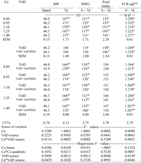
evaluated. Four-hundred males Ross-708 day-old chicks weredistributed among 80 cages in four Petersime batteries. Interaction effects (P < 0.05) on BW, BW gain, feed intake, and FCR were observed at 14d. BW and BW gain were reduced (P < 0.05) in chicks fed 1α(OH)D3-supplemented
diets containing 1.4% dietary Ca. Moreover, 1α(OH)D3 supplementation improved FCR up to 7
points, when dietary Ca content was 0.8% as compared to chicks fed non-supplemented diets. A quadratic effect (P < 0.05) due to dietary Ca level in the diet was observed on FCR at 14d. An interaction effect (P < 0.05) was detected on flock uniformity at 14d. Chicks fed dietary Ca levels of 0.95% and 1.10% and supplemented with 1α(OH)D3 had the best flock uniformity (5.88% and
6.56%, respectively). Dietary supplementation of 1α(OH)D3 reduced the RA of tibia length by
0.38 points (P < 0.05). Chickens fed non-supplemented diets with 1.25 % dietary Ca had greater tibia ash than chicks fed non-supplemented diets containing 0.80 and 1.40% dietary Ca (36.15 vs. 32.65 and 32.72 %, respectively). Interaction effects (P < 0.05) were detected on Ca, P, Mn, and Zn digestibilities. Ca and P digestibility was improved by 1α(OH)D3 supplementation in diets
22 decreased BW and BWG. However, 1α(OH)D3 supplementation at low dietary Ca content (0.8%
Ca) ameliorated the negative effect of on FCR up to 7 point at 14d. Also, 1α(OH)D3
23 INTRODUCTION
Ca and Vitamin D (VitD) are two important nutrients involved in overall body functionality and physiology (Delezie et al., 2012). These two nutrients work together; VitD regulates intestinal Ca absorption, and dietary Ca levels modulates the required hydroxylation to form the active VitD3
metabolite (Wasserman and Fullmer, 1995). Therefore, VitD is required by broilers for a proper Ca and P metabolism (Henry, 2011). It is known that bone mineralization and mineral digestibility could be also affected by dietary Ca level, VitD metabolites, and its bioavailability (Roberson et al., 1994; Mutucumarana et al., 2014a; Paiva et al., 2013, 2014).
The dietary levels of VitD3, Ca, and P used in commercial poultry diets is not consistant with the NRC (1994) recommendations. The NRC (1994) recommends 200 IU/kg of VitD3,
although different authors reported higher levels of inclusion (1,000-2,000 IU/kg) are necessary for adequate bone development. Different research groups (Fritts and Waldroup., 2003; Whitehead
24
Regardless of the dietary requirements of today’s broilers for Ca, variability in dietary Ca concentration is a very common problem in feed ingredients and commercial least-cost feed formulation (Li et al., 2017). There are many situations presented in the industry can affect the final dietary Ca concentration in the feed that chickens received. Failures in ingredient handling, mixing, and feed manufacturing often results in suboptimal dietary Ca levels, or insufficient or inconsistant doseing or application of phytase (Van Kempen et al., 2001; Adedokun and Adeola, 2013; Li et al., 2017), which all increases risk of effect Ca formulation errors.
Ca particle size and solubility, are characteristics that can also affect broiler responses. Better BWG, FI and FCR, as well an improve tibia ossification have been observed with the utilization of Ca carbonate particles over 150 microns (Guinotte et al., 1991). Also, coarse particle size have been demonstrated to obtain a better nutrient digestibility. In an study performed by Anwar et al., (2016), a higher digestibility, and lower Ca in vitro solubility (0.33), was obtained with the utilization of coarse limestone particle size (1-2 mm). Lower solubility of Ca particle seems increase Ca absorption due to a slow mineral release, while the opposite occurs with the high solubility Ca with a faster Ca absorption (Zhang and Coon, 1997). There are reports indicating that the utilization of high Ca levels in broiler diets leads to bad results of P duodenal digestibility (Mutucumarana et al., 2014a).
An effective dietary VitD source could help to minimize any possible marginal deficiency
25 increases the ability of phytate phosphorus utilization in chickens (Mohammed et al., 1991; Edwards, 1993).
The 1α(OH)D3 is a synthetic VitD analogue that requires one hydroxylation at C25 to form
biologically active VitD3, and it has been demonstrated to have positive effects on the growth
performance and bone development of broiler chickens (Edwards Jr. et al., 2002; Snow et al., 2004). Its effect on preventing tibia dyschondroplasia and rickets were demonstrated as well (Rennie and Whitehead, 1996; Edwards Jr. et al,. 2002; Driver et al., 2005). Dietary supplementation of 1α(OH)D3 help to improve P utilization in broilers (Han et al., 2009) as well
as other minerals like Mg and Zn (Biehl et al., 1995). Bone mineralization and ash percentage is also positively affected by 1α(OH)D3 (Edwards Jr, 2002).
It was hypothesized that 1α(OH)D3 could be affected by Ca levels and it can affect chicken
26 MATERIALS AND METHODS
Treatments and Chicken Husbandry
One experiment was conducted using a randomized complete block design of 2 x 5 factorial arrangement of two dietary levels of 1α(OH)D3 (0 and 5 µg/kg feed (1,600 IU/kg VitD3/kg feed))
and 5 levels of Ca (0.80, 0.95, 1.10, 1.25, and 1.40 %) to obtain 10 treatment combinations. Chicks were placed in 80 Petersime battery cages with 8 replicate cages per treatment, and all were located in a room with controlled temperature. A total of 400 Ross 708 male-chicks were placed with 5 male chicks per cage.Room temperature was recorded and adjusted daily to guarantee thermoneutral temperatures. Feed and water were offered ad libitum to the chicks during the whole experimental period. Galvanized metal linear feeder and drinker were used; one of each were placed in a side of each cage.
Diets
Chicks received corn-soybean meal crumble diets formulated to meet Ross 708 requirements (Aviagen, 2014). The basal diet (Table 2.1) was supplemented with VitD3 as
cholecalciferol (4,000 IU/kg). A commercial phytase (Natuphos®) was used in all diets up to 500
FTU/kg, expecting to release 0.12% Ca and 0.12% P. The levels of Ca and P were obtained adding proportions of limestone, dicalcium phosphate and sand. The limestone particle sized used as Ca source had a granulometry of 1129µ and a solubility of 81.72% in 60 minutes of dissolution in EDTA.
Live Performance
27 Bone development and mineralization
At 16 days of age, three chicks per pen were selected, euthanized and leg bones removed. After defleshing, the weights and lengths of femurs, tibia and tarsus-metatarsus (shanks) from both legs were recorded to calculate bone Relative Asymmetry (RA). The RA was calculated (|L-R|/[(L+R)/2]x100) as described by Møller et al. (1995, 1999). Subsequently, femur bones were placed in porcelain crucible and incinerated in a muffle furnace at 600℃ for 8 hours. The resulting bone ashes were weighted, and ash percentage was calculated as a measure of bone mineralization. Mineral digestibility and retention
Between 11 and 13 d of age, excreta samples were collected from all cages. Ileal content from three birds per cage was collected at 16 days of age. Jejunum content was washed with deionized water and collected in Falcon tubes. Ileal samples were freeze-dried and further analyzed by inductively coupled plasma optical emission spectroscopy (Optima 8000, Perkin Elmer Corporation, Waltham, MA) and acid insoluble ash methodology, with Celite® as marker, for
obtaining Ca and P digestibility. Ileal digestibility was calculated for dry matter, Ca and P. From excreta samples mineral retention of Ca, P, Mn, and Zn were assessed on dry matter basis.
All the procedures involving the birds used in the present experiment were approved by the North Carolina State University Institutional Animal Care and Use Committee.
Statistical Analysis
28 and the inclusion levels of 1α(OH)D3. Each of the four battery units were considered as a block
(random effect). Mean separation was done using Tukey’s or Student’s t tests. All percentage data were transformed using the best Box-Cox transformation prior to analyses. All data were evaluated using JMP 13 software (SAS Inst. Inc., Cary, NC)
RESULTS
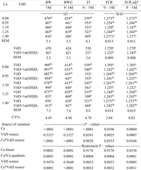
Performance
Significant interaction effects (P < 0.05) were detected on BW, BW gain, feed intake, and FCR at 14d. Feed intake, BW and BW gain were reduced (P < 0.05) in chicks fed 1α(OH)D3
-supplemented diets when dietary Ca content was 1.4%. (Table 2.2). Moreover, dietary supplementation 1α(OH)D3 improved FCR up to 7 points, when dietary Ca content was 0.80% as
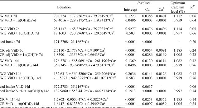
compared to chicks fed non-supplemented diets (Table 2.3). In addition, a quadratic effect (P < 0.05) of dietary Ca levels was observed on FCR at 14d (Table 2.4), with the best FCR achieved at 1.01% and 1.19% of Ca for 1α(OH)D3 supplemented and non-supplemented diets, respectively. A
significant interaction effect (P < 0.05) was detected on flock uniformity (CV%) at 14d. Groups of chicks fed dietary Ca levels of 0.95% and 1.10% and supplemented with 1α(OH)D3 had the
lowest coeficient of variation (5.88% and 6.56%, respectively). Bone development and mineralization
A treatment interaction effect (P < 0.05) was observed for tibia ash at 16 d (Table 2.5). A positive quadratic effect of Ca was observed for chicks fed diets without 1α(OH)D3. The regression
29 32.72 %, respectively). The supplementation with 1α(OH)D3 resulted in intermediate responses,
therefore supporting bone mineralization regardless of the dietary content of Ca.
No interaction effects were detected for bone RA. However, an effect of Ca was detected for tibia and shank weight RA (P < 0.05) at 16 d (Table 2.5). Regression analysis indicated a negative quadratic effect of Ca on tibia weight RA with a calculated optimum RA at 1.06% of Ca (Table 2.7). Furthermore, the lowest shank RA was detected at 1.10% Ca inclusion.
Mineral digestibility and retention
Results obtained in ileal digestibility analysis suggested that the supplementation with
1α(OH)D3 increased Ca (51.22 vs. 38.75%) and P (58.63 vs. 46.11%) ileal digestibility when
dietary Ca level was 0.95% when compared to diets without 1α(OH)D3 inclusion (Table 2.6).
Moreover, a linear effect was detected on Ca ileal digestibility due to dietary Ca level, only in chicks fed 1α(OH)D3 supplemented diets (Figure 2.2).
For mineral retention analysis, interaction effects (P<0.05) were observed on the apparent digestibility coefficient of dry matter (CDADM), Ca, P, Mn, and Zn at 14 d (Table 2.8). The supplementation of 1α(OH)D3 in diets containing 0.95% dietary Ca increased Ca and P retention,
whereas diets containing 0.80% dietary Ca showed greater (P < 0.001) Mn retention (89.83 vs 91.62 %). In addition, a positive quadratic effect (P < 0.05) was detected for chicks fed diets regardless of 1α(OH)D3 for CDADM with optimum estimated at 1.19 and 1.29% of Ca for
1α(OH)D3 supplemented and no supplemented diets, respectively (Table 2.10). Positive quadratic
effects were also observed in diets without 1α(OH)D3 on Mn and Zn retention (Figure 2.6 ; 2.7).
30 A comparison of ileal digestibility and retention percentage was calculated for each one of the treatments. This comparison was defined as the difference of ileal digestibility vs retention (ileal digestibility % - retention %). The differences obtained in each treatment were statistically analyzed. (Mutucumarana et al., 2014b; Perryman et al., 2016).
An interaction effect (P < 0.05) was detected for Ca and P difference between Ca ileal digestibility and retention (Table 2.9). The difference between Ca ileal digestibility and retention increased significantly with the addition of 1α(OH)D3 at 1.40% Ca Figure 2.10). Furthermore, an
effect of Ca was detected (P < 0.01) for Ca and P. It was observed that a negative difference between the digestion and retention tends to become positive as the dietary Ca level increase. Ca and P digestibility vs. retention regressions had a quadratic effect of Ca levels. No effect (P > 0.05) of 1α(OH)D3 in the difference between digestibility and retention was observed (Table 2.9).
DISCUSSION
Previous studies indicated that the utilization of 1α(OH)D3 in poultry diets, improved chick
BW gain and FCR during the first three weeks of chick age (Biehl and Baker 1997; Snow et al., 2004; Driver et al., 2005). Furthermore, Han et al. (2012) proposed that the addition of 1α(OH)D3
can correct Ca deficiencies in diets, increasing BW gain in birds during the period of 21 to 42 d of age. Same effect was detected in the present study for the first two weeks of chicken life, where FCR and BWG improved at low Ca levels with the supplementation of 1α(OH)D3.
The main objective of the present study was to determine the effect of 1α(OH)D3 under
31 Regression analysis showed a quadratic effect of Ca levels on the performance of chickens and growth. It is proposed that this quadratic effect could be due to effects on feed intake caused by the hormonal regulation of Ca absorption and it homeostasis at the bone level (Proszkowiec-Weglarz and Angel, 2013).
At low Ca inclusion, the organism would not have a source of Ca to supply for bone mineralization and development. On the other side, in an excess of Ca level, the body cannot regulate Ca as it normally would, generating an inefficient bone development in chicks at this early age. Also, as proposed by Shafey (1993), an excess of Ca can reduce soluble fraction of other minerals used for growth. In this way their availability for its absorption is reduced, generating growth depression in chickens.
Rath et al. (2000) mentioned that in addition to the importance of the presence of collagen crosslinks in the bone for mechanical strenght, Ca homeostasis is an essential driving force in the maintenance of bone strength. This statement can be reinforced by the present bone mineralization results that indicated that bone ash content increased by dietary Ca levels fitting to a quadratic effect. This means that at the lowest or highest Ca level, where Ca homeostasis with P is harder to obtain, the mineralization of the bone seems to be more affected. Likewise, the results of the present experiment suggested a positive influence of 1α(OH)D3 on bone mineralization, increasing
the percentage of bone ash. This result agrees with many previous studies (Biehl and Baker, 1997; Edwards et al., 2002; Snow et al., 2004; Han et al., 2009, 2012) that have shown that the inclusion
of 1α(OH)D3 used at different doses (from 5 to 20 μg/kg) increased tibia ash percentage.
32
1α(OH)D3 to regulate the deficit or excess of dietary Ca, guaranteeing in this way bone
mineralization (Edwards Jr, 2002; Han et al., 2012).
The RA is a parameter that indicates the fitness of bilateral development, and in bones it has been studied to evaluate bird responses to their environment. The RA has been suggested as a tool to indicate animal wellbeing (Møller et al., 1999; Van Poucke et al., 2007). RA have been positively related with chicken growth rate too. (Møller, et al., 1995, 1999). There was not found previous studies that analyzed the effect of 1α(OH)D3 in bone RA data, so this research and the
data presented in it becomes the first study relating these data. The 1α(OH)D3 inclusion showed to
have a positive effect in the symmetric development of bone size, being reflected in the decrease of tibia RA length. This confirm that 1α(OH)D3 had a positive influence in bone quality
development. Also, in the present study, dietary Ca levels showed to have an influence on the RA of tibia and shank weight at 16 days. Tibia weight RA fitted to a quadratic regression indicating an optimum level of Ca at 1.06%, for the best tibia symmetry, while shank RA of weight indicated lower relative asymmetry at 1.10 and 1.25% of dietary Ca.
Mineral retention analyses were obtained for Ca, P, Cu, Mn and Zn in excreta. For all the minerals, the dietary Ca level exert a significant effect. For Ca retention, the dietary Ca produced a negative linear effect in diets without 1α(OH)D3, decreasing the retention as the dietary Ca
concentration increased. With the inclusion of 1α(OH)D3, this effect became quadratic. The
estimated optimum level for Ca retention with 1α(OH)D3 was 0.84% Ca. Suggesting that at this
point, levels of Ca absorbed and retained in the organism are increased by 1α(OH)D3
supplementation. Similar results were obtained with P retention. The inclusion of 1α(OH)D3 generated higher P retention than in groups without 1α(OH)D3. This increment of retention of Ca
33 cholecalciferol produced and increment on Ca and P retention compared to a diet with 66 µg/kg of cholecalciferol. The interaction effect indicated quadratic effects with and without 1α(OH)D3
supplementation and the optimum levels of Ca were 1.11% and 1.16%, respectively.
Ca and P digestibility showed similar patterns, increasing mineral ileal digestibility with
1α(OH)D3 inclusion, and improving the digestibility in the groups with 0.95%. It can be said that 1α(OH)D3 promotes a favorable effect increasing the Ca and P intestinal absorption and utilization.
Bielh et al, (1995), detected a trend (P < 0.09) of increasing plasma cholecalciferol with the utilization of 1α(OH)D3 regardless the utilization or not of 120 FTU phytase.
Ca levels had a quadratic effect on Mn and Zn retention when diets had no 1α(OH)D3. For
Mn, the addition of 1α(OH)D3 caused a negative linear effect, that decreased the retention as the
Ca levels increased. This effect allows to increase Mn retention when diets are deficient of Ca. (Figure 2.5)
34 One interesting detail detected in these analyses is that the differences calculated goes from negative to positive values in both groups. This means that at lower Ca levels the mineral digested is lower than the retained. This suggested that in a situation of mineral deficiency, chickens started to reabsorb and retained higher levels of minerals to equilibrate the homeostasis generated by this low Ca levels. Focusing on how the chick could retain additional Ca after ileum stage, it is suggested that in chicks there also exist a kind of reabsorption happening at cecum and kidney levels. Svihus et al., (2013) mentioned that the nutritional significance of the caeca in chickens remains unclear. However, previous studies suggested based on the proximal ceca surface structure, with similarities to the jejunum, that nutrient absorption may take place there (Ferrer et al., 1991). Ceca Ca absorption is not well documented in chicks, nevertheless, a research done in rats by Nellans and Goldsmith (1981), concluded that cecum possess the highest density of Ca transport sites and receptors in the intestine. Further research in ceca mineral absorption in chicks would be necessary. On the other side, it also must be considered renal Ca and P metabolism that can be affecting these results. In addition to intestinal absorption and bone resorption, it is known that Ca homeostasis is obtained by renal excretion or reabsorption of Ca (Wideman, 1987). In situations of high demand of Ca, this excretion can be reduced, increasing the reabsorption from renal tubes to try to reach the required Ca levels by the organism (Proszkowiec-Weglarz and Angel 2013).
CONCLUSION
1α(OH)D3 supplementation in addition to the premix cholecalciferol during the starter
phase is effective for better chick performance at low dietary Ca levels. Supplementation with
1α(OH)D3 in starter phase resulted in higher Ca, P, Mn and Zn retention when low levels of Ca
35 Furthermore, 1α(OH)D3 allows better ossification of tibia especially at low Ca levels with better