EFFECT OF SILVER-BASED PRODUCTS ON SIMULATED BODY FLUIDS
PARISA SANATI
A dissertation submitted in partial fulfillment of the requirements for the award of degree of
Master of Science (Biotechnology)
Faculty of Biosciences and Medical Engineering Universiti Teknologi Malaysia
I would like to dedicate this thesis to special people in my life:
I am ever grateful to my beloved husband, Mehdi, for his encouragement, patience and affection.
My genuine appreciation goes to my beloved parents, Ahmad and Parivash who always believe in their daughter and never let me down. Without their f supports even a word of this thesis has not been written
ACKNOWLEDGEMENT
I would like to dedicate this thesis to special people in my life. I am ever grateful to my beloved husband, Mehdi, for his encouragement, patience and affection. My genuine appreciation goes to my beloved parents, Ahmad and parivash who always believe in their daughter and never let me down. Without their f supports even a word of this thesis has not been written. To my brother Mohamad and my sister Azita for their inspiration and love. My sincere thanks goes to my supervisor Dr Nik Ahmad Nizam Bin Nik Malek for his continuous guide lines , motivation, advices, and valuable support during my master period. I appreciate all my lecturers and the staff of Faculty of Biosciences and Bioengineering, University Technology Malaysia.
ABSTRACT
ABSTRAK
TABLE OF CONTENTS
CHAPTER TITLE PAGE
TITLE PAGE i
AUTHOR'S DECLARATION ii
DEDICATION iii
ACKNOWLEDGEMENT iv
ABSTRACT v
ABSTRAK vi
TABLE OF CONTENTS vii
LIST OF TABLES xi
LIST OF FIGURES xiii
LIST OF ABBREVIATIONS AND SYMBOLS xvii
1 INTRODUCTION
1.1 Background of Study 1
1.2 Statement of Problem 6
1.3 Objective of Study 7
1.4 Scope of Study 7
1.5 Significance of Study 8
1.6 General Overview of the Research 8
2 LITERATURE REVIEW
2.2 Escherichia coli as Microbial Model 12 2.3 Silver-Based Products Applications 13 2.3.1 Silver as an Antimicrobial Agent 15 2.3.2 Action of Silver Ions/AgNO3 on
Microorganisms 17
2.4 Zeolite and its Properties 18
2.5 Mineral Clay and its Properties 20 2.5.1 Bentonite and its Properties 22 2.5.2 Kaolinite and Its Properties 24 2.6 Silver-Zeolites as Antibacterial Agents 24 2.7 Silver Clay as Antibacterial Agent 26
2.8 Simulated Body Fluid 27
2.8.1 Simulated Body Fluid (SBF) 28 2.8.2 Simulated Gastric Fluid (SGF) 30
3 RESEARCH METHODOLOGY
3.1 Preparation of Materials 32
3.1.1 Preparation of Zeolites and
Silver-Clay 32
3.2 Characterization of Materials 34
3.2.1 Infrared Spectroscopy (FTIR) 34 3.2.2 Energy Dispersive X-ray (EDX)
Analyzer 35
3.3 Preparation of Simulated Body Fluids 35 3.3.1 Preparation of Simulated Body Fluid 35 3.3.2 Preparation of Simulated Gastric Fluids 38
3.4 Antibacterial Assay 39
3.4.1 Preparation of Antibacterial Pellet 39 3.4.2 Antibacterial Assay by Disk Diffusion
Technique 39
3.4.2.2 Preparation of the Contaminated SBF by Escherchia coli
40
3.4.2.3 Preparation of the Standard
Turbidity for Samples 41
3.4.2.4 Preparation of Sterile Normal
Slain 0.9% 41
3.4.2.5 Preparation of Inoculums 42 3.4.2.6 Inoculation on the NA Plate
42 3.5 Preparation of Different Concentrations of SBP
in SBF and SGF 43
3.5.1 Preparation of Different Concentrations
of SBP in SGF 43
3.5.2 Preparation of Different Concentrations
of SBP in SBF 46
4 RESULTS AND DISCUSSION
4.1 Elemental Studies of Samples by EDX 48 4.2 Characterization of Ag-Modified Zeolites and
Clays by FTIR 54
4.3 Characterization of Samples after Contact with
SGF and SBF 57
4.3.1 FTIR and pH Variations after Contact
with SBF 58
4.3.2 FTIR and pH Variations after Contact
with SGF 63
4.4 The Pepsin Activity Assay 71
5 CONCLUSION
5.1 Conclusion 77
5.2 Suggestion
80
LIST OF TABLES
TABLE NO. TITLE PAGE
2.1 Some literatures about silver action as an antimicrobial
agent. 18
2.2 Silver zeolite as an antibacterial agent 26
3.1 Concentrations of mixture solution, volume of SGF for each sample and amounts of silver-zeolite A in every sample.
44
3.2 The concentrations of mixture solution of SBP added to SGF,volume of SGF for each sample, and amounts of Ag-zeolite Y,Ag-clinoptilolite, Ag-kaolinite, and Ag-bentonite.
44
3.3 The abbrevitaions of treated sugarcane bagasse samples 46 4.1 The percent of chemical elements (%) in zeolites and
Ag-zeolites 52
4.2 The percent of chemical elements (%) in clays and
Ag-clays 53
4.3 The pH variation of SBF after combination with
silver-kaolinite and silver-bentonite 60
4.4 The pH variation of SBF after combination with silver-clinoptilolite and silver-zeolite A and silver- zeoliteY in different concentrations
62
4.5 The pH changes of SGF after mixing with Ag-bentonite
samples in varied concentration 64
samples in different concentrations
4.7 The contrast between changes in pH of SGF after mixing with different concentrations of synthesized zeolite A and Y
69
4.8 The pH changes of SGF after adding to Ag-clinoptilolite in
different concentrations 70
4.9 The inhibition zone on direct bacterial sample of E. coli
pure culture 72
4.10 The inhibition zone on bacterial cultured from
LIST OF FIGURES
FIGURE NO. TITLE PAGE
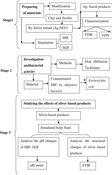
1.1 Research methodology design 10
2.1 Image of E. coli from scanning electron microscope
(SEM) 13
2.2 The structure of synthetic zeolite 20
2.3 Crystalline structure of bentonite 23
2.4 Mechanism of silver (Ag) ions adsorbed on zeolite 25 3.1 diagram for the preparation of zeolites and
silver-clays 33
3.2 FTIR model Nicolet iS5 applied in study 34
3.3 The diagram for SBF preparation
37 3.4 The diagram of SGF preparation
38 3.5 Steps for the preparation 0.5% concentration of
silver-zeolite A combined with SGF.
45 3.6 Steps for preparation 0.5% concentration of Ag-zeolite A
combined with SBF 47
4.1 EDX spectrum of zeolite Y 49
4.3 EDX spectrum of zeolite A 49
4.4 EDX spectrum of Ag-zeolite A 50
4.5 EDX spectrum of clinoptilolite 50
4.6 EDX spectrum of Ag-clinoptilolite 50
4.7 EDX spectrum of kaolinite 51
4.8 EDX spectrum of Ag-kaolinite 51
4.9 EDX spectrum of bentonite 51
4.10 EDX spectrum of Ag-bentonite 52
4.11 FTIR spectra of zeolite A (zA) and Ag-zeolite A (zA.Ag) 55 4.12 FTIR spectra of zeolite Y (zY) and Ag-zeolite Y(zY.Ag) 55 4.13 FTIR spectra of clinoptilolite (clin) and Ag-clinoptilolite
(Ag.clin)
56 4.14 FTIR spectra of kaolinite (Kao) and Ag-kaolinite (Ag.Ka) 56 4.15 FTIR spectra of bentonite (Ben) and Ag- bentonite
(Ag.Ben)
57 4.16 The FTIR spectrums of silver-kolinite which was mixed
with SBF in different concentrations from 0.5% to 10%, and spectrum of silver-kaolinite which was not added to SBF (0%)
58
4.17 The FTIR spectra of silver-bentonite which was mixed with SBF in different concentrations from 0.5% to 10%, and spectrum of silver bentonite which was not added to SBF (0%)
59
4.18 The FTIR spectrum of Ag-clinoptilolite after combination with SBF, in different concentrations from 0.5% to 10% and spectrum of Ag-clinoptilolite which was not added to SBF (0%)
61
4.19 The FTIR spectrum of Ag-zeolite A after combination with SBF in different concentrations from 0.5% to 10%,
and spectrum of Ag-zeolite A which was not added to SBF (0 %)
4.20 The FTIR spectrum of Ag-zeolite Y after combination with SBF in different concentrations from 0.5% to 10% and spectrum of Ag-zeolite Y which was not added to SBF (0 %)
62
4.21 The FTIR spectra of Ag-bentonite and their spectrum after combination with SGF in different concentration from 0.5% to 10%, and spectrum of silver-bentonite which was not added to SGF (0%)
63
4.22 The FTIR spectra of Ag-kaolinite and their spectrum after combining with SGF in different concentrations ranged from 0.5% to 10%, and spectrum of silver-kaolinite which was not added to SGF (0 %)
65
4.23 The FTIR spectra of Ag-zeolite A and their range after mixing with SGF in different concentrations from 0.5% to 10%, and spectrum of silver-zeolite A which was not added to SGF (0 %)
67
4.24 The FTIR spectra of Ag-zeolite Y and their range after mixing with SGF in different concentrations from 0.5% to 10%, and spectrum of silver-zeolite Y which was not added to SGF (0 %)
68
4.25 The FTIR spectra of Ag-clinoptilolite and their range after mixing with SGF in different concentrations from 0.5% to 10%, and spectrum of silver-clinoptilolite which was not added to SGF (0 %)
70
4.26 Negative control of non-modified zeolite/clay 74
4.27 Positive control of silver nitrate 74
4.28 The inhibition zones around antibacterial agents of silver kaolinite and silver zeolite A
silver zeolite A and silver zeolite Y
4.30 The inhibition zones around antibacterial agents disks of silver clinoptilolite, silver zeolite Y, silver bentonite, and negative control
LIST OF ABBREVIATIONS AND SYMBOLS
Ag Silver
Al alumina
AgNo3 silver nitrate
°C Degree Centigrade Celsius
CaCl2 calcium chloride
C6H10O5 3.Deoxyglucosone
Ca Calcium
Cd Cadmium
Cl chloride
Cm-1 Centimetre (for energy unit)
CO2 Carbon dioxide
C-O-C Carbon- Oxygen- Carbon
CONAB National Food Supply Agency in brazil
Cr(VI) Chromium six
CFU Colony Forming Unit
Cu Copper
E. coli Escherichia coli ECF extracellular fluid
EDX Energy Dispersive X-Ray
FTIR Fourier Transform Infrared Spectroscopy
g Gram
(CH2OH)3CNH2 Hydroxymethyl-aminomethane
H Hour
H2SO4 Sulphuric acid
H3PO4 Phosphoric acid
HCl Hydrogen chloride acid
HNO3 Nitric acid
HCO3 Carbonic Acid
IR Infrared spectroscopy
K2HPO4.3H2O dipotassium phosphate anhydrate
KCl potassium chloride
KOH Potassium hydroxide
MIC Minimum Inhibition Concentration MgCl2.6H2O chloride hexahydrate
Mg Magnesium
ml Milliliter
mm Millimetre
Mmol/gr Millimole/ gram
Mol\L Moll/litre
NaCl sodium chloride
NaHCO3 sodium bicarbonate
NaOH Sodium hydroxide
Na+ Sodium Possitive Ion
OH group Hydroxyl group
Pb Lead
rpm Rotations per minute
Si silicon
SBF Simulated Body Fluid
SGF Simulated Gastric fluid SBPs Silver Based Products
SO4 Sulfate
Si-O-Al Silicon-Oxygen-Aluminium Si-O-Si Silicon-Oxygen-Silicon
w/w weight/weight
CHAPTER 1
INTRODUCTION
1.1 Background Information
Silver (signified chemically as Ag) that is commonly known as a heavy metal is widely used in many products in our daily life. It is generally a solid material with various chemical and physical properties. Different unique features of silver make it proper to be applied in various industrial and medical cases. In optical and electrical industries, the silver positive ions are used. Silica core-silver shell nanoparticles (silver nanoshells) produces a silver outer layer was used in UV−vis spectroscopy and transmission electron microscopy (Jackson and Halas, 2001). Additionally, in the medical field, silver could be employed as an antimicrobial agents. They can react with the critical bacterial enzymes in order to neutralize their function, disrupt the electron transport system, and attach to their DNA (Baker et al., 2003). Silver ions also can modify the construction of the cell envelope of the bacteria. Among metal ions, silver ion has the maximum antimicrobial action, and a range of silver complexes have been used as agents for treatment (Kawahara et al., 2000). In the current study, silver nitrate (AgNo3) has been used in the preparation of silver based
products (SBPs) because of its higher solubility in water and other aqueous solutions in comparison with other silver compounds.
Silver can be harmful for human health. Prolonged exposure to that metal can cause some illnesses like argyria (reversible pigmentation of the skin) and/or argyrosis (reversible pigmentation of the eyes). The affected area becomes bluish-gray or ash bluish-gray, and it is most prominent in areas of the body exposed to sunlight (Drake and Hazelwood, 2005). Also, silver particles have been shown to have some of cytotoxicity effects in some human cells, and they possess a high affinity for the thiol groups in the liver and reduce glutathione, and consequently, it leads to depleting the amount of glutathione available for biochemical pathways. Reduced glutathione plays an important role in maintaining proper structure and function of red blood cells, as well as eliminating organic peroxides (Baldi and Nucci, 1988).
engineering fields. Today, many well-known SBPs are globally produced, including silver based colours, silver nanoparticles, silver sulfadiazine, silver nitrate, silver chloride, silver zeolites, and silver clays. SBPs are potentially applicable in producing the antibacterial agents. Silver-loaded zeolite X shows antimicrobial action, thus, it can be used as antibacterial agent against Staphylococcus aureus and Pseudomonas aeruginosa (Kwakye‐Awuah et al., 2008). The silver-based nanoclay
demonstrated strong antibacterial activity against Salmonella spp.(Busolo et al., 2010). The antimicrobial properties of silver-modified montmorillonites are also examined in growth inhibition of Escherichia coli ( E. coli) and the results proved its positive effects (Magana et al., 2008).
There are much more to be said about the practical applications of SBPs in engineering and industries. Applications such as the in vitro efficacy of silver sulfadiazine and chlorhexidine against bacteria usually found in burn wounds and some kinds of silver-based dressings that are commercially available can be mentioned as follow. Aquacel-Ag , Contreet Foam which are used for wounds infection treatment (Burd et al., 2007), ultrasensitive silver-based color which is applied for staining of polypeptides in polyacrylamide gels (Sammons et al., 1981), using silverized activated carbon filters beside the ion-exchange resins (Gupta, 1998) for water purification units and distribution systems of hospitals and hotels to manage contagious agents (Li et al., 2005), using polylactic acid (PLA) biocomposites which obtained by solvent casting, with a new silver-based antibacterial layered silicate additive which is suitable for food packaging requests and etc.
molecular sieving with high ion-exchanged capacity (Takaishi et al., 1975). Zeolites do not have antibacterial activity but it can be induced with adding the silver ions and surfactant molecules (Rivera et al, 2000). Zeolites that are structurely modified with silver are capable to function as an antibacterial agent. Those matrixes could be appropriately used as carriers because of their high ion exchange capacity, high surface area, negative surface charge, chemical inertness, and low toxicity. Zeolites can play a significant role as a supportive material for silver ions to create an alternative substance to overcome the antibiotic resistance problem through improving some antibacterial agents’ abilities.
Another supporting material is natural clay minerals that are widely used because of their low expense and abundance. Clay materials are important as adsorbents because of their unique framework which have eminent sorption specifications and high capacity for ion exchange. Clay minerals have a layered structure of silica (Si), alumina (Al), and water. They can be divided in several groups such as montmorillonite group , kaolinite group, chlorite group, and illite group, as well as bentonite clay which is a marketable material made up of montmorillonite and smectite clay (Nayak and Singh, 2007). Clays like zeolites provide a physically stable surface for silver nanoparticles and silver ions to be a supporting material for them(Etris and Cappel, 2003). Silver ion is released from zeolites and clays in the infectious area (Matsumura et al, 2003), therefore, silver-modified mineral clay can be used as an antibacterial agent. Despite widespread use of silver-based products, there are very few studies on the effects of them on human organs and fluids such as body, gastric, and intestine fluids.
current study consists of different ions like K+, HCO3, Na+, Cl-, Mg+2, Ca+2, SO4, and
HPO4 (Marques et al., 2011). Moreover, the simulated body fluid could be applied
in evaluating the impact of drug on the human body fluid and providing an environment the same as the human body in order to estimate mutual influences of these fluids with drugs or materials.
Some studies have reported the usefulness of the SBF in testing bioactivity. (Kokubo and Takadama, 2006). Another body examined in this study in terms of the SBPs is the simulated gastric fluid (SGF) with a pH of below 2, during fasting state. It is noticeable that its pH can be ranged from 1 up to 7. 5 after food intaking. Based on the meal contents, the fed-state gastric pH will rise to values ranged between 4 and 7 (Marques et al., 2011).
Similar to SBF, the simulated gastric fluid was initially developed for estimating the effect of drug on the human gastric fluid and providing a situation similar to the real human stomach. It should be performed in order to evaluate the reciprocal impacts of SGF with the structures of the drugs or materials.
1.2 Statement of Problem
Antibiotics and antibacterial agents have been produced and applied for treatment of infectious diseases, and also they have been used in industry sector. In medical cases, these compounds have several unwanted impacts on human body that could be either harmless or dangerous.
In spite of the global spreading of SBPs as an antibacterial agent in medical, pharmacy, and industry, little researches were conducted on their effects on human body fluids. Each kind of chemical compound (e. g., drug or food) could have certain interactions with both target and non-target fluids and organs in human body. Consequently, some changes occur in SBPs framework which cause some chemical changes in human body fluids and also in their own structures.
Lots of researches about the effects of antibacterial agents on human organs as medicines have been carried out, for example the effect of silver sulfadiazine on the burned skins. It is essential for human health to find out the interactions between SBP and human body fluids or gastric fluids, and the effects on these fluids resulted from the mentioned interaction are significant.
1.3 Objectives of Study
The objectives of this study are listed below:
1. To prepare and characterize silver-based products.
2. To investigate the antibacterial activity of Silver-based products
3. To study the effect of silver based products on simulated body fluid and simulated gastric fluid
1.4 Scope of Study
1.5 Significance of Study
SBPs have been used as antibacterial agents for treating some kinds of infectious diseases. Body fluid and gastric fluid are important fluids in body that can be considered as target for SBP.
Investigation of antibacterial activity besides the harmless or dangerous effects of SBP on body fluids is crucial for human health condition. On the other hand, structural changes which were happened on SBP after being added to SBF and SGF are important from pharmaceutical and medical aspect.
SBP were added to body fluids in different concentrations. Among them, those of them that show least pH changes in the body fluids and more stability in SBP structure could be used in treating infectious body fluids with SBP in a way to have the least unwanted effect. Thus, these results can help the pharmaceutical and medical industry to produce proper medicine to treat the infectious diseases.
1.6 General Overview of the Research
Figure 1.1 Research methodology design. Simulation SBF SGF Preparing of materials
Modification Ag- based products
By Silver nitrate (Ag-NO3) Clay and Zeolite
Characterization EDX FTIR Investigation antibacterial activity Methods Material
Disk Diffusion Technique
Escherichia coli
Contaminated SBF by infective bacteria
Studying the effects of silver based products
Simulated body fluid
Analysis the pH changes of SBF, SGF
Analysis the structural changes of silver based products
Silver based products
pH meter FTIR
Stage 3 Stage 2
REFERENCES
Andre, M., Vernby, A., Odenholt, I., Stalsby Lundborg, C., Axelsson, I., & Eriksson, M. (2008). Diagnosis-prescribing surveys in 2000, 2002 and 2005 in Swedish general practice: Consultations, diagnosis, diagnostics and treatment choices. Scandinavian Journal of Infectious Diseases, 40(8), 648-654.
Azam, A., Ahmed, A. S., Oves, M., Khan, M., & Memic, A. (2012). Size-dependent antimicrobial properties of CuO nanoparticles against Gram-positive and-negative bacterial strains. International Journal of Nanomedicine, 7, 3527-3535.
Baker, G., Smith, J., & Cowan, D. A. (2003). Review and re-analysis of domain-specific 16S primers. Journal of Microbiological Methods, 55(3), 541-555.
Baldi ,C., & Nucci, A. (1988). Effects of silver in isolated rat hepatocytes. Toxicology Letters, 41, 261-268.
Blanco, M., Blanco, J., Gonzalez, E., Mora, A., & Prado, C. (1996). Prevalence and characteristics of Escherichia coli serotype O 157: H 7 and other verotoxin-producing E. coli in healthy cattle. Epidemiology and Infection, 117(2), 251-258.
Bond, H., & Philip Ho, H. (1970). Solid maniature silver-silver chloride electrodes for chronic implantation. Electroencephalography and Clinical Neurophysiology, 28(2), 206-208.
Bozkaya, O., & Yalcın, H. (2005). Diagenesis and very low-grade metamorphism of the Antalya unit: mineralogical evidence of Triassic rifting, Alanya-Gazipaşa, central Taurus belt, Turkey. Journal of Asian Earth Sciences, 25(1), 109-119.
Breidt, J. F., Hayes, J., & McFeeters, R. (2004). Independent effects of acetic acid and pH on survival of Escherichia coli in simulated acidified pickle products. Journal of Food Protection, 67(1), 12-18.
Brett, D. W. (2006). A discussion of silver as an antimicrobial agent: alleviating the confusion. Ostomy/Wound Management, 52(1), 34-38.
Brosius, R., Bazin, P., Thibault-Starzyk, F., & Martens, J. A. (2005). Operando FTIR study of reaction pathways of selective catalytic reduction of NOx with decane in the presence of water on iron-exchanged MFI-type zeolite. Journal of Catalysis, 234(1), 191-198.
Burd, A., Kwok, C. H., Hung, S. C., Chan, H. S., Gu, H., & Lam, W. K. (2007). A comparative study of the cytotoxicity of silver based dressings in monolayer cell, tissue explant, and animal models. Wound Repair and Regeneration, 15(1), 94-104.
Busolo, M. A., Fernandez, P., Ocio, M. J., & Lagaron, J. M. (2010). Novel silver-based nanoclay as an antimicrobial in polylactic acid food packaging coatings. Food Additives and Contaminants, 27(11), 1617-1626.
isolates from a multinational trial of complicated skin and skin structure infections. Journal of Clinical Microbiology, 46(2), 678-684.
Carretero, M. I., & Pozo, M. (2010). Clay and non-clay minerals in the pharmaceutical and cosmetic industries Part II. Active ingredients. Applied Clay Science, 47(3), 171-181.
Chang, D. E., Smalley, D. J., Tucker, D. L., Leatham, M. P., Norris, W. E. &, Stevenson, S. J. (2004). Carbon nutrition of Escherichia coli in the mouse intestine. Proceedings of the National Academy of Sciences of the United States of America, 101(19), 7427-7432.
Chopra, I. (2007). The increasing use of silver-based products as antimicrobial agents: a useful development or a cause for concern. Journal of Antimicrobial Chemotherapy, 59(4), 587-590.
Cowan, M. M., Abshire, K. Z., Houk, S. L., & Evans, S. M. (2003). Antimicrobial efficacy of a silver-zeolite matrix coating on stainless steel. Journal of Industrial Microbiology & Biotechnology, 30(2), 102-106.
Do Rosario, J. A., De Moura, G. B. , Gusatti, M., & Riella, H. G. (2009). Synthesis of silver-treated bentonite: evaluation of its antibacterial properties. Chemical Engineering, 17, 1795-1801.
Donnenberg, M. S., & Whittam, T. S. (2001). Pathogenesis and evolution of virulence in enteropathogenic and enterohemorrhagic Escherichia Coli. Journal of Clinical Investigation, 107(5), 539-548.
Dunn, K., & Edwards-Jones, V. (2004). The role of Acticoat with nanocrystalline silver in the management of burns. Burns: Journal of the International Society for Burn Injuries, 30, 1-9.
Erb, A., Sturmer, T., Marre, R., & Brenner, H. (2007). Prevalence of antibiotic resistance in Escherichia coli: overview of geographical, temporal, and methodological variations. European Journal of Clinical Microbiology & Infectious Diseases, 26(2), 83-90.
Etris SF, & Cappel, CR. (2003). Silver Compounds. In Kirk-Othmer Encyclopedia Of Chemical Technology (4Th ed.), New York, John Wiley & Sons, Inc.
Farrar Jr, G. E., & Bower, R. J. (1967). Gastric Juice and Secretion: Physiology and Variations in Disease. Annual Review of Physiology, 29(1), 141-168.
Feng, Q., Wu, J., Chen, G., Cui, F., Kim, T., & Kim, J. (2000). A mechanistic study of the antibacterial effect of silver ions on Escherichia coli and Staphylococcus aureus. Journal of Biomedical Materials Research, 52(4), 662-668.
Ferey, G. (1998). The new microporous compounds and their design. Comptes Rendus de l'Académie des Sciences-Series IIC-Chemistry, 1(1), 1-13.
Gupta, A. (1998). Silver as a biocide: will resistance become a problem. Nature Biotechnology, 16, 888-895.
Hall, J. E. (2010). Guyton and Hall Textbook of Medical Physiology: Enhanced E-book: Elsevier Saunders.
He, H., Yang, D., Yuan, P., Shen, W., & Frost, R. L. (2006). A novel organoclay with antibacterial activity prepared from montmorillonite and Chlorhexidini Acetas. Journal of Colloid and Interface Science, 297(1), 235-243.
Huth, T. S., Burke, J. P., Larsen, R. A., Classen, D. C., & Stevens, L. E. (1992). Randomized trial of meatal care with silver sulfadiazine cream for the prevention of catheter-associated bacteriuria. Journal of Infectious Diseases, 165(1), 14-18. Inoue, Y. & H. Hamashima (2012). Effect of Silver-Loaded Zeolite on the Susceptibility
of Escherichia coli against Antibiotics. Journal of Biomaterials and Nanobiotechnology 3(1), 114-117.
Jackson, J., & Halas, N. (2001). Silver nanoshells: variations in morphologies and optical properties. The Journal of Physical Chemistry B, 105(14), 2743-2746.
Jun, S. K., Kim, JS., Kuk, E., Yu, KN., Kim, JH., & Park, SJ. (2007). Antimicrobial effects of silver nanoparticles. Nanomedicine: Nanotechnology, Biology and Medicine, 3, 95–101.
Kaduk, J. A., & Faber, J. (1995). Crystal structure of zeolite Y as a function of ion exchange. The Rigaku Journal, 12(2), 14-34.
Kang, S. W., Wei, W. C., Tsai, S. H., & Yang, S. Y. (2006). Experimental investigation of silver nano-fluid on heat pipe thermal performance. Applied Thermal Engineering, 26(17), 2377-2382.
Kim, J. S., Kuk, E., Yu, K. N., Kim, J. H., Park, S. J., & Lee, H. J. (2007). Antimicrobial effects of silver nanoparticles. Nanomedicine: Nanotechnology, Biology and Medicine, 3(1), 95-101.
Kirov, G., Pechigargov, V., & Landzheva, E. (1979). Experimental crystallization of volcanic glasses in a thermal gradient field. Chemical Geology, 26(1), 17-28.
Klasen, H. (2000). A historical review of the use of silver in the treatment of burns. II. Renewed interest for silver. Burns, 26(2), 131-138.
Kokubo, T., & Takadama, H. (2006). How useful is SBF in predicting in vivo bone bioactivity. Biomaterials, 27(15), 2907-2915.
Kothari, V., & Seshadri, S. (2010). In vitro antibacterial activity in seed extracts of Manilkara zapota, Anona squamosa, and Tamarindus indica. Biological Research, 43(2), 165-168.
Kulprathipanja, S. (2010). Zeolites in Industrial Separation and Catalysis: John Wiley & Sons Inc., Online Library.
Kuo, Y., & Shanbour, L. (1980). Effects of cyclic AMP, ouabain and furosemide on ion transport in isolated canine gastric mucosa. The Journal of Physiology, 309(1), 29-43.
Kwakye-Awuah, B., Williams, C., Kenward, M., & Radecka, I. (2008). Antimicrobial action and efficiency of silver-loaded zeolite X. Journal of Applied Microbiology, 104(5), 1516-1524.
Lechert, H. (2000). Possibilities and limitations of the prediction of the Si/Al ratios of zeolites from the batch composition. Microporous and Mesoporous Materials, 40(1), 181-196.
Li, Y., Moon, K. S., & Wong, C. (2005). Monolayer-protected silver nano-particle-based anisotropic conductive adhesives: Enhancement of electrical and thermal properties. Journal of Electronic Materials, 34(12), 1573-1578.
Magana, S., Quintana, P., Aguilar, D., Toledo, J., Angeles-Chavez, C., & Cortés, M. (2008). Antibacterial activity of montmorillonites modified with silver. Journal of Molecular Catalysis A: Chemical, 281(1), 192-199.
Marques, M. R. C., Loebenberg, R., & Almukainzi, M. (2011). Simulated biological fluids with possible application in dissolution testing. Dissolution Technology, 18(3), 15-28.
Matsumura, Y., Yoshikata, K., Kunisaki, S., & Tsuchido, T. (2003). Mode of bactericidal action of silver zeolite and its comparison with that of silver nitrate. Applied and Environmental Microbiology, 69(7), 4278-4281.
Miranda-Trevino, J. C., & Coles, C. A. (2003). Kaolinite properties, structure and influence of metal retention on pH. Applied Clay Science, 23(1), 133-139.
Nandkumar, M. A., Ranjit, M., Kumar, S. S. P., Hari, P., Ramesh, P., & Sreenivasan, K. (2010). Antimicrobial Silver oxide incorporated urinary catheters for Infection resistance. Trends in Biomaterials and Artificial Organs, 24(3), 85-91.
O'Connell, D., & Tursky, B. (1960). Silver-silver chloride sponge electrodes for skin potential recording. The American Journal of Psychology, 73(2), 302-304.
Ojha, K., Pradhan, N. C., & Samanta, A. N. (2004). Zeolite from fly ash: synthesis and characterization. Bulletin of Materials Science, 27(6), 555-564.
Park, H. J., Kim, J. Y., Kim, J., Lee, J. H., Hahn, J. S., & Gu, M. B. (2009). Silver-ion-mediated reactive oxygen species generation affecting bactericidal activity. Water Research, 43(4), 1027-1032.
Park, Y., Ayoko, G. A., & Frost, R. L. (2011). Application of organoclays for the adsorption of recalcitrant organic molecules from aqueous media. Journal of Colloid and Interface Science, 354(1), 292-305.
Piper, D., & Fenton, B. H. (1965). pH stability and activity curves of pepsin with special reference to their clinical importance. Gut, 6(5), 506-508.
Rai, M., Yadav, A., & Gade, A. (2009). Silver nanoparticles as a new generation of antimicrobials. Biotechnology Advances, 27(1), 76-83.
Rettig, R., & Virtanen, S. (2009). Composition of corrosion layers on a magnesium rare-earth alloy in simulated body fluids. Journal of Biomedical Materials Research Part A, 88(2), 359-369.
Rivera-Garza, M., Olguın, M., Garcıa-Sosa, I., Alcántara, D., & Rodrıguez-Fuentes, G. (2000). Silver supported on natural Mexican zeolite as an antibacterial material. Microporous and Mesoporous Materials, 39(3), 431-444.
Saga, T., & Yamaguchi, K. (2009). History of Antimicrobial Agents and Resistant Bacteria. . Japan Medical Association Journal, 52(2), 103-108.
Saladin, K. S., & Miller, L. (1998). Anatomy & Physiology: McGraw-Hill Companies.
Sammons, D. W., Adams, L. D., & Nishizawa, E. (1981). Ultrasensitive silver-based color staining of polypeptides in polyacrylamide gels. Electrophoresis, 2(3), 135-141.
Santos, M., Oliveira, C., Tachinski, C., Fernandes, M., Pich, C., & Angioletto, E. (2011). Bactericidal properties of bentonite treated with Ag+ and acid. International Journal of Mineral Processing, 100(1), 51-53.
Schmitz, R. M., Schroeder, C., & Charlier, R. (2004). Chemo–mechanical interactions in clay: a correlation between clay mineralogy and Atterberg limits. Applied Clay Science, 26(1), 351-358.
Schulze, K. (2006). Imaging and modelling of digestion in the stomach and the duodenum. Neurogastroenterology & Motility, 18(3), 172-183.
Sherman, J. D. (1999). Synthetic zeolites and other microporous oxide molecular sieves. Proceedings of the National Academy of Sciences, 96(7), 3471-3478.
Silver, S. (2006). Bacterial silver resistance: molecular biology and uses and misuses of silver compounds. FEMS Microbiology Reviews, 27(23), 341-353.
Sposito, G., Skipper, N. T., Sutton, R., Park, S., Soper, A. K., & Greathouse, J. A. (1999). Surface geochemistry of the clay minerals. Proceedings of the National Academy of Sciences, 96(7), 3358-3364.
Stankovic, N., Logar, M., Lukovic, J., Pantić, J., Miljevic, M., & Babic, B. (2011). Characterization of bentonite clay from “Greda” deposit. Processing and Application of Ceramics, 5(2), 97-101.
Tadesse, D. A., Zhao, S., Tong, E., Ayers, S., Singh, A., & Bartholomew, M. J. (2012). Antimicrobial drug resistance in Escherichia coli from humans and food animals, United States, 1950–2002. Emerging Infectious Diseases, 18(5), 741.
Takaishi, T., Yatsurugi, Y., Yusa, A., & Kuratomi, T. (1975). Changes in the sieving action and thermal stability of zeolite a produced by ion-exchange. Journal of the Chemical Society, Faraday Transactions 1: Physical Chemistry in Condensed Phases, ( 71), 97-105.
Thakur, S., Zhao, S., McDermott, P. F., Harbottle, H., Abbott, J., & English, L. (2010). Antimicrobial resistance, virulence, and genotypic profile comparison of Campylobacter jejuni and Campylobacter coli isolated from humans and retail meats. Foodborne Pathogens and Disease, 7(7), 835-844.
Vertzoni, M., Dressman, J., Butler, J., Hempenstall, J., & Reppas, C. (2005). Simulation of fasting gastric conditions and its importance for the in vivo dissolution of lipophilic compounds. European Journal of Pharmaceutics and Biopharmaceutics, 60(3), 413-417.
Wiegand, I., Hilpert, K., & Hancock, R. E. W. (2008). Agar and broth dilution methods to determine the minimal inhibitory concentration (MIC) of antimicrobial substances. Nature Protocols, 3(2), 163-175.
Yonemoto, E. H., Kim, Y. I. Schmehl, R. H. Wallin, J. O. Shoulders, B. A. Richardson, B. R. Haw J. F.& Mallouk T. E. (1994). Photoinduced Electron Transfer Reactions in Zeolite-Based Donor-Acceptor and Donor-Donor-Acceptor Diads and Triads. Journal of the American Chemical Society, 116(23), 10557-10563.
Yoon, KY., Hoon, J., Byeon, J., Park, H., & Hwang, J. (2007). Susceptibility constants of Escherichia coli and Bacillus subtilis to silver and copper nanoparticles. Science of the Total Environment, 37(3), 572–575.