Developmental biology of the leech Helobdella
DAVID A. WEISBLAT*
,1and DIAN-HAN KUO
21Dept. of Molecular and Cell Biology, University of California, Berkeley, USA and 2Dept. of Life Science, National Taiwan University, Taiwan.
ABSTRACT Glossiphoniid leeches of the genus Helobdella provide experimentally tractable mod-els for studies in evolutionary developmental biology (Evo-Devo). Here, after a brief rationale, we will summarize our current understanding of Helobdella development and highlight the near term prospects for future investigations, with respect to the issues of: D quadrant specification; the tran-sition from spiral to bilaterally symmetric cleavage; segmentation, and the connections between segmental and non-segmental tissues; modifications of BMP signaling in dorsoventral patterning and the O-P equivalence group; germ line specification and genome rearrangements. The goal of this contribution is to serve as a summary of, and guide to, published work.
KEY WORDS:
embryonic patterning, Helobdella, leech, Lophotrochozoa, spiralian
Introduction
Two types of questions motivate the field of Evo-Devo. First, what developmental mechanisms governed embryogenesis in now-extinct metazoan species at various nodes of the phylogenetic tree? And second, what changes in development have occurred in the lineages leading from these ancient species to their modern descendants? These questions are based on the underlying truism that evolutionary changes in adult body plan arise from changes in the developmental processes by which the body plan arises. Interest in these questions is heightened by the discovery that animals ranging from cnidarians to vertebrates are remarkably similar in both the numbers and kinds of genes in their genomes. Thus, it appears that the dramatic differences in animal body plans arise from differences in how largely conserved sets of genes are regulated and deployed during development.
Lacking a time machine to recover ancestral species for study, the accepted strategy for investigating these questions is to compare the development of extant species, interpreting similarities and differences in light of the phylogenetic tree by which they arose. Similarities are candidates for features present in the ancestor of the species under comparison, while differences reflect changes that have occurred in one or more of the lineages leading from the ancestor to the modern descendants.
This strategy brings up another challenge, that of reconstructing the phylogenetic tree required for interpreting the data emerging from developmental comparisons among species. The problem is particularly acute for the large numbers of soft-bodied species and the expanses of evolutionary history for which no good fossil
www.intjdevbiol.com
*Address correspondence to: David A. Weisblat . Dept. of Molecular & Cell Biology, 385 LSA, University of California, Berkeley, CA 94720-3200, USA. Tel: +1-510-642-8309 . Fax: +1-510-643-6791. E-mail: weisblat@berkeley.edu - web: http://mcb.berkeley.edu/labs/weisblat
Accepted: 18 September 2014.
ISSN: Online 1696-3547, Print 0214-6282 © 2014 UBC Press
Printed in Spain
Abbreviations used in this paper: BMP, bone morphogenetic protein; DV, dorsoventral; G1, gap phase-1 of cell cycle; G2, gap phase-2 of cell cycle; MAPK, mitogen-activated protein kinase; SYC, syncytial yolk cell; TGFb, transforming growth factor beta. record is available. Traditional phylogenetic methods of grouping animals based on similarities and differences in morphology or embryology build on assumptions about the nature of the evolu-tionary changes we seek to elucidate, which introduces an inher-ently circular logic to the Evo-Devo undertaking. This problem is avoided by using molecular sequence comparisons to construct phylogenies without references to morphological or developmental features. Such analyses have led to the recognition of three super-phyla of bilaterally symmetric animals, Deuterostomia, Ecdysozoa and Lophotrochozoa, which emerged before the Cambrian era. Of these, Lophotrochozoa is at once the largest in terms of recog-nized phyla and also the least well represented in terms of current experimental systems. While the precise phylogenetic structure and characteristics of this group remain in flux, Lophotrochozoa appears to have spiral cleavage as an ancestral feature (Giribet, 2008; Struck, 2014). Thus, in comparing the mechanisms by which, for example, bilateral mesoderm arises from the 4d micromere in annelids, flatworms, mollusks and their allies, we are peering back through the mists arising from roughly 600 million years of evolutionary tinkering at exactly homologous cells and cell lineages.
Fig. 1. Photographs of Helobdella austinensis adult (Kutschera et al., 2013). The trunk of Helobdella is an-nulated, however annulation is not prominent. Anterior and posterior suckers are located on the ventral side at the anterior and posterior ends. A pair of pigmented eyes can be found in the non-segmental prostomial region of the anterior end. Internal organs, such as the digestive tract, are visible through the body wall. Darker food material outlines the midgut (crop, intestine and ceca). Embryos are encased in transparent cocoons attached to the posterior ventral body wall of a brood-ing leech. Scale bar: 2 mm.
100
68
150
50
N
40
0
M
12
Q
O/P
48
Mesoderm Ectoderm
and Savage, 1997): the rostral end of the animal comprises four segments, designated R1-R4, whose ganglia are fused to make the head brain; the midbody region comprises 21discrete seg-ments designated M1-M21, each with its own segmental ganglion; the caudal region comprises 7 segments, designated C1-C7, whose ganglia are fused to make the tail brain.
Embryogenesis in clitellate annelids (leeches
Fig. 2. Segmental mesoderm and ectoderm in Helobdella arise by determinate cell lineages from a posterior growth zone composed of five bilateral pairs of lineage-restricted stem cells (M, N, O/P and Q teloblasts). For clarity, only the mesoderm is shown on the left and only the ectoderm is shown on the right; gastrulation move-ments are omitted. The time line (left) shows the approximate clonal age (hours) of the blast cell clones. Individual blast cell clones (for the M and O/P teloblasts) or pairs of clones (for the N and Q teloblasts) are highlighted by colors at three time points: 1) as undivided cells; 2) at their first mitosis (12 hrs for m blast cells, 40-48 hrs for the ec-todermal blast cells; 3) as sets of definitive progeny (>150 hrs). Note that for the M, O and P lineages the clone of a single blast cell makes one segment’s worth of definitive progeny, although the individual clones are distributed across more than one segment in each case; thus, a typical m blast cell contributes muscles to one segment, a nephridium to the next posterior segment, and a few neurons to the segment after that. For both the N (ventral) and Q (dorsal) ectodermal lineages, alternating blast cells undertake distinct patterns of division and differentiation and one clone of each type is required to make one segment’s worth of progeny.
oligochaetes and leeches together form a clade designated as Clitellata, which arises from within the polychaetes (McHugh, 2000; Siddall, 2001; Struck et al., 2011). Some unsegmented taxa previously regarded as distinct phyla also arise from within the polychaetes (McHugh, 2000). This finding provides further evidence of evolutionary plasticity of the body plan, and of the problems arising from traditional phylogenetic methods.
Overview of Helobdella development
Compared to polychaetes and oligochaetes, leeches are characterized by an absence of segmental bristles (chaetae), and by modification of their anterior and posterior ends to form suckers, correlated with a fixed number of body segments. (Fig. 1). Neurobiologists have established a simplified nomenclature for leech segments based on the gross anatomy of the seg-mental ganglia (there is also an anterior-dorsal ganglion that is not segmental in origin (Weisblat et al., 1984; Shankland
that is highly conserved among leeches, between leeches and oligochaetes, and to a lesser extent in the polychaete outgroups (Dohle, 1999). For glossiphoniid leeches, an embryonic staging system was devised, accompanied by a non-standard nomencla-ture based on knowledge of prospective cell fates in the embryo (Bissen and Weisblat, 1989; Fernández and Stent, 1980; Weisblat and Huang, 2001; Huang et al., 2002; Table 1; Fig. 3). While this nomenclature appears at first to complicate comparisons with spiralian taxa outside Clitellata, it also enables the naming of cells without making controversial assumptions about which cells are homologous across widely separated species, in which cell divisions have been described with varying degrees of precision.
Hermaphroditism and unequal cleavage are ancestral traits for the clitellates. So far as is known, Helobdella is unique for the clade in that several species are capable of self-fertilization as well
as cross-fertilization (Wedeen et al., 1990; Tan et al., 2004; Cho
et al., 2014; DAW unpublished observations). Next to the asexual
reproduction by adult fission seen in some flatworms and oligo-chaetes, self-fertilization provides the ultimate ability for recovering from population bottlenecks associated with relatively ephemeral freshwater habitats. In addition, cross-fertilization in glossiphoniid leeches is achieved by random implantation of a spermatophore in the body wall, which can be traumatic and even lethal for a small individual (DAW personal observation). We speculate that this risk associated with cross-fertilization would tend to balance the genetic disadvantages associated with self-fertilization in Helobdella spe-cies, whose individuals are small (1-2 cm in length).
Fertilization and cleavage
As for all leeches, fertilization in Helobdella is internal and initi-ates meiosis of the egg nucleus. The fertilized eggs (~400 microns
diameter) arrest at metaphase of meiosis I until they are deposited in cocoons, from which they are easily removed and cultured in dilute salt solutions. Thus, while in vitro fertilization has yet to be achieved for Helobdella, developmental events beginning with polar body formation are readily accessible. In clitellates such as
Helobdella, unequal cleavage entails the formation of yolk-deficient,
mRNA- and organelle-rich domains of cytoplasm (teloplasm) at the animal and vegetal poles of the zygote. Two unequal, roughly meridional cleavages segregate teloplasm to macromere D (Figs. 3, 4). Then the first, highly unequal, dextrorotatory spiral cleav-age generates an animal pole quartet of micromeres (a’-d’) and a vegetal quartet of macromeres (A’-D’), corresponding to 1a-1d and 1A-1D in standard spiralian nomenclature. The A, B and C quadrants each form three micromeres (a’-a’’’, b’-b’’’, c’-c’’’) after which macromeres A’’’, B’’’ and C’’’ cease dividing. Curiously, the Helobdella Standard Spiralian
D' 1D
d' 1d
DM 2D
DNOPQ 2d
DM' 3D
dm' 3d
DM'' 4d
dm'' 4D
DNOPQ''' 2d222
dnopq''' 2d221
TABLE 1
CORRESPONDENCE BETWEEN THE NAMES USED TO DENOTE KEY HELOBDELLA BLASTOMERES
AND STANDARD SPIRALIAN NOMENCLATURE, AS REPRESENTED IN SANDIG AND DOHLE (1988)
stage 1 stage 2
AB
CD
A B
C
D
A''' B'''
C'''
stage 3 stage 4a stage 4b stage 4c
A B
D C D’ C’
B’ A’
DNOPQ DM
A’’’ B’’’
DNOPQ’’’ C’’’ M
stage 6a stage 6b
stage 5 stage 7
N OPQ OPQ
N
OPQ QOP
O/P O/P
A''' B'''
C'''
NOPQ Fig. 3. Schematic depicting key events during
cleav-age (stcleav-ages 1-7). Animal pole views, micromeres are not labeled. Fertilization is internal, but the embryos arrest in female meiosis until zygote deposition. During stage
(1), after formation of the two polar bodies (tiny circles), cytoplasmic rearrangements form yolk-deficient domains of cytoplasm (teloplasm) at the animal and vegetal poles. Animal pole teloplasm originates as a concentric ring (gray circle), which closes at the animal pole as the female pro-nucleus shifts to the center of the zygote after meiosis. In stages (2,3), asymmetric cleavages segregate teloplasm (gray circles) to the D macromere. Stage (4a) is marked by the formation of the first micromere quartet. In stage
(4b), macromere D’ undergoes an obliquely horizontal cleavage to form cells DNOPQ and DM (2d and 2D in standard nomenclature). After forming additional micro-meres, stage (4c) is marked by the division of cell DM’’ into left and right M teloblasts. Only the left M teloblast is visible from the animal pole. After forming yet more micromeres, stage (5) marks the division of cell DNOPQ’’’ into left and right NOPQ cells (teloplasm is still present, but not shown here). More micromeres arise, then the NOPQ’’ cells form N teloblasts and OPQ proteloblasts in stage (6a). After yet more micromeres are formed, stage
B quadrant in glossiphoniid leeches has either lost its spiral cleav-ages or makes them in anti-phase to those in the other quadrants (Sandig and Dohle, 1988; Bissen and Weisblat, 1989). The three macromeres contribute to midgut endoderm as will be described in more detail later. First quartet micromeres contribute the non-segmental, dorsal anterior ganglion of the nervous system and to the epidermis of a provisional body wall that forms during epiboly (Weisblat et al., 1980; Weisblat et al., 1984). The secondary and tertiary micromere trios contribute progeny to other non-segmental fates (Smith and Weisblat, 1994; Huang et al., 2002).
Cell divisions are asynchronous and vary in duration in a cell-specific manner. Cycle times during cleavage vary from one to several hours in duration; equivalent divisions are faster in the D quadrant lineage. G1 phase is absent during cleavage and most of the variation in cell cycle duration results from changes in the duration of the G2 phase (Bissen and Weisblat, 1989).
Macromere D’ undergoes an idiosyncratic series of modified spiral cleavages, starting with an obliquely equatorial fourth cleavage
to form ectodermal and mesodermal precursors of roughly equal size, cells DNOPQ (2d) at the animal pole and DM (2D) at the vegetal pole (Figs. 3, 4). Further divisions lead to formation of the five bilateral pairs of teloblasts and 15 much smaller cells, which are designated as micromeres by virtue of their small size rather than the orientation of the cleavage by which they arise (Figs. 3, 4). These micromeres also contribute to various non-segmental tissues in a lineage-specific manner (Smith and Weisblat, 1994; Huang et al., 2002).
A single pair of mesodermal (M) teloblasts arises at seventh cleavage by the bilateral division of a large vegetal blastomere designated DM’’ (Figs. 3, 4). This lineage is presumably homologous to micromere 4d in standard spiralian nomenclature, but appears more similar to macromere 4D in size and position. One might ask whether this difference reflects a reassignment of the mesodermal fate from 4d to 4D, or rather a change in the relative sizes and positions of 4d and 4D. In any case, this modification to spiral cleavage further warrants the use of non-standard nomenclature
ns
cellularization of SYC to form gut A/B
fusion of teloblasts and supernumerary blast cells with A/B/C to form SYC
M M
Fig. 4. Summary of early lineages in the Helobdella embryo. In general, large cells are designated with capital letters and small cells are designated with lower case letters. Teloblasts are highlighted by shaded circles. Cell DNOPQ is the homolog of micromere 2d in standard spiralian nomenclature and DM’’ is the presumptive homolog of micromere 4d. Cells we designate as micromeres are highlighted by colored rectangles, and cells that give rise to equivalent or bilaterally sym-metric clones in H. robusta (Huang et al., 2002) are designated by the same color (i.e., a’ and b’, c’ and d’, c’’’ and dm’, dnopq’ and dnopq’’, left and right nopq’ and nopq’’, left and right opq’, left and right opq’’, left and right n’). Segmental founder cells for each lineage are indicated by colored type. Note that each M and N teloblast generates cells (em1-em6 and micromere n’, respectively) that do not contribute segmental progeny (Zhang and Weisblat, 2005; Gline et al., 2011), while the OP proteloblast makes op blast cells that
to refer to blastomeres in leech embryo. Four pairs of teloblasts arising from DNOPQ are the precursors of segmental ectoderm, and are designated as N, O, P and Q (or N, O/P, O/P and Q, as will be explained below; Fig. 4). Note that the M, N, Q and O/P teloblasts are born at different times, which raises questions as to how the production of segmental founder cells is coordinated among them (see below).
Germinal band formation and epiboly
A’’’-C’’’ macromeres) and gradually coalesce in anteroposterior progression like a zipper, with the n bandlets in direct apposition along the prospective ventral midline of the embryo (Figs. 5, 6). Throughout their ventrovegetal migration, the germinal bands and the territory behind them remain covered by the micromere-derived epithelium, which thus spreads to eventually cover the entire em-bryo. This exemplifies the epithelial movement known as epiboly. The micromere-derived epithelium, reinforced at its basal surface by contractile fibers of mesodermal origin, provides a provisional integument for the embryo.
Fig. 5. Formation of germinal bandlets and germi-nal bands. (A) Schematic depicting a late stage 7 embryo (animal pole view), showing the relationship of teloblasts, blast cells, bandlets and germinal bands. The left M, N and O/P teloblasts are labeled. Not all teloblasts are visible. The micromere-derived epithelium that covers the germinal bands and the dorsal territory between them is indicated by an irregular meshwork.
(B) A more detailed view of the left germinal band, showing the initial mitotic patterns within the m and n bandlets. Colors indicate the phase of the cell cycle: green = S phase; purple = G2 phase; red = M phase; yellow = G1 phase. (C) Details regarding the formation of the O and P lineages. In stage 6b, the OP teloblast precursor makes four op blast cells, which contribute O and P pattern elements to segments R1-R4, comprising the head of the leech and then divides symmetrically to form the ipsilateral pair of bipotent O/P teloblasts.
N
early stage 8
mid stage 8
late stage 8
gp
Fig. 6. Epiboly at early, mid and late stage 8 (L-to-R, respectively). Top row, animal pole views. Bottom row, lateral views with the forming germinal plate (ventral side) at right. Germinal bands and germinal plate are depicted in light gray. The micromere-derived epithelium of the provisional integument is depicted as an irregular dark gray meshwork. The migrating germinal bands and epibolizing epithelium gradually encloses the syncytial yolk cell (SYC), which comprises the bulk of the embryo; teloblast remnants within the SYC are depicted by black circles. Arrows indicate the direction of germinal band movements and epibolic spreading of the provisional integument. Abbreviations: A, anterior; gb, germinal band; gp, germinal plate; P, posterior.
Each teloblast undergoes relatively rapid (~1 hour cell
cycle), repeated, highly asymmetric divisions, giving rise to coherent columns (bandlets) of segmental founder cells (blast cells). Curiously, the OP proteloblasts each make four “op” blast cells before dividing equally to generate the ipsilateral pairs of O/P teloblasts (Figs. 3, 4).
The blast cell cycles are prolonged 10 to 40-fold rela-tive to those of the teloblasts, again by cell type-specific prolongation of the G2 phase (Bissen and Weisblat, 1989; Fig. 5). On each side of the embryo, the five columns of blast cells form parallel arrays (germinal bands), whose distal ends interact in a poorly understood manner with micromeres near the animal pole at the future anterior end of the embryo. Within each germinal band, the four ecto-dermal bandlets lie superficial to the single mesoecto-dermal (m) bandlet, as described in C.O. Whitman’s pioneering 19th century investigations and are designated as n, o,
p and q respectively.
The space between the left and right germinal bands, corresponding to prospective dorsoanterior territory, is occupied in part by other micromere-derived cells. The germinal bands and the cells separating them are covered by a layer of squamous epithelial cells derived from specific micromeres. Collectively, this assemblage of cells is designated as the micromere cap.
Germinal band coalescence results in formation of a narrow, bilaterally symmetric sheet of cells called the germinal plate, from which segmental mesoderm and ectoderm will arise. During subsequent stages, as cells proliferate within the germinal plate, it expands dorsolaterally, displacing the provisional integument. In parallel, the yolk is elongated as the midgut forms. Eventually, the edges of the germinal plate meet at the dorsal midline, closing the body tube of the developing leech (Fig. 7).
Normally, the developing embryos hatch from their fertilization envelopes and from their cocoons and yet remain attached to the ventral body wall of the parent until their yolk is exhausted and development is complete. Curiously, the hatching occurs well be-fore either the anterior or posterior suckers are functional. A subtle prominence at the anterior end of the germinal plate appears to secrete both the hatching enzyme (because that is the site over which the fertilization membrane is first opened) and also adhesive substances by which the embryos stick to the ventral body wall of the parent. Later, the posterior suckers mature and the embryos use those to fix themselves to the parental body wall. This occurs before the anterior sucker has developed, so during this time, the embryos are dangling from the parental body wall like Medusa’s serpentine hair.
Lineage-driven segmentation of mesoderm and
ectoderm
The first applications of microinjected marker substances as embryonic cell lineage tracers (Weisblat et al., 1978; Weisblat et
al., 1980) were in the Helobdella embryo, and were undertaken
to test Whitman’s proposal that the N teloblasts and n bandlets in glossiphoniid leeches are the exclusive progenitors of the neurons of the ventral nerve cord. The situation proved more complicated than Whitman had proposed—each of the five bandlets contributes a spatially stereotyped pattern comprising diverse cell types to each segment (Fig. 2). The specific sets of segmentally iterated cells arising from the five teloblasts are designated the M, N, O, P and
Q kinship groups, respectively; thus, for mesoderm and ectoderm, the left and right halves of each segment comprise the summation of one each of the five kinship groups (Fig. 2).
In addition to a few peripheral neurons and epidermal cells in ventral territory, the N kinship group includes most of the neurons for the ganglia of the ventral nerve cord, which are individually identifiable in glossiphoniid leeches as in the medicinal leeches of the genus Hirudo (Kramer and Weisblat, 1985; Muller et al., 1981). In contrast, the Q kinship groups include primarily epidermal cells and peripheral neurons in dorsal territory, while the O and P kinship groups comprise distinct mixed sets of epidermal cells and peripheral neurons in ventrolateral and dorsolateral territory, respectively (Fig. 2). However, the O, P and Q lineages also con-tribute specific sets of ganglionic neurons and glia as well, thanks to the migration of specific precursor cells (Torrence and Stuart, 1986; Braun and Stent, 1989a; Braun and Stent, 1989b).
The occurrence of these lineage-restricted stem cells contribut-ing spatially stereotyped sets of segmental progeny is a prominent feature of leech development, and studies in clitellate embryos have revealed clearly homologous kinships groups (Storey, 1989; Goto et al., 1999a; Goto et al., 1999b; Nakamoto et al., 2000). But subsequent studies revealed further and even more intriguing details of what proved to be a highly lineage-driven segmentation process, including some features that so far appear to be unique to the clitellate annelids.
Segmentation in vertebrates and some arthropods (e.g.,
Dro-sophila) entails the imposition of boundaries upon fields of cells.
Cell movements across the boundaries are restricted and cell division patterns within the territories delimited by boundaries are indeterminate (Fig. 8). In contrast to these boundary-driven processes, segmentation in Helobdella appears to result almost as an epiphenomenon of the highly stereotyped blast cell lineages (Weisblat and Shankland, 1985). The existence of the teloblast-specific kinship groups is strongly suggestive of tightly controlled blast cell lineages, and it was initially assumed that each kinship group would be the clone of a single blast cell from the
correspond-early stage 9 (lateral)
mid stage 11 (dorsal) early stage 11
(dorsal) stage 10
(lateral) late stage 9
(lateral)
juvenile (dorsal)
2 1
2 3
4 5
7
10 9 8
11
12 6
ing lineage, but this proved not to be. Individual m, o and p blast cell clones are as stereotyped as predicted but they extend across multiple segments—two in the case of the o and p blast cell clones and three in the case of the m blast cell clones (Fig. 2). Thus, each M, O and P kinship group (i.e., the cells in one segment derived from a given teloblast) is formed by the longitudinal interdigitation of clones of two or more m, o or p blast cells, respectively (Fig. 2). The N and Q lineages exhibit an unexpected variant on the process of lineage-driven segmentation, in that two distinct types of blast cell clones in each of these lineages are required to generate a complement for cells for their respective kinship groups (Figs. 2, 4). This means that the bandlets of blast cells emanating from the N teloblasts (for example) come to comprise two distinct types of cell in exact alternation, which can be distinguished as nf and ns blast cells by differences in cell cycle duration, and also by differ-ences in the orientation and degree of asymmetry of their initial mitoses (Zackson, 1984; Bissen and Weisblat, 1987; Bissen and Weisblat, 1989; Zhang and Weisblat, 2005) and subsequent mitotic patterns (Zhang and Weisblat, 2005). A key question is whether the nf and ns blast cells have distinct identities from birth, which implies some sort of flip-flop mechanism for cell fate assignment in the parent teloblast. Alternatively, initially equipotent blast cells could be patterned by signaling interactions operating on the bandlets. Ablation experiments suggest that the blast cells are committed to distinct f and s fates several hours before their first mitoses, and other studies suggest that there may be subtle differences in the
boundaries of Hox gene expression that are shared across the various teloblast lineages (Nardelli-Haefliger and Shankland, 1992) turn out to be established in a lineage-dependent manner (Nardelli-Haefliger et al., 1994).
Another set of open questions in leech segmentation is how the embryos count to large numbers with high precision. No natural variation in segment number has been observed for these animals. Thus, the posterior growth zone invariably generates exactly 32 segments in leeches (Euhirudinea). Sawyer (1986) reports that two deeply branching groups Branchiobdellida and Acanthobdellida make different, but also fixed, numbers of segments. And many oligochaete embryos exhibit indeterminate segmentation--new segments are added throughout life from a posterior growth zone that persists after the teloblasts are no longer evident. This differ-ence in segmentation correlates roughly with the presdiffer-ence and absence of regenerative capacities in oligochaetes and leeches, respectively (Bely, 2006; Bely and Nyberg, 2010), and is certainly one of the greatest questions confronting those interested in an-nelid development. A possible hint at the answer emerges from the observations that teloblasts in Helobdella ultimately fuse with a syncytial yolk cell that is the precursor of the midgut endoderm (Fig. 4 and see below), whereas in the oligochaete Tubifex, no such fusions or syncytial yolk cells are reported (Shimizu, 1982a) For Helobdella, counting out the segments is not controlled at the level of blast cell production--in addition to the 32 or 64 segmental founder cells, each teloblast makes a variable number
Fig. 8. Two modes of generating segmental patterning. (A) In boundary-driven segmentation, a critical step is the imposition of boundaries (indicated by arrow) on pre-existing fields of cells (column of circles at left) to establish segment primordia (bracketed sets of figures in column at right). This may occur either sequentially as in vertebrates or simultaneously as in Drosophila. Cells do not normally cross the boundaries and cell division patterns (lines connecting middle column to right column, possibility of cell death represented by x) may vary among segment primordia, as long as the overall pattern-ing process is conserved. (B) In lineage-driven segmentation, exemplified by clitellate annelids, longitudinally arrayed founder cells (left column) undergo highly determinate division patterns to generate spatially stereotyped clones of progeny (indicated by grey, dotted and black lines). In this case, repeating units (bracketed figures in column at right) will be generated even if adjacent clones interdigitate as shown.
cyokinetic processes by which nf and ns blast cells arise from the teloblast (Bissen and Weisblat, 1987), suggestive of a flip-flop mechanism. At the molecular level, it appears that the ns blast cells express higher levels of a cdc42 homolog than do nf cells (Zhang et
al., 2009), but these differences were not evident in
the cells most proximal to the teloblasts, so we regard this question as still open.
In either case, the fact that each N and Q teloblast makes two blast cells per segment indicates that they are a fundamentally different type of stem cell than the M and O/P teloblasts, each of which makes just one blast cell per segment. Moreover, the fact that blast cell production rates are roughly the same for all teloblasts means that the N and Q teloblasts are producing “segmental equivalents” at half the rate of the M and O/P teloblasts. One consequence of this disparity is that blast cells in the n and q bandlets must move past those in the m, o and p bandlets within the germinal band, and the m, o and p blast cell clones elongate relative to those of individual n and q blast cells, so that the blast cell clones destined to contribute to any particular segment eventually come into register (Weisblat and Shankland, 1985; Shankland, 1999). Another consequence is that the clones from different lineages contributing to any given segment are born at different times; moreover, the time difference between the birthdate of the n and q versus m, o and p blast cells contributing to a given segment increases with more posterior segments (Lans et al., 1993). The different clones seem to proliferate and differentiate according to distinct lineage-dependent clocks rather than sharing a segment-specific clock. Even the segment-specific
x x
x
A) boundary-driven segmentation
B) lineage-driven segmentation
of supernumerary blast cells (Zackson, 1982; Shankland, 1984; Desjeux and Price, 1999; Fig. 4).
Despite various critiques and counter-examples (see Duboule, 2007), the generalization persists that clustered Hox genes are synonymous with a segmented body plan. Prior to the sequencing of the Helobdella genome, an extensive PCR and in situ hybridiza-tion survey of hox gene occurrence and expression in Helobdella gave results that were consistent with co-linearity between the domains of expression and the inferred architecture of the cluster (Kourakis et al., 1997). Recent whole genome sequencing gave some surprising new results, however. A mollusc-annelid comple-ment of 11 hox genes has been inferred from the genome of a mollusc Lottia gigantea (this unsegmented animal contains all 11 genes in a single cluster) and a polychaete annelid Capitella teleta (where 11 orthologous genes are present in at most three clusters); in contrast, and notwithstanding its beautifully segmented body plan, Helobdella contains 19 hox genes: several of the canoni-cal 11 genes have undergone one or more duplications; others appear to have been lost; and no scaffold contains more than 4 contiguous hox genes (Simakov et al., 2013). Clearly, revisiting the expression and regulation of the Helobdella hox genes should provide for informative tests of various aspects of the Hox dogma, such as co-linearity and posterior dominance.
Segmentation and embryonic origins of endoderm
In contrast to the condition in Drosophila and vertebrates, the leech midgut is also segmented; morphological segmentation of the midgut is presaged at the molecular level by iterated stripes of expression of the mRNA encoding homeodomain protein Lox10 (Nardelli-Haefliger and Shankland, 1993). Whitman (1878) proposed the A, B and C quadrant macromeres as endodermal precursors, but here again the situation is more complicated. After the production of D quadrant-derived precursors of segmental mesoderm and ectoderm is complete, the supernumerary blast cells and the teloblasts themselves are incorporated by cell fu-sion into a syncytial yolk cell (SYC), which has formed earlier in development by the stepwise fusion of macromeres A’’’, B’’’ and C’’’ during epiboly (Liu et al., 1998; Isaksen et al., 1999; Desjeux and Price, 1999; Gwendolen Y. Chang and DAW unpublished ob-servations; Fig. 4); in other spiralians, macromere 4D is presumed to contribute to the midgut in parallel with the A, B and C quadrant macromeres. In Helobdella, the midgut epithelium forms by cel-lularization of the SYC (Nardelli-Haefliger and Shankland, 1993); and proper patterning of endoderm depends on interactions with mesoderm (Wedeen and Shankland, 1997). Whether or not the original three macromere nuclei proliferate within the SYC prior to cellularization remains to be determined, but no additional nuclei were observed prior to teloblast and blast cell fusion (G.Y. Chang and D.A.W., unpublished observations).More recently, results obtained from a detailed analysis of the mesodermal lineage raised further questions about the origins of endoderm in Helobdella (Gline et al., 2011). Using injections of plasmids and mRNA encoding a histone2B:GFP fusion protein as a nuclear lineage tracer, it was found that the first six blast cells produced by each M teloblast (now designated as em1 through em6) do not contribute canonical clones of segmental mesoderm, but rather distinct sets of progeny to a variety of other tissues. In particular, the em1 and em2 cells contribute dispersed clones of
roughly 10-14 cells each in the region between the two germinal bands at early stage 8. As segmentation and organogenesis proceed (stage 10-11) cells in the em1 and em2 clones are found widely distributed along the lumenal surface of the gastrointestinal tract from the proboscis through at least the posterior midgut, which seems at odds with the previously reported origins of endoderm from the SYC (Nardelli-Haefliger and Shankland, 1993). Whether the SYC and the em1/2 clones contribute equivalent or comple-mentary sets of cells to the endoderm remains to be determined.
Recent progress in Helobdella developmental biology
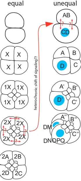
D quadrant formation
In equal cleaving spiralian embryos, the four quadrants of the embryo are initially equipotent, and the critical transition from fourfold radial to bilateral symmetry (D quadrant specification) occurs by inductive interactions some time after the 8-cell stage, depending on the species (Fig. 9). By contrast, unequal cleavage entails the
X
X
X X
AB
CD
1X
1X 1X
1X
A
2X
2X
2X
2X
2A 2B
2D 2C
B
D
C
A’
C’
D’
B’
A’’
DM
C’’
B’’
DNOPQ
?
?
?
?
??
gni
la
ngi
s f
o tf
ih
s ci
nor
hc
or
et
eh
equal
?
?
unequal
segregation of D quadrant determinants in the first and second divisions, so that the embryo never passes through a state of true fourfold symmetry (Fig. 9). A landmark paper by Freeman and Lundelius (1992) made a compelling case for the assertion that equal cleavage was the ancestral condition for spiralian embryos. Whatever the condition of the ur-annelid, the ancestral condition for clitellate annelids is a version of unequal cleavage in which animal and vegetal domains of yolk-deficient cytoplasm (teloplasm) arise within the zygote following fertilization and meiosis. Teloplasm is segregated to the prospective D quadrant macromere by unequal first and second cleavages.
Notwithstanding the apparent homology of this process among clitellate annelids, the underlying cell biological mechanisms dif-fer in several key points between Helobdella and the oligochaete
Tubifex; these differences illustrate the evolutionary plasticity of
developmental mechanisms in general and those regulating D quadrant specification in particular. First, teloplasm formation is dependent on microtubule cytoskeleton in Helobdella but on the microfilament cytoskeleton in Tubifex (Astrow et al., 1989; Shimizu, 1982b). Second, the unequal cleavage of the Helobdella zygote is driven by a transient down-regulation of one centrosome during late metaphase (as judged by loss of gamma-tubulin immunoreactivity) and then the associated astral microtubules, in what was initially a symmetrical, bipolar mitotic apparatus; as in many embryos, the centrosome pair arises by duplication of the paternally derived centrosome (Ren and Weisblat, 2006). In Tubifex by contrast, the centrosome is maternally derived and fails to duplicate; thus the monastral spindle is highly asymmetric from the onset of mitosis (Ishii and Shimizu, 1995; Ishii and Shimizu, 1997). During second cleavage in Helobdella, both poles of a symmetric, biastral mitotic apparatus become closely associated with the cell cortex at the AB-CD interface and is then either pulled or pushed toward the right side of the embryo in an actomyosin-dependent process (Lyons and Weisblat, 2009, Fig. 10). For the corresponding mitosis in Tubifex, the AB-CD interface is small compared to the mitotic apparatus; only one pole of the mitotic apparatus is associated with the cortex and the mitotic apparatus is markedly asymmetric (Takahashi and Shimizu, 1997)
Whatever the mechanisms involved, segregation of teloplasm to a single blastomere at the 4-cell stage is critical for D quadrant specification in Helobdella. Any cell inheriting substantial amounts
of teloplasm gains the capacity of executing the D quadrant-specific series of cleavages. It was originally assumed that animal and vegetal domains of teloplasm might contain specific ectodermal and mesodermal determinants, respectively, but this appears not to be the case: 1) vegetal teloplasm migrates to the animal pole and mixes with animal teloplasm prior to third cleavage in normal development (Holton et al., 1989); 2) experiments combining cyto-plasmic extrusion with centrifugation show that vegetal teloplasm can confer ectodermal fates, apparently subject to interaction with animal pole cortex (Nelson and Weisblat, 1992). Teloplasm is enriched in polyadenylated mRNAs (Holton et al., 1994), includ-ing maternally inherited transcripts such as the leech homologs of
nanos, piwi, vasa and numerous other genes (Pilon and Weisblat,
1997; Kang et al., 2002; Cho et al., 2014). Consistent with the ap-parent equivalence of animal and vegetal domains of teloplasm, the transcripts examined so far have been observed in both the animal and vegetal domains. We also note that teloplasm is not a limiting factor in D quadrant specification--in experimentally manipulated embryos, the presumptive C and D macromeres can both give rise to full complements of mesodermal and ectodermal teloblasts (Astrow et al., 1987).
The full molecular mechanism of D quadrant determination is yet to be determined in detail for any spiralian species. Activation of a MAPK pathway appears to be a conserved feature across some, but not all species (Lambert and Nagy, 2001; Lambert and Nagy, 2003; Koop et al., 2007; Henry and Perry, 2008; Amiel et al., 2013); immunostaining for the phosphorylated MAPK reveals activation of a MAPK pathway in cells 3D or 4d and treatment with inhibitors radializes the embryos. But MAPK pathways provide for intracel-lular signal transduction. A priori, some sort of cell-to-cell signaling must operate upstream or downstream of the MAPK cascade in equal cleavers, so that one and only one of the four quadrants assumes the D fate. It is tempting to postulate the involvement of canonical transmembrane receptor signaling pathways of the sort already known to mediate lateral inhibition and inductive signaling in numerous developmental contexts.
In Helobdella, activation of MAPK signaling is seen in the AB and CD cells during the 2-cell stage (Gonsalves and Weisblat, 2007), at least 4 cell cycles earlier than in the other spiralian systems where MAPK activation has been implicated in D quadrant specification (and inhibitor treatment does not radialize the Helobdella embryo).
T I P M A T
I P M
T
Zygote
In Helobdella, MAPK activation alternates between the CD and AB cells during the 70 minutes of interphase with a set time course. Intriguingly, notch transcripts show a dynamic pattern of accumula-tion and degradaaccumula-tion in the AB and CD cells that parallels that of MAPK activation (Fig. 10). The pattern of notch accumulation is associated with stabilization of maternal and early zygote notch transcripts and appears to be mediated by AU-rich elements (AREs) in the 3’-UTR; AREs confer MAPK-switchable mRNA (in)stability in mammalian systems (Chen and Shyu, 1995; Lasa et al., 2000). Several wnt genes shows similarly dynamic patterns of transcript accumulation and degradation; and for Wnt7, immunostaining has shown that protein expression follows the in situ hybridiza-tion pattern (Huang et al., 2001; Cho et al., 2010). Moreover, the pattern of Wnt7 immunostaining exhibits features expected for a situation where lateral inhibition is operative—reducing the extent of contact between the AB and CD blastomeres (by separating the two cells at first cleavage, or simply by removing the physical constraint of the fertilization membrane prior to first cleavage) results in up-regulation of Wnt7 expression, with positive immu-nostaining in both cells. Many questions remain concerning this set of observations. Does MAPK signaling regulate the stability (or even the transcription) of the other genes showing dynamic patterns of transcript accumulation during the 2-cell stage? How many genes exhibit this pattern of dynamic early expression? More importantly, what is the embryological significance of this pattern? And perhaps most importantly, if early activation of MAPK signaling in Helobdella represents a heterochronic acceleration of events associated with stochastic specification of D quadrant in an equally cleaving spiralian (Fig. 9), does it also mean that Wnt and/or Notch signaling is involved in the inductive interaction of D quadrant specification in extant equal cleavers?
Transition from spiral cleavage to bilaterality
Spiral cleavage in early development is the defining character-istic of spiralian taxa. Micromere-producing spiral cleavage starts at the third cleavage and typically continues on for three more rounds of division. At the end of spiral cleavage, cells in the 2d and 4d lineages undergo equal divisions that yield left/right pairs of homologous ectodermal and mesodermal precursor cells--in leech it is cells 4d (DM’’ in Helobdella terminology), followed by the great-granddaughter of 2d (2d222, or DNOPQ’’’ in Helobdella
terminology) that undergo the bilateral divisions. It is generally accepted that these bilateral divisions are the first landmark of bilaterality in spiralian development. However, as is in many as-pects of spiralian development, little is known how the transition from spiral cleavage to bilaterality occurs exactly. A recent finding in the leech has brought insight into molecular mechanism that regulates this spiral-to-bilateral transition (Schmerer et al., 2013).
It was shown that blocking Pax family transcription factor activity by expressing dominant negative constructs, as well as blocking transcription activity by inhibitor treatment, caused extra rounds of spiral cleavage in place of bilateral divisions of the ectodermal DNOPQ’’’ cell and the mesodermal DM’’ cell. This suggests that transcriptional activating activity of Pax is required for the transi-tion from spiral cleavage to bilateral division. Furthermore, the key Pax protein involved in this process belongs to a subfamily that is unique to spiralian taxa, Paxb (Schmerer et al., 2009). This finding
brings up the possibility that Paxb is evolutionarily and functionally
linked to spiral cleavage, and this also brings up more questions
about the regulation of spiral cleavage and cell lineage in the leech. For example, since these Pax genes were expressed as maternal transcripts, what are the factors that regulate the onset of their translation? What are the additional factors involved in the choice between the highly asymmetric, micromere-producing cell division and the more equal, teloblastogenic division? Answers to these questions may have important implications for the molecu-lar mechanisms underlying the evolutionarily conserved spiralian developmental program.
Connecting segmental and non-segmental tissues
As mentioned above, the boundaries of the blast cell clones do not match the segmental boundaries in the M, O and P lineages, as each blast cell clone spans two segments in O and P lineages and three in the M lineage (Fig. 2). Even the nf blast cell clone, which contributes cells primarily to the posterior portion of each segmental ganglion, has been shown to contribute a small number of cells to the anterior margin of the next ganglion back, before the ganglionic primordia separate (Shain et al., 1998). This disposition of blast cell clones raises a question concerning the anteriormost and the posteriormost segments--what becomes of the cells that would normally arise from or be contributed to the “missing” seg-ments? Clearly, there must be some modification to the cell lineage patterns in these terminal segments and at the interface between segmental and non-segmental tissues.
Using high-resolution lineage tracing techniques, we have begun to understand how the connection between non-segmental tissue and the anterior segmental tissue is made. In the mesoderm, the M teloblast produces six ‘em’ blast cells before it starts to produce standard, purely segmental ‘sm’ blast cells. The first sm cell, sm1, contributes to segments 1-3, sm2 to segments 2-4, sm3 to segments 3-5, and so on. Interestingly, the two posteriormost em cells, em5 and em6, contribute to both non-segmental prostomium tissue and segments 1 and 2, and their segmental contribution appears to be homologous to the pattern elements arising from segmental sm blast cells in the more posterior segments (Gline et al., 2011; Fig. 4). In the N lineage, the first blast cell made by the N teloblast is termed n0, as it makes a unique contribution that serves as a link
between the segmental ganglia and prostomium tissue (Zhang and Weisblat, 2005); after making n0, the N teloblast sequentially
generates one nf cell, one ns cell and then the n’ micromere (which contributes squamous epithelium to the micromere cap (Smith and Weisblat, 1994; Smith et al., 1996) before resuming blast cell production. Thus, in the M and N lineages, some of the first few blast cells arising from the teloblast differ from the more posterior, standard segmental blast cells in that they make special contributions at the connection between the segmental tissues and the micromere-derived prostomium.
The O and P lineages follow yet another scenario. First of all, the OP proteloblast undergoes four rounds of teloblast-like asym-metric cell division, producing four ‘op’ blast cells before dividing equally to produce the ipsilateral pair of O/P teloblasts (Fig. 5). These four op blast cells do make segmental contributions, mainly to the four rostral segments (R1-R4); within these segments, the set of pattern elements arising from each op blast cell appears is largely identical to the sum of a set of o blast cell-derived pattern elements plus a set of p blast cell-derived pattern elements. The clone of the first op blast cell, op1, straddles segments R1 and R2,
R4 and M1 (Shankland, 1987; Kuo and Shankland, 2004a). This leaves some otherwise segmentally iterated pattern elements absent from the R1 segment. It is not clear whether these pattern elements are completely missing in the R1 segment or are still present but come from the micromere lineage(s) instead. In any case, the OP lineage represents a different scheme to deal with the issue of serial homology in the anterior terminus of segmental tissue. A further consequence of these various modifications to the M and N lineages is that even though the teloblasts are born across a time span of 7 hours, the blast cells contributing to the first segment, R1, are born at about the same time, starting in stage 6 (Gline et al., 2011; Fig. 4).
The O-P equivalence group and the evolutionary plasticity of BMP-mediated dorsoventral patterning systems
In Drosophila and vertebrates, dorsoventral patterning entails formation of a morphogen gradient--a continuously graded activity of some signaling molecule across a field of cells; individual cells assume one of several fates as specified by various concentra-tion thresholds for the signal. Cell lineage is usually of negligible importance for cell fate decisions within a morphogen field. In cell lineage-driven development by contrast, cell fate specification is typically coupled to mitosis; each fate decision is therefore binary, and cell-cell signaling is usually local and contact-dependent. Thus, by comparison with morphogen systems, the O-P equivalence group in leech provides a model for elucidating the evolution of dorsoventral patterning mechanisms.
Among the five bilateral pairs of teloblasts, the fates of the M, N and Q teloblasts appear to be specified by cell autonomous pro-cesses, such that these cells are committed to their particular fates at birth. In contrast, the progenitor teloblasts of O and P lineages in
Helobdella are equivalent in their developmental potential, and thus
they are designated ‘O/P’ to reflect their developmental plasticity. The primary blast cells in the ipsilateral o and p bandlets consti-tute a developmental equivalence group, in which fates of initially
equipotent blast cells are specified by external positional cues. Over the years, cell ablation experiments have identified several cues that are involved in patterning the O-P equivalence group. First, ablation of the P lineage caused an O-to-P fate change, but O lineage ablation did not induce a P-to-O fate change (Shankland, 1984; Weisblat and Blair, 1984). This suggests that the p bandlet normally prevents cells of the o bandlet from adopting the P fate. Second, ablation of the micromere-derived provisional integument that covers the germinal band induced an O-to-P fate change (Ho and Weisblat, 1987), suggesting that this covering epithelium also prevents cells in the o bandlet from adopting the P fate. Finally, Q lineage ablation resulted in a P-to-O fate change, indicating that cells of the q bandlet induce the P fate in adjacent o/p blast cells (Huang and Weisblat, 1996).
While the O-P equivalence group is well studied at the cellular and embryological levels, we are only beginning to understand the underlying molecular mechanisms. It turns out that the O-P equivalence group is patterned by a complex network of BMP sig-naling (Kuo and Weisblat, 2011). Among the five genes encoding BMP-type TGFb superfamily ligands in the leech genome, four are
expressed in the germinal band. Hau-admp, Hau-bmp2/4a and
Hau-bmp2/4b are broadly expressed in all bandlets. In contrast, Hau-bmp5-8 is specifically expressed in the q bandlet and is both
necessary and sufficient for the specification of P fate. This discovery was consistent with the previous Q lineage ablation experiment, and suggests that the q blast cells instruct the adjacent o/p blast cells to adopt the P fate by producing Hau-BMP5-8. Intriguingly, Hau-BMP5-8 signaling in the O-P equivalence group appears to signal in a short-range, contact-dependent manner (Kuo and Weisblat, 2011).
Patterning of the O-P equivalence group also involves a BMP antagonist, gremlin. Models in which positional information is conveyed by opposing gradients of agonist and antagonist predict that the BMP antagonists would be expressed in ventral territory of the germinal band, i.e., the n or o bandlet. Surprisingly, however,
q blast cell o/p blast cell o/p blast cell n blast cell
BMP2/4s BMP2/4s BMP2/4s BMP2/4s
gremlin gremlin
q blast cell p blast cell o blast cell n blast cell BMP2/4s BMP2/4s BMP2/4s BMP2/4s
gremlin
BMP5-8
gremlin
1
2
3
P fate determinants Fig. 11. A model for Helobdella O-P
equivalence group patterning (based on data from Kuo and Weisblat, 2011). (A) In the absence of input from BMP5-8, homeostatic feedback between gremlin and the two BMP2/4s (Hau-BMP2/4a and Hau-BMP2/4b) maintains low levels of BMP signaling activity along the DV axis.
(B) A short-range BMP5-8 signaling from the dorsalmost q bandlet (1 in lower panel) elevates BMP signal activity specifically in the adjacent dorsolateral p bandlet and not in the ventrolateral o bandlet. Elevated levels of BMP signaling in the p bandlet up-regulate gremlin and genes that specify the P fate, such as Hau-six1/2a (2 in lower panel). Up-regulation of gremlin in the p bandlet further dampens BMP signal activity in the adjacent o bandlet to permit normal O fate development (3 in lower panel), while signaling in the p bandlet re-mains high due to the gremlin-insensitive BMP5-8 signal from the q bandlet.
A
Hau-gremlin is expressed at highest level in the p blast cells, which
happen to experience the highest level of BMP activity in the germinal band (Kuo and Weisblat, 2011). The resolution to this paradox is as follows. First, localized Hau-BMP5-8 up-regulates Hau-gremlin in the p bandlet. Second, functional analysis indicated that the broadly expressed BMP2/4s, but not Hau-admp, are involved in O-P patterning, and Hau-gremlin only inhibits BMP2/4s, not BMP5-8. Together, these data suggest that a homeostatic feedback circuit may operate within the O-P equivalence group--BMP2/4 signaling up-regulates Hau-gremlin expression and Hau-gremlin inhibits the BMP2/4 signal. Superimposed on this, the gremlin-insensitive BMP5-8 from the Q lineage drives the set point of BMP signaling activity higher in the p bandlets; high BMP signaling in the p bandlet then up-regulates gremlin expression; this then represses BMP2/4 signaling in the o bandlet, which is not affected by the short-range, contact-dependent BMP5-8 signal (Kuo and Weisblat, 2011; Fig. 11). Based on this model, the function of gremlin is to enhance the O-P difference in BMP signaling activity initially induced by localized BMP5-8 signaling. Indeed, both Hau-gremlin knockdown and over-expression failed to induce a complete fate change, indicating an auxiliary, rather than dominant, role for gremlin in O-P patterning (Kuo and Weisblat, 2011).
Elucidating the function of Hau-gremlin helps to interpret the earlier P lineage ablation experiments, in which the O lineage transfates into P after the original P lineage is lost (Shankland, 1984; Weisblat and Blair, 1984). Two (non-exclusive) explana-tions for this result were as follows: (1) that the p bandlet sends an inhibitory signal to repress P fate in the o bandlet, and (2) that the physical presence of the p bandlet serves as a steric block to prevent the o bandlet from contacting the q bandlet (and thus the P fate inducing signal). Now, molecular data indicate that there is truth in both explanations. The normal O fate appears to require up-regulation of inhibitory gremlin in the p bandlet. However, over-expression of gremlin alone is not sufficient for O fate specification. Since BMP5-8 arising from the q bandlet is not sensitive to gremlin, keeping the o blast cell from contacting the q bandlet is another essential condition for normal O fate.
Thus, results of the functional analyses of BMP5-8 and gremlin provide cogent molecular explanations for the Q ablation and P ablation experiments. But, what about the ablation experiments involving the provisional integument? Since no transcripts of BMP-specific antagonists were detected in the provisional integument, we speculate that integumental repression of BMP signaling may be mediated by other multi-functional molecules such as extracellular matrix proteins (Zhu et al., 1999; Ohkawara et al., 2002; Takada
et al., 2003; Wang et al., 2008; Olivares et al., 2009; Ramirez and
Rifkin, 2009).
The axial polarity of BMP signaling in the O-P equivalence group is consistent with the evolutionarily conserved BMP axis, i.e., higher in the dorsal and lower in the ventral for all protostome taxa where BMP signaling is involved in dorsoventral patterning (De Robertis and Sasai, 1996). This suggests that the O-P patterning mechanism was derived from the dorsoventral BMP morphogen gradient. However, the deployment of BMP ligands and antagonists has been dramatically reorganized in the O-P equivalence group, presumably as an adaptation to the emergence of cell lineage-driven embryogenesis in the leech.
Further evidence that the O-P patterning mechanisms are evo-lutionarily flexible over a relatively short evolutionary span comes
from the fact that Q lineage ablation experiments performed on different clitellate species yielded different results (Arai et al., 2001; Kuo and Shankland, 2004b). Interestingly, BMP5-8 knockdown produces robust P-to-O fate change in Helobdella austinensis, in which a ‘redundant’ m-derived P fate inducing signal was also identified by cell ablation experiments (Kuo and Shankland, 2004b). The discrepancy between the Q ablation and BMP5-8 knockdown experiments could be explained if this m-derived signal only operates when the q bandlet is physically removed from the germinal band. While it is not clear what molecular signal is responsible for this conditional, m-derived, P fate-inducing signal, the q bandlet-derived BMP5-8 signal is nevertheless the primary P fate inducing signal normally operating in H. austinensis, just like in other Helobdella species. Interspecies difference in Q ablation experiments, how-ever, still reflects the yet-to-be-uncovered diversity in patterns and organizations of cell-cell interaction within the genus Helobdella.
Germ line specification and genome rearrangements
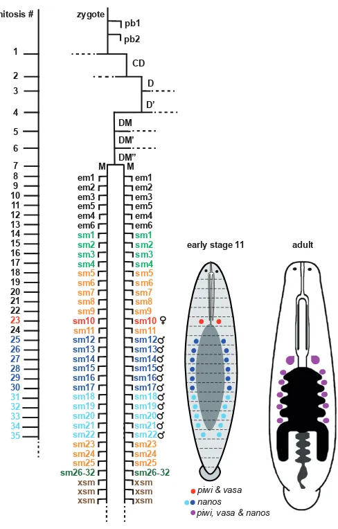
The embryonic origins of the germline in spiralians were enigmatic until relatively recently. The situation has improved considerably with the combination of lineage tracing and in situ hybridization techniques, building on the discovery that genes such as nanos,
vasa, and piwi are broadly conserved markers of primordial germ
cells (PGCs) in diverse taxa (Juliano et al., 2010). In Helobdella as in many other animals, it turns out that these markers are initially broadly expressed during early development, with expression be-coming restricted to PGCs during stages 9-11. [As a simultaneous hermaphrodite, Helobdella species generate both male and female gametes, associated with one pair of ovaries in midbody segment 6 (M6) and 4-6 pairs of testisacs in segments M8 through M11 or M13, respectively]. An unexpected discovery was that the male and female PGCs initially express these markers differentially;
nanos is expressed preferentially in the male PGCs at the same
time that piwi and vasa are expressed preferentially in the female PGCs (Kang et al., 2002; Cho et al., 2014). A second conclusion of interest is that male and female PGCs arise from mesodermal blast cell of the segment with which they are associated. That is, the female PGCs arise in the m blast cell clone whose main contri-bution is to segment M6 (Cho et al., 2014), and presumptive male PGCs arise from 11 m blast cell clones contributing to segments M8-M18 (Kang et al., 2002). This latter result is discrepant with the fact that only with the final numbers of only 4-6 pairs of testisacs in Helobdella species in segments M8-M13. It was proposed that the 11 pairs of nanos-positive cells or cell clusters in the stage 11 embryo represent an ancestral set of pre-PGCs that is then pruned to the varying numbers of testisacs seen in modern leeches.
such as the formation of germ plasm. The relative contributions of inductive and cell autonomous fate decision processes to germline specification in Helobdella remain to be determined.
In any event, the fact that ipsilateral male and female PGCs
share the same lineage for 23 rounds of mitosis increases the probability that any translocations that do occur will be shared between the male and female PGCs. In the case of a self-fertile hermaphrodite, this provides the opportunity to rescue a translo-cation in one gamete with a matching translotranslo-cation in the other gamete, so that normal gene dosage is maintained. All clitellate annelids are hermaphroditic, but self-fertilization is rare. To our knowledge, three species of Helobdella are the only clitellates for which self-fertilization has been documented. We have recently speculated that this combination of developmental cell lineage and reproductive history may account for the dramatic loss of macrosynteny (the conserved linkage of orthologous genes) be-tween Helobdella and other species (for example, as described above for the Hox complex genes). Testing this explanation for the observed genome rearrangements would require denser sampling of genome architecture and reproductive strategies among leeches and related annelids.
Conclusions: prospects for future studies
The material presented above is intended to give a quick overview of Helobdella development as we know it today, and to outline some emerging questions for those who might be tempted to investigate further. Leeches such as Helobdella and other annelids provide interesting material for studying diverse topics, such as cell fate determination, developmental dynamics of the transcriptome, evolutionary dynamics of the genome, lineage-driven segmenta-tion, stem cell mechanisms, and regeneration.
A PubMed search on the keyword Drosophila yields more than 85,000 entries and searching on Caenorhabditis yields more than 21,000--yet no one would claim that our understanding of these exhaustively studied embryos is anywhere near complete. By contrast, searching on Helobdella yields 134 entries--clearly we have only begun to outline the main features of development in this embryo. Given the fact that annelids, molluscs and other lo-photrochozoans have been evolving independently of ecdysozoans and deuterostomes for the better part of one billion years, it seems reasonable to expect that they have explored different regions of the ill-defined, highly-dimensional space of possible developmental mechanisms. We anticipate that the apparent dynamic activation of MAPK, Notch and Wnt signaling pathways in the 2-cell stage, novel deployment of the BMP signaling pathway in DV patterning and a segmentation process driven by stereotyped cell lineages rather than imposition of boundaries in Helobdella are but the first of many interesting observations to be made in this system.
Acknowledgements
The authors thank members of their laboratories for helpful discus-sion and Dr. Elizabeth Heath-Heckman for valuable editorial comments. Research from DAW’s laboratory reported here has been supported by grants from the National Science Foundation and the National Institutes of Health. DHK’s research is funded by grants from Ministry of Science and Technology (Taiwan) and National Taiwan University.
References
AMIEL A R, HENRY J Q and SEAVER E C (2013). An organizing activity is required for head patterning and cell fate specification in the polychaete annelid Capitella teleta: new insights into cell-cell signaling in Lophotrochozoa. Dev Biol. 379: 107-122. ARAI A, NAKAMOTO A, SHIMIZU T (2001). Specification of ectodermal teloblast
zygote
lineages in embryos of the oligochaete annelid Tubifex: involvement of novel cell-cell interactions. Development 128: 1211-1219.
ASTROW S, HOLTON B, WEISBLAT D A (1987). Centrifugation redistributes factors determining cleavage patterns in leech embryos. Dev Biol 120: 270-283. ASTROW S H, HOLTON B and WEISBLAT D A (1989). Teloplasm formation in a leech,
Helobdella triserialis, is a microtubule-dependent process. Dev Biol 135: 306-319. BELY A E, NYBERG K G (2010) Evolution of animal regeneration: re-emergence of
a field. Trends Ecol Evol 25:161-170.
BELY A E (2006). Distribution of segment regeneration ability in the Annelida. Integr Comp Biol 46:508-518
BISSEN S T, WEISBLAT D A (1987). Early differences between alternate n blast cells in leech embryo. J Neurobiol 18: 251-269.
BISSEN S T, WEISBLAT D A (1989). The durations and compositions of cell cycles in embryos of the leech, Helobdella triserialis. Development 106: 105-118. BRAUN J, STENT G S (1989a). Axon outgrowth along segmental nerves in the leech.
I. Identification of candidate guidance cells. Dev Biol 132: 471-485.
BRAUN J, STENT G S (1989b). Axon outgrowth along segmental nerves in the leech. II. Identification of actual guidance cells. Dev Biol 132: 486-501.
CHEN C Y, SHYU A B (1995). AU-rich elements: characterization and importance in mRNA degradation. Trends Biochem Sci 20: 465-470.
CHO S J, VALLÈS Y, GIANI V C JR, SEAVER E C, WEISBLAT D A (2010). Evolution-ary dynamics of the Wnt gene family: a lophotrochozoan perspective. Mol Biol Evol 27: 1645-1658.
CHO S J, VALLÈS Y, WEISBLAT D A (2014). Differential expression of conserved germ line markers and delayed segregation of male and female primordial germ cells in a hermaphrodite, the leech Helobdella. Mol Biol Evol 31: 341-354. DE ROBERTIS E M, SASAI Y (1996). A common plan for dorsoventral patterning in
Bilateria. Nature 380: 37-40.
DESJEUX I, PRICE D J (1999). The production and elimination of supernumerary blast cells in the leech embryo. Dev Genes Evol 209: 284-293.
DOHLE W (1999). The ancestral cleavage pattern of the clitellates and its phylogenetic deviations. Hydrobiologia 402: 267-283.
DUBOULE D (2007). The rise and fall of Hox gene clusters. Development 134: 2549-2560.
FERNANDEZ J, STENT G S (1980). Embryonic development of the glossiphoniid leech Theromyzon rude: structure and development of the germinal band. Dev Biol 78: 407-434.
FREEMAN G, LUNDELIUS J W (1992). Evolutionary implications of the mode of D quadrant specification in coelomates with spiral cleavage. J Evol Biol 5: 205-247. GIRIBET G (2008). Assembling the lophotrochozoan (=spiralian) tree of life. Philos
Trans R Soc Lond B Biol Sci 363:1513-1522.
GLINE S E, NAKAMOTO A, CHO S J, CHI C, WEISBLAT D A (2011). Lineage analysis of micromere 4d, a super-phylotypic cell for Lophotrochozoa, in the leech Helob-della and the sludgeworm Tubifex. Dev Biol 353: 120-133.
GONSALVES F C, WEISBLAT D A (2007). MAPK regulation of maternal and zygotic Notch transcript stability in early development. Proc Natl Acad Sci USA 104: 531-536. GOTO A, KITAMURA K, ARAI A, SHIMIZU T (1999a). Cell fate analysis of teloblasts in
the Tubifex embryo by intracellular injection of HRP. Dev Growth Diff 41: 703-713. GOTO A, KITAMURA K, SHIMIZU T (1999b). Cell lineage analysis of pattern formation
in the Tubifex embryo. I. Segmentation in the mesoderm. Int J Dev Biol 43: 317-327. HENRY J J, PERRY K J (2008). MAPK activation and the specification of the D
quadrant in the gastropod mollusc, Crepidula fornicata. Dev Biol 313: 181-195. HO R K, WEISBLAT D A (1987). A provisional epithelium in leech embryo: cellular
origins and influence on a developmental equivalence group. Dev Biol 120: 520-534. HOLTON B, ASTROW S H, WEISBLAT D A (1989). Animal and vegetal teloplasms
mix in the early embryo of the leech, Helobdella triserialis. Dev Biol 131: 182-188. HOLTON B, WEDEEN C J, ASTROW S H, WEISBLAT D A (1994). Localization of
polyadenylated RNAs during teloplasm formation and cleavage in leech embryos. Roux’s Arch Dev Biol 204: 46-53.
HUANG F Z, BELY A E, WEISBLAT D A (2001). Stochastic WNT signaling between nonequivalent cells regulates adhesion but not fate in the two-cell leech embryo. Curr Biol 11: 1-7.
HUANG F Z, KANG D, RAMIREZ-WEBER F A, BISSEN S T, WEISBLAT D A (2002).
Mi-cromere lineage in the glossiphoniid leech Helobdella. Development 129: 719-732. HUANG F Z, WEISBLAT D A (1996). Cell fate determination in an annelid equivalence
group. Development 122: 1839-1847.
ISAKSEN D E, LIU N J L, WEISBLAT D A (1999). Inductive regulation of cell fusion in leech. Development 126: 3381-3390.
ISHII R, SHIMIZU T (1995). Unequal first cleavage in the Tubifex egg: involvement of a monastral mitotic apparatus. Dev Growth Diff 37: 687-701.
ISHII R, SHIMIZU T (1997). Equalization of unequal first cleavage in the Tubifex egg by introduction of an additional centrosome: implications for the absence of cortical mechanisms for mitotic spindle asymmetry. Dev Biol 189: 49-56.
JULIANO C E, SWARTZ S Z, WESSEL G M (2010). A conserved germline multipo-tency program. Development 137: 4113-4126.
KANG D, PILON M, WEISBLAT D A (2002). Maternal and zygotic expression of a nanos-class gene in the leech Helobdella robusta: primordial germ cells arise from segmental mesoderm. Dev Biol 245: 28-41.
KOOP D, RICHARDS G S, WANNINGER A, GUNTER H M, DEGNAN B M (2007). The role of MAPK signaling in patterning and establishing axial symmetry in the gastropod Haliotis asinina. Dev Biol 311: 200-212.
KOURAKIS M J, MASTER V A, LOKHORST D K, NARDELLI-HAEFLIGER D, WEDEEN C J, MARTINDALE M Q, SHANKLAND M (1997). Conserved anterior boundaries of Hox gene expression in the central nervous system of the leech Helobdella. Dev Biol 190: 284-300.
KRAMER A P, WEISBLAT D A (1985). Developmental neural kinship groups in the leech. J Neurosci 5: 388-407.
KUO D-H, SHANKLAND M (2004a). A distinct patterning mechanism of the O and P cell fates in the development of the rostral segments of the leech Helobdella robusta: implication for the evolutionary dissociation of developmental pathway and morphological outcome. Development 131: 105-115.
KUO D-H, SHANKLAND M (2004b). Evolutionary diversification of specification mechanisms within the O/P equivalence group of the leech genus Helobdella. Development 131: 5859-5869.
KUO D-H, WEISBLAT D A (2011). A new molecular logic for BMP-mediated dorso-ventral patterning in the leech Helobdella. Curr Biol 21: 1282-1288.
KUTSCHERA U, LANGGUTH H, KUO D-H, WEISBLAT D A, SHANKLAND M (2013). Description of a new leech species from North America, Helobdella austinensis n. sp. (Hirudinea: Glossiphoniidae), with observations on its feeding behaviour. Zoosyst Evol 89: 239-246.
LAMBERT J D, NAGY L M (2001). M