Differential expression of the Brunol/CELF family genes
during Xenopus laevis early development
JINGYANG WU
1,2,#, CHAOCUI LI
1, #, SHUHUA ZHAO
1,2and BINGYU MAO*
,11CAS-Max Planck Junior Scientist Group, State Key Laboratory of Genetic Resources and Evolution, Kunming Institute of Zoology, Chinese Academy of Sciences, Kunming, China and
2Graduate University of Chinese Academy of Sciences, Beijing, China
ABSTRACT The BRUNOL/CELF family of RNA-binding proteins plays important roles in post-transcriptional regulation and has been implicated in several developmental processes. In this study, we describe the cloning and expression patterns of five Brunol genes in Xenopus laevis. Among them, only Brunol2 is maternally expressed and the zygotic expression of the other four Brunol genes starts at different developmental stages. During Xenopus development, Brunol1, 4-5 are exclusively expressed in the nervous system including domains in the brain, spinal cord, optic and otic vesicles. Brunol2 and 3 are expressed in both the somatic mesoderm and the nervous system. Brunol2 is also extensively expressed in the lens. In transfected Hela cells, BRUNOL1, 2 and 3 proteins are localized in both the cytoplasm and the nucleus, while BRUNOL4 and 5 are only present in the cytoplasm, indicating their different functions.
KEY WORDS:
Brunol, CELF, Xenopus, expression pattern
The BRUNOL (Bruno-like) family of RNA-binding proteins has been implicated in several post-transcriptional regulatory pro-cesses including pre-mRNA alternative splicing, mRNA stability and translation. The family was named due to their protein sequence similarity to Drosophila melanogaster Bruno (Good et al., 2000). The same family was also named CELF (CUG-BP1 and ETR-3 Like Factors) (Ladd et al., 2001). For the correspondence between these two systems please refer to Barreau et al. (2006) and Brimacombe and Ladd (2007). BRUNOL family proteins contain three RNA recognition motifs (RRMs), two in the N-terminal region and one in the C-N-terminal region. The RRMs show a high degree of sequence similarity but the sequences of the linker region between the second and third RRMs are less conserved (Barreau et al., 2006).
Mammalian BRUNOL family of proteins contains six members. A phylogenetic analysis suggests that the human BRUNOL pro-teins fall into two distinct subfamilies: one contains BRUNOL2, 3 (CUG-BP1 and ETR-3), and the other BRUNOL1, 4-6 (CELF3-6) (Ladd and Cooper, 2004). Brunol2 and 3 are broadly expressed in adult human and mice tissues, with strong expression in the heart, skeletal muscle, and brain (Good et al., 2000; Ladd et al., 2001; Ladd et al., 2004). In contrast, Brunol1, 4, 5 and 6 are mainly
BIOLOGY
www.intjdevbiol.com*Address correspondence to: Dr. Bingyu Mao. Kunming Institute of Zoology, Chinese Academy of Sciences, 32 East Jiao Chang Road, Kunming 650223, China. Tel: +86-871-5198-989. Fax: +86-871-519-3137. e-mail: mao@mail.kiz.ac.cn
#Note: The indicated authors contributed equally to this work.
Accepted: 29 May 2008. Final author corrected PDF published online: 14 August 2009. Edited by: Makoto Asashima.
ISSN: Online 1696-3547, Print 0214-6282
© 2009 UBC Press Printed in Spain
Abbreviations used in this paper: PBS, phosphate buffered saline; PCR, polymerase chain reaction; RRM, RNA recognition motif; WMISH, whole mount in situ hybridization.
Drosophila and mouse have revealed the involvement of BRUNOL proteins in the neural, muscle and germ cell development (Loria et al., 2003; Milne and Hodgkin, 1999; Timchenko et al., 2004; Kim-Ha et al., 1995; Kress et al., 2007; Dev et al., 2007).
In Xenopus, two members of Brunol genes have been reported previously. Brunol1, also named etr-1, was found in a screen of genes expressed in noggin-induced neural tissue (Knecht et al., 1995). Xenopus BRUNOL2 (also known as EDEN-BP) was firstly isolated as a maternal factor mediating sequence-specific mRNA deadenylation in early Xenopus embryos (Paillard et al., 1998). Recently, more than 150 maternal mRNAs from Xenopus tropicalis egg extracts have been identified as potential BRUNOL2 targets (Graindorge et al., 2008), suggesting an important role of BRUNOL2 in early embryonic development. Xenopus Brunol2 is also implicated in the segmentation process. Inactivation of Brunol2 causes severe segmentation defects, and the expression of segmentation markers in the Notch signalling pathway is disrupted (Gautier-Courteille et al., 2004).
In this study, we have cloned three new Brunol genes (Brunol3-5) as well as different paralogs of Brunol1 and 2 of Xenopus laevis and systematically studied their expression patterns during early embryogenesis. The sub-cellular localization of the different BRUNOL proteins was also compared. The results suggest overlapping but different roles of the Brunol genes in Xenopus development.
Results and Discussion
Isolation and phylogenetic analysis of Xenopus Brunol genes
The Xenopus laevis Brunol1a (etr-1) and Brunol2a (EDEN-BP) have been reported previously (Knecht et al., 1995; Paillard et al., 1998). Since X. laevis is pseudotetraploid, there often exit two closely related paralogs for each gene (Hellsten et al., 2007). In our attempt to clone the X. laevis Brunol genes, we have found different paralogs for both Brunol1 and 2, designated Brunol1b and 2b, showing 98% and 92% identity with Brunol1a and 2a respectively. Two Brunol3 paralogs, designated Brunol3a and 3b, were isolated, with 88% sequence identity. Compared with BRUNOL3a, BRUNOL3b missed about 44 amino acids in the linker region, likely due to alternative splicing. X. laevis Brunol4 and 5 were cloned by PCR. One additional isoform of Brunol5
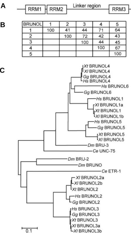
(Brunol5a) was also isolated which missed one 49 base-pair exon to produce a premature stop codon, resulting in a truncated protein of only 132 amino acids. We failed to clone the Xenopus Brunol6 using primers according to conserved sequence among vertebrate Brunol6 mRNAs. Using mouse Brunol mRNA se-quence queries, we could find only 5 chromosome loci in X. tropicalis genome. No ESTs of potential Brunol6 could be found either in X. laevis or tropicalis. The predicted partial sequence listed as X. tropicalis BRUNOL6 (CELF6) in Brimacombe and Ladd (2007) was proved to be the C-terminal part of BRUNOL1. It is possible that Brunol6 is missing in Xenopus. In chick, Brunol1 (CELF3) is missing due to a deletion event. The Xenopus BRUNOL proteins all contained three conserved RRMs with a linker region between the second and third one (Fig. 1A). Among the 5 Xenopus BRUNOL proteins, BRUNOL2 and BRUNOL3 proteins are more closely related with an overall sequence identity of 72%. BRUNOL1, 4 and 5 form another sub-group and the average sequence identity among them was about 66%. The overall sequence identity between the two sub-groups was below 45% (Fig. 1B).
Using MEGA program (Kumar et al., 2004), we studied the phylogenetic relationship of the BRUNOL proteins in human,
Fig. 1. Phylogenetic analysis of Xenopus BRUNOL proteins. (A) The representative structure of Xenopus BRUNOL proteins. (B) Sequence identities of the X. laevis BRUNOL proteins. For BRUNOL proteins that have paralogs, the average values of the paralogs were shown. (C)
Phylogenetic analysis of BRUNOL family proteins. ClustalW alignment, Poisson correction model, and Bootstrap test were used for the neigh-bor-joining (NJ) tree construction. The sequences of X. laevis BRUNOL3b, 4, 5, 5a have been deposited in GenBank under accession numbers EU743743-EU743746. Other accession nos. were as follows: Xenopus laevis (Xl) BBRUNOL1a AAA81375, BRUNOL1b AAH46942, BRUNOL2a AAH70706, BRUNOL2b NP_001084196, BRUNOL3a AAB09041; Xeno-pus tropicalis (Xt) BBRUNOL1 BC154063, BRUNOL2 NP_001017152, BRUNOL3 NP_001096417, BRUNOL4 AAI21491, BRUNOL5 AAI25740; Drosophila melanogaster (Dm) Bruno NP_723738, BRU-2 NP_788039, BRU-3 NP_729923. The BRUNOL protein sequences of human (Homo sapiens, Hs), chicken (Gallus gallus, Gg) and Caonorhabditis elegans(Ce) were used as in Brimacome and Ladd (2007).
B
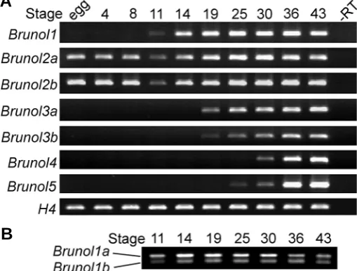
Fig. 2. Temporal expression of Xenopus Brunol genes.(A) RT-PCR analysis of the developmental expression of the Xenopus Brunol genes. egg: unfertilized egg. Histone 4 (H4) was used as a loading control. (B)
The Brunol1 PCR products after NcoI digestion, showing the differential expression levels of Brunol1a and 1b.
B
A
chick, X. laevis and tropicalis, Drosophila and C. elegans (Fig. 1C). The BRUNOL proteins fell into two sub-groups based on their overall sequence similarity, one contained BRUNOL2 and 3, the other contained BRUNOL1 and 4-6. The Drosophila and C. elegans BRUNO proteins also fitted into the two BRUNOL sub-families, suggesting an early diversification of the two subfamilies during evolution.Temporal expression of Xenopus Brunol genes
To investigate the temporal expression patterns of the Brunol genes in X. laevis, a series of RT-PCR analysis was carried out (Fig. 2A). Brunol2a and b were detected strongly maternally as reported previously (Gautier-Courteille et al., 2004). Their ex-pression was weak at the beginning of the neurula stage and became stronger at later stages. All the other Brunol genes were not maternally expressed and their expression started at different developmental stages. Brunol1 began to express at about stage 11 and increased with development. From stage 19 on, it kept strongly expressed. The transcripts of Brunol3a and b could be detected weakly at stage 19 and kept expressed at later stages. Brunol4 and 5 started to express at stage 30 and 25 respectively and were both strongly expressed at later stages.
Different paralogs of the same Brunol genes showed the same temporal expression patterns during development, although their expression levels were slightly different. Since the sequences of Brunol1a and b were 98% identical, it was difficult to design PCR primers to distinguish them. They differed in one fragment in which Brunol1b contained two additional NcoI restriction sites. This fragment was amplified by PCR and after NcoI digestion, it was able to distinguish the two paralogs. At stages 11 to 30, the expression level of Brunol1a was stronger than Brunol1b with a ratio of about 2.5:1. At stages 36 and 43, the two paralogs were expressed at similar levels (Fig. 2B).
Spatial expression of Brunol genes during Xenopus laveis early embryogenesis
To avoid cross reaction, the sequences corresponding to the less conserved linker regions between RRM2 and 3 were chosen for probes preparation. For the different paralogs of the same gene, it was unable to distinguish the expression patterns of individual paralogs using these probes due to their high sequence similarities.
The temporal expression patterns of the Brunol genes from the WMISH results fitted well with the RT-PCR analysis. Brunol2 was the only one maternally expressed and no expression was de-tected for Brunol1, 3, 4 and 5 till the end of gastrulation.
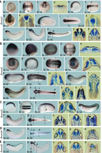
As reported previously, Brunol1 was specifically expressed in the neural tissue and stronger in the anterior region (Fig. 3A, Knecht et al., 1995). At stage 13 and 14, weak expression of Brunol1 was detected in the anterior neural plate region as well as two patches lateral to the brain region, representing the presump-tive trigeminal placodes (Fig. 3Aa-d). From stage 16 to 25, Brunol1 was more abundantly expressed in the whole neural tube but weak in the roof plate and floor plate regions (Fig. 3Ae-k). At stage 30, Brunol1 was strongly expressed in the forebrain, mid-brain, hindmid-brain, the epiphysis, spinal cord and optic vesicles, but not at mid-hindbrain boundary (Fig. 3Al). At stage 36, its expres-sion remained strong in the above regions and appeared also in the otic vesicles (Fig. 3Am-p). In the eyes, it was strongly
ex-pressed in the retinas (Fig. 3 Ao).
Maternal Brunol2 mRNA has been reported to distribute homo-geneously in the early Xenopus embryos (Gautier-Courteille et al., 2004) and vegetally in zebrafish (Suzuki et al., 2000). In our study, however, Brunol2 transcripts were detected at the animal poles in the unfertilized eggs and cleavage and blastula stage embryos (Fig. 3Ba and data not shown). During gastrulation, it was expressed in the ectoderm but not in the yolk plug region (Fig. 3Bb). At stage 13, it was broadly expressed in the whole embryo (Fig. 3Bc). At stages 14 and 16, its expression became confined in the para-axial mesoderm and the neural plate (Fig. 3Bd-g). At stages 19 and 26, it was expressed in the somites and neural tube (Fig. 3Bh-m). At stage 19, it was also detected in the trigeminal placodes and the presumptive eye fields (Fig. 3Bi). At tailbud stages, Brunol2 was expressed in the brain, spinal cord, optic and otic vesicles and the branchial arches (Fig. 3Bn-s). It was exten-sively expressed in the lens (Fig. 3Bq), which was also the case for zebrafish Brunol2 (Suzuki et al., 2000), suggesting a con-served role for BRUNOL2 in lens development. Brunol2 remained weakly expressed in the somites in the trunk region and strongly in the tail region (Fig. 3Bo,p,s).
G
B
C
D
E
F
H
A
G'
B'
C'
D'
E'
F'
H'
A'
G''
B''
C''
D''
E''
F''
H''
A''
Fig. 3. Embryonic expression of the X. laevis Brunol genes at the indicated stages.(A) Brunol1, (a,c,g), frontal views, dorsal to the top; (b,f,j,n), dorsal views, anterior to the left; (e,i,l,m), lateral views, anterior to the left. (B) Brunol2, (a,b), lateral views, animal pole to the top; (c,d,h,l,p), dorsal views, anterior to the left; (f,i), frontal views, dorsal to the top; (g,k,n,o), lateral views, anterior to the left. (C)Brunol3, (a,c,e,f), lateral views, anterior to the left; (b,g), dorsal views, anterior to the left. (D) Brunol4, (a), lateral view, anterior to the left; (b), dorsal view, anterior to the left. (E)Brunol5, (a,b), lateral view, anterior to the left. The positions of the sections were indicated.
B
C
D
(Fig. 3Cg,h,i). At stage 36, it was also detected in the facial epibranchial placode (Fig. 3Cf).
The expression of Bruno4 and 5 were both restricted in the nervous system. Brunol5 began to express at about stage 25 while Brunol4 started at stage 30. At stage 30, Brunol5 was detected in the ventral part of the brain and anterior half of the spinal cord as well as the optic and otic vesicles (Fig. 3Ea). At stage 36, Brunol4 and Brunol5 showed overlapping expressing domains in the ventral midbrain, hindbrain, anterior spinal cord, optic and otic vesicles (Fig. 3 D,E). Brunol5 but not Brunol4 was detected in the dorsal forebrain and epiphysis (Fig. 3 D,E).
Post-transcriptional regulation exerted by RNA-binding pro-teins plays important roles in the development and maintenance of the nervous system (reviewed in Perrone-Bizzozero and Bolognani, 2002). For example, the BRUNOL related ELAV/Hu family proteins are also differentially expressed during neurogenesis and are essential for proper neural development (Perron et al., 1999; Perrone-Bizzozero and Bolognani, 2002). Our data showed that the Xenopus Brunol genes were all strongly expressed in the nervous system, adding more complexity to the post-transcriptional control of neural development. The five Brunol genes were all expressed in the neural retinas in the developing eye, with Brunol2 also strongly in the lens, suggesting an impor-tant role of BRUNOLs in eye development.
Subcellular localization of BRUNOL proteins
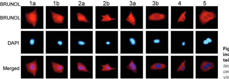
The full length Brunol open reading frames (ORF) were fused with a C-terminal Flag tag coding sequence and the subcellular localization of the BRUNOL proteins was detected using anti-Flag antibody in transfected Hela cells. BRUNOL1a, 1b, 2a, 2b and 3a were all localized in both the cytoplasm and the nucleus, while BRUNOL3b, 4 and 5 distributed only in the cytoplasm (Fig. 4). Interestingly, BRUNOL3a and 3b showed different localization. In the nucleus, BRUNOLs have been shown to regulate the alterna-tive splicing of some pre-mRNAs through binding to a U/G rich sequence element (Barreau et al., 2006). In the cytoplasm, the BRUNOL proteins control the translation or stability of target mRNAs through binding to sequence elements in the 5’- or the 3’-regions (Iakova et al., 2004; Timchenko et al., 1999). The different subcellular localization of the BRUNOL proteins might indicate their different functions.
Materials and Methods
Cloning of Xenopus laevis Brunol genes and phylogenetic analysis
Through database searches using mouse Brunol mRNA sequences
Fig. 4. Subcellular localization of the indicated Xenopus BRUNOL pro-teins. BRUNOL proteins were de-tected using anti-FLAG antibody. The cells were also stained with DAPI to visualize the nuclei.
as queries, unigene clusters were identified for X. laevis Brunol1a
(Xl.986), 1b (Xl.12160), 2a (Xl.12254), 2b (Xl.25921) and 3 (Xl.1068).
PCR primers were designed according to the EST sequences to amplify the full open reading frames. For X. laevis Brunol4 and 5, primers were designed according to the corresponding X. tropicalis EST sequences. The primers used were:
Brunol1: forward 5’- TGTTGGGTGGTGGGAGTCTGTTCCTT -3’ and
reverse 5’- ATGTTCTCCTTGGTCCAGCAGCAGT -3’,
Brunol2a: forward 5’- CTGATCAAGAAAACGCTAATGGGGGATA-3’ and
reverse 5’- ATGGACAAATGGCAGGAAGGGATCAG -3’,
Brunol2b: forward 5’- ATAATGAATGGCACAATGGACCACCC -3’ and
reverse 5’- ATGGACAAATGGCAGGAAGGGATCAG -3’,
Brunol3: forward 5’- GCTTCTGTGGCATTGATGTTTGAGC -3’ and
reverse 5’- CTGGGGTTAGGATCAGTAAGGTTTGC -3’,
Brunol4: forward 5’- CAGAGAGACCAGGCAGCTTCCTCT TG -3’ and
reverse 5’-TCAGTAAGGGCGGTTGGCATCCTTCG -3’,
Brunol5: forward 5’- ATGGCCAGACTGACGGAGAGA GAG -3’ and
reverse 5’- TCAGTATGGTTGGGTTGTGTCCTTAGGC-3’.
cDNAs were reverse transcribed using total RNA from stage 13 to 36 as template and oligo-T 18 as primer (ThermpScript RT-PCR System, Invitrogen). The PCR products were then cloned into pGEM-T vectors (Promega) and the inserts were full length sequenced.
Phylogenetic tree construction was performed as described (Zhao et
al., 2007).
RT-PCR assay and restriction enzyme digestion
Total RNAs were extracted using TRIzol regent (Invitrogen) from X.
laevis embryos at different stages, and the first strand cDNAs were
synthesized using random hexamer nucleotide as RT primers (RevertAid H minus first strand cDNA Synthesis kit, Fermentas). The amount of the templates was adjusted according to Histone 4 (H4) expression levels. PCR assays were carried out in the linear phase.
Primers used were:
Brunol1: forward 5’- CGCACAMAGCGCTTACCTCAAT -3’ and
reverse 5’- GTGGCAGTGAGTGTTGGTGGAG -3’,
Brunol2a: forward 5’- TGGGGTAACCTAGCTGGACTCG -3’and
reverse 5’- GGGGACTGCTGGAAGAAGTGAG -3’,
Brunol2b: forward 5’- TGGGGTAACCTGACTGGACTGA -3’ and
reverse 5’- GGGGGCTGCTGGAAGAAGTGAG -3’,
Brunol3a: forward 5’- GACCATGGAGGGCTGTTCTTCA -3’ and
reverse 5’- ACCCAGGTTACTGGGGGTGGTA -3’,
Brunol3b: forward 5’- GACCATGGAGGGCTGTTCTTCA -3’ and
reverse 5’- ATGCACCCAGGTTACKGGAGGA -3’,
Brunol4: forward 5’- CCAGCTGTGCCTAGCATTCCAT -3’ and
reverse 5’- GGCTGTGGAAAGGCTTGACTGA -3’,
Brunol5: forward 5’- AGACTATGCCTGGGGCCTCATC -3’ and
reverse 5’- GTAGCTCCCATGCGAAGTGGAC -3’.
PCR conditions: 94°C for 2 min; 30 cycles of 94°C for 30 sec, 55°C for 30 sec, 72°C for 30 sec; followed by one cycle of 72°C for 5 min.
digested with NcoI before agarose gel electrophoresis, and the software Quantity One (Bio-Rad) was used to analyze the bands’ density.
Embryos, in situ hybridization and section
In vitro fertilization, embryo culture, and whole mount in situ
hybridiza-tion (WMISH) of Xenopus embryos were carried out as described (Zhao
et al., 2007). Stained embryos of selected stages were embedded in
paraffin, sectioned at 30μm, and photographed using a light microscope (Leica).
Cell transfection and immunohistochemistry
The ORFs of Brunol genes were cloned in frame into a eukaryotic expression vector pCS2+ with a Flag tag coding sequence. Cultured Hela cells were then transfected with these vectors using Lipofectamine2000 (Invitrogen) in 96-well plates. At 24h after transfection, cells were fixed with 3.7% formaldehyde for 20min, permeablized in 0.1% Triton X-100/ PBS for 10min, washed with PBS, blocked in 1% BSA/PBS for 1h, and then treated with mouse anti-FLAG antibody (Sigma) for 1.5h. After washing with 1% BSA/PBS for 5 times, cells were treated with Cy3-conjugated anti-mouse secondary antibody (Sigma) for 1.5h. The cells were washed and stained with 1μg/ml DAPI (Sigma) for 5 min and then visualized using a fluorescence microscope (Leica).
Acknowledgements
This work was supported by grants from the National Natural Science Foundation of China (30425011 and 305303800), the Key State Re-search Program from the Ministry of Science and Technology of China (2006CB943902 and 2007CB947201) and the Innovation Project of Chinese Academy of Sciences (KSCX2-YW-R-090).
References
BARREAU, C., PAILLARD, L., MEREAU, A. and OSBORNE, H. B. (2006). Mamma-lian CELF/Bruno-like RNA-binding proteins: molecular characteristics and biologi-cal functions. Biochimie 88: 515-525.
BRIMACOMBE, K. R. and LADD, A. N. (2007). Cloning and embryonic expression patterns of the chicken CELF family. Dev Dyn 236: 2216-2224.
DELAUNAY J., LE MEE G., EZZEDDINE N., LABESSE G., TERZIAN C., CAPRI M. AND AIT-AHMED O. (2004). The Drosophila Bruno paralogue Bru-3 specifically binds the EDEN translational repression element. Nucleic Acids Res 32: 3070-3082.
DEV, A., NAYERNIA, K., MEINS, M., ADHAM, I., LACONE, F. and ENGEL, W. (2007). Mice deficient for RNA-binding protein brunol1 show reduction of spermatogen-esis but are fertile. Mol Reprod Dev 74: 1456-1464.
GAUTIER-COURTEILLE, C., LE CLAINCHE, C., BARREAU, C., AUDIC, Y., GRAINDORGE, A., MANIEY, D., OSBORNE, H. B. and PAILLARD, L. (2004). EDEN-BP-dependent post-transcriptional regulation of gene expression in Xeno-pus somitic segmentation. Development 131: 6107-6117.
GOOD, P. J., CHEN, Q., WARNER, S. J. and HERRING, D. C. (2000). A family of human RNA-binding proteins related to the Drosophila Bruno translational regu-lator. J Biol Chem 275: 28583-28592.
GRAINDORGE, A., LE TONQUEZE, O., THURET, R., POLLET, N., OSBORNE, H. B. and AUDIC, Y. (2008). Identification of CUG-BP1/EDEN-BP target mRNAs in Xenopus tropicalis. Nucleic Acids Res 36: 1861-1870.
HELLSTEN, U., KHOKHA M.K., GRAMMER, T.C., HARLAND, R.M., RICHARDSON, P. and ROKHSAR, D.S. (2007). Accelerated gene evolution and subfunctionalization in the pseudotetraploid frog Xenopus laevis. BMC Biol. 5: 31.
IAKOVA, P., WANG, G. L., TIMCHENKO, L., MICHALAK, M., PEREIRA-SMITH, O. M., SMITH, J. R. and TIMCHENKO, N. A. (2004). Competition of CUGBP1 and calreticulin for the regulation of p21 translation determines cell fate. EMBO J 23: 406-417.
KIM-HA, J., KERR, K. and MACDONALD, P. M. (1995). Translational regulation of oskar mRNA by bruno, an ovarian RNA-binding protein, is essential. Cell 81: 403-412.
KNECHT, A. K., GOOD, P. J., DAWID, I. B. and HARLAND, R. M. (1995). Dorsal-ventral patterning and differentiation of noggin-induced neural tissue in the absence of mesoderm. Development 121: 1927-1935.
KRESS, C., GAUTIER-COURTEILLE, C., OSBORNE, H. B., BABINET, C. and PAILLARD, L. (2007). Inactivation of CUG-BP1/CELF1 causes growth, viability, and spermatogenesis defects in mice. Mol Cell Biol 27: 1146-1157.
KUMAR, S., TAMURA, K. and NEI, M. (2004). MEGA3: Integrated software for Molecular Evolutionary Genetics Analysis and sequence alignment. Brief Bioinform 5: 150-163.
LADD, A. N., CHARLET, N. and COOPER, T. A. (2001). The CELF family of RNA binding proteins is implicated in cell-specific and developmentally regulated alternative splicing. Mol Cell Biol 21: 1285-1296.
LADD, A. N. and COOPER, T. A. (2004). Multiple domains control the subcellular localization and activity of ETR-3, a regulator of nuclear and cytoplasmic RNA processing events. J Cell Sci 117: 3519-3529.
LADD, A. N., NGUYEN, N. H., MALHOTRA, K. and COOPER, T. A. (2004). CELF6, a member of the CELF family of RNA-binding proteins, regulates muscle-specific splicing enhancer-dependent alternative splicing. J Biol Chem 279: 17756-17764.
LORIA, P. M., DUKE, A., RAND, J. B. and HOBERT, O. (2003). Two neuronal, nuclear-localized RNA binding proteins involved in synaptic transmission. Curr Biol 13: 1317-1323.
MILNE, C. A. and HODGKIN, J. (1999). ETR-1, a homologue of a protein linked to myotonic dystrophy, is essential for muscle development in Caenorhabditis elegans. Curr Biol 9: 1243-1246.
PAILLARD, L., OMILLI, F., LEGAGNEUX, V., BASSEZ, T., MANIEY, D. and OSBORNE, H. B. (1998). EDEN and EDEN-BP, a cis element and an associ-ated factor that mediate sequence-specific mRNA deadenylation in Xenopus embryos. EMBO J 17: 278-287.
PERRON, M., FURRER, M.P., WEGNEZ, M. and THODORE, L. (1999). Xenopus elav-like genes are differentially expressed during neurogenesis. Mech Dev 84: 139-142.
PERRONE-BIZZOZERO, N. and BOLOGNANI, F. (2002). Role of HuD and other RNA-binding proteins in neural development and plasticity. J Neurosci Res 68: 121-126.
SUZUKI, H., MAEGAWA, S., NISHIBU, T., SUGIYAMA, T., YASUDA, K. and INOUE, K. (2000). Vegetal localization of the maternal mRNA encoding an EDEN-BP/Bruno-like protein in zebrafish. Mech Dev 93: 205-209.
TIMCHENKO, N. A., PATEL, R., IAKOVA, P., CAI, Z. J., QUAN, L. and TIMCHENKO, L. T. (2004). Overexpression of CUG triplet repeat-binding protein, CUGBP1, in mice inhibits myogenesis. The J Biol Chem 279: 13129-13139.
TIMCHENKO, N. A., WELM, A. L., LU, X. and TIMCHENKO, L. T. (1999). CUG repeat binding protein (CUGBP1) interacts with the 5' region of C/EBPbeta mRNA and regulates translation of C/EBPbeta isoforms. Nucleic Acids Res 27: 4517-4525.
YANG, Y., MAHAFFEY, C. L., BERUBE, N., MADDATU, T. P., COX, G. A. and FRANKEL, W. N. (2007). Complex seizure disorder caused by Brunol4 defi-ciency in mice. PLoS Genet 3: e124.
Further Related Reading, published previously in the Int. J. Dev. Biol.
See our recent Special Issue Fertilization, in honor of David L. Garbers and edited by Paul M. Wassarman and Victor D. Vacquier at: http://www.ijdb.ehu.es/web/contents.php?vol=52&issue=5-6
Differential expression of two TEF-1 (TEAD) genes during Xenopus laevis development and in response to inducing factors François Naye, Karine Tréguer, Fabienne Soulet, Corinne Faucheux, Sandrine Fédou, Nadine Thézé and Pierre Thiébaud
Int. J. Dev. Biol. (2007) 51: 745-752
Identification of a second Xenopus twisted gastrulation gene
Michael Oelgeschläger, Uyen Tran, Kristina Grubisic and Edward M De Robertis Int. J. Dev. Biol. (2004) 48: 57-61
Heterogeneous nuclear ribonucleoprotein complexes from Xenopus laevis oocytes and somatic cells
A Marcu, B Bassit, R Perez and S Piñol-Roma Int. J. Dev. Biol. (2001) 45: 743-752
Misexpression of the RNA-binding protein ELRB in Xenopus presumptive neurectoderm induces proliferation arrest and programmed cell death
M Perron, M P Furrer, M Wegnez and L Théodore Int. J. Dev. Biol. (1999) 43: 295-303
Zinc finger proteins in early Xenopus development T Hollemann, E Bellefroid, R Stick and T Pieler Int. J. Dev. Biol. (1996) 40: 291-295
Expression of GTP-binding protein gene drg during Xenopus laevis development S Kumar, M Iwao, T Yamagishi, M Noda and M Asashima
Int. J. Dev. Biol. (1993) 37: 539-546