High dose dietary vitamin D
3
increases bone mass and strength in mice
Liam Williamson
a, Alan Hayes
b,c, Erik D Hanson
b,d, Peter Pivonka
e, Natalie A Sims
a,f, Jonathan H. Gooi
a,⁎
aDepartment of Medicine, St. Vincent's Hospital Melbourne, The University of Melbourne, Melbourne, VIC 3065, Australia
b
Institute for Sport, Exercise and Active Living (ISEAL), College of Health and Biomedicine, Victoria University, Melbourne, VIC 8001, Australia
cAustralian Institute for Musculoskeletal Science (AIMSS), Western Health, Melbourne, VIC 3021, Australia d
Department of Exercise & Sport Science, University of North Carolina, Chapel Hill, NC, USA
e
St Vincent's Department of Surgery, The University of Melbourne, Melbourne, VIC 3065, Australia
f
St. Vincent's Institute of Medical Research, 9 Princes St, Fitzroy, VIC 3065, Australia
a b s t r a c t
a r t i c l e i n f o
Article history:
Received 23 November 2016
Received in revised form 18 January 2017 Accepted 8 February 2017
Available online 10 February 2017
Vitamin D plays a critical role in skeletal homeostasis. Vitamin D supplementation is used worldwide to maintain optimal bone health, but the most appropriate level of supplementation remains controversial. This study aimed to determine the effects of varying doses of dietary vitamin D3on the mechanical properties and morphology of growing bone.
Eight-week-old female mice were supplied with one of 3 diets, each containing a different dose of vitamin D3: 1000 IU/kg (control), 8000 IU/kg or 20,000 IU/kg. Mice hadad libitumaccess to the specialty diet for 4 weeks be-fore they were culled and their tibiae collected for further analysis. The collected tibia underwent three-point bending and reference-point indentation from which their mechanical properties were determined, and cortical and trabecular morphology determined by micro computed tomography.
Dietary supplementation with 20,000 IU/kg vitamin D3resulted in greater ductility (~ 200%) and toughness (~150%) compared to the 1000 IU/kg control. The 20,000 IU/kg diet was also associated with significantly greater trabecular bone volume fraction and trabecular number. The 8000 IU/kg diet had no significant effect on trabec-ular bone mass.
We conclude that vitamin D3supplementation of 20,000 IU/kg during early adulthood leads to tougher bone that is more ductile and less brittle than that of mice supplied with standard levels of dietary vitamin D3(1000 IU/kg) or 8000 IU/kg. This suggests that dietary vitamin D3supplementation may increase bone health by improving bone material strength and supports the use of vitamin D3supplementation, during adolescence, for achieving a higher peak bone mass in adulthood and thereby preventing osteoporosis.
© 2017 The Authors. Published by Elsevier Inc. This is an open access article under the CC BY-NC-ND license (http://creativecommons.org/licenses/by-nc-nd/4.0/).
Keywords:
Vitamin D3 3 point bending Cholecalciferol Bone strength
1. Introduction
It is generally established that vitamin D3is crucial for bone health through its actions as a regulator of minerals, and in turn, skeletal ho-meostasis in vertebrates (Anderson et al., 2011). Deficiencies in vitamin D3during childhood can have significant health consequences such as growth retardation (Rajakumar, 2003) and detrimental effects on bone mineral acquisition (Lehtonen-Veromaa et al., 2002) and bone re-modelling (Outila et al., 2001; Cheng et al., 2003; Fares et al., 2003) lead-ing to rickets (O'Riordan and Bijvoet, 2014). These consequences in adolescence are also a significant risk factor for the development of osteoporosis later in life (Dawson-Hughes et al., 1991; Lips, 2001).
The effects of vitamin D3as a treatment for osteoporosis in adult-hood are controversial. Conflicting reports suggests vitamin D3
supplementation in adulthood reduces (Bischoff-Ferrari et al., 2005; Tang et al., 2007), has no effect (Michaëlsson et al., 2003), or increases the incidence of osteoporotic fractures (Smith et al., 2007; Sanders et al., 2010). By comparing and normalising 23 separate studies,Reid et al. (2013)found that vitamin D supplementation was not effective in re-ducing fracture risk in those not experiencing vitamin D deficiency.
An alternative strategy for preventing osteoporosis is to optimise peak bone mass during growth via vitamin D3supplementation during adolescence. A number of intervention studies examining the effect of vitamin D3supplementation in adolescents have reported significant in-creases in bone mineral content (BMC) (El-Hajj Fuleihan et al., 2006; Viljakainen et al., 2006) and bone mineral density (BMD) (Du et al., 2004), while other studies have reported no beneficial effects (Andersen et al., 2008). Recent systematic reviews and meta-analysis (Winzenberg et al., 2010; Winzenberg et al., 2011) have concluded that vitamin D3supplementation during adolescence had no significant effect on BMC and BMD, however there has been no randomised con-trolled trial to assess the effect of vitamin D3supplementation on bone ⁎ Corresponding author at: Melbourne Medical School, The University of Melbourne, 28
Regent Street, Fitzroy, VIC 3065, Australia.
E-mail address:jgooi@unimelb.edu.au(J.H. Gooi).
http://dx.doi.org/10.1016/j.bonr.2017.02.001
2352-1872/© 2017 The Authors. Published by Elsevier Inc. This is an open access article under the CC BY-NC-ND license (http://creativecommons.org/licenses/by-nc-nd/4.0/). Contents lists available atScienceDirect
Bone Reports
health. Currently the recommended daily intake of vitamin D3in infants, children and adolescents is 400 IU (Wagner and Greer, 2008). This dos-age is based on clinical trials measuring biomarkers of vitamin D3status and the indirect observations that 400 IU of vitamin D3prevents and treats rickets (Wagner et al., 2006; Rajakumar and Thomas, 2005).
It is clear that there is a need for further investigation into the effect of vitamin D3dietary supplementation on bone mass in a model of growing bone where subjects are vitamin D3replete. Therefore, our ob-jective was to determine whether increasing dietary vitamin D3levels (8000 and 20,000 IU/kg) above the standard levels (1000 IU/kg) signif-icantly alters bone mass and strength in growing mice.
2. Methods and animals
2.1. Animals
7 week old female C57Bl/6 J mice were obtained from Monash Ani-mal Services, Victoria and housed in a temperature and humidity con-trolled environment on a 12 h light/dark cycle. All animal work was approved by Victoria University Animal Ethics Committee. All animals were fed on standard growth diet (AIN-93G, containing 0.47% calcium (Ca), 0.35% phosphate (PO4), vitamin D31000 IU/kg) from weaning until 8 weeks of age, at which point the animals were randomly allocat-ed to one of three diets for a period of 4 weeks. These diets were mod-ified AIN-93G diets supplemented with set concentrations of cholecalciferol (vitamin D3) as follows: control (1000 IU/kg, n = 10), 8000 IU/kg (n = 10) or 20,000 IU/kg (n = 7). The animals had ad libitum access to both food & water. All animals consumed the same amount of food (average of 2.5 g per day, no significant difference be-tween groups). At 12 weeks of age, the mice were deeply anaesthetized with pentobarbital sodium (60 mg/kg injection i.p.), killed by cervical dislocation, and their tibiae harvested. Tibiae were carefully dissected free from soft tissues; lengths measured with a digital caliper, and stored at−80 °C prior to mechanical testing and further analysis.
2.2. Micro computed tomography (μCT)
Tibiae were analyzed by micro-computed tomography as described previously (Johnson et al., 2014) using the SkyScan 1076 System (Bruker-microCT, Kontich, Belgium). Images were acquired using the following settings: 9μm voxel resolution, 0.5 mm aluminiumfilter, 44 kV voltage, and 220μA current, 2300 ms exposure time, rotation 0.5°, frame averaging = 1. The images were reconstructed and analyzed using SkyScan Software programs NRecon (version 1.6.3.3), DataViewer (version 1.4.4), and CT Analyser (version 1.12.0.0) as previously de-scribed (Johnson et al., 2014).
CTAn software was then used to select the regions of interest for both the cortical (CTAn version 1.15.4.0) and trabecular (CTAn version 1.11.8.0) bone of each scan. Trabecular region of interest (ROI) was se-lected as a 2 mm region starting 0.5 mm below the proximal growth plate. Cortical ROI was selected as a 1 mm region starting 7 mm below the growth plate. The cortical ROI was chosen such that the middle of the ROI corresponded with the point at which load was applied to the bones in the three-point bending experiments.
The analysis of bone structure was completed using adaptive thresholding (mean of min and max values) in CT Analyser. The thresh-olds for analysis were determined based on multilevel Otsu thresholding of the entire data set, and were set at 45–255 for trabecular bone and 71–255 for cortical bone.
2.3. Three point bending
Each tibia was rehydrated overnight in phosphate buffered saline (PBS) at room temperature prior to testing. To determine the mechan-ical properties of cortmechan-ical bone each tibia was loaded to failure at 0.5 mm/s using a Bose Biodynamic 5500 Test Instrument (Bose, DE,
USA). The span between the lower supports was 10 mm. Prior to test-ing, the tibiae were kept moist in gauze swabs soaked in PBS. Bones were positioned such that the load was applied 8.75 mm from the top of distal condyle in the anterior-posterior (AP) direction with distal condyle facing downwards (Supplementary Fig. 1). Wintest software (WinTest 7) was used to collect the load-displacement data across 10 s with a sampling rate of 250 Hz. Structural properties including Ul-timate force (FU; N), yield force (FY; N), stiffness (S; N/mm), and energy (work) to failure (U; mJ) (Johnson et al., 2014) endured by the tibia were calculated from the load and displacement data as outlined in Jepsen et al. (2015). The yield point was determined from the load dis-placement curve at the point at which the curve deviated from linear. Widths of the cortical mid-shaft in the medio-lateral (ML) and antero-posterior (AP) directions, moment of inertia (Imin), and the average cor-tical thickness were determined byμCT in the cortical region described above. Tibial material properties, i.e., stress–strain curves were calculat-ed from the structural properties (i.e., load-displacement curve) in combination with morphological data fromμCT as outlined inTurner and Burr (1993). The obtained stress–strain curves reflect the stiffness, strength and failure properties of the bone material itself without the influence of geometry.
2.4. Reference-point indentation
Local bone material properties at the tibial mid-shaft were examined by reference point indentation (RPI) as previously described (Tang et al., 2007) using a BP2 probe assembly apparatus (Biodent Hfc, Active Life Scientific Inc., Santa Barbara, CA, USA). The BP2 assembly includes a 90-degree cono-spherical test probe with a≤5μm radius point and a
flat bevel reference probe with ~ 5 mm cannula length and friction b0.1 N. Each sample was indented 5 times with the initial indentation occurring on the anterior surface of the bone, 6 mm from the tibia-fibula joint along the midline of the bone. Subsequent indentations were taken by moving the sample left, right, forwards or backwards approximately 1 mm in each direction from the initial indentation. The machine was used with the following settings; indentation force 2 N, 2 indentations per sec (Hz), 10 indentation cycles per measurement and touchdown force of 0.1 N. The distance the probe travels into the bone (total inden-tation distance [TDI]) is a measure of the bone's resistance to fracture; indentation distance increase (IDI) is the indentation distance in the last cycle relative to thefirst cycle and is correlated to bone tissue rough-ness; average unloading slope indicates the compressibility of the bone and can be used as a measure of stiffness (Johnson et al., 2014).
2.5. Statistics
All graphs are represented as the mean of all biological replicates. The number of animals (n) is reported on the graph or in thefigure leg-ends. All error bars are standard error of the mean. Significant differ-ences were identified by one-way ANOVA with Tukey's post-hoc test (GraphPad Prism 6.0 software). Statistical significance was considered pb0.05.
3. Results
3.1. Effects of vitamin D3dietary intervention on tibial structural properties
The highest level of dietary vitamin D3(20,000 IU/kg), but not the 8000 IU/kg dose was associated with a significantly greater (116%) failure displacement when compared to the control group (Fig. 1A). The 20,000 IU/kg diet was also associated with significantly greater post-yield displacement compared to the control (206%), and the 8000 IU/kg (154%) group (Fig. 1B). Tibiae from mice supplied with 20,000 IU/kg dietary vitamin D3also showed a significantly greater work-to-failure compared control tibiae (153%) (Fig. 1C). Varying die-tary vitamin D3levels had no significant effect on ultimate load, ultimate 45
displacement, stiffness, yield load, yield displacement or tibial failure load compared to the control group (Table 1,Fig. 1D). Furthermore, di-etary vitamin D3levels did not lead to statistically significant differences in tibial length, tibial antero-posterior width or tibial medio-lateral width (Table 1).
3.2. Effects of vitamin D3dietary intervention on tibial material properties
The dietary level of 20,000 IU/kg vitamin D3was associated with sig-nificantly lower (80%) stress at the failure point, compared to the con-trol (Fig. 2A). This high level of dietary vitamin D3was also associated with a significantly greater strain at the failure point, compared to con-trol (144%) (Fig. 2B) and a significantly greater post-yield strain com-pared to both the control (210%), and the 8000 IU/kg group (152%) (Fig. 2C). The 20,000 IU/kg diet of vitamin D3was associated with a sig-nificantly greater (145%) tibial toughness compared to the control (Fig. 2D). In contrast, none of these parameters were altered in mice given the 8000 IU/kg vitamin D3diet (Fig. 2A–D). No statistically significant differences between groups were observed in maximum stress, strain at maximum stress or elastic modulus related to dietary levels of vita-min D3(Table 2). Nor was any statistically significant change observed in the stress or strain at the yield point of any samples, when subjected to vitamin D3dietary intervention (Table 2).
No dietary intervention was associated with any significant changes in the reference point indentation parameters (Table 3).
3.3. Effects of vitamin D3 dietary intervention on bone morphology
μCT scans of the trabecular secondary spongiosa of the tibiae re-vealed that a diet containing 20,000 IU/kg vitamin D3resulted in a sig-nificantly greater trabecular bone volume fraction when compared to the control (146%) and the 8000 IU/kg group (131%) (Fig. 3A). There Table 1
Structural properties.
Control (1000 IU/kg)
8000 IU/kg 20,000 IU/kg
Tibial length (mm) 18.08 ± 0.2 17.58 ± 0.2 17.58 ± 0.1 Tibial anterior-posterior
width (mm)
1.209 ± 0.01 1.245 ± 0.02 1.238 ± 0.02
Tibial medial-lateral width (mm) 1.061 ± 0.03 1.068 ± 0.02 1.081 ± 0.04 Ultimate force (N) 9.99 ± 0.3 10.78 ± 0.2 10.02 ± 0.3 Ultimate displacement (mm) 0.561 ± 0.03 0.518 ± 0.02 0.491 ± 0.03 Stiffness (N/mm) 27.54 ± 0.9 29.73 ± 2.0 29.58 ± 1.8 Yield load (N) 9.48 ± 0.3 10.04 ± 0.3 9.28 ± 0.2 Yield displacement (mm) 0.421 ± 0.03 0.405 ± 0.03 0.354 ± 0.02 Failure force (N) 8.92 ± 0.4 9.26 ± 0.4 7.51 ± 0.5a Failure displacement (mm) 0.76 ± 0.04 0.85 ± 0.05 1.03 ± 0.07bb
Tibiae from 12 week old female mice that had been on a vitamin D3dietary intervention for 4 weeks were subjected to three-point-bending and structural properties determined. Values are presented as group mean ± SEM. n = 10 Control, 8000 IU/kg; n = 7 20,000 IU/kg.
a
pb0.05 vs 8000 IU. bb
pb0.01 vs control.
was no significant change in trabecular thickness due to vitamin D3 sup-plementation (Fig. 3B). This high dosage of vitamin D3was also associ-ated with a significantly lower trabecular separation (Fig. 3C) and significantly greater trabecular number (Fig. 3D) compared to the con-trol. None of these parameters were changed in the 8000 IU/kg group. The combination of all changes in all parameters contributing to the overall greater trabecular bone volume fraction can be seen in the models reconstructed from theμCT scans (Fig. 4).
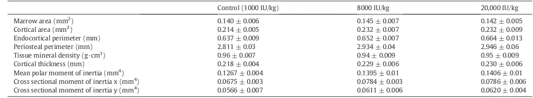
Varying dietary vitamin D3levels did not lead to statistically signifi -cant differences in cortical tissue mineral density, morphology, includ-ing cortical area, marrow area, endocortical perimeter, cortical
thickness, polar moment of inertia or cross sectional moment of inertia in either the x or y-direction (Table 4).
4. Discussion
This study investigated the effects of dietary vitamin D3on the me-chanical properties and morphology of murine tibiae. We found that provision of 20,000 IU/kg to 8 week old female mice for 4 weeks was as-sociated with greater bone strength, greater structural and material ductility, and greater toughness. This level of dietary vitamin D3was also associated with significantly higher trabecular bone volume frac-tion but no detectable change in cortical structure.
Table 3
Reference-point indentation test results.
Control (1000 IU/kg)
8000 IU/kg 20,000 IU/kg
1st cycle indentation distance (μm)
32.62 ± 1.4 31.19 ± 1.8 32.34 ± 1.9
Total indentation distance (μm) 35.61 ± 1.4 34.38 ± 2.1 34.25 ± 1.8 Indentation distance
increase (μm)
5.65 ± 0.6 5.43 ± 0.5 4.41 ± 0.2
Avg unloading (N/μm) 0.204 ± 0.006 0.205 ± 0.011 0.235 ± 0.011 Avg loading slope (N/μm) 0.168 ± 0.007 0.163 ± 0.008 0.183 ± 0.009 Avg energy dissipated (μJ) 4.88 ± 0.3 5.09 ± 0.4 4.27 ± 0.3
Tibiae from 12 week old female mice that had been on a vitamin D3dietary intervention for 4 weeks were subjected to reference point indentation and the results recorded. Values are presented as group mean ± SEM. n = 10 Control, 8000 IU/kg; n = 7 20,000 IU/kg. Table 2
Material properties.
Control (1000 IU/kg)
8000 IU/kg 20,000 IU/kg
Elastic modulus (MPa) 6224.03 ± 191.0 6090.18 ± 511.5 5682.51 ± 160.4 Ultimate stress (MPa) 160.59 ± 5.6 159.02 ± 7.8 144.89 ± 6.6 Ultimate strain 0.038 ± 0.04 0.039 ± 0.002 0.037 ± 0.002 Yield stress (MPa) 152.51 ± 5.7 148.24 ± 8.2 134.35 ± 6.4 Yield strain 0.0306 ± 0.002 0.0303 ± 0.002 0.0263 ± 0.001 Failure stress (MPa) 143.72 ± 6.9 136.77 ± 8.9 108.48 ± 8.0b Failure strain 0.0548 ± 0.003 0.0638 ± 0.004 0.0771 ± 0.006bb
Tibiae from 12 week old female mice that had been on a vitamin D3dietary intervention for 4 weeks were subjected to three-point-bending and the material properties were de-termined. Values are presented as group mean ± SEM. n = 10 Control, 8000 IU/kg; n = 7 20,000 IU/kg.
b
pb0.05. bb
pb0.01 vs control.
Fig. 2.Dietary interventions with vitamin D3 were shown to have an effect on the material properties of bone. Dietary intervention with 20,000 IU/kg vitamin D3 is associated with reduced Failure Stress, greater failure strain, post-yield strain, and toughness when compared to the control. (A) Failure stress, (B) failure strain, (C) post-yield strain and (D) toughness were determined by 3-point-bending. For all graphs (Control (1000 IU/kg) n = 10, 8000 IU/kg n = 10, 20,000 IU/kg n = 7), columns represent mean/group and error bars indicate SEM, where *pb0.05, **pb0.01 and ***pb0.001. MPa = megapascals.
47
The greater trabecular bone mass in mice administered the highest level of vitamin D3is consistent with previous studies (Lee et al., 2010), where vitamin D interventions resulted in greater effects on tra-becular but not cortical bone.Lee et al. (2010)demonstrated byμCT that increasing dietary vitamin D3levels administered to 10 week old rats (from approximately 100 IU/kg to 600 IU/kg), led to greater femoral and vertebral trabecular bone mass with the higher doses. Again, consis-tent with our observations, they did not observe any change in cortical bone dimensions; indicating that vitamin D dietary interventions
predominantly modify trabecular rather than cortical bone structure. They observed a significant effect at a lower Vitamin D3dosage com-pared to our study, likely due to the use of a vitamin D3deficient rat model and a significantly longer intervention period (20 weeks com-pared to 4 weeks).
Although we observed no significant change in cortical dimensions, the mechanical properties of cortical bone were greatly altered by the highest level of vitamin D3supplementation, indicating a likely change in bone composition. The greater post-yield displacement, post-yield
Fig. 4.Reconstructed images, using ParaView (version 3.14.1), of the sample from each group; (A) Control (1000 IU/kg), (B) 8000 IU/kg and (C) 20,000 IU/kg, with the trabecular bone volume fraction closest to that of the group mean.
strain, failure displacement and failure strain in mice on the 20,000 IU/ kg dietary intervention indicated that both the structural and material ductility were greater than in mice supplied with the control diet. Fur-thermore, dietary levels of 20,000 IU/kg vitamin D3resulted in the need for a higher energy requirement for bone fracture, both over the whole bone structure (work-to-failure) and the material (toughness). Previous studies reported that treatment of ovariectomised rats with 0.5μg/kg/day 1,25-dihydroxyvitamin D3(1,25OHD3) significantly in-creased toughness compared to both the ovariectomized control and non-ovariectomized control (Aerssens et al., 1994; Zimmermann et al., 2015). While our study did not measure 1,25OHD3levels, previous re-ports (Aerssens et al., 1994; Zimmermann et al., 2015) suggest that it is likely that the greater toughness observed with the 20,000 IU/kg die-tary intervention is due to an increased level of circulating 1,25OHD3, resulting in a more ductile and tougher bone; however this will need fu-ture confirmation.
The material properties of bone, specifically the ease with which col-lagenfibrils slide over each other can increase toughness since in-creased ability to slide will allow the bone to deform and absorb any applied loads (Zimmermann et al., 2015). Through reference point micro-indentation (RPI) we examined the material properties of the bone but observed no differences between any of our dietary interven-tion groups. Paschalis et al., (2016) (Paschalis et al., 2016) reported that vitamin D and calcium supplementation for a three year period in post-menopausal osteoporosis significantly altered bone mineral and organic matrix quality. The lack of an obvious change using RPI may be due to insufficient precision of this method; this could be overcome through the use of a nano- or pico-indentor. Additionally, a limitation of RPI is that it only examines the material properties on the periosteal surface. It is possible that our observed changes in material properties may be due to the incorporation of mineral at a deeper level or on the endocortical surface. Future studies using Raman microspectroscopy (as used by Paschalis et al.) may permit these questions to be addressed. The greater ductility and toughness observed in the bones of our growing mice suggest that dietary supplementation with vitamin D3 may be a suitable method to increase peak bone mass during adoles-cence to prevent development of osteoporosis later in life. This strategy of decreasing fracture risk in the elderly by optimising bone health dur-ing bone development was previously suggested byRizzoli et al. (2010), and was supported by a positive correlation between childhood bone health and bone health in adulthood. Further evidence to support this strategy comes from a number of intervention studies in adolescents which have reported significant increases in bone mineral content (BMC) and bone mineral density (BMD) with vitamin D3 supplementa-tion.Viljakainen et al. (2006)reported that daily tablets of 5 and 10μg vitamin D3supplementation (approximately 200–400 IU/d) for 1 year in adolescent girls resulted in significant increases in femoral bone BMC. LikewiseEl-Hajj Fuleihan et al. (2006)treated adolescent girls with 14,000 IU (equivalent to 2000 IU/d) for 1 year and observed an in-crease in hip BMC. Finally,Du et al. (2004)observed in 10–12 year old girls that daily consumption of milk fortified with 8μg vitamin D3
(320 IU/d) for a period of 2 years resulted in size adjusted increases in BMC and BMD. Our experiments were conducted on 8-week-old female mice, which are post-puberty, but still growing, and in these mice we saw an increase in bone strength through a 20,000 IU/kg dietary supple-mentation, suggesting that clinical dietary vitamin D3supplementation may be an avenue to increase bone health in young, growing bones, and could result in decreased fracture risk in adulthood. However, this re-quires additional research.
In conclusion, we observed that high levels of dietary vitamin D3in adult mice resulted in greater tibial ductility, toughness and greater tra-becular bone volume fraction. Our results suggest that high levels of di-etary vitamin D3may be suitable for achieving a higher peak bone mass in adulthood and thereby preventing osteoporosis.
Supplementary data to this article can be found online athttp://dx. doi.org/10.1016/j.bonr.2017.02.001.
References
Aerssens, J., Van Audekercke, R., Talalaj, M., Van Vlasselaer, P., Bramm, E., Geusens, P., Dequeker, J., 1994.Effect of 1 alpha-vitamin D3 on bone strength and composition in growing rats with and without corticosteroid treatment. Calcif. Tissue Int. 55 (6), 443–450.
Andersen, R., Molgaard, C., Skovgaard, L.T., Brot, C., Cashman, K.D., Jakobsen, J., Lamberg-Allardt, C., Ovesen, L., 2008.Effect of vitamin D supplementation on bone and vitamin D status among Pakistani immigrants in Denmark: a randomised double-blinded pla-cebo-controlled intervention study. Br. J. Nutr. 100, 197–207.
Anderson, P.H., Atkins, G.J., Turner, A.G., Kogawa, M., Findlay, D.M., Morris, H.A., 2011. Vi-tamin D metabolism within bone cells: effects on bone structure and strength. Mol. Cell. Endocrinol. 347 (1–2), 42–47.
Bischoff-Ferrari, H.A., Willett, W.C., Wong, J.B., Giovannucci, E., Dietrich, T., Dawson-Hughes, B., 2005.Fracture prevention with vitamin D supplementation: a meta-anal-ysis of randomized controlled trials. JAMA 293 (18), 2257–2264.
Cheng, S., Tylavsky, F., Kroger, H., Karkkainen, M., Lyytikainen, A., Koistinen, A., Mahonen, A., Alen, M., Halleen, J., Vaananen, K., Lamberg-Allardt, C., 2003.Association of low 25-hydroxyvitamin D concentrations with elevated parathyroid hormone concentra-tions and low cortical bone density in early pubertal and prepubertal Finnish girls. Am. J. Clin. Nutr. 78, 485–492.
Dawson-Hughes, B., Dallal, G.E., Krall, E.A., Harris, S., Sokoll, L.J., Falconer, G., 1991.Effect of vitamin D supplementation on wintertime and overall bone loss in healthy postmen-opausal women. Ann. Intern. Med. 115, 505–512.
Du, X., Zhu, K., Trube, A., Zhang, Q., Ma, G., Hu, X., Fraser, D.R., Greenfield, H., 2004. School-milk intervention trial enhances growth and bone mineral accretion in Chinese girls aged 10–12 years in Beijing. Br. J. Nutr. 92, 159–168.
El-Hajj Fuleihan, G., Nabulsi, M., Tamim, H., Maalouf, J., Salamoun, M., Khalife, H., Choucair, M., Arabi, A., Vieth, R., 2006.Effect of vitamin D replacement on musculo-skeletal parameters in school children: a randomized controlled trial. J. Clin. Endocrinol. Metab. 91, 405–412.
Fares, J.E., Choucair, M., Nabulsi, M., Salamoun, M., Shahine, C.H., Fuleihan GEl, H., 2003. Effect of gender, puberty, and vitamin D status on biochemical markers of bone re-modeling. Bone 33, 242–247.
Jepsen, K.J., Silva, M.J., Vashishth, D., Guo, X.E., van der Meulen, M.C., 2015.Establishing Biomechanical Mechanisms in Mouse Models: Practical Guidelines for Systematically Evaluating Phenotypic Changes in the Diaphyses of Long Bones. pp. 1523–4681. Johnson, R.W., Brennan, H.J., Vrahnas, C., Poulton, I.J., McGregor, N.E., Standal, T., Walker,
E.C., Koh, T.T., Nguyen, H., Walsh, C.C., Forwood, M.R., Martin, T.J., Sims, N.A., 2014. The primary function of gp130 signaling in osteoblasts is to maintain bone formation and strength, rather than promote osteoclast formation. J. Bone Miner. Res. 29 (6), 1492–1505.
Lee, A.M., Anderson, P.H., Sawyer, R.K., Moore, A.J., Forwood, M.R., Steck, R., Morris, H.A., O'Loughlin, P.D., 2010.Discordant effects of vitamin D deficiency in trabecular and Table 4
Bone morphology.
Control (1000 IU/kg) 8000 IU/kg 20,000 IU/kg
Marrow area (mm2
) 0.140 ± 0.006 0.145 ± 0.007 0.142 ± 0.005
Cortical area (mm2
) 0.214 ± 0.005 0.232 ± 0.007 0.232 ± 0.009
Endocortical perimeter (mm) 0.637 ± 0.009 0.652 ± 0.007 0.664 ± 0.013
Periosteal perimeter (mm) 2.811 ± 0.03 2.934 ± 0.04 2.946 ± 0.06
Tissue mineral density (g·cm3) 0.96 ± 0.007 0.94 ± 0.009 0.95 ± 0.009
Cortical thickness (mm) 0.218 ± 0.004 0.229 ± 0.006 0.230 ± 0.006
Mean polar moment of inertia (mm4
) 0.1267 ± 0.004 0.1395 ± 0.01 0.1406 ± 0.01
Cross sectional moment of inertia x (mm4
) 0.0675 ± 0.003 0.0784 ± 0.003 0.0786 ± 0.006
Cross sectional moment of inertia y (mm4
) 0.0566 ± 0.007 0.0611 ± 0.006 0.0620 ± 0.004
Tibiae from 12 week old female mice that had been on a vitamin D3dietary intervention for 4 weeks had their length measured with calipers and then were imaged with aμCT scanner from which their bone morphology was determined. Values are presented as group mean ± SEM. n = 10 Control, 8000 IU/kg; n = 7 20,000 IU/kg.
49
cortical bone architecture and strength in growing rodents. J. Steroid Biochem. Mol. Biol. 121 (1–2), 284–287.
Lehtonen-Veromaa, M.K., Mottonen, T.T., Nuotio, I.O., Irjala, K.M., Leino, A.E., Viikari, J.S., 2002.Vitamin D and attainment of peak bone mass among peripubertal Finnish girls: a 3-y prospective study. Am. J. Clin. Nutr. 76, 1446–1453.
Lips, P., 2001.Vitamin D deficiency and secondary hyperparathyroidism in the elderly: consequences for bone loss and fractures and therapeutic implications. Endocr. Rev. 22, 477–501.
Michaëlsson, K., Melhus, H., Bellocco, R., Wolk, A., 2003.Dietary calcium and vitamin D in-take in relation to osteoporotic fracture risk. Bone 32 (6), 694–703.
O'Riordan, J.L., Bijvoet, O.L., 2014.Rickets before the discovery of vitamin D. Bonekey Rep. 3, 478.
Outila, T.A., Karkkainen, M.U., Lamberg-Allardt, C.J., 2001.Vitamin D status affects serum parathyroid hormone concentrations during winter in female adolescents: associa-tions with forearm bone mineral density. Am. J. Clin. Nutr. 74, 206–210.
Paschalis, E.P., Gamsjaeger, S., Hassler, N., Fahrleitner-Pammer, A., Dobnig, H., Stepan, J.J., Pavo, I., Eriksen, E.F., Klaushofer, K., 2016. Vitamin D and calcium supplementation for three years in postmenopausal osteoporosis significantly alters bone mineral and or-ganic matrix quality. Bonehttp://dx.doi.org/10.1016/j.bone.2016.11.002.
Rajakumar, K., 2003.Vitamin D, cod-liver oil, sunlight, and rickets: a historical perspec-tive. Pediatrics 112, e132–e135.
Rajakumar, K., Thomas, S.B., 2005.Reemerging nutritional rickets: a historical perspective. Arch. Pediatr. Adolesc. Med. 159, 335–341.
Reid, I.R., Bolland, M.J., Grey, A., 2013.Effects of vitamin D supplements on bone mineral density: a systematic review and meta-analysis. Lancet 383 (9912), 146–155. Rizzoli, R., Bianchi, M.L., Garabedian, M., McKay, H.A., Moreno, L.A., 2010.Maximizing
bone mineral mass gain during growth for the prevention of fractures in the adoles-cents and the elderly. Bone 46 (2), 294–305.
Sanders, K.M., Stuart, A.L., Williamson, E.J., Simpson, J.A., Kotowicz, M.A., Young, D., Nicholson, G.C., 2010.Annual high-dose oral vitamin D and falls and fractures in older women: a randomized controlled trial. JAMA 303, 1815–1822.
Smith, H., Anderson, F., Raphael, H., Maslin, P., Crozier, S., Cooper, C., 2007.Effect of annual intramuscular vitamin D on fracture risk in elderly men and women–a population-based, randomized, double-blind, placebo-controlled trial. Rheumatology (Oxford) 46, 1852–1857.
Tang, B.M., Eslick, G.D., Nowson, C., Smith, C., Bensoussan, A., 2007.Use of calcium or cal-cium in combination with vitamin D supplementation to prevent fractures and bone loss in people aged 50 years and older: a meta-analysis. Lancet 370 (9588), 657–666. Turner, C.H., Burr, D.B., 1993.Basic biomechanical measurements of bone: a tutorial. Bone
14 (4), 595–608.
Viljakainen, H.T., Natri, A.M., Karkkainen, M., Huttunen, M.M., Palssa, A., Jakobsen, J., Cashman, K.D., Molgaard, C., Lamberg-Allardt, C., 2006.A positive dose-response ef-fect of vitamin D supplementation on site-specific bone mineral augmentation in ad-olescent girls: a double-blinded randomized placebo-controlled 1-year intervention. J. Bone Miner. Res. 21, 836–844.
Wagner, C.L., Greer, F.R., 2008.Prevention of rickets and vitamin D deficiency in infants, children, and adolescents. Pediatrics 122, 1142–1152.
Wagner, C.L., Hulsey, T.C., Fanning, D., Ebeling, M., Hollis, B.W., 2006.High-dose vitamin D3 supplementation in a cohort of breastfeeding mothers and their infants: a 6-month follow-up pilot study. Breastfeed. Med. 1, 59–70.
Winzenberg, T.M., Powell, S., Shaw, K.A., Jones, G., 2010.Vitamin D supplementation for improving bone mineral density in children. Cochrane Database Syst. Rev., CD006944 Winzenberg, T., Powell, S., Shaw, K.A., Jones, G., 2011.Effects of vitamin D supplementa-tion on bone density in healthy children: systematic review and meta-analysis. BMJ 342, c7254.