R E V I E W
Monitoring of Minimal Residual Disease (MRD) in
Chronic Myeloid Leukemia: Recent Advances
This article was published in the following Dove Press journal: Cancer Management and Research
Cosimo Cumbo *
Luisa Anelli * Giorgina Specchia Francesco Albano
Department of Emergency and Organ Transplantation (D.E.T.O.), Hematology Section, University of Bari, Bari 70124, Italy
*These authors contributed equally to this work
Abstract:Chronic myeloid leukemia (CML) is a myeloproliferative neoplasm caused by the BCR-ABL1fusion gene generation as a consequence of the t(9;22)(q34;q11) rearrangement.
The identification of the BCR-ABL1 transcript was of critical importance for both CML
diagnosis and minimal residual disease (MRD) monitoring. In this review, we report the recent advances in the CML MRD monitoring based on RNA, DNA and protein analysis.
The detection of theBCR-ABL1transcript by the quantitative reverse-transcriptase
polymer-ase chain reaction is the gold standard method, but other systems bpolymer-ased on digital PCR or on GeneXpert technology have been developed. In the last years, DNA-based assays showed
high sensitivity and specificity, and flow cytometric approaches for the detection of the
BCR–ABL1 fusion protein have also been tested. Recently, new MRD monitoring systems
based on the detection of molecular markers other than theBCR-ABL1fusion were proposed.
These approaches, such as the identification of CD26+ leukemic stem cells, microRNAs and
mitochondrial DNA mutations, just remain preliminary and need to be implemented. In the precision medicine era, the constant improvement of the CML MRD monitoring practice could allow clinicians to choose the best therapeutic algorithm and a more accurate selection of CML patients eligible for the tyrosine kinase inhibitors discontinuation.
Keywords:chronic myeloid leukemia, minimal residual disease, MRD monitoring
Introduction
Chronic myeloid leukemia (CML) is a clonal myeloproliferative disorder driven by the chimeric BCR-ABL1 oncoprotein, resulting from a t(9;22)(q34;q11) balanced reciprocal translocation. The rearrangement produces the Philadelphia (Ph) chro-mosome where theBCR-ABL1oncogene is generated; its chimeric transcript is the marker of the disease.1,2 Tyrosine kinase inhibitors (TKIs) therapy targets
BCR-ABL1 positive cells and induces hematologic and molecular remission in
80–90% of CML patients, with a survival rate comparable to that of age-matched healthy individuals.3–5 Response to TKI treatment is assessed by hematologic, cytogenetic, and molecular testing performed at specific time-points during follow-up. Detection of theBCR-ABL1transcript level by quantitative reverse-transcriptase polymerase chain reaction (RQ-PCR) is the gold standard method for monitoring CML minimal residual disease (MRD) and the optimal CML patient management.6 In fact, standardized and regular MRD monitoring in CML patients is essential for defining the response to treatment and choosing the best therapeutic strategy (as well as providing prognostic information) and also for selecting patients in sus-tained deep molecular response who are eligible for TKI discontinuation.7 This gains relevance in the era of targeted therapy, where the introduction of MRD
Correspondence: Francesco Albano Department of Emergency and Organ Transplantation (D.E.T.O.), Hematology Section, University of Bari, P.zza G. Cesare, 11, Bari 70124, Italy Tel +39 080-5478031 Fax +39 080-5508369 Email francesco.albano@uniba.it
Cancer Management and Research
Dove
press
open access to scientific and medical research
Open Access Full Text Article
Cancer Management and Research downloaded from https://www.dovepress.com/ by 118.70.13.36 on 24-Aug-2020
monitoring has profoundly transformed patients management.8 Efficient methods for disease monitoring should guarantee fast, inexpensive and sensitive disease detection. In fact, even if in the last two decades the standardization of CML monitoring has remained one of the most laborious procedures, the efficacy of different new approaches has recently been tested. The main strate-gies developed in the last years, are based on BCR-ABL1
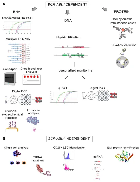
chimeric gene or transcript or protein detection, although some alternative strategies have been made (Figure 1). In this review we summarize the recent advances in the CML MRD monitoring, considering the advantages and disad-vantages of each approach and focusing on future perspectives.
BCR-ABL1-Dependent MRD
Monitoring
RNA-Based Approaches
RQ-PCR Monitoring and Standardization of the Experimental Procedure
CML molecular monitoring by RQ-PCR is based on total RNA extraction from peripheral blood (PB) or bone mar-row (BM) cells, reverse-transcription of RNA into cDNA, and quantitative co-amplification of the BCR-ABL1 tran-script and of an internal housekeeping gene. Molecular monitoring in CML should be performed according to the established Europe Against Cancer criteria, defining specific primer/probe systems for both BCR-ABL1 and
ABL1 genes.9 As many experimental steps and technical details can cause variability and heterogeneity in RQ-PCR analysis, the EUropean Treatment Outcome Study (EUTOS) program in Europe and the LabNet network in Italy, promoted the standardization of RQ-PCR procedures and establishment of the expression of BCR-ABL1 tran-script level as“international scale (IS)”.10–13The baseline
BCR-ABL1 RNA level (100% IS) was defined as the
median BCR-ABL1 transcript level to reference gene ratio in 30 newly diagnosed CML patients in the IRIS study.14,15 The most commonly used reference genes are
ABL1, GUSB orBCR; ABL1is used by most laboratories
worldwide,GUSBis used by some European laboratories, whereasBCRis employed as reference gene in Australasia and some US laboratories.12,14,16 In the IRIS study, the second BCR-ABL1 IS level corresponds to a 1000-fold (3-log) reduction in the BCR-ABL1 transcript level compared to the IRIS baseline, defining a major molecular response (MMR). There are two possible ways of
calculating the IS: according to the Conversion Factor (CF) or using the BCR-ABL1 reference standard method. At the time of the IRIS trial, the Adelaide laboratory served as central reference laboratory, and sample exchange was performed with 38 different international laboratories to attribute the specific CF expressing the
BCR-ABL1transcript level according to the IS.17To deter-mine the CF, each set of data generated by a specific laboratory was compared with that obtained by the refer-ence laboratory, using the statistical comparison procedure of Bland and Altman.17 In this method theBCR-ABL1 IS value is expressed as follows: [(sum of BCR-ABL1
copies)/(sum of reference gene copies)] × CF × 100.13,17 However, this system is time-consuming and laborious due to the need for many samples exchange between different laboratories. The second method for standardization to the IS, that will probably become the main system in the future, is the use of secondaryBCR-ABL1 reference stan-dard samples, now commercially available although they are not yet FDA-approved.18 These reference standards have been calibrated to the World Health Organization (WHO) primary reference standards for BCR-ABL1
RNA, four reference samples that correspond to the BCR-ABL1 IS values of 10%, 1%, 0.1%, and 0.01%. Primary standards were obtained in limited quantities by diluting the K562 cell line (BCR-ABL1-positive) in the HL60 cell line (BCR-ABL1-negative).18 Another reference standard is ERM-AD623, a certified plasmid developed for the standardization of BCR-ABL1 RQ-PCR19 which contains fragments of e14-a2 BCR-ABL1 fusion transcripts, BCR
andGUSB. Six different concentrations of linearized plas-mid were quantified by digital PCR and tested in several laboratories. The use of ERM-AD623 allows the calibra-tion of internal reference materials to improve the accu-racy of the results. According to the European Leukemia Net (ELN) recommendations, RQ-PCR analysis should be performed every 3 months until the achievement of MMR and even after the MMR is confirmed, as a close monitor-ing is required in view of a possible treatment discontinuation.20 If MMR is lost during follow-up, the occurrence ofABL1 mutations should be investigated and molecular monitoring should be carried out more frequently.10,21,22 Follow up evaluation can be performed using PB or BM samples, and typically, both BCR-ABL1
and the reference gene are tested in duplicate.13The BCR-ABL1quantification is considered positive when any of the three replicates are positive, and thefinal number of BCR-ABL1 and reference gene copies is the total across
Cancer Management and Research downloaded from https://www.dovepress.com/ by 118.70.13.36 on 24-Aug-2020
Figure 1Methods for CML MRD monitoring. The strategies are based on the identification ofBCR-ABL1fusion (A) or on the detection of molecular markers independent fromBCR-ABL1(B).
Abbreviations:bkp, breakpoint; PLA, proximity ligation assay; LSC, leukemic stem cells.
Cancer Management and Research downloaded from https://www.dovepress.com/ by 118.70.13.36 on 24-Aug-2020
replicates sum. When different housekeeping genes are used by different laboratories, the results can be compared using the ERM-AD623 plasmid as a RQ-PCR calibrator.19
Molecular Response (MR) Milestones
A recent version of ELN recommendations reformulated crucial aspects of CML patients molecular monitoring.20 In the past years, RQ-PCR monitoring and therapeutic improvements have led to the definition of given MR milestones at specific time-points during patient treatment. Based on the achievement of the cytogenetic and/or mole-cular milestones, the ELN guidelines divide CML patients into three groups: optimal response, warning and failure.10,20Thefirst crucial time-point is the achievement of aBCR-ABL1 IS transcript level < 10% (a reduction by at least 1 log of BCR-ABL1 transcript levels from the standardized IS baseline) after 3 months of therapy,14 defining Early Molecular Response (EMR). EMR is con-sidered to be a crucial treatment response, predicting the outcome of CML patients receiving either imatinib or second generation TKI, influencing event-free survival (EFS), progression-free survival (PFS), and overall survi-val (OS).23,24 However, not achieving EMR is considered a “warning” rather than therapy “failure” by recent ELN and European Society of Medical Oncology (ESMO) recommendations.10,25 Recent evidence showed that the kinetics ofBCR-ABL1transcripts during thefirst 3 months is much more informative than the achievement of EMR at 3 months. The definition of “BCR-ABL1 halving time” was introduced by Branford et al in 2014, showing that patients with a halving time of less than 76 days had a better outcome, even if they did not achieve EMR at 3 months.26 These data were subsequently confirmed by other studies of CML patients receiving second generation
TKI.27,28 However, the study of BCR-ABL1 transcript
kinetics has not yet been incorporated in follow-up recom-mendations, as the experimental procedure needs to be developed, such as the need for two or more consecutive quantitative analyses within thefirst three months and the switch to a control gene other than ABL1.14 The second crucial molecular milestone is achieved when the BCR-ABL1IS transcript level is < 1% (2 log reduction) after 6 months of therapy. The third molecular milestone is the achievement of MMR, consisting in the reduction of the
BCR-ABL1 transcript level by at least 3 logs (MR3) after 12 months. The definition of MMR was introduced during the IRIS (International Randomized Study of Interferon and STI-571) study, that led to imatinib registration as
first line treatment.29 The availability of more potent second generation TKI allowed the identification of Deep Molecular Response (DMR), defined as a BCR-ABL1IS transcript level of≤0.01%. DMR can be further subdivided into MR4, MR4.5, or MR5when the logarithmic reduction is 4, 4.5, or 5 logs, yieldingBCR-ABL1≤0.01%,
≤ 0.0032%, and ≤ 0.001%, respectively.13 The sample is considered good quality whenABL1 transcript copies are more than 10.000 (corresponding to 24.000GUSBcopies) at molecular response MR4, but the minimumABL1copies have increased to 32.000 and 100.000 at MR4.5, and MR5, respectively.13 RQ-PCR monitoring allowed the stratifi ca-tion of CML patients into “optimal responders” that can continue the same TKI,“warning cases”to be considered for a possible TKI switch, and“failed patients”who do not reach molecular milestones at 3, 6, or 12 months and need an immediate change of therapy.10,30,31The introduction of more potent and efficient second generation TKI in front-line therapy is allowing a more rapid achievement of each milestone, with a sustained deep MR.32,33
Molecular Monitoring During TKI Discontinuation
Therapeutic success of the last years allowed the achieve-ment for most newly diagnosed CML patients of a normal life expectancy, comparable to that of age matched indivi-duals in the general population.10,20After survival, another crucial objective in CML clinical management is repre-sented by the achievement of a stable DMR after long term TKI therapy, that is the prerequisite for Treatment Free Remission (TFR). The achievement of a stable DMR after long term TKI therapy is the prerequisite for Treatment Free Remission (TFR) that is the most important objective in CML clinical management. Stop Imatinib (STIM1) and TWISTER were the first two TFR pivotal studies, which enrolled patients with at least 3 years of imatinib therapy and more than 2 years of confirmed undetectable BCR-ABL1, with a MR4.5 or MR5 sensitivity test.34,35 About 50–60% of cases in both studies showed molecular relapse (re-emergence of BCR-ABL1 transcripts or MMR loss), mostly during thefirst 6 months after imatinib suspension and nearly all cases regained MR after restarting treat-ment. Several subsequent TKI suspension studies were performed in CML patients treated with dasatinib or nilo-tinib for at least 2 years and maintaining durable deep MR. These studies showed TFR without molecular relapse in about 50 to 69% of cases.34,36-38As regards recommenda-tions on discontinuation criteria, the National Comprehensive Cancer Network (NCCN) guidelines and
Cancer Management and Research downloaded from https://www.dovepress.com/ by 118.70.13.36 on 24-Aug-2020
other expert reviews suggested the selection of patients with a non-high Sokal score, showing a typicalBCR-ABL1
transcript (b2a2 or b3a2) and a MR4 or MR4.5 level of molecular response for a minimum of 1–2 years under TKI therapy.11,39,40 More stringent TFR criteria were defined by recent ELN recommendations, requiring typical BCR-ABL1 transcripts, a minimal TKI therapy duration of 4–5 years, and a DMR (MR4or better) duration of more than 2 years.20 A durable DMR is considered a more important selection criterion than the duration of TKI treatment, ensuring a greater TKI discontinuation success.39 Because of the high frequency of molecular relapse, TFR trials require monthly RQ-PCR monitoring for the first 6 to 12 months, with a possible gradual reduction thereafter.20,34,41,42 However, RQ-PCR might not be the best approach for selecting CML cases to include in TKI discontinuation trials and also for molecular monitoring during TFR, as 50–60% of patients with undetectable DMR are expected to lose MMR.43–45 Several studies have demonstrated the persistence of leukemic cells in the BM niche of CML patients treated with TKI and with undetectable BCR-ABL1 transcript levels by RQ-PCR. A recent flow cytometry study showed the presence of circulating CD26+ leukemic stem cells (LSC) in about 30% of CML patients with undetectable BCR-ABL1 by RQ-PCR.46 Failure to detect the BCR-ABL1 transcript could be attributed to low RQ-PCR sensitivity or to absent gene transcription in quiescent LSC that reside in a hypoxic niche.47To address this issue, new more sensi-tive methodologies have been investigated to identify pos-sible residual LSC and to allow a more accurate selection of CML patients likely to benefit from TFR.
BCR-ABL1Transcript Monitoring by Digital PCR (dPCR)
In the last years, dPCR has revolutionized the molecular monitoring of MRD in hematological diseases.48–52dPCR is the most accurate and sensitive method for measuring the abundance of specific nucleic acids, providing their absolute quantification and offering a greater precision and reproducibility compared to RQ-PCR.53,54Recent stu-dies showed that dPCR is more efficient than RQ-PCR for CML MRD monitoring and for a more accurate selection of patients eligible for TFR.55–59 In dPCR the biological sample analyzed is divided into thousands or millions of separate reactions and amplification is performed with a higher efficiency in many separate microscopic parti-tions, in wells or droplets (ddPCR).60 Each partition is evaluated as positive or negative, according to the
presence or absence of template molecules and the nucleic acids copy number in the starting sample is identified by Poisson statistics.61In this way, an absolute quantification can be performed with a greater sensitivity and without the need for a standard curve.62 dPCR is considered able to detect a single BCR-ABL1 positive cell out of 107and is less subject to inhibitory agents and nonspecific amplification.55,56 Several independent studies demon-strated a greater sensitivity of dPCR as compared to RQ-PCR, demonstrating a significant percentage of CML cases that lost TFR and were found to beBCR-ABL1positive by dPCR. Moreover, cases in follow-up showing the loss of DMR by RQ-PCR were identifiable by dPCR within 3 months before.57,59,63,64 Recent studies, such as Life After Stopping Tyrosine Kinase Inhibitors (LAST) and the Imatinib Suspension and Validation (ISAV) are inves-tigating the efficacy of dPCR for the molecular monitoring of patients with undetectable BCR-ABL1 who have sus-pended TKI therapy.65,66 Because of several advantages and high sensitivity, dPCR has been used to evaluate the circulating tumor burden in cancer research and for the quantification of circulating tumor DNA in plasma from solid neoplasms.67Recently, dPCR has been employed for the quantification of theBCR-ABL1transcript in exosomes from PB of CML patients under TKI treatment,68 demon-strating its success as a possible innovative monitoring system based on exosomes analysis. Exosomes are extra-cellular vesicles (EVs) secreted by living cells exocytosis mediating intracellular and microenvironment communica-tion. They are lipid nanoparticles (50-100 nm) produced either constitutively or upon induction, and their molecular composition depends on the parent cell origin.69,70To date, exosomes have been especially employed for the diagnosis of solid tumors on the basis of their internal proteins and nucleic acids, or on the membrane surface expression of cancer-specific antigens. Moreover, exosomes have also been isolated from leukemic cells and can be utilized as prognostic, diagnostic and therapeutic biomarkers in dif-ferent hematologic neoplasms, such as acute myeloid leu-kemia, multiple myeloma and chronic lymphocytic leukemia.71–74 Microvesicles released by the K562 cell line have been reported to contain bothBCR-ABL1 DNA and transcript and are able to induce the CML phenotype in transplanted rats, or can activate BM mesenchymal stem cells promoting leukemic cells proliferation.75,76Few data are available on the identification and quantification of
BCR-ABL1 in exosomes from CML patients, generally
isolated from PB. In the explorative study by Bernardi
Cancer Management and Research downloaded from https://www.dovepress.com/ by 118.70.13.36 on 24-Aug-2020
et al, ten CP-CML patients were analyzed by RQ-PCR and dPCR on both PB cells and on tumor-enriched exosomes, demonstrating the presence of theBCR/ABL1transcript in exosomes derived from CML patients. However these are very preliminary data, therefore the clinical and prognostic significance ofBCR-ABL1positive exosomes and their use in CML molecular monitoring remains to be investigated.68 However, despite several advantages and applications, dPCR shows some limits as it generally requires a longer experimental time and can be condi-tioned by errors in pre-analytical phases such as sampling, RNA extraction and cDNA synthesis. Moreover, positive and negative control samples are needed, the quantification of a reference gene is still useful to evaluate the sample quality, and the use of a CF is required for the expression of results according to the IS.77 Therefore, international standardization is needed before dPCR can be adopted as a routine molecular method for CML monitoring.
The GeneXpert System
The GeneXpert system is a cartridge-based RQ-PCR auto-mated method developed by Cepheid (California, USA), that is able to detect BCR-ABL1 p210 transcripts when directly processing PB samples. The GeneXpert instrument utilizes microfluidics in a cartridge and performs RNA extraction, RT-PCR andBCR-ABL1 fluorescence detection in one reaction.78This is a very simple, fast system, requir-ing a reduced technical expertise. The RQ-PCR is per-formed within a cartridge not reporting BCR-ABL1
transcript levels on the IS; therefore a specific CF needs to be developed and used in addition to a cartridge lot-specific calibration, in order to obtain the BCR-ABL1
estimation on IS.79,80 Recent studies demonstrated a good correlation between the standardized RQ-PCR and the GeneXpert systems for BCR-ABL1 monitoring,79,81 and a new cutoff of 1.5% at 3 months has been proposed as predictive of both cytogenetic and molecular response at 12 months.82Secondly, the initial accuracy of the BCR-ABL1
test on the GeneXpert, fails at very low transcript levels, below 0.01% BCR-ABL1 (MR4);79,83 however, soon after Cepheid introduced a GeneXpert assay with an enhanced sensitivity at a level of MR4.5 on the IS.84 Moreover, atypical rareBCR-ABL1 transcripts cannot be identified by the current GeneXpert BCR-ABL1 assay; therefore, all negative suspected CML cases have to be screened by other molecular methods.85 Another development of the GeneXpert system is the BCR-ABL1 transcript quantifi ca-tion on a paper template consisting of dried blood spots
(DBSs).86About 200 µL of PB were spotted onfilter paper and analyzed after about 40 days; this study showed a high concordance between DBSs and fresh blood samples, with a high correlation (R2=0.94), revealing that this assay can work on RNA, despite it being less stable than DNA. An accurate BCR-ABL1 transcript quantification can be obtained on DBSs, in addition to a possibleABL mutation detection on DNA. This system is especially useful for low-resource regions in the world, that could send samples to specialized centers at very low cost.86
The Attomolar Electrochemical Detection
A new system for the detection of BCR-ABL1 chimeric transcript has recently been developed using an electro-chemical DNA biosensor based on polyaniline-gold nanoparticles.87cDNA samples from CML cases exposed to the biosensor surface, hybridize with fusion gene-specific DNA probes and induce changes in the ampero-metric current. The hybridization between the biosensor and the target cDNA can be evidenced by Atomic force microscopy (AFM) images, that show dramatic changes in the surface of the nanostructured platform after exposure to CML samples. This electrochemical biosensor is a fast, simple, and innovative tool for the molecular detection of the BCR-ABL1 transcript, displaying high specificity and sensitivity. It can detect concentrations as low as 0.0694 fM of recombinant plasmid containing the BCR-ABL1
fusion gene. Even if it is difficult to compare this detection limit with the BCR-ABL1 levels on IS, this is the lowest concentration at which the BCR-ABL1 transcript was detected; the biosensor can therefore be considered a useful method for MRD monitoring in CML. Other
BCR-ABL1 biosensors had previously been developed,
but featured a longer platform construction time and higher limit of fusion gene detection.88,89
Atypical BCR-ABL1Transcripts
About 2% of CML patients show atypicalBCR-ABL1 tran-scripts, different from the most common e13a2 (b2a2) and e14a2 (b3a2) isoforms, that can be identified by the con-comitant contribution of conventional cytogenetics, FISH, and RT-PCR. VariantBCR-ABL1transcripts are often e1a2 or e18/e19a2. Some studies suggested that CML patients with variant isoforms had a worse outcome than cases with the typicalBCR-ABL1fusion, but few data are available on their long-term outcomes because of the rarity of these cases.90 A study was recently performed on 4750 CML patients and a total of 19 uncommon transcripts were
Cancer Management and Research downloaded from https://www.dovepress.com/ by 118.70.13.36 on 24-Aug-2020
detected in 83 (1.7%) patients.91 The three most frequent types were e19a2, e13a3/e14a3 and e1a2. Patients carrying e19a2 (n = 16) and e1a2 (n = 11) transcripts, had signifi -cantly reduced probabilities of 1-year complete cytogenetic response and MMR compared with patients with common e13a2 and e14a2 transcripts, receiving frontline imatinib therapy. Moreover, cases with the e19a2 transcript had low probabilities of 2-year EFS and PFS and patients with the e1a2 transcript had low probability of 2-year EFS.91In other words, the rare uncommon BCR-ABL1 fusion tran-scripts may be relevant for TKI therapy outcomes. In view of these data, the MRD monitoring of these patients gains relevance. Most laboratories do not carry out RQ-PCR for atypical BCR-ABL1transcripts, as standardization to the IS has not yet been performed for variant isoforms, so the MMR evaluation cannot be accurately performed in these CML patients. In these cases, follow-up monitoring is usually performed by FISH or by conventional nested RT-PCR; otherwise, patient-specific primers and probe may be employed in RQ-PCR or dPCR.90,92A new multiplex RQ-PCR method that is able to detect at least 14 different possible isoforms of the BCR-ABL1 fusion gene in one reaction with high sensitivity and specificity, has recently been developed to allow both precise CML diagnosis and MRD monitoring.93This method is fast, simple and cheap, shows sensitivity up to 10−6 copies and is able to detect several possible major (M-bcr), minor (m-bcr), micro (μ -bcr), and nano (n-bcr) bcr BCR-ABL1 isoforms.93 On the other hand, most of the RQ-PCR methods currently used are able to detect only a fewBCR-ABL1isoforms at a time, such as e13a2, e14a2 and e1a2. Other multiplex RT-PCR methods have previously been developed, but show low sensitivity or are laborious to carry out.94,95
Alternatively Spliced BCR-ABL1Variants
Conventional and standardized BCR-ABL1 monitoring by RQ-PCR cannot distinguish between the classic BCR-ABL1 transcript and different isoforms or abnormal splicing variants. It is therefore crucial to identify conven-tionalBCR-ABL1transcripts in order to obtain an accurate MRD evaluation and to distinguish variant isoforms pos-sibly responsible for therapy resistance. Recently, a new sensitive system has been developed, based on long-range PCR amplification and ultra-deep sequencing (UDS), that is able to identify and quantify both conventional and variant or mutated BCR-ABL1transcripts.96An outer and an inner set of BCR and ABL1 primers were designed to amplify several possible mutant or variant fusion isoforms;
these large amplicons ofBCR-ABL1transcripts were used for library preparation and subjected to UDS. This analysis allowed the identification of the mutatedBCR-ABL1Ins35pb
isoform causing a conformational change in the chimeric protein, in all CML cases showing a suboptimal response to TKI therapy. This isoform cannot be distinguished from the conventional BCR-ABL1 transcript by standardized RQ-PCR, thus causing an improper categorization of the molecular response. Moreover, it has been hypothesized that theBCR-ABL1Ins35pbisoform is expressed in quiescent LSC, that continue to proliferate slowly.
DNA-Based Approaches
In the last decade, the use of RNA as input material for MRD molecular monitoring has many times been recon-sidered in favor of genomic DNA. In fact, CML patients shown by RQ-PCR to maintain a complete molecular response after stopping imatinib treatment were demon-strated to have evidence of persistent leukemic cells by DNA PCR.97 The DNA-based assay was found to have a greater sensitivity than the standardized RNA quantifi ca-tion, yielding a lower detection limit of 10−6and this is the major advantage of the use of DNA-based MRD monitor-ing in CML. In fact, the quantification of MRD by geno-mic DNA enables an estimation of cells number, unaffected by mRNA expression levels and mRNA stability.97However, even if more sensitive, the choice of DNA as input material requires a two-steps approach for the molecular MRD monitoring of CML patients. Thefirst laborious step is the patient-specificBCR-ABL1breakpoint characterization (Table 1). This is the main obstacle, in fact, unlike theBCR gene, theABL1gene possible break-point sites span a region of about 150 Kbps. The second step consists in the molecular quantification of leukemic cells in the CML patient using different methodological strategies. In the last years, several strategies have been tested for this two-steps approach, as reported below.
Patient-SpecificBCR-ABL1Breakpoint Characterization
Different methodological approaches have been developed with the aim to simplify the laborious step of patient-specific
BCR-ABL1breakpoint characterization. A protocol based on long-range PCR (using a constantBCR-primer and 10ABL1 -primers at a distance of approximately 15 kb each) and automated DNA sequencing has been proposed.98 Other authors used a PCR system originally developed for genome walking99,100 based on DNA fragmentation, adaptors liga-tion, nested-PCR amplification using adaptor-specific
Cancer Management and Research downloaded from https://www.dovepress.com/ by 118.70.13.36 on 24-Aug-2020
primers and Sanger sequencing (SS).101,102Subsequently, the Sequential Hybrid Primer PCR (SHP-PCR) was developed. The strategy combines a primary highly multiplexed PCR (six forward primers mapping on BCR and 302 reverse primers mapping on ABL1) followed by two rounds of nested-PCR (to removing nonspecific amplification) and SS.103–105Breakpoint characterization was also performed by long-range PCR (using a single forward primer inBCR
and a panel of 20 reverse primers spanning both alternative first exons ofABL1) and SS.97,106To simplify this method, narrowing the genomic area of analysis by FISH assays (preliminarily performed using seven overlapping fosmid clones) was proposed, reducing from 20 to three the number ofABL1reverse primers to be used in three subsequent long-range PCRs.107An analogous approach was used to charac-terize and compare breakpoint sequences in p210 CML, p210 ALL and p190 ALL.108In the pediatric CML context, a two-round multiplex long-range PCR was developed, designing five nested primer pairs covering the major breakpoint cluster region (M-BCR) of theBCRgene, and 20 nested primer pairs covering the ABL1 breakpoint cluster region.109,110 The advent of next generation sequencing (NGS) has greatly facilitated the breakpoint characterization step, even if it offers this opportunity only to laboratories that can afford a major investment in this type of technology. Some authors set up a strategy based on the generation of large amplicons by a two-round multiplex long-range PCR97,98 and subse-quent ultra-wide sequencing using 454 NGS technology.111 Two other groups performed an approach based on short-read sequencing (Illumina technology).112,113The study of struc-tural variants by conventional NGS approaches, remains affected by the limit of the reads length. For this reason, for the BCR-ABL1 breakpoint characterization, the MinION, a third generation long-read nanopore-based sequencer, already tested in the hematological field,114–118 has been
proposed. The length of the reads produced and the platform accessibility in term of costs and ease of use, make the technology ideal for this intent.107
Leukemic Cells Molecular Quantification
Sequencing of the region spanning the BCR-ABL1 geno-mic breakpoint is aimed at designing a patient-specific pair of primers for the molecular quantification of leukemic cells in the CML patient PB. The first attempts were based on real time quantitative PCR (qPCR)101,102,105,109 that some authors performed as nested qPCR.97,104 The comparison of qPCR on genomic DNA with the standar-dized RQ-PCR on RNA, shows an excellent correlation (r = 0.884;p< 0.0001)105but had better precision at low minimal residual disease levels,97,102,104,105 where geno-mic DNA could be more informative than mRNA for the detection of residual disease or for very immature selected progenitors.98 Furthermore, the use of DNA presents two other technical advantages: the handling of a stable mole-cule instead of RNase sensitive RNA and the use of patient-specific primers thus minimizing the PCR contam-ination risk.98 In the last years, different approaches have been made to test the dPCR performances on geno-mic BCR-ABL1 quantification.106,107,110,112 Recently, a comparison between RQ-PCR, qPCR, RT-dPCR and dPCR in the detection of low levels ofBCR-ABL1positive disease was made.112The authors demonstrated that dPCR
for BCR-ABL1 DNA is the most sensitive method
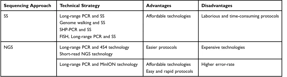
avail-able for residual-disease detection in CML and may prove useful in the management of TKI withdrawal. Furthermore, dPCR on DNA and RQ-PCR on RNA were performed in parallel to determine the relative contribution of the leukemic cell number to molecular response in 516 paired samples from 59 newly-diagnosed patients treated withfirst-line imatinib in the TIDEL-II study.106The study Table 1Different Approaches forBCR-ABL1Genomic Breakpoint Characterization
Sequencing Approach Technical Strategy Advantages Disadvantages
SS Long-range PCR and SS Affordable technologies Laborious and time-consuming protocols Genome walking and SS
SHP-PCR and SS
FISH, Long-range PCR and SS
NGS Long-range PCR and 454 technology Easier protocols Expensive technologies Short-read NGS technology
Long-range PCR and MinION technology Affordable technologies Easy and rapid protocols
Higher error-rate
Abbreviations:SS, sanger sequencing; NGS, next generation sequencing; SHP-PCR, sequential hybrid primer PCR; FISH,fluorescent in situ hybridization.
Cancer Management and Research downloaded from https://www.dovepress.com/ by 118.70.13.36 on 24-Aug-2020
clarified the kinetics of molecular response in CML, show-ing that in the first three months of treatment, the rapid decline inBCR-ABL1mRNA is due to a reduction in both cells number and transcripts level per cell. On the contrary, beyond three months, the BCR-ABL1 mRNA decrease is proportional to the leukemic cells depletion. Moreover, the results confirmed the greater sensitivity of dPCR, in fact
BCR-ABL1 DNA was quantifiable in 48% of undetectable
BCR-ABL1mRNA samples, making MRD quantifiable for
an additional 5–18 months (median 12 months).106 The greater sensitivity of the DNA-based assay, that has been widely demonstrated in the last years, could be especially interesting in the case of the TKI-discontinuation in CML. In this crucial step of MRD monitoring, this approach may help to distinguish those patients who can from those who should not undergo discontinuation.119 In fact one of the primary aims of the MRD monitoring is to avoid relapse, that occurs in about 50% of patients achieving and main-taining deep molecular response who stop TKI therapy.120
Protein-Based Approaches
The chimeric BCR-ABL1 protein was considered as a possible detection target to detect for CML MRD monitor-ing. For this purpose, aflow cytometric immunobead assay for the detection of BCR–ABL1 fusion proteins (p190, p210 and p230) in cell lysates was developed.121The approach, using a bead-bound anti-BCR catching antibody and a fluorochrome-conjugated anti-ABL1 detection antibody, was tested on 145 samples (BM or PB) and showed full concordance with RT-PCR of fusion gene transcripts. The limit of detection calculated analyzing serial dilutions of three BCR–ABL1 positive cell lines (TOM-1, K562 and AR230) showed a sensitivity of at least 1%, higher than karyotyping (around 5%) and FISH (2–4%), but lower than the PCR technique.121The assay, commonly named the CBA (cytometric bead array) system, was subsequently adopted by other authors and tested in different BCR-ABL1 positive hematological diseases.122,123 It was demonstrated to be a rapid, easy technique for specific detection of BCR–ABL1 proteins in leukemic cells. The technique is independent of the
BCRgene breakpoint, does not need special laboratory facil-ities other than a routineflow cytometer, and yields results in around four hours. This could be useful in countries where molecular diagnostics is not readily available to support clin-ical practice.121 Furthermore, a novel approach to detecting and enumerating cells positive for the BCR-ABL1 fusion protein was recently proposed.124 The technique combines the in situ proximity ligation assay with flow cytometry as
readout (PLA-flow). The BCR and ABL1 parts of the chimeric protein are targeted with an antibody each, the fluorescent signals are amplified by rolling circle amplification and the BCR-ABL1 positive cells are detected at frequencies as low as one in 10.000. The PLA-flow test is strongly correlated to the RQ-PCR technique, both at disease diagnosis and for MRD monitoring during follow-up. Since it allows simultaneous immunostaining of additional cell markers, it makes it possible to identify specific cell populations involved in CML disease progression.124
BCR-ABL1
Independent MRD
Monitoring
Residual LSC Identi
fi
cation
Single cell sequencing (SCS) technologies, based on single cells isolation and amplification of genetic material, have recently been developed.125 It is now possible to analyse genomic, transcriptomic, and epigenomic information in sin-gle cancer cells, better defining the basis of clonal hetero-geneity. SCS may also allow MRD monitoring in hematological malignancies by sequencing circulating tumor cells (CTCs) from PB. In a recent study a whole transcriptome approach, Smart-seq2, was employed to detect theBCR-ABL1 gene in single cells isolated from the K562 cell line.126This study showed that CML LSC resistant to TKI had a different transcriptome from that of normal hema-topoietic stem cells (HSCs) and drug resistance specific mutations were also revealed.126Moreover, further single-cell studies showed that TKI therapy induced genetic changes in CML LSC, as different subclones were observed in CML patients at diagnosis and after TKI treatment.127 Single cell methods allowed a better identification and char-acterization of CML LSC distinguishing them from healthy stem cells, by combining gene expression analysis and the screening of cell surface markers. These systems allowed a Lin−CD34+CD38−CD45RA−CKIT−CD26+ population to be identified as a potential therapeutic target in cases of CML relapse.127These kinds of approach may be useful to identify and study the rare CML cases characterized by the presence of concomitant driver genes mutations.128,129
CD26+ LSC Detection
The CD26 antigen (dipeptidyl-peptidase IV, DPPIV) has been identified as a potential biomarker for the isolation of LSC in BM samples of CML cases, as it is specifically expressed on all leukemic cells from CML patients but not present on normal BM cells or on leukemic cells from other
Cancer Management and Research downloaded from https://www.dovepress.com/ by 118.70.13.36 on 24-Aug-2020
neoplasms.130CD26 has been proposed to have a crucial role in the interaction between LSC and the BM niche and the resistance to TKI therapy.131Moreover, it has been demon-strated that CD26+ LSC from BM of CML patients are characterized by the expression of BCR-ABL1 transcripts and their number decreases during TKI treatment,130 sug-gesting that they can be used as a useful marker for MRD monitoring.132However, the CD26+ LSC number was inde-pendent of the BCR-ABL1expression, as an increase from diagnosis to the third month of follow-up was revealed, and then a reduction during imatinib therapy.133A recent study demonstrated that a more practical monitoring can be per-formed by CD26+ LSCflow cytometry on CML patients PB samples. CD26 expression was detected on CD45+/CD34 +/CD38- blood samples within 24 h, at diagnosis, during follow-up, and during TFR,46 showing the persistence of residual circulating CD26+ LSC even in most of the CML patients with undetectable BCR-ABL1. No correlation was revealed between LSC persistence and the use of specific TKI or between the absolute number of PB CD26+ LSC and the BCR-ABL1 transcript level according to the IS. These data showed that quiescent residual CML LSC may be tran-scriptionally silent and so not detectable by RQ-PCR or by other molecular systems investigating the BCR-ABL1 tran-script. However, the opportunity of monitoring residual PB CD26+ LSC, in addition to standardBCR-ABL1molecular detection, is being investigated in other currently ongoing studies.
Polycomb BMI1 Protein Identi
fi
cation
The Polycomb gene BMI1 is expressed in Ph negative chronic myeloid neoplasms, in follicular lymphoma, in acute myeloid leukemia, and in advanced CML phases and is associated with a poor prognosis. BMI1 supports the self-renewal of both HSC and LSC, cooperating withBCR-ABL1 in stimulating cell proliferation.134 BMI1 is overexpressed in CML compared to healthy individuals and shows an increased expression during disease progression.135A recent study showed thatBMI1could be a new, valid marker of response to TKI therapy in CML, independently of the BCR-ABL1 transcript level.133It has been observed that the BMI1 expression increases after diagnosis, whereas a good correlation with standard mole-cular response is revealed after the third month of therapy. BMI1 protein expression was detected by immunofl uores-cence assays and confocal microscopy in CD26+ LCS from CML cases expressing the BCR-ABL1 protein, therefore not allowing a quantitative determination.
MicroRNAs (miRNAs) Quanti
fi
cation
To determine alternative biomarkers to discriminate which patients can safely and successfully discontinue imatinib use, miRNAs expression quantification has also been con-sidered. Using a TaqMan miRNA array on PB mononuc-lear cells (PBMCs), the miRNAs expression profile was studied in five patients who had discontinued imatinib (STOP-IM group), seven CML patients receiving imatinib (IM group), and five healthy volunteers (HV).136Among 22 differently expressed miRNAs, three of them (let-7b, miR-148b, and miR-326) were selected for a further vali-dation by RQ-PCR in 16 patients of the STOP-IM group, 33 of the IM group, and 15 HV. Downregulation of miR-148b was observed in the STOP-IM group patients and in a subset of the IM group with a higher sustained undetect-able minimal residual disease (UMRD) and a higher per-centage of natural killer cells. These preliminary results support the idea that circulating PBMCs miR-148b may contribute to immune surveillance in STOP-IM patients and may therefore have a potential role as a biomarker for the safe discontinuation of IM.136Later, the exosome and plasma miRNAs contribution was also considered. A preliminary screening of candidate miRNAs in seven STOP-IM group patients compared with seven HV, identi-fied a downregulation of exosome and plasma miR-215 in the STOP-IM group compared to the controls.137 Furthermore, the plasma miRNA-215 level was signifi -cantly downregulated in 20 STOP-IM cases (p < 0.0001) compared to 32 patients with UMRD in the IM group and 28 HV. The low plasma miR-215 level was also signifi -cantly correlated (p = 0.0229) to a higher total IM intake. These observations suggest a possible role of plasma miR-215 (mirror of exosome miR-miR-215) in successful IM discontinuation.137 Further studies will be required to reveal the biological and clinical significance of miRNAs in CML pathogenesis and the potential role of these mole-cules as alternative biomarkers for the molecular monitor-ing of patients in treatment discontinuation.Mitochondrial DNA (mtDNA) Mutations
Detection
Recent evidences showed that somatic mtDNA mutations frequently occur in many human solid and hematologic neoplasms.138 A study by Pagani et al, developed a new approach based on long-range PCR and NGS for the iden-tification of mtDNA mutations in CML cases under TKI therapy.138 Because of the low frequency and extreme
Cancer Management and Research downloaded from https://www.dovepress.com/ by 118.70.13.36 on 24-Aug-2020
heterogeneity of these mutations, specific software for var-iant identification must be used to reduce error rate and false-positive results. Several somatic mutations were found in CML diagnosis samples at a higher average rate than that reported in other cancers; about 48% of mutations were detected in noncoding DNA or were synonymous muta-tions. Many of the identified variants showed a frequency too low to be validated by SS. Some somatic mutations were detected in both diagnosis and remission samples, probably because of the persistence of residual leukemic cells. These preliminary data showed that the identification of somatic mtDNA mutations in CML patients could be useful for monitoring the response to TKI therapy and for a better selection of patients eligible for TFR.
Closing Remarks
The introduction of TKI in CML treatment has totally revolutionized patient management but demands ever more efficient MRD monitoring strategies. Although
BCR-ABL1transcript quantification by RQ-PCR remains the gold standard method, several approaches have been developed in the last decade with the aim of improving this practice. One of the main limits of CML MRD monitoring is the poor ability to detect LSC persistent in the BM of patients with sustained undetectable mole-cular residual disease.139 In fact, the BM microenviron-ment may provide survival signals that contribute to the failure to eliminate these residual cells that could cause disease relapse.140 To overcome this drawback, different new monitoring strategies have been developed and tested. The introduction of new techniques and the pos-sibility to detect targets other than the BCR-ABL1 chi-meric transcript, can improve the disease detection, in terms of accuracy, sensitivity and specificity, but require a long validation step, as well as a standardization pro-cess before their introduction into clinical practice. In the precision medicine era, the constant improvement of the CML MRD monitoring practice could allow clinicians to choose the best therapeutic algorithm, and in particular aid the selection of patients eligible for TKI discontinua-tion. In fact, an ever more efficient monitoring protocol could reduce the high percentage of relapses in treat-ment-free remission cases, thus improving the disease management and the life expectancy of CML patients.
Acknowledgments
The authors would like to thank Ms. MVC Pragnell, B.A., for language revision of the manuscript.
Funding
This work was supported by“Associazione Italiana contro le Leucemie (AIL)-BARI”.
Disclosure
The authors declare no competing interests.
References
1. Nowell PC, Hungerford DA. Chromosome studies on normal and leukemic human leukocytes.J Natl Cancer Inst.1960;25:85–109. 2. Melo JV, Barnes DJ. Chronic myeloid leukaemia as a model of
disease evolution in human cancer. Nat Rev Cancer. 2007;7 (6):441–453. doi:10.1038/nrc2147
3. Hehlmann R, Lauseker M, Saußele S, et al. Assessment of imatinib asfirst-line treatment of chronic myeloid leukemia: 10-year survi-val results of the randomized CML study IV and impact of non-CML determinants. Leukemia. 2017;31(11):2398–2406. doi:10.1038/leu.2017.253
4. Chereda B, Melo JV. Natural course and biology of CML.Ann Hematol.2015;94(2):107–121. doi:10.1007/s00277-015-2325-z 5. Rossi AR, Breccia M, Abruzzese E, et al. Outcome of 82 chronic
myeloid leukemia patients treated with nilotinib or dasatinib after failure of two prior tyrosine kinase inhibitors. Haematologica.
2013;98(3):399–403. doi:10.3324/haematol.2012.064337 6. Soverini S, De Benedittis C, Mancini M, Martinelli G. Best
prac-tices in chronic myeloid leukemia monitoring and management. Oncologist. 2016;21(5):626–633. doi:10.1634/theoncologist.2015-0337
7. Ross DM, Hughes TP. How I determine if and when to recommend stopping tyrosine kinase inhibitor treatment for chronic myeloid leukaemia.Br J Haematol.2014;166(1):3–11. doi:10.1111/bjh.12892 8. Ben Lassoued A, Nivaggioni V, Gabert J. Minimal residual disease testing in hematologic malignancies and solid cancer.Expert Rev Mol Diagn.2014;14(6):699–712. doi:10.1586/14737159.2014.927311 9. Gabert J, Beillard E, van der Velden VHJ, et al. Standardization
and quality control studies of “real time” quantitative reverse transcriptase polymerase chain reaction of fusion gene transcripts for residual disease detection in leukemia - A Europe Against Cancer Program.Leukemia.2003;17(12):2318–2357. doi:10.1038/ sj.leu.2403135
10. Baccarani M, Deininger MW, Rosti G, et al. European LeukemiaNet recommendations for the management of chronic myeloid leukemia: 2013.Blood.2013;122(6):872–884. doi:10.11 82/blood-2013-05-501569
11. Radich JP, Deininger M, Abboud CN, et al. Chronic myeloid leukemia, version 1.2019, NCCN clinical practice guidelines in oncology. J Natl Compr Cancer Netw. 2018;16(9):1108–1135. doi:10.6004/jnccn.2018.0071
12. Cross NCP, White HE, Müller MC, Saglio G, Hochhaus A. Standardized definitions of molecular response in chronic myeloid leukemia. Leukemia. 2012;26(10):2172–2175. doi:10.1038/leu.20 12.104
13. Cross NCP, White HE, Colomer D, et al. Laboratory recommenda-tions for scoring deep molecular responses following treatment for chronic myeloid leukemia. Leukemia. 2015;29(5):999–1003. doi:10.1038/leu.2015.29
14. Hughes T, Deininger M, Hochhaus A, et al. Monitoring CML patients responding to treatment with tyrosine kinase inhibitors: review and recommendations for harmonizing current methodology for detecting BCR-ABL transcripts and kinase domain mutations and for expressing results.Blood.2006;108(1):28–37. doi:10.1182/ blood-2006-01-0092
Cancer Management and Research downloaded from https://www.dovepress.com/ by 118.70.13.36 on 24-Aug-2020
15. Branford S, Cross NCP, Hochhaus A, et al. Rationale for the recom-mendations for harmonizing current methodology for detecting BCR-ABL transcripts in patients with chronic myeloid leukaemia. Leukemia.2006;20(11):1925–1930. doi:10.1038/sj.leu.2404388 16. Cross NCP. Standardisation of molecular monitoring for chronic
myeloid leukaemia. Best Pract Res Clin Haematol. 2009;22 (3):355–365. doi:10.1016/j.beha.2009.04.001
17. Branford S, Fletcher L, Cross NCP, et al. Desirable performance characteristics for BCR-ABL measurement on an international reporting scale to allow consistent interpretation of individual patient response and comparison of response rates between clinical trials.Blood.2008;112(8):3330–3338. doi:10.1182/blood-2008-04-150680
18. White HE, Hedges J, Bendit I, et al. Establishment and validation of analytical reference panels for the standardization of quantitative BCR-ABL1 measurements on the international scale.Clin Chem.
2013;59(6):938–948. doi:10.1373/clinchem.2012.196477 19. White H, Deprez L, Corbisier P, et al. A certified plasmid reference
material for the standardisation of BCR-ABL1 mRNA quantifi ca-tion by real-time quantitative PCR.Leukemia.2015;29(2):369–376. doi:10.1038/leu.2014.217
20. Hochhaus A, Baccarani M, Silver RT, et al. European LeukemiaNet 2020 recommendations for treating chronic myeloid leukemia. Leukemia.2020:966–984. doi:10.1038/s41375-020-0776-2. 21. Soverini S, De Benedittis C, Polakova KM, et al. Next-generation
sequencing for sensitive detection of BCR-ABL1 mutations rele-vant to tyrosine kinase inhibitor choice in imatinib-resistant patients. Oncotarget. 2016;7(16):21982–21990. doi:10.18632/ oncotarget.8010
22. Soverini S, Bavaro L, De Benedittis C, et al. Prospective assess-ment of NGS-detectable mutations in CML patients with non-optimal response: the NEXT-in-CML study.Blood.2019;135: 8. doi:10.1182/blood.2019002969
23. Marin D, Ibrahim AR, Lucas C, et al. Assessment of BCR-ABL1 transcript levels at 3 months is the only requirement for predicting outcome for patients with chronic myeloid leukemia treated with tyrosine kinase inhibitors. J Clin Oncol. 2012;30(3):232–238. doi:10.1200/JCO.2011.38.6565
24. Hjorth-Hansen H, Stenke L, Söderlund S, et al. Dasatinib induces fast and deep responses in newly diagnosed chronic myeloid leu-kaemia patients in chronic phase: clinical results from a randomised phase-2 study (NordCML006). Eur J Haematol.
2015;94(3):243–250. doi:10.1111/ejh.12423
25. Hochhaus A, Saussele S, Rosti G, et al. Chronic myeloid leukae-mia: ESMO clinical practice guidelines for diagnosis, treatment and follow-up†.Ann Oncol. 2017;28(suppl_4):iv41–iv51. doi:10. 1093/annonc/mdx219
26. Branford S, Yeung DT, Parker WT, et al. Prognosis for patients with CML and >10% BCR-ABL1 after 3 months of imatinib depends on the rate of BCR-ABL1 decline. Blood. 2014;124 (4):511–518. doi:10.1182/blood-2014-03-566323
27. Iriyama N, Fujisawa S, Yoshida C, et al. Shorter halving time of BCR-ABL1 transcripts is a novel predictor for achievement of molecular responses in newly diagnosed chronic-phase chronic myeloid leukemia treated with dasatinib: results of the D-first study of Kanto CML study group. Am J Hematol. 2015;90 (4):282–287. doi:10.1002/ajh.23923
28. El Missiry M, Hjorth-Hansen H, Richter J, et al. Early BCR-ABL1 transcript decline after 1 month of tyrosine kinase inhibitor therapy as an indicator for treatment response in chronic myeloid leukemia. Dello Sbarba P, ed.PLoS One.2017;12(1):e0171041. doi:10.1371/ journal.pone.0171041
29. Hughes TP, Kaeda J, Branford S, et al. Frequency of major mole-cular responses to imatinib or interferon alfa plus cytarabine in newly diagnosed chronic myeloid leukemia. N Engl J Med.
2003;349(15):1423–1432. doi:10.1056/NEJMoa030513
30. Baccarani M, Castagnetti F, Gugliotta G, Rosti G. A review of the European LeukemiaNet recommendations for the management of CML. Ann Hematol.2015;94 Suppl 2(S2):S141–7. doi:10.1007/ s00277-015-2322-2
31. Pallera A, Altman JK, Berman E, et al. NCCN guidelines insights: chronic myeloid leukemia, version 1.2017. J Natl Compr Canc Netw.2016;14(12):1505–1512. doi:10.6004/jnccn.2016.0162 32. Cortes JE, Saglio G, Kantarjian HM, et al. Final 5-year study
results of DASISION: the dasatinib versus imatinib study in treat-ment-naïve chronic myeloid leukemia patients Trial.J Clin Oncol.
2016;34(20):2333–2340. doi:10.1200/JCO.2015.64.8899
33. Hochhaus A, Saglio G, Hughes TP, et al. Long-term benefits and risks of frontline nilotinib vs imatinib for chronic myeloid leukemia in chronic phase: 5-year update of the randomized ENESTnd trial. Leukemia.2016;30(5):1044–1054. doi:10.1038/leu.2016.5 34. Mahon F-X, Réa D, Guilhot J, et al. Discontinuation of imatinib in
patients with chronic myeloid leukaemia who have maintained complete molecular remission for at least 2 years: the prospective, multicentre Stop Imatinib (STIM) trial. Lancet Oncol. 2010;11 (11):1029–1035. doi:10.1016/S1470-2045(10)70233-3
35. Ross DM, Branford S, Seymour JF, et al. Safety and efficacy of imatinib cessation for CML patients with stable undetectable mini-mal residual disease: results from the TWISTER study. Blood.
2013;122(4):515–522. doi:10.1182/blood-2013-02-483750 36. Hochhaus A, Masszi T, Giles FJ, et al. Treatment-free remission
following frontline nilotinib in patients with chronic myeloid leu-kemia in chronic phase: results from the ENESTfreedom study. Leukemia.2017;31(7):1525–1531. doi:10.1038/leu.2017.63 37. Imagawa J, Tanaka H, Okada M, et al. Discontinuation of dasatinib
in patients with chronic myeloid leukaemia who have maintained deep molecular response for longer than 1 year (DADI trial): a multicentre Phase 2 trial. Lancet Haematol. 2015;2(12):e528– e535. doi:10.1016/S2352-3026(15)00196-9
38. Takahashi N, Nishiwaki K, Nakaseko C, et al. Treatment-free remis-sion after two-year consolidation therapy with nilotinib in patients with chronic myeloid leukemia: STAT2 trial in Japan.Haematologica.
2018;103(11):1835–1842. doi:10.3324/haematol.2018.194894 39. Hughes TP, Ross DM. Moving treatment-free remission into
main-stream clinical practice in CML. Blood. 2016;128(1):17–23. doi:10.1182/blood-2016-01-694265
40. Breccia M, Foà R. Current information and recommendations on the discontinuation of TKI inhibitors in chronic myeloid leukemia. Curr Oncol Rep.2018;20(3):23. doi:10.1007/s11912-018-0669-y 41. Rousselot P, Charbonnier A, Cony-Makhoul P, et al. Loss of major
molecular response as a trigger for restarting tyrosine kinase inhibitor therapy in patients with chronic-phase chronic myelogenous leukemia who have stopped imatinib after durable undetectable disease.J Clin Oncol.2014;32(5):424–430. doi:10.1200/JCO.2012.48.5797 42. Rea D, Nicolini FE, Tulliez M, et al. Discontinuation of dasatinib
or nilotinib in chronic myeloid leukemia: interim analysis of the STOP 2G-TKI study. Blood. 2017;129(7):846–854. doi:10.1182/ blood-2016-09-742205
43. Cortes J, Rea D, Lipton JH. Treatment-free remission withfi rst-and second-generation tyrosine kinase inhibitors.Am J Hematol.
2019;94(3):346–357. doi:10.1002/ajh.25342
44. Guru Murthy GS, Atallah E. Treatment-free remission in CML: the US perspective. Curr Hematol Malig Rep. 2019;14(1):56–61. doi:10.1007/s11899-019-0496-8
45. Hughes TP, Ross DM. Targeted therapies: remembrance of things past - discontinuation of second-generation TKI therapy for CML. Nat Rev Clin Oncol. 2017;14(4):201–202. doi:10.1038/nrclinonc. 2017.11
46. Bocchia M, Sicuranza A, Abruzzese E, et al. Residual peripheral blood CD26+ leukemic stem cells in chronic myeloid leukemia patients during TKI therapy and during treatment-free remission. Front Oncol.2018;8:194. doi:10.3389/fonc.2018.00194
Cancer Management and Research downloaded from https://www.dovepress.com/ by 118.70.13.36 on 24-Aug-2020
47. Rovida E, Marzi I, Cipolleschi MG, Dello Sbarba P. One more stem cell niche: how the sensitivity of chronic myeloid leukemia cells to imatinib mesylate is modulated within a “hypoxic” environment. Hypoxia (Auckland, NZ).2014;2:1–10. doi:10.2147/HP.S51812 48. Link-Lenczowska D, Pallisgaard N, Cordua S, et al. A comparison
of qPCR and ddPCR used for quantification of the JAK2 V617F allele burden in Ph negative MPNs. Ann Hematol. 2018;97 (12):2299–2308. doi:10.1007/s00277-018-3451-1
49. Brunetti C, Anelli L, Zagaria A, et al. Droplet digital PCR is a reliable tool for monitoring minimal residual disease in acute promyelocytic leukemia. J Mol Diagn. 2017;19(3):437–444. doi:10.1016/j.jmoldx.2017.01.004
50. Drandi D, Ferrero S, Ladetto M. Droplet digital PCR for minimal residual disease detection in mature lymphoproliferative disorders. Methods Mol Biol.2018:1768:229–256. doi:10.1007/978-1-4939-7778-9_14
51. Del Giudice I, Raponi S, Della Starza I, et al. Minimal residual disease in chronic lymphocytic leukemia: a new goal?Front Oncol.
2019;9:689. doi:10.3389/fonc.2019.00689
52. Coccaro N, Anelli L, Zagaria A, et al. Droplet digital PCR is a robust tool for monitoring minimal residual disease in adult philadelphia-positive acute lymphoblastic leukemia.J Mol Diagn.
2018;20(4):474–482. doi:10.1016/j.jmoldx.2018.03.002
53. Hindson CM, Chevillet JR, Briggs HA, et al. Absolute quantifi ca-tion by droplet digital PCR versus analog real-time PCR. Nat Methods.2013;10(10):1003–1005. doi:10.1038/nmeth.2633 54. Baker M. Digital PCR hits its stride. Nat Methods. 2012;9
(6):541–544. doi:10.1038/nmeth.2027
55. Jennings LJ, George D, Czech J, Yu M, Joseph L. Detection and quantification of BCR-ABL1 fusion transcripts by droplet digital PCR.J Mol Diagn.2014;16(2):174–179. doi:10.1016/j.jmoldx.20 13.10.007
56. Goh H-G, Lin M, Fukushima T, et al. Sensitive quantitation of minimal residual disease in chronic myeloid leukemia using
nano-fluidic digital polymerase chain reaction assay. Leuk Lymphoma.
2011;52(5):896–904. doi:10.3109/10428194.2011.555569 57. Franke GN, Maier J, Wildenberger K, et al. Comparison of real-time
quantitative PCR and digital droplet PCR for BCR-ABL1 monitoring in patients with chronic myeloid leukemia.J Mol Diagn.2020;22 (1):81–89. doi:10.1016/j.jmoldx.2019.08.007
58. Maier J, Franke G-N, Schubert K, et al. A comparison of droplet digital PCR and quantitative RT-PCR for low level BCR-ABL in CML patients with molecular responses. Blood. 2014;124 (21):1792. doi:10.1182/blood.v124.21.1792.1792
59. Bernardi S, Malagola M, Zanaglio C, et al. Digital PCR improves the quantitation of DMR and the selection of CML candidates to TKIs discontinuation.Cancer Med.2019;8(5):2041–2055. doi:10. 1002/cam4.2087
60. Vogelstein B, Kinzler KW. Digital PCR.Proc Natl Acad Sci U S A.
1999;96(16):9236–9241. doi:10.1073/pnas.96.16.9236
61. Sykes PJ, Neoh SH, Brisco MJ, Hughes E, Condon J, Morley AA. Quantitation of targets for PCR by use of limiting dilution. Biotechniques.1992.
62. Whale AS, Huggett JF, Cowen S, et al. Comparison of microfluidic digital PCR and conventional quantitative PCR for measuring copy number variation.Nucleic Acids Res.2012;40(11):e82. doi:10.10 93/nar/gks203
63. Nicolini FE, Dulucq S, Boureau L, et al. Evaluation of residual disease and TKI duration are critical predictive factors for mole-cular recurrence after stopping imatinibfirst-line in chronic phase CML patients.Clin Cancer Res.2019;25(22):6606–6613. doi:10.11 58/1078-0432.CCR-18-3373
64. Wang WJ, Zheng CF, Liu Z, et al. Droplet digital PCR for BCR/ ABL(P210) detection of chronic myeloid leukemia: a high sensitive method of the minimal residual disease and disease progression. Eur J Haematol.2018;101(3):291–296. doi:10.1111/ejh.13084
65. Mori S, Vagge E, le Coutre P, et al. Age and dPCR can predict relapse in CML patients who discontinued imatinib: the ISAV study.Am J Hematol.2015;90(10):910–914. doi:10.1002/ajh.24 120
66. Atallah E, Schiffer CA, Weinfurt KP, et al. Design and rationale for the life after stopping tyrosine kinase inhibitors (LAST) study, a prospective, single-group longitudinal study in patients with chronic myeloid leukemia.BMC Cancer.2018;18(1):359. doi:10. 1186/s12885-018-4273-1
67. van Ginkel JH, Huibers MMH, van Es RJJ, de Bree R, Willems SM. Droplet digital PCR for detection and quantification of circulating tumor DNA in plasma of head and neck cancer patients. BMC Cancer. 2017;17(1):428. doi:10.1186/s12885-017-3424-0
68. Bernardi S, Foroni C, Zanaglio C, et al. Feasibility of tumor-derived exosome enrichment in the onco-hematology leuke-mic model of chronic myeloid leukemia.Int J Mol Med.2019;44 (6):2133–2144. doi:10.3892/ijmm.2019.4372
69. Simpson RJ, Jensen SS, Lim JWE. Proteomic profiling of exo-somes: current perspectives. Proteomics. 2008;8(19):4083–4099. doi:10.1002/pmic.200800109
70. Zocco D, Ferruzzi P, Cappello F, Kuo WP, Fais S. Extracellular vesicles as shuttles of tumor biomarkers and antitumor drugs.Front Oncol.2014;4(SEP). doi:10.3389/fonc.2014.00267
71. SharifiH, Shafiee A, Molavi G, et al. Leukemia-derived exosomes: bringing oncogenic signals to blood cells. J Cell Biochem.
2019;120(10):16307–16315. doi:10.1002/jcb.29018
72. Wojtuszkiewicz A, Schuurhuis GJ, Kessler FL, et al. Exosomes secreted by apoptosis-resistant Acute Myeloid Leukemia (AML) blasts harbor regulatory network proteins potentially involved in antagonism of apoptosis. Mol Cell Proteomics. 2016;15 (4):1281–1298. doi:10.1074/mcp.M115.052944
73. Wang J, De Veirman K, Faict S, et al. Multiple myeloma exosomes establish a favourable bone marrow microenvironment with enhanced angiogenesis and immunosuppression.J Pathol.2016;23 9(2):162–173. doi:10.1002/path.4712
74. Yeh YY, Ozer HG, Lehman AM, et al. Characterization of CLL exosomes reveals a distinct microRNA signature and enhanced secretion by activation of BCR signaling. Blood. 2015;125 (21):3297–3305. doi:10.1182/blood-2014-12-618470
75. Fu FF, Zhu XJ, Wang HX, et al. BCR-ABL1-positive microvesicles malignantly transform human bone marrow mesenchymal stem cells in vitro.Acta Pharmacol Sin.2017;38(11):1475–1485. doi:10. 1038/aps.2017.116
76. Cai J, Wu G, Tan X, et al. Transferred BCR/ABL DNA from K562 extracellular vesicles causes chronic myeloid leukemia in immuno-deficient mice. Loges S, ed.PLoS One.2014;9(8):e105200. doi:10. 1371/journal.pone.0105200
77. Izzo B, Gottardi EM, Errichiello S, Daraio F, Baratè C, Galimberti S. Monitoring chronic myeloid leukemia: how molecu-lar tools may drive therapeutic approaches.Front Oncol.2019;9. doi:10.3389/fonc.2019.00833.
78. Dufresne SD, Belloni DR, Levy NB, Tsongalis GJ. Quantitative assessment of the BCR-ABL transcript using the cepheid Xpert BCR-ABL monitor assay. Arch Pathol Lab Med. 2007. doi:10.1043/1543-2165(2007)131[947:QAOTBT]2.0.CO;2 79. Enjeti A, Granter N, Ashraf A, et al. A longitudinal evaluation of
performance of automated BCR-ABL1 quantitation using cartridge-based detection system.Pathology.2015;47(6):570–574. doi:10.1097/PAT.0000000000000293
80. Gerrard G, Foong HE, Mudge K, Alikian M, Apperley JF, Foroni L. Cepheid xpert monitor platform for the confirmation of BCR-ABL1 IS conversion factors for the molecular monitoring of chronic myeloid leukaemia.Leuk Res.2016;49:47–50. doi:10.1016/ j.leukres.2016.08.007
Cancer Management and Research downloaded from https://www.dovepress.com/ by 118.70.13.36 on 24-Aug-2020
81. López-Jorge CE, Gómez-Casares MT, Jiménez-Velasco A, et al. Comparative study of BCR-ABL1 quantification: xpert assay, a feasible solution to standardization concerns. Ann Hematol.
2012;91(8):1245–1250. doi:10.1007/s00277-012-1468-4
82. García-Gutiérrez V, Gómez-Casares MT, Puerta JM, et al. A BCR-ABL1 cutoff of 1.5% at 3 months, determined by the GeneXpert system, predicts an optimal response in patients with chronic myeloid leukemia. Speletas M, ed.PLoS One.2017;12(3): e0173532. doi:10.1371/journal.pone.0173532
83. O’Dwyer ME, Swords R, Nagler A, et al. Nilotinib 300mg BID as frontline treatment of CML: prospective analysis of the Xpert BCR-ABL Monitor system and significance of 3-month molecular response. Leuk Res. 2014;38(3):310–315. doi:10.1016/j.leukres.20 13.11.016
84. Day G-J, Lockwood C, Payton JE, et al. Development of Xpert® BCR-ABL ultra, an automated and standardized multiplex assay with required performance characteristics for BCR-ABL1 quanti-tative measurement on an international reporting scale. Blood.
2015;126(23):2793. doi:10.1182/blood.v126.23.2793.2793 85. Sharplin K, Altamura H, Taylor K, Wellwood J, Taylor D,
Branford S. Chronic myeloid leukaemia: the dangers of not know-ing your BCR-ABL1 transcript. Leuk Res. 2019;87:106231. doi:10.1016/j.leukres.2019.106231
86. Torra OS, Beppu L, Smith JL, et al. Paper or plastic? BCR-ABL1 quantitation and mutation detection from dried blood spots.Blood.
2016;127(22):2773–2774. doi:10.1182/blood-2015-12-689059 87. Avelino KYPS, Frias IAM, Lucena-Silva N, et al. Attomolar
elec-trochemical detection of the BCR/ABL fusion gene based on an amplifying self-signal metal nanoparticle-conducting polymer hybrid composite. Colloids Surf B Biointerfaces. 2016;148:57 6–584. doi:10.1016/j.colsurfb.2016.09.029
88. Chen J, Zhang J, Wang K, Lin X, Huang L, Chen G. Electrochemical biosensor for detection of BCR/ABL fusion gene using locked nucleic acids on 4-aminobenzenesulfonic acid-modified glassy carbon electrode. Anal Chem. 2008;80(21):8028–8034. doi:10.1021/ac801040e
89. EnsafiAA, Taei M, Rahmani HR, Khayamian T. Sensitive DNA impe-dance biosensor for detection of cancer, chronic lymphocytic leukemia, based on gold nanoparticles/gold modified electrode.Electrochim Acta.
2011;56(24):8176–8183. doi:10.1016/j.electacta.2011.05.124 90. Zagaria A, Anelli L, Coccaro N, et al. BCR–ABL1 e6a2 transcript
in chronic myeloid leukemia: biological features and molecular monitoring by droplet digital PCR. Virchows Arch. 2015;467 (3):357–363. doi:10.1007/s00428-015-1802-z
91. Qin YZ, Jiang Q, Jiang H, et al. Prevalence and outcomes of uncommon BCR-ABL1 fusion transcripts in patients with chronic myeloid leukaemia: data from a single centre. Br J Haematol.
2018;182(5):693–700. doi:10.1111/bjh.15453
92. Duan MH, Li H, Cai H. A rare e13a3 (b2a3) BCR-ABL1 fusion transcript with normal karyotype in chronic myeloid leukemia: the challenges in diagnosis and monitoring minimal residual disease (MRD).Leuk Res.2017;59:8–11. doi:10.1016/j.leukres.2017.05.009 93. Tong YQ, Zhao ZJ, Liu B, et al. New rapid method to detect BCR-ABL fusion genes with multiplex RT-qPCR in one-tube at a time.Leuk Res.2018;69:47–53. doi:10.1016/j.leukres.2018.04.001 94. Burmeister T, Reinhardt R. A multiplex PCR for improved detec-tion of typical and atypical BCR-ABL fusion transcripts.Leuk Res.
2008;32(4):579–585. doi:10.1016/j.leukres.2007.08.017
95. Link-Lenczowska D, Sacha T, Zawada M, Czekalska S, Florek I, Skotnicki AB. Nietypowe odmiany transkryptu BCR-ABLu chorych na przewlekłąbiałaczkęszpikową- schemat postępowania diagnos-tycznego w monitorowaniu minimalnej choroby resztkowej [Atypical BCR-ABL transcripts in patients with chronic myeloid leukemia–the scheme for the diagnosis and monitoring of minimal residual disease]. Przegl Lek.2014;71(5):258–262. Polish.
96. Yuda J, Miyamoto T, Odawara J, et al. Persistent detection of alternatively spliced BCR-ABL variant results in a failure to achieve deep molecular response. Cancer Sci. 2017;108(11):22 04–2212. doi:10.1111/cas.13353
97. Ross DM, Branford S, Seymour JF, et al. Patients with chronic myeloid leukemia who maintain a complete molecular response after stopping imatinib treatment have evidence of persistent leu-kemia by DNA PCR.Leukemia.2010;24(10):1719–1724. doi:10. 1038/leu.2010.185
98. Waller CF, Dennebaum G, Feldmann C, Lange W. Long-template DNA polymerase chain reaction for the detection of the bcr/abl translocation in patients with chronic myelogenous leukemia.Clin Cancer Res.1999;5(12):4146–4151.
99. Mattarucchi E, Guerini V, Rambaldi A, et al. Microhomologies and interspersed repeat elements at genomic breakpoints in chronic myeloid leukemia.Genes Chromosom Cancer.2008;47(7):625–63 2. doi:10.1002/gcc.20568
100. Siebert PD, Chenchik A, Kellogg DE, Lukyanov KA, Lukyanov SA. An improved PCR method for walking in uncloned genomic DNA.Nucleic Acids Res.1995;23(6):1087–1088. doi:10.1093/nar/23.6.1087 101. Mattarucchi E, Spinelli O, Rambaldi A, et al. Molecular monitoring
of residual disease in chronic myeloid leukemia by genomic DNA compared with conventional mRNA analysis. J Mol Diagn.
2009;11(5):482–487. doi:10.2353/jmoldx.2009.080150
102. Pagani IS, Spinelli O, Mattarucchi E, et al. Genomic quantitative real-time PCR proves residual disease positivity in more than 30% samples with negative mRNA-based qRT-PCR in chronic myeloid leukemia.Oncoscience.2014;1(7):510–521. doi:10.1863 2/oncoscience.65
103. Bartley PA, Martin-Harris MH, Budgen BJ, Ross DM, Morley AA. Rapid isolation of translocation breakpoints in chronic myeloid and acute promyelocytic leukaemia: research paper. Br J Haematol.
2010;149(2):231–236. doi:10.1111/j.1365-2141.2009.08071.x 104. Bartley PA, Ross DM, Latham S, et al. Sensitive detection and
quantification of minimal residual disease in chronic myeloid leu-kaemia using nested quantitative PCR for BCR-ABL DNA. Int J Lab Hematol. 2010;32(6PART 1):222–228. doi:10.1111/ j.1751-553X.2010.01236.x
105. Bartley PA, Latham S, Budgen B, et al. A DNA real-time quanti-tative PCR method suitable for routine monitoring of low levels of minimal residual disease in chronic myeloid leukemia. J Mol Diagn.2015;17(2):185–192. doi:10.1016/j.jmoldx.2014.10.002 106. Pagani IS, Dang P, Kommers IO, et al. BCR-ABL1 genomic DNA
PCR response kinetics during first-line imatinib treatment of chronic myeloid leukemia. Haematologica. 2018;103(12):20 26–2032. doi:10.3324/haematol.2018.189787
107. Cumbo C, Impera L, Minervini CF, et al. Genomic BCR-ABL1 breakpoint characterization by a multistrategy approach for“ perso-nalized monitoring”of residual disease in chronic myeloid leuke-mia patients.Oncotarget.2018;9(13):10978–10986. doi:10.18632/ oncotarget.23971
108. Score J, Calasanz MJ, Ottman O, et al. Analysis of genomic breakpoints in p190 and p210 BCR-ABL indicate distinct mechanisms of formation. Leukemia.2010;24(10):1742–1750. doi:10.1038/leu.2010.174 109. Krumbholz M, Karl M, Tauer JT, et al. Genomic BCR-ABL1
break-points in pediatric chronic myeloid leukemia.Genes Chromosom Cancer.2012;51(11):1045–1053. doi:10.1002/gcc.21989
110. Krumbholz M, Goerlitz K, Albert C, Lawlor J, Suttorp M, Metzler M. Large amplicon droplet digital PCR for DNA-based monitoring of pediatric chronic myeloid leukaemia. J Cell Mol Med.2019;23(8):4955–4961. doi:10.1111/jcmm.14321
111. Linhartova J, Hovorkova L, Soverini S, et al. Characterization of 46 patient-specific BCR-ABL1 fusions and detection of SNPs upstream and downstream the breakpoints in chronic myeloid leu-kemia using next generation sequencing. Mol Cancer. 2015;14 (1):1–5. doi:10.1186/s12943-015-0363-8
Cancer Management and Research downloaded from https://www.dovepress.com/ by 118.70.13.36 on 24-Aug-2020