1 /
Role of Rho GTPases and PTEN in the Migration
of Human Glioma Cells
Myrto Raftopoulou
A thesis submitted to the University o f London
for the Degree o f Doctor o f Philosophy
October 2003
MRC Laboratory for Molecular Cell B iology and Cell Biology Unit
University College London
ProQuest Number: U642412
All rights reserved
INFORMATION TO ALL USERS
The quality of this reproduction is dependent upon the quality of the copy submitted.
In the unlikely event that the author did not send a complete manuscript and there are missing pages, these will be noted. Also, if material had to be removed,
a note will indicate the deletion.
uest.
ProQuest U642412
Published by ProQuest LLC(2015). Copyright of the Dissertation is held by the Author.
All rights reserved.
This work is protected against unauthorized copying under Title 17, United States Code. Microform Edition © ProQuest LLC.
ProQuest LLC
789 East Eisenhower Parkway P.O. Box 1346
Abstract
Rho GTPases play a key role in regulating the migration of many cell types including astrocytes. Astrocytes are relatively poor migrating cells, whereas, gliomas, can be highly invasive, infiltrating the surrounding tissue and spreading diffusely in the brain due, in part, to their highly motile behaviour. This thesis investigates the aberrant migration of three human glioma cell lines (U373, U138, U87) and, using microinjection techniques, it is shown that the small GTPase Rac is essential for the migration of astrocytes as well as for the three glioma cell lines. In agreement with a higher rate of migration, it was demonstrated that the level of active Rac (Rac-GTP) is higher in U373 than in astrocytes. Surprisingly, however, the levels of Rac-GTP in the more motile cell lines, U138 and U87, are much lower than U373 cells.
Table of Contents
A bstract... 2
Table of Contents... 3
Table of Figures... 10
Table of Tables... 13
Chapter 1 - Introduction...14
1.1 Summary... 14
1.2 Gliomas... 15
1.2.1 Classification and general characteristics... 15
1.2.2 Glioma mutations in signaling pathways... 18
1.2.2.1 Cell cycle dysregulation...18
1.2.2.2 Growth factor upregulation... 20
1.2.2.3 Mutations on chromosome lOq...22
1.2.3 Tumour invasion...23
1.2.3.1 Migratory phenotype... 23
1.2.3.2 Factors promoting invasion...25
1.3 Cell migration and Rho GTPases... 27
1.3.1 The Rho GTPase cycle... 29
1.3.2 Rho GTPase signaling to the actin cytoskeleton...31
1.3.2.2 Rho...33
1.3.3 Rho GTPases and microtubules...34
1.3.4 Extracellular Control... 36
1.3.4.1 Integrin-matrix interactions...36
1.3.4.2 Soluble factors... 37
1.3.5 PI 3-kinase signaling in cell migration... 41
1.3.5.1 PI(3,4,5)P3, a signal for polarised chemotaxis... 41
1.3.5.2 Downstream targets of PI(3,4,5)P3 signaling... 43
1.3.5.3 PTEN in directional sensing...45
1.4 PTEN...46
1.4.1 Discovery of PTEN... 46
1.4.2 PTEN homologues...47
1.4.2 PTEN; a tumour suppressor... 48
1.4.3.1 PTEN is the major susceptibility gene in two human hamartoma syndromes...48
1.4.3.2 PTEN is mutated in many sporadic cancers... 49
1.4.3.3 PTEN controls cell growth, cell cycle arrest and apoptosis... 51
1.4.4 An antagonist to PI 3-kinase... 54
1.4.4.1 Downregulation of Akt/PKB... 55
1.4.4.2 Control of cell cycle...55
1.4.4.3 Regulation of apoptosis... 56
1.4.4.4 Additional roles for the lipid phosphatase activity of PTEN... 59
1.4.5 A role for PTEN in cell spreading, migration and invasion... 62
1.4.5.1 PTEN and cell spreading... 62
1.4.5.3 PTEN and cell invasion...64
1.4.6 Linking structure to function...65
1.4.7 Regulation of PTEN... 69
1.4.7.1 Phosphorylation... 69
1.4.7.2 Localisation... 71
1.4.7.3 Expression... 72
1.5 Conclusions... 73
Chapter 2 - Materials and Methods... 74
2.1 Molecular Biology...74
2.1.1 Polymerase chain reactions...74
2.1.2 DNA constructs... 74
2.1.3 Restriction digests and purification of DNA fragments...82
2.1.4 Ligations...82
2.1.5 Preparation of CaClz competent E.Coli...83
2.1.6 Transformation of competent E.Coli... 83
2.1.7 Purification of DNA...83
2.2 Cell Biology... 84
2.2.1 Cell lines and culture conditions...84
2.2.1.1 COS-7 and MDA-MB-435 cells...84
2.2.1.2 U373, U138 and U 87... 84
2.2.1.3 Astrocytes...85
2.2.2 The wound-healing assay... 85
2.2.3 Microinjection of U373, U138, U87 and astrocytes... 85
2.2.4 Transfection of COS-7 cells...86
2.2.6 Reagents... 86
2.2.6.1 Antibodies used for immunofluorescence staining... 87
2.2.6.2 Inhibitors...87
2.2.7 Video time-lapse microscopy...87
2.3 Protein Biochemistry...87
2.3.1 Preparation of GST fusion proteins... 87
2.3.1.1 Purification of recombinant Rac/ Rho/ Cdc42 for GEF assays... 87
2.3.1.2 Purification of GST-PAK CRIB and GST-Rhotekin for pull-down assays... 88
2.3.2 GTPase Pull-down assays...89
2.3.3 GEF assays (ON-Rates)...90
2.3.4 Immunoprécipitation assays... 90
2.3.5 Kinase assays...91
2.3.5.1 PAK kinase assay... 91
2.3.5.2 PKC^ Kinase assay...92
2.3.6 PTEN Phosphatase assay...92
2.3.7 SDS Polyacrylamide gel electrophoresis (SDS-PAGE)... 92
2.3.8 Immunoblotting/ Western blotting...93
2.3.9 Labelling of cells with [^^P]-orthophosphate and phosphatase assay 93 2.3.10 Peptide maps and phosphoamino acid analysis... 94
2.4 Yeast Two-Hybrid... 95
2.4.1 Yeast Plates... 96
2.4.2 Cloning...96
2.4.3 Yeast transformations...96
2.4.5 Yeast-two hybrid screen...97
2.4.6 Isolation of yeast DNA...98
2.4.7 Transformation of yeast DNA into DHSa... 98
2.4.8 Minipreps and digests... 98
2.4.9 Fast tranformation of yeast... 99
2.4.10 p-galactosidase filter transfer assay... 99
Chapter 3 - Glioma Migration and the Rho GTPases...101
3.1 Summary... 101
3.2 Introduction... 102
3.3 Results...102
3.3.1 General characteristics of glioma behaviour... ...102
3.3.2 Increased malignancy of glioma cells corresponds to an increase in their migration rate... 104
3.3.3 Analysis of cell migration after microinjection - Role of Rac and Cdc42... 108
3.3.3.1 Assay for U373 cells...108
3.3.3.2 Assay for U87 cells...111
3.3.4 Relative levels of active Rac and Cdc42 in gliomas... ... 114
3.3.5 Determination of cellular GEF activity...115
3.3.6 PAK and PKC^ activity as downstream effectors of Rac and Cdc42... 116
3.4 Discussion... 118
3.4.1 Glioma migration is different from that of astrocytes...119
3.4.2 AroleforCdc42?... 119
3.4.3 A role for Rac?... 121
Chapter 4 - The Role of PTEN in Glioma Cell M igration...123
4.1 Summary...123
4.2 Introduction...124
4.3 Results... 124
4.3.1 PTEN is not expressed in U373, U138 andU87 cells... 124
4.3.2 PTEN overexpression inhibits the migration of gliomas but not astrocytes... 126
4.3.3 The protein phosphatase activity of PTEN is necessary to inhibit migration... 127
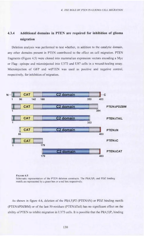
4.3.4 Additional domains in PTEN are required for inhibition of glioma migration... 130
4.3.5 The inhibition of migration by the C-terminus of PTEN is not unique to U373 cells... 135
4.4 Discussion...137
4.4.1 PTEN’s role in cell migration...137
4.4.2 Distinct functions for the different PTEN domains... 137
4.4.3 A function for the C2 domain...138
Chapter 5 - Regulation of PTEN’s C2 Domain... 141
5.1 Summary... 141
5.2 Introduction... 141
5.3 Results...142
5.3.1 Post-translational modification of PTEN regulates its function... 142
5.3.2 Phosphorylation of a single PTEN residue renders the protein inactive... 144
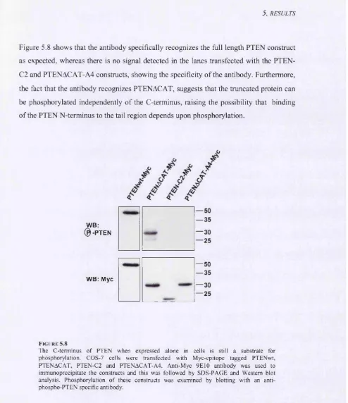
5.3.3 The N-terminus of PTEN binds its C-terminus... 147
5.4 Discussion...156
5.4.1 PTEN phosphorylation as key regulator of its function...156
5.4.2 Is PTEN a protein phosphatase acting autocatalytically?...157
Chapter 6 - Additional Roles for the Carboxy-terminus of PTEN...159
6.1 Summary...159
6.2 Introduction...160
6.3 Results... 160
6.3.1 The role of the C-terminus of PTEN in cell spreading...160
6.3.2 Binding partners for the C-terminus of PTEN... 164
6.3.2.1 Yeast-two hybrid screen...164
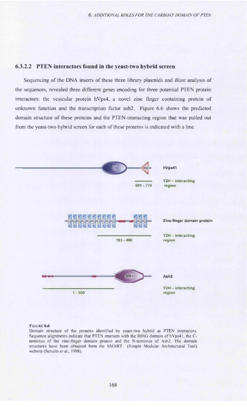
6.3.2.2 PTEN interactors found in the yeast-two hybrid screen...168
6.4 Discussion...169
6.4.1 PTEN and cell spreading... 169
6.4.2 Novel, potential binding partners of PTEN... 171
Chapter 7 - Discussion...174
7.1 Rho GTPases... 174
7.2 PTEN...175
7.3 Regulation of cell migration... 178
Acknowledgements... 179
Table of Figures
Figure 1.1...16
Figure 1.2... 19
Figure 1.3... 21
Figure 1.4... 24
Figure 1.5... 26
Figure 1.6...28
Figure 1.7...29
Figure 1.8... 30
Figure 1.9...32
Figure 1.10...35
Figure 1.11... 39
Figure 1.12... 41
Figure 1.13... 44
Figure 1.14... 48
Figure 1. 15...54
Figure 1.16...58
1. In t r o d u c t i o n
Figure 1.18...64
Figure 1.19...66
Figure 1.20...69
Figure 1.21...70
Figure 3 .1 ... 103
Figure 3 .2 ... 106
Figure 3 .3 ... 107
Figure 3 .4 ... 110
Figure 3 .5 ... 113
Figure 3 .6 ... 115
Figure 3 .7 ... 116
Figure 3 .8 ... 117
Figure 3.9... 118
Figure 4 .1 ... 125
Figure 4 .2 ... 126
Figure 4 .3 ... 127
Figure 4 .4 ... 128
Figure 4 .5 ... 130
Figure 4 .6 ... 132
Figure 4 .7 ... 133
Figure 4 .8 ... 134
Figure 4 .9 ... 136
Figure 4.10... 140
Figure 5 .1... 143
Figure 5 .3 ...146
Figure 5 .4 ... 147
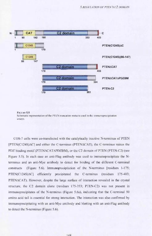
Figure 5 .5... 148
Figure 5 .6... 149
Figure 5.7... 150
Figure 5 .8...151
Figure 5 .9... 152
Figure 5.10...154
Figure 5.11...155
Figure 5.12...158
Figure 6 .1... 162
Figure 6 .2 ... 164
Figure 6 .3 ... 165
Figure 6 .4 ...166
Figure 6 .5 ... 167
Figure 6 .6 ...168
Figure 6.7... 171
Figure 7.1...176
Table of Tables
Table 1.1...50
Table 1.2...67
Table 2.1... 82
Table 3.1... 104
Table 4.1... 129
Chapter 1
Introduction
1.1
Summary
Tumour cells possess a plethora of mechanisms to escape from their site of origin and invade normal neighbouring tissue or metastasize to distant organs. This property of tumour cells is the major cause of the ineffectiveness of the current treatment therapies. Cancers of the central nervous system (CNS) in particular, are considered to be some of the most invasive tumours, and although they lack the ability to use the lymphatic system to metastasize to sites outside the CNS, they acquire properties that allow them to infiltrate normal brain tissue very effectively (Le et al., 2003). The most common brain tumours originate from glial cells and are termed gliomas. Glial cells are subdivided into microglia (cells that fimction as circulating leukocytes of the brain) and into macroglia. Macroglia cells include astrocytes, oligodendrocytes and ependymal cells. Gliomas can arise from all glial cell types, but the most common and aggressive are thought to derive from astrocytes (Kleihues et al., 1995).
L In t r o d u c t i o n
cell to migrate, it needs to elongate a protrusion (spread) towards the direction of movement, form new adhesion sites with the substratum at the front, contract the cell body, and move the rear by dissolving the extracellular -matrix adhesions at the back. Signaling cascades are activated to coordinate cell spreading and migration, most notably, pathways that regulate actin cytoskeleton remodeling. At the centre of the actin rearrangement lies the Rho family of small GTPases that regulate many cellular processes involved in cell movement (Ridley, 2001a).
Rho GTPases are tightly regulated and can be activated by a variety of upstream signals, including growth factors binding to tyrosine kinase receptors, agonists binding to seven-pass membrane receptors, or integrin-matrix interactions. Although there is little evidence to implicate Rho GTPase dysregulation directly with cellular transformation, aberrant upstream signaling to Rho GTPases may play an important role in human cancer formation, particularly with respect to cell invasion and metastasis (Jaffe and Hall, 2002; Malliri and Collard, 2003). Gliomas, for example, upregulate EGF and PDGF tyrosine kinase receptor signaling, which can lead to Rac activation (Ridley et al., Cell 1992, Nobes et al.. Cell 1995). Phosphatidylinositol 3-kinase (PI 3-kinase) is also regulated downstream of receptor tyrosine kinases and the PI 3-kinase signaling pathway is frequently a target in tumors, as it confers a cell growth and proliferative advantage. PTEN, the primary antagonist to PI 3-kinase signaling, is mutated in many primary human cancers, including gliomas. It plays a role in the insulin signaling pathway and has been shown to regulate cell growth, apoptosis, migration and invasion (Knobbe et al., 2002). PTEN inactivation lies therefore at the heart of human tumour formation.
This thesis addresses the aberrant signaling pathways of glioma cells that affect cell migration, with particular emphasis on the roles played by the Rho family of GTPases and PTEN.
1.2
Gliomas
1.2.1 Classification and general characteristics
L In t r o d u c t i o n
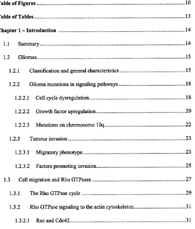
characterised by nuclear atypia and cellular pleomorphism. Grade III tumours are known as anaplastic astrocytomas and exhibit a high mitotic rate. Finally, grade IV tumours are highly malignant and are referred to as glioblastoma multiforme (GBM). GBMs have a high proliferation rate and are accompanied by increased angiogenesis and tissue necrosis (Figure 1.1) (Maher et al., 2001; Rao, 2003). Malignant gliomas are highly invasive and are termed diffuse gliomas because o f their property to infiltrate normal brain tissue and quickly spread in the CNS. This makes them incurable by surgery and glioblastomas are invariably fatal.
FIGU R E 1.1
Two exam ples o f GBM s with different clinical history. Reproduced from Maher et al., 2001. (A ) Grade II astrocytoma from a young woman w hose tumour progressed to GBM 5 years after diagnosis. Arrows indicate neoplastic astrocytes with hyperchromatic nuclei. (B ) GBM from an older man with a short clinical history. Nrnecrosis, P: palisading nuclei, MVP: microvasculature proliferation.
L In t r o d u c t i o n
protein (GFAP) and astrocytes are therefore regarded as the most common cell of origin. Astrocytes perform a variety of functions in the CNS, including the regulation of neuronal homeostasis, growth and survival, guidance of neuronal axonal migration and formation of synapses. They are also involved in immune responses and in repair of tissue after injury, in a process known as gliogenesis. Some astrocyte precursor cells retain the ability to proliferate well after CNS development, a property that may make them more prone to transformation compared to other cells of the CNS (Wechsler-Reya and Scott,
2001).
Astrocytes are produced from multipotent neural stem cells (NCSs) that have the capacity to self renew and generate neurons, astrocytes and oligodendrocytes. As differentiation progresses along the cell lineages, more restrictive precursor cells are generated. First, NCSs produce neural precursor cells that only give rise to neurons, and glial precursor cells that can only give rise to astrocytes and oligodendrocytes (Rao and Mayer-Proschel, 1997; Rao et al., 1998). Glial-precursor cells then generate astrocyte or oligodendrocyte progenitor cells that produce astrocytes or oligodendrocytes respectively. The proliferation and differentiation of these progenitor cells are tightly controlled by extracellular stimuli. Astrocyte differentiation, for example, is promoted by EGF (Bachoo et al., 2002) and EGF receptor (EGFR) knockout mice exhibit a delay in astrocyte differentiation and have a fewer number of astrocytes in several parts of the brain (Komblum et al., 1998). Other astrocyte differentiation factors include the cytokines of the ciliary neurotrophic factor (CNTF) and leukemia inhibitory factor (LIF) that use the Jak/Stat signaling pathway, and bone morphogenetic proteins (BMPs) that can promote astrocyte differentiation on their own or in combination with LIF (Wechsler-Reya and Scott, 2001).
1. In t r o d u c t i o n
1.2.2 Glioma mutations in signaling pathways
Gliomas arise either de novo (primary) or as low-grade tumours that progress to a more malignant phenotype (secondary). It is thought that they develop along different genetic pathways and are thus clinically distinct. Gliomas exhibit losses of parts of chromosomes or amplification of genes and most of the genetic mutations identified target either cell cycle arrest and apoptotic pathways or signal transduction cascades downstream of receptor tyrosine kinases.
1.2.2.1 Cell cycle dysregulation
1. In t r o d u c t i o n
M
INK4 CKIs \P 1 9 , p 1 6 /
G 1/G 0 G2
E2F
S
R b
%
C ip/K ip
(CyclinI
Çyclind
CDK4/6 CDK2
*
14/p19
MDM2 A p o p to s is
Fi g u r e 1. 2
Cell cycle com ponents mutated in gliomas. Proteins involved in the regulation o f G1 to S phase transition are depicted. 1NK4 family CKIs inhibit cyclinD /C D K 4/6 com plexes, whereas Cip/Kip CKIs act on both C yclinD /C D K 4/6 and Cyclin E/CDK2 com plexes. This prevents phosphorylation o f Rb and subsequent release o f E2F and entry into S phase. p l4 /p l9 A R F negatively regulates the p53 inhibitor M DM 2, and promotes apoptosis. A red asterisk indicates the proteins frequently mutated in gliomas.
L In t r o d u c t i o n
regulator MDM-2. MDM-2 promotes the proteosomal degradation of p53 by directly binding to p53 and acting as an E3 ubiquitin ligase (Kubbutat et al., 1997; Lohrum et al.,
2000).
1.2.2.2 Growth factor upregulation
Growth factors and their receptors play an important role in glial development and are often targets in gliomagenesis. Amplification of the EGFR occurs in 40-50% of glioblastomas resulting in abnormally elevated levels of EGFR, and is an event more frequently associated with high-grade glioblastomas (Fenstermaker and Ciesielski, 2000). Studies in mice suggest a critical role for EGFR during CNS development and although EGFR has been shown to be involved in astrocyte differentiation, it also appears to be necessary for sustained proliferation of the neural stem cell pool (Maher et al., 2001). Several EGFR ligands, including EGF and TGF-a, have been shown to promote proliferation of astrocytes in culture and growth of astrocyte precursor cells (Wechsler- Reya and Scott, 2001).
There is evidence to suggest that glioblastomas express EGF and TGF-a together with EGFR and thus growth of these tumours can be sustained through both paracrine and autocrine stimulatory loops of the EGFR (Figure 1.3). Another consequence of receptor amplification (which occurs in 40-60% of GBMs) is that often this results in the expression of mutated forms of the receptor. Approximately 60% of glioblastomas express truncated EGFR, which lacks part of the extracellular domain and can no longer bind ligand, but is constitutively tyrosine phosphorylated and is thus always active (Ekstrand et al., 1994; Nagane et al., 2001; Nishikawa et al., 1994). The constitutively active EGFR is not efficiently downregulated, unlike the wild-type form, and introduction of the truncated active EGFR into glioma cells, dramatically enhances their tumourigenicity (Ciesielski and Fenstermaker, 2000), proliferative capacity and resistance to apoptosis (Nagane et al., 1996). In addition, mice expressing the viral oncogene derived from the EGFR, v-erbB, have been shown to develop oligodendrogliomas (Weiss et al., 2003).
1. In t r o d u c t i o n
PDGF in neural progenitors and astrocytes developed oligodendrogliomas and oligoastrocytomas respectively (Dai et al., 2001). In addition, the pattern o f PDGF expression closely correlates with p53 mutations in low-grade tumours, suggesting a possible genetic interaction (Hermanson et al., 1996).
TGFaJ
DGI
EGF
E G FR
E G F R P D G F R
CPI3I
Src Src
+ve feedback
loop c o n stitu tiv e
a ctivation
MYC
DGI
Ras
EGF.
+ve feedback loop PKC
|m a p k|
AP1 DGI
EGF) ^
Fi g u r e 1.3
Growth factor receptor upregulation in gliom as. A few o f the signaling pathways downstream o f EGFR and PDGFR are depicted. EGFR amplification results in EGF upregulation and the subsequent autocrine and paracrine loop o f receptor activation (sim ilarly for PDGF/PDGFR). A truncated EGFR (denoted by a red cross) is constitutively active. The Ras/M APK and PI 3-kinase/PKB are two o f the many pathways activated downstream o f the PDGF and EGF receptors. PKC and M YC are also targets o f the tyrosine kinase receptor signaling.
L In t r o d u c t i o n
gliomas. Studies with transgenic mice showed that expression of active Ras or v-Src in astrocytes induced the formation of glioblastomas and astrocytomas respectively. Mice with germline genetic mutations, however, resulting in active Ras and Src, develop normally and any gliomas formed are due to secondary mutations (Guha et al., 1997). In addition, although the combined activation of Ras and Akt signaling pathways have been reported to induce glioblastomas, no activating mutations in either protein have been identified in human gliomas (Dai and Holland, 2001). It is likely that die abnormal activation of the receptor tyrosine kinase receptors EGFR, PDGFR and FGF results in the simultaneous activation of several downstream pathways that together lead to the formation of glioblastomas or increase the tumourigenicity of low-grade tumours.
1.2.2.3 Mutations on chromosome lOq
A large number of gliomas (75-90%) have part of the long arm of chromosome 10 missing. This region is also often deleted in other tumours, including breast, prostate and endometrial carcinoma. PTEN was identified as one of the tumour suppressor genes frequently lost or inactivated in this region. Approximately 40% of high-grade gliomas exhibit PTEN mutations and PTEN inactivation occurs in both primary and secondary gliomas, suggesting that it may be interacting with the Rb or EGFR pathways. The induction of tumour growth by the combined activation of active Ras and Akt can be blocked by overexpression of PTEN (Cheney et al., 1998; Fumari et al., 1997). PTEN induces growth suppression by causing a Gl cell-cycle block in these glioblastomas (discussed in detail later) (Myers et al., 1998).
L In t r o d u c t i o n
1.2.3 Tumour invasion
1.23.1 Migratory phenotype
The migratory behaviour of gliomas and their ability to invade normal tissue and spread diffusely is a key component of their malignant phenotype. Invasion is a property of both low-grade astrocytomas and high-grade glioblastomas and resembles the migratory phenotype of glial cells during CNS development. It is possible that glioma invasion follows the same mechanisms of early glial migration during embryogenesis through the reactivation of the same signaling pathways. In the adult, neurons, oligodendrocytes and astrocytes do not migrate. However, it has been shown that in response to injury, nestin-positive cells, most probably neural stem cells and glial progenitors, migrate to the site of injury and there undergo terminal differentiation into astrocytes. It is not clear whether these pathways can be disrupted during tumourigenesis, but they provide a likely source for malignant transformation. It was demonstrated that neural stem cells and glioma cells co-cluster following in vivo injection, suggesting that they might use the same routes when migrating in the CNS (Johansson et al., 1999; Magavi et al., 2000). Migration and invasion of gliomas is very different to other tumours that do not spread locally to the extent that gliomas do, but rather use the lymphatic system and the blood vessels to metastasize to distant sites. Gliomas lack the ability to cross the blood brain barrier, but rather have acquired properties that enable them to spread in the surrounding normal tissue (Rao, 2003).
1. In t r o d u c t i o n
myeloid leukemia cells (Farina et a l, 1998). Leukocytes and Dictyostelium use a type of movement that is generated by cortical filamentous actin and is based on short-lived interactions with the substratum. Small-cell lung and prostate carcinomas use this type of movement to undergo early detachment and spread from a small primary tumour.
A m o eb o id >
Individual m ig ratio n
M ese n ch y m al '
C h a in s /c lu s te rs
S h e e ts
C ollective m ig ratio n
Fi g u r e 1 . 4
The different types o f tumour cell migration. Individual cells display either an am oeboid type o f migration {D ictyostelium , leukemia or lymphoma cells) or a m esenchym al migration (gliom as, fibrosarcomas). C ollective migration is subdivided into chain-like m ovement (m elanom a cells, epithelial tumours) and migration as sheets (epithelial tumours, vascular tumours).
1. In t r o d u c t i o n
than amoeboid migration and one of the reasons is that focal adhesion turnover is relatively slow (Webb et al., 2002).
1.2.3.2 Factors promoting invasion
The extracellular matrix is the main component being destroyed in a tissue during tumour cell invasion and differs for each cell type within an organ. Tumour cells of the CNS possess a unique set of proteolytic enzymes that enables them to infiltrate normal brain tissue (Lakka et al., 2003). Anaplastic astrocytomas and glioblastoma cells express higher levels of the receptor for the serine protease urokinase-type plasminogen activator (uPA), uPAR, than do normal cells or low-grade gliomas (Yamamoto et al., 1994). Transfection of low-grade gliomas with uPAR was shown to increase their invasive capacity in a matrigel assay, compared to parental cells, in a uPA-dependent manner (Mohanam et al., 2002; Mohanam et al., 1998; Mohanam et al., 1999). Furthermore, studies with antisense-uPA, showed that the increased cell motility conferred by uPA/uPAR signaling is dependent on the PI 3-kinase/Akt pathway (Chandrasekar et al., 2003). In glioblastoma cells, uPAR colocalises at focal contacts with the avp3 integrin, and downregulation of uPAR increases the expression of a3 p i integrin, induces changes in cell morphology and disorganization of the cytoskeleton (Gladson et al., 1995; Rao, 2003). Studies in nude mice demonstrated that stably transfected glioblastoma cells with antisense uPAR undergo apoptosis when injected in the mouse brain (Adachi et al., 2002; Mohanam et al., 2001). The observed cell death correlates with an increase in BAX, release of cytochrome c and activation of caspase-9.
1. In t r o d u c t i o n
EGFR overexpression. EGFR signaling was shown to induce MMP-9 activation, an effect dependent on PI 3-kinase, and hyaluronic acid-mediated secretion o f M MP-9 and subsequent activation o f the MAPK pathway in human gliomas, was inhibited by re- introduction of PTEN (Choe et ah, 2002; Park et ah, 2002).
Cytokines Growth factors ECM
plasmln plasminogen
MMPs
plasmin
P1) -► Transcription
Fi g u r e 1.5
The uPA /uPA R and MMP pathw ays. Pro-uPA bound to its receptor is cleaved m ore efficiently by plasmin to produce uPA. uPA, in turn, cleaves plasm inogen into plasmin in a positive feedback loop. The tail o f uPAR can activate transcription via the Jak/Stat pathway. uPAR colocalises with integrins at focal contacts and is involved in the activation o f several pathways downstream o f integrins, like the Ras/M APK pathway. M MPs are released as inactive pro-M MPs and are cleaved into active M MPs by a variety o f com ponents, including growth factors and plasmin. ECM: extracellular matrix.
1. In t r o d u c t i o n
that facilitate movement within the tissue, growth factors, such as EGF, PDGF and IGFl also promote cell migration and invasion through the activation of several signal transduction pathways (Lakka et al., 2002)). EGF induces migration and process extension of both normal astrocytes and astrocytic tumour cells, whereas IGF-1 promotes chemotaxis of breast cancer cells in an integrin-dependent mechanism (Hu et al., 2003; Wang et al., 2003). PDGF, on the other hand, has been shown to be important in low- grade glioma migration through activation of PI 3-kinase and PLC-y pathways. A common mediator of growth factor and integrin stimulated migration and tumour cell invasion is the family of Rho GTPases. Gliomas and tumour cells in general, display many common features of normal migrating cells, when migrating during development. To gain a better insight in the mechanisms of glioma invasion, it is important, therefore, to examine cell migration more closely.
1.3
Cell migration and Rho GTPases
1. In t r o d u c t i o n
Cytokine
G p ro tein -co u p led
® 1 re c e p to rs
R ece p to r ty ro sin e
re c e p to rs k in a se s
\
/
Protein kinases (MARK, PKC)
Lipid kinases
P hospholipases (p l d)
Rho GTPases
\
M em branetraffic M icrotubu e s
A ctin T ran scrip tio n
A dhesion
Cell M igration
Fi g u r e 1 .6
Regulation o f cell migration. The main pathways that regulate cell migration are shown. These include PKCs, PLC, PI 3-kinase, MAPK and Rho GTPases.
1. In t r o d u c t i o n
o n tra c tio n (R hgj.
^ T B ! r e 3 [ o n o f m o v e m e n t/P o la h t^ jC g g ^ g ^
\ —
~
»1."Direction o f m ov em en t/ P olarity (C d c4 j)T
2 /tâ m e llip o d ia /M e m b ra n e ruffles (Rac) ^
K N ew a d h e s io n s w ith s u b s tr a tu m
"K D w solution o f old a d h e s io n s a n d tail re tra c tio n
Fi g u r e 1 .7
Schem atic representation o f the key steps o f cell migration (seen from the top and the side). A migrating cell polarises in the direction o f m ovement in a Cdc42 dependent manner and polym erises actin to form filopodia and Rac-dependent lamellipodia. The cell also spreads and makes new adhesions with the substratum (focal contacts). Finally, it contracts in a R h o -dependent way, and the tail retracts follow ing the dissolution o f the adhesions at the rear.
Multiple intracellular signaling molecules have been implicated in cell migration, MAPK cascades, lipid kinases, phospholipases, Ser/Thr kinases (such as protein kinase Cs [PKC] and p65PAK) and tyrosine kinases (such as Abl and Src). However, one particular family o f proteins seems to play a pivotal role in regulating the biochemical pathways most relevant to cell migration; Rho GTPases (Figure 1.7) (Ridley, 2001a).
1.3.1 The Rho G TPase cycle
1. In t r o d u c t i o n
(GEFs), promote the exchange o f GDP for GTP to activate the GTPase, GTPase- activating proteins (GAPs) negatively regulate the switch by enhancing the intrinsic GTPase activity and guanine nucleotide dissociation inhibitors (GDIs) are thought to block the GTPase cycle by sequestering and solubilizing the GDP-bound form (Schmidt and Hall, 2002).
U p stream s ig n a ls
GEF
e ffe c to r
S caffold
GAP
GDI d o w n stre a m
sig n a lin g p ath w a y s
Fi g u r e 1.8
The Rho GTPase cycle. Rho GTPases cycle betw een an inactive GDP-bound form and an active G TP-bound form. The cy cle is tigh tly regulated m ainly by guanine nu cleotid e exch ange factors (G E Fs), G TPase activating proteins (G A P s) and guanine nu cleotid e dissociation inhibitors (G D Is). In their active form, Rho G TPases can bind to effector m olecules and mediate their effect, for example, on the actin cytoskelton.
L In t r o d u c t i o n
but later in many other cell types) using constitutively active and dominant negative, interfering forms, have shown that Rho regulates the assembly of contractile, actimmyosin filaments, while Rac and Cdc42 regulate the polymerisation of actin to form peripheral lamellipodial and filopodial protrusions, respectively (Hall, 1998). In addition, all three GTPases promote the assembly of integrin-based, matrix adhesion complexes (Nobes and Hall, 1995; Ridley and Hall, 1992). It is perhaps not surprising, therefore, that these three regulatory proteins play such an important part in controlling cell migration. Furthermore, and in addition to their effects on actin, Rho, Rac and Cdc42 can influence a wide range of other biochemical activities. Most notably, Cdc42 is required for the establishment of cell polarity in all eukaryotic cells, including yeast, while all three can, in distinct ways, affect the microtubule cytoskeleton and gene transcription (Van Aelst and D'Souza-Schorey, 1997).
1.3.2 Rho GTPase signaling to the actin cytoskeleton
1.3.2.1 Rac and Cdc42
Rac and Cdc42 are both required at the front of migrating cells. Rac is required to generate protrusive force at the front, while Cdc42 is required to recognize external directional cues and polarise protrusive activity accordingly. Cdc42 also induces filopodia. Their role is not entirely clear, but they have been suggested to probe the extracellular milieu. The cellular targets of Rac and Cdc42 that promote changes to the actin cytoskeleton have been the subject of intense investigation. The Ser/Thr kinase p65PAK is commonly activated upon either Rac or Cdc42 activation and is believed to play an important role in regulating actin dynamics during cell adhesion and migration. p65PAK regulates focal adhesion turnover with the help of PIX and GITl (GRK interactor 1) but how it does so is not known (Manabe Ri et al., 2002; Obermeier et al., 1998). In addition, p65PAKl phosphorylates and activates LIM kinase (LIMK), which in turn phosphorylates and inactivates cofilin (Figure 1.9) (Arber et al., 1998; Edwards et al., 1999). Cofilin, which facilitates depolymerisation at the pointed end of actin filaments, cycles between an active and inactive form, and this is essential for promoting filament treadmilling at the front of migrating cells (Hamburg, 1999).
L In t r o d u c t i o n
are able to activate the Arp2/3 complex, which can initiate actin polymerisation, either de novo or at the tips or sides o f pre-exisiting filaments. In this way the dendritic morphology o f lamellipodial actin is thought to be generated (Amann and Pollard, 2001; W eaver et al., 2003). WASpAVAVE can also bind to profilin, which acts synergistically with Arp2/3 to speed-up actin polymerisation (Blanchoin et al., 2000; Yang et al., 2000). Rac and Cdc42 are thought to activate members o f this family directly (Eden et al., 2002; Rohatgi et al., 2000), although activation could also be indirect through the Rac and Cdc42 effector IRSp53 (Figure 1.9) (Krugmann et al., 2001; Miki and Takenawa, 2002; Miki et al., 2000).
Cdc42 Rac Rho
IRSp53 ROCI mDia
MLC
phosphatase IRSp53 WASPA/VAVE/Scar LIMK
i
Arp2/3 Profilin
Actin polymerisation
Cofilin
Actin F-actin polymerisation stabilisation
phospho-MLC
actimmyosin Actin crosslinking polymerisation
\ 7
Filopodia Lamellipodia S tress Fibres
F i g u r e 1.9
7. I n t r o d u c t i o n
1.3.2.2 Rho
Rho activity in migrating cells is associated with focal adhesion assembly and cell contractility. One important Rho target involved in stimulating actin:myosin filament assembly and therefore contractility is the Ser/Thr kinase pl60ROCK. hi migrating leukocytes, for example, Rho and pl60ROCK are required for proper rear cell detachment (Alblas et al., 2001). ROCK plays an essential role during migration of P cells in the larval development of C. elegans and during dorsal closure and gastrulation in Drosophila (Barrett et al., 1997; Magie et al., 1999; Spencer et al., 2001). In its active State, plôOROCK, like p65PAK, can phosphorylate and activate LIMK, which in turn phosphorylates and inactivates cofilin leading to stabilization of actin filaments within the actimmyosin filament bundles (Maekawa et al., 1999; Sumi et al., 2001). pl60ROCK interacts with and phosphorylates the myosin binding subunit (MBS) of myosin light chain phosphatase and thereby inactivates it (Kawano et al., 1999). This leads to increased levels of myosin phosphorylation which then can cross-link actin filaments and generate contractile force. At the rear of a migrating cell, this can promote movement of the cell body and detachment of the cell rear (Mitchison and Cramer, 1996). Clearly, however, this activity is incompatible with membrane protrusion and hence mechanisms must be in play to inhibit this activity at the leading edge. One way this might occur is through the Rac/p65PAK pathway - p65PAK can phosphorylate and inactivate myosin light chain kinase (MLCK), leading to decreased levels of myosin phosphorylation (Kaback et al., 1984; Kiosses et al., 1999). Conflicting results have shown that p65PAK can phosphorylate and activate MLC thus enhancing cell contractility (Sells et al., 1999).
1. In t r o d u c t i o n
1.3.3 Rho GTPases and microtubules
Although the effects of Rho GTPases on the actin cytoskeleton have received most attention to date, it is now clear that they can also modulate the microtubule cytsokeleton and this may play an important role during the migration of at least some cell types (Wittmann and Waterman-Storer, 2001). The first clue for a link was the observation that disruption of microtubules with nocodazole activates Rho, but when washed out of cells it leads to Rac activation (Liu et al., 1998). Later Rho was shown to promote the stabilization of microtubules through its target mDia that directly interacts with microtubules and promotes their capping (Figure 1.10) (Ishizaki et al., 2001). Another possible pathway to regulate microtubule dynamics in mammalian cells could well be the one that is known to operate in yeast (Gundersen, 2002). It has been shown, by direct imaging studies of the budding cortex, that yeast regulate capture and shrinkage of microtubules by a mechanism that involves the formin Bnil, Kar9 and Biml/Yebl, a homologue of the mammalian microtubule tip protein EBl (Adames and Cooper, 2000; Schuyler and Pellman, 2001). Rac, on the other hand, has been shown to modify microtubule dynamics through p65PAK-dependent phosphorylation and inactivation of the microtubule destabilising protein stathmin (Figure 1.10) (Daub et al., 2001; Kuntziger et al., 2001).
1. In t r o d u c t i o n
o f microtubules and contribute to the regulation o f cell polarisation (Etienne-Manneville and Hall, 2003).
C dc42 Rho
Par6/PKCÇ p65PAK mDia
GSK-3 Stathmin
APC
M ic ro tu b u le + e n d c o rtic a l
a s s o c ia ti o n
M ic ro tu b u le + e n d e lo n g a tio n
M ic ro tu b u le s ta b iliz a tio n
Fi g u r e 1 .1 0
Sim plified representation o f the effects o f Rho G TPases on microtubules. Cdc42 regulates microtubule dynamics by becom in g recruited to a Par6/ PKCÇ com plex. Formation o f the com plex leads to the inhibition o f GSK-3 and the release o f APC, which can associate with the plus-end o f microtubules at the leading edge. Rac also regulates microtubules by activating p65PAK to phosphorylate and inactivate stathmin, a m icrotubule-destabilising protein. Little is known on how Rho activation stabilises microtubules, other than it involves its effector mDia.
L In t r o d u c t i o n
evidence to indicate that APC can bind to microtubules and move along them probably through binding to the microtubule-associated protein EBl and the kinesin KAP3, thus playing a similar role to the yeast protein Kar9 (Gundersen, 2002; Jimbo et al., 2002; Nakamura et al., 2001). The movement of APC towards the plus ends of microtubules in migrating cells could serve to localise ASEF to sites of actin-rearrangement, where Rac could be recruited in a similar way and be activated only locally (Bienz, 2002; Jimbo et al., 2002). GEFHl, a GEF that has been shown to induce GTP loading on both Rac and Rho, could be acting in an analogous way to ASEF, as there is evidence to suggest that it localises to microtubules too (Ren et al., 1998).
1.3.4 Extracellular Control
A variety of extracellular signals have been implicated in the initiation or regulation of cell migration and broadly fall into two categories: soluble components, and extracellular matrix. These mediate signaling cascades that drive the Rho GTPase cycle by modifying any of the GTPase regulatory proteins.
1.3.4.1 Integrin-matrix interactions
L IN T R O D U C T IO N
et al., 2001). Thus during spreading of fibroblasts on fibronectin, which has some similarities to migration, Cdc42 and Rac are activated upon engagement of the integrins to the substratum (as observed by looking at the phosphorylation of several downstream kinases like FAK or Src), leading to actin polymerisation at the periphery and protrusion of the membrane (Berrier et al., 2002; Clark et al., 1998; O'Connor and Mercurio, 2001). At later times, these two GTPases are downregulated and Rho activity is increased, as the cells stabilize their morphology and put down firm focal adhesions (Arthur and Burridge, 2001; Ren et al., 1999). Activation of Rac and transient inhibition of Rho seem to be critical events at the front of migrating cells, whereas activation of Rho is required in the cell body and at the rear.
The mechanisms by which these two GTPases are differentially activated within a single cell are not clear, however recent studies have given us some insights (Sander et al., 1999). Integrin a3 p l engagement by laminin (10/11) preferentially activates Rac through phosphorylation of plSOCas and activation of the CrkQ / DOCK 180 pathway but engagement of the integrin aSpl to fibronectin seems to activate Rho (Gu et al., 2001). Elegant FRET studies by the Schwartz lab have shown that integrins control the translocation of Rac to the plasma membrane at the front of the migrating cell and enhance its association with effectors by causing its dissociation from Rho-GDI (Del Pozo et al., 2002). Finally, activation of integrin a6(34 by laminin has been shown to play a role in cell migration of carcinoma cells by inducing the activation and translocation of Rho (rather than Rac) to ruffles, in a cAMP dependent way, presumably for the formation o f new adhesions (O'Connor et al., 2000).
1.3.4.2 Soluble factors
L In t r o d u c t i o n
cell periphery. Recently a new technique for visualizing activated Rac in living cells, FLAIR (fluorescence activation indicator for Rho proteins), has been used with cells induced to migrate along a gradient of PDGF. In this case, PDGF induces an intracellular gradient of active Rac, the highest levels being at the leading edge and the lowest at the rear of the migrating cell (Kraynov et al., 2000).
Interestingly, PDGF also leads to the activation of Rho, apparently through the inactivation of pl90RhoGAP, a negative regulator of Rho GTP levels (Chiarugi et al., 2000). It is expected that active Rho will be excluded from the leading edge of migrating cells, but this has not yet been shown directly. PDGF signaling provides a nice example of how the same stimulus activates different GTPases to produce a coordinated response; the mechanisms by which the two GTPase are differently localised is, however, completely unknown.
Two peptide factors acting through tyrosine kinase receptors, EGF and Pvfl (homologous to VEGF and PDGF) are thought to act as chemoattractants directing border cell migration during oocyte maturation in Drosophila. The PVR (the receptor for Pvfl) is thought to control F-actin accumulation in the border cells by signaling to Rac via mbc (myoblast city), the fly orthologue of mammalian DOCK 180 (Duchek and Rorth, 2001; Duchek et al., 2001).
1. In t r o d u c t i o n
LPA, S1P, chemokines FN, laminin, PS, coliagen
GPCRs EGF, VEGF, PDGF
Integrins RTKs
f130Cas;
SHP2 FAK P190
RhoGAP
CrkII Vav, Sos
Pix, pREX1, TIAM1
DOCK180 »130Ci
ELMO, I0CK18(
CrkII
ELMO,
Rho G TPases
Fi g u r e 1 .1 1
Sim plified representation o f som e o f the upstream regulatory pathways o f Rho GTPases. Ligand binding to either tyrosine kinase receptors or G-protein coupled receptors or integrins results in the activation o f signaling com ponents like FAK, Src, She, Fyn, PI 3-kinase that ultimately lead to the activation or inactivation o f GEFs and G APs respectively. Activation o f PH-domain GEFs (e.g. TIA M -1, pREX, Vav) leads to the activation o f Rac and Cdc42.
L In t r o d u c t i o n
Fyn. pl30Cas translocates to the plasma membrane and activates Rac (Okamoto et al., 2000a; Okamoto et al., 2000b; Vouret-Craviari et al., 2002). It has also been reported that SIP stimulates endothelial cell migration and cortical actin assembly through activation of PI 3-kinase and subsequently protein kinase B (PKB/Akt), which in turn phosphorylates the SIP receptor EDGl and promotes Rac activation (Lee et al., 2001).
In addition to attractive cues, there is growing evidence that repulsive cues also play an important role in directing cell migration. This has been studied best in the context of neuronal axon extension and growth cone guidance, which shows many similarities to the protrusive front of a migrating cell, although in this case the cell body does not move. Studies in C. elegans^ Drosophila and Xenopus have clearly identified a crucial role for Rho GTPases in these aspects of neuronal behavior, as well as in neuronal cell migration itself (Luo, 2000). Work carried out by many groups has come to the conclusion that Rho acts antagonistically to Rac/Cdc42, the former causing growth cone and axon retraction and the latter causing extension (Kozma et al., 1997). As an example, the ephrins are membrane-bound repulsive ligands for dorsal root ganglion-derived neuronal growth cones that act through the family of Eph tyrosine kinase receptors (Schmucker and Zipursky, 2001). Upon ligand binding, EphA activates Rho through the GEF ephexin and promotes growth cone collapse, in part through activation of its downstream Ser/Thr kinase target, plbOROCK (Figure 1.12) (Shamah et al., 2001; Wahl et al., 2000).
1. In t r o d u c t i o n
Plexin
Rac-GTP
PDZ-RhoGEF Ephexin
Rho-GTP
1
ROCK
Rho-GTP
Growth c o n e repulsion/ co llap se
F i g u r e 1.12
Rho G TPases are regulators o f growth cone migration. Rho is activated downstream o f the repulsive growth cone cues, ephrins and semaphorins. Ephrins bind to their receptor EphR to activate the Rho-GEF ephexin, whereas semaphorins bind to plexins to activate PDZ-Rho GEF. Rac-GTP is also known to bind to the cytoplasm ic tail o f plexin.
1.3.5 PI 3-kinase signaling in cell migration
1.3.5.1 PI(3,4,5)P3, a signal for polarised chemotaxis
colony-L In t r o d u c t i o n
1 (CSF-1), whereas type IB PI 3-kinases are required for neutrophil chemotaxis to a variety of inflammatory mediators that signal via G-protein coupled receptors (Hirsch et al., 2000; Li et al., 2000; Sasaki et al., 2000; Vanhaesebroeck et al., 1999). PI 3-kinases are heterodimers of a regulatory and a catalytic subunit. The p85 regulatory subunit is a target for phosphorylation by a variety of cytoplasmic and receptor tyrosine kinases and binds constitutively to the pi 10 catalytic subunit. Type I PI 3-kinases phosphorylate phosphatidylinositol phospholipids at position 3 of the inositol ring (Vivanco and Sawyers, 2002). Their main product is PI(3,4,5)P3, which is believed to primarily act as a ligand to recruit PH domain containing proteins to the plasma membrane. The property of certain PH domains to specifically translocate to localised pools of PI(3,4,5)P3 was exploited by several groups to visualize bursts of PI(3,4,5)P3 on the plasma membrane, using pleckstrin homology (PH) domains fused to GFP (GFP-PH). Studies in Dictyostelium^ neutrophils and fibroblasts demonstrated co-localisation of the GFP-PH construct with centers of actin polymerisation at the front of the migrating cell (Haugh et al., 2000; Parent et al., 1998; Servant et al., 1999).
The role of PI 3-kinase in chemotaxis has also been examined through PI 3-kinase knock-out studies in mice and Dictyostelium. Mouse PI 3-kinasey ^ neutrophils demonstrated an impaired ability to chemotax towards a gradient of fMLP, C5a or interleukin-8 and fail to activate PKB, the main downstream target of PI(3,4,5)P3 (Hirsch et al., 2000; Li et al., 2000; Sasaki et al., 2000). Close examination of these neutrophils revealed that they were unable to efficiently sustain a pseudopod extension exclusively towards the chemotactic gradient. Similarly, Dictyostelium cells that had the PI 3-kinaseI and PI 3-kinase2 genes inactivated, showed an impaired directionality, migrated more slowly and in a less polarised manner towards a gradient of either cAMP or folate, compared to parent cells (Funamoto et al., 2002; Funamoto et al., 2001). The importance of PI 3-kinases was supported further by studies in macrophages using antibodies raised specifically against class lA PI 3-kinasep and PI 3-kinaseô isoforms and several studies using the PI 3-kinase inhibitors wortmannin and LY294002 (Chung et al., 2001; Sadhu et al., 2003; Vanhaesebroeck et al., 1999).
L In t r o d u c t i o n
restricted to the leading edge of migrating cells during chemotaxis. The kinetics of relocalisation of PI 3-kinases closely resembles the relocalisation kinetics of PH domain proteins (Funamoto et al., 2002). Importantly, the same group demonstrated that relocalisation of PI 3-kinases to the plasma membrane is independent of their activation. These results suggest that although PI 3-kinases are key regulators of cell polarity, additional mechanisms that lie upstream of PI3-kinases must be responsible for establishing the initial cell asymmetry, in response to a chemotactic gradient.
1.3.5.2 Downstream targets of PI(3,4,5)P3 signaling
1. In t r o d u c t i o n
contains a PH domain (Cote and Vuori, 2002; Meller et al., 2002), Studies in Drosophila
and lymphocytes have revealed a role for DOCK 180 in cell migration and chemotaxis and these effects might be modulated through PI(3,4,5)?3 targeting and subsequent activation of Rac (Duchek and Rorth, 2001; Duchek et ah, 2001).
In teg rin s
T y ro sin e k in a se re c e p to rs G -protein c o u p le d re c e p to rs
PTEN
PI 3-kinase
PI(3,4,5)P3
+ve feedback loop
R ecru itm en t of P H -dom ain p ro te in s
Cell polarity
Dbl fam ily DOCK180 GEF
I
RacGTPV
S patial ac tiv a tio n of actin p o ly m erisatio n
Fi g u r e 1 .1 3
L In t r o d u c t i o n
The relationship between Rac and PI 3-kinase during cell migration may be more interesting, since the two are able to interact directly with each other and Rac activation stimulates PI 3-kinase leading to the production of PI(3,4,5)P3 (Benard et al., 1999; Bokoch et al., 1996; Genot et al., 2000; Hawkins et al., 1995; Servant et al., 2000). This would provide an opportunity for a positive feedback loop. To explore this possibility, Bourne and co-workers made use of neutrophils expressing the PH domain of Akt coupled to fluorescent green protein (PH-Akt-GFP) as a probe for the spatial distribution of PI(3,4,5)Ps (Wang et al., 2002; Weiner et al., 2002). They found that in response to a chemoattractant or an exogenously delivered bolus of PI(3,4,5)P], neutrophils asymmetrically accumulate PI(3,4,5)Pg and filamentous actin at the leading edge and this involves a positive feedback loop operating between PI 3-kinase and Rac. Disruption of this feedback loop results in a jerky, non-polarised cell response to chemoattractants. The exchange factors regulating Rac during neutrophil chemotaxis are not known, although the RacGEF PREX-1, has recently been shown to be activated by PI(3,4,5)P3 in neutrophils leading to Rac-dependent stimulation of the NADPH oxidase (Welch et al.,
2002).
1.3.5.3 PTEN in directional sensing
Elegant studies in Dictyostelium by the Firtel and Devreotes laboratories have provided important evidence for a role of the PI(3,4,5)P3 phosphatase PTEN in cell chemotaxis. The two laboratories have used complementary approaches to show that chemotaxis of Dictyostelium cells is dependent upon the interplay of PTEN and PI 3- kinases. These studies revealed that PTEN null cells exhibit chemotactic defects reciprocal to those of PI 3-kinase 1/PI 3-kinase 2 null cells or to wild type cells treated with PI 3-kinase inhibitors. PTEN null cells have numerous filopodia and pseudopodia, move more rapidly and appear to have multiple polarity axes thus exhibiting a slower overall response to the chemoattractant compared to wild type cells (lijima and Devreotes, 2002). On the other hand, PI 3-kinase 1/PI 3-kinase 2 null cells have a reduced number of pseudopodia compared to wild type cells and had chemotactic defects due to an overall loss in polarity (Funamoto et al., 2002). In addition, PTEN knock-in cells displayed a slower rate of chemotaxis and a decrease in cell polarity.
L In t r o d u c t i o n
although polarity is impaired, it is not completely lost. PTEN knock-out cells, however, had a more sustained accumulation of PH domains at the plasma membrmie upon chemoattractant stimulation (lijima and Devreotes, 2002). Funamoto et al., observed that PTEN had a reciprocal localisation to PI 3-kinase. PTEN transiently translocates from the plasma membrane to the cytosol with kinetics that mirror those of PI 3-kinase relocalisation. In chemotaxing cells, PTEN is localised mostly at the edges and the posterior part of the cells, whereas PI 3-kinase accumulates at the leading edge of the migrating cell, towards the chemotactic gradient (Funamoto et al., 2002). Li addition, PTEN null cells, displayed a prolonged accumulation of F-actin relative to wild type cells, suggesting that actin polymerisation is a direct downstream event in PI 3-kinase signaling. Overall these observations demonstrate that PI 3-kinases and PTEN through their antagonistic effects on PI(3,4,5)P] are key players in the maintenance, and also possibly in the establishment of cell polarity.
1.4
PTEN
1.4.1 Discovery o f PTEN
L In t r o d u c t i o n
groups demonstrated the ability of PTEN to dephosphorylate position D3 of phosphoinosityl phospholipids, with highest affinity for PI(3,4,5)?3 (Maehama and Dixon, 1998; Myers et al., 1998).
1.4.2 PTEN homologues
1. In t r o d u c t i o n
5 4_ 84
PTEN
445 TPIPa
—
326 TPIP pPTEN2/TPTE
432
TPTE homolog 1
Fi g i i r e 1.14
Domain structure o f the mammalian PTEN hom ologues PTEN2, TPIP, TPTE. The domain predictions were performed using the SM ART (Sim ple Modular Architectural T ool) w ebsite and m odified according to Walker et al., 2001. Phosphatase domain: pink, C2 domain: green, TM domain: blue, PDZ binding motif: purple.
1.4.2 PTEN: a tumour suppressor
1.4.3.1 PTEN is the major susceptibility gene in two human hamartoma syndromes
L In t r o d u c t i o n
epithelial thyroid glands, as well as the presence of hamartomas of the skin, oral mucosa, intestinal and endometrial epithelium (Liaw et ah, 1997; Marsh et ah, 1998a; Marsh et ah,
1997a; Marsh et ah, 1997b).
Around 80% of Cowden disease cases have been found to harbor germline PTEN mutations, two thirds of which have been mapped to exons 5, 7 and 8 (Marsh et ah, 1998a). Mutations of the PTEN gene include deletions and insertions, resulting in nonsense or missense events, and occur over the entire length of PTEN, with 43% mapping to the phosphatase domain (exon 5). When patients with Cowden disease were examined for the importance of PTEN in the development of the various hamartomas, it was found that loss of heterozygosity (LOH) had occurred in 25% of thyroid and breast cancers (Gimm et ah, 2000a; Simpson and Parsons, 2001).
Germline PTEN mutations have also been described in families with BRRS. The disease is characterised by polyposis, lipomatosis, hemangiomatosis, macrocephaly and high birth weight (Marsh et ah, 1998b). PTEN germline mutations have been identified in 60% of patient cases with BRRS. These include loss of region 10q23 and mutations resulting from chromosome rearrangements. BRRS, unlike CD, is not associated with an increased risk in malignant carcinomas, although breast tumours, lipomas and fibroadenomas have all been associated with the disease. The clinical manifestations of CD and BRRS are very similar and their mutational patterns seem to overlap. They are therefore believed to represent variable penetrance of the same disorder. Thus, the syndromes that harbor PTEN mutations have all been grouped together and referred to as the PTEN Hamartoma Tumour Syndrome (PHTS), and these also include Proteus syndrome (PS) and Lhermitte-Duclos syndrome (LDS) (Gimm et ah, 2000a; Waite and Eng, 2002).
1.4.3.2 PTEN is mutated in many sporadic cancers
1. In t r o d u c t i o n
frequency in those adenocarcinomas that exhibit a non-aggressive phenotype (Mutter, 2001). The same is also true in some ovarian cancers (Sato et al., 2000). Endometriod endometrial carcinomas display PTEN mutations in 45% o f the cases, o f which 61% show complete loss o f PTEN expression and more than 90% show reduced protein expression, whereas endom etrioid ovarian tumours exhibit PTEN m utations at a frequency of 21%.
Tissue T um or type
Frequency of PTEN Inactivation
Average/ Range
P rim ary cause
Brain Glioblastoma 48% (17-70%) Mostly LOH
Breast Ductal carcinoma 37% (15-48%) Mostly LOH
Endometrium Endometrioid carcinoma
42% (34-83%) LOH and mutation
Prostate Adenocarcinoma 33% (17-41%) Mostly LOH
Ovary Cyst
adenocarcinoma
33% (6-45%) LOH and mutation
Skin Melanoma 33% (32-33%) Mostly LOH
Thyroid Carcinoma 37% Moslty LOH
Ta b l e 1.1
PTEN mutations and deletions in sporadic human cancers. Adapted from Mutter, AJP 2001.
1. In t r o d u c t i o n
levels of PTEN were reduced in 50% of the cases, and that this occurred predominantly in advanced cancers. Similarly, in melanomas, the frequency of PTEN mutations in the primary tumour is much lower than that in which the cancer has metastasized. Examination of 4 primary and 30 metastatic melanomas revealed very low or no expression of PTEN in 65% of the cases (Zhou et al., 2000). Therefore, it is likely that loss of PTEN is associated with progression of these tumours to a more malignant phenotype.
Gliomas that have lost PTEN are primarily the aggressive glioblastomas, and it has been shown that PTEN deletion is not a frequent occurrence among tumours that undergo a progressive increase in malignancy grade, but is rather a characteristic of primary glioblastomas. Indeed, the mutation rate of PTEN in glioblastomas was found to be 44%, whereas in low grade gliomas and astrocytomas PTEN mutations are rare (Duerr et al., 1998). It seems, therefore, that PTEN loss plays an important role in the progression of this and other tumours to a more aggressive phenotype.
1.4.3.3 PTEN controls ceU growth, cell cycle arrest and apoptosis
In order to examine the role of PTEN in development and tumour progression, mice were generated that were homozygous or heterozygous deficient for PTEN. These experiments demonstrated an important role for PTEN during development. Homozygous PTEN deficient mice were not viable and died within embryonic days 6.5 and 9.5, and the embryos appeared disorganised. One group targeted exons 3 to 5 of the PTEN gene, another group targeted exons 4 to 6, whereas a third group targeted exon 5, which coded for the phosphatase domain of PTEN. In all cases the mutant mice showed abnormal patterning with expanded cephalic and caudal regions, and developed a variety of tumours, including prostate endometrium and thyroid carcinomas (Di Cristofano et al.,
1998; Podsypanina et al., 1999; Suzuki et al., 1998). Some of the observed neoplasias and hamartomas resembled those observed for CD and BRRS, but these mice also developed a variety of other tumours that were not characteristic of human hamartoma syndromes (Podsypanina et al., 1999).
1. In t r o d u c t i o n
epithelium cells, primordial germ cells, kératinocytes and neurons (Backman et al., 2001; Crackower et al., 2002; Kwon et al., 2001; Suzuki et al., 2001). Surprisingly, loss of PTEN in these cell types did not induce carcinomas immediately, but after a long follow- up, a subpopulation of PTEbT^ cells was observed to transform into malignancies. When Cre-LoxP was used to inactivate PTEN in the mouse brain, mice developed enlarged brains, and in particular, the cerebellum and neuronal cell bodies were almost double the size of their wild type counterparts, thus contributing to the observed brain phenotype. However, no tumours were observed, and depending on which promoter was used to inactivate PTEN, there were significant differences in the observed phenotypes (Morrison, 2002). Groszner et al., used the nestin promoter to delete PTEN throughout the brain, which resulted in an increase in proliferation of neural stem cells and a decrease in cell death of PTEN" " neurons (Groszer et al., 2001). However, when Kwon et al., and Backman et al., used the glial fibrillary acidic protein (GFAP) promoter to induce a more restricted deletion of PTEN and avoid targeting of neuronal stem cells, the levels of proliferation and apoptosis appeared normal (Backman et al., 2001; Kwon et al., 2001). These groups did, however, observe a progressive increase in neuronal soma cell size.
To analyse the role of PTEN more closely in cellular processes such as cell growth and proliferation, mouse embryonic stem cells and fibroblasts lacking PTEN expression were generated. PTEN'^' cells exhibited elevated levels of PI(3,4,5)P3, phosphorylated- PKB and P70 S6 kinase (S6K). Furthermore, an increased rate in proliferation and a reduced sensitivity to apoptotic stimuli was also observed for PTEN'^ T and B cells, neurons, kératinocytes and mammary epithelium. An increase in cell size was also observed in T cells, neurons and cardiac myocytes, which also exhibited defects in contractility (Backman et al., 2001; Crackower et al., 2002). Réintroduction of PTEN in PTEN'' fibroblasts or carcinoma cells restored the cells’ ability to respond to apoptotic stimuli and inhibited cell cycle progression in PTEN'^' glioblastoma cells (Cheney et al., 1998; Fumari et al., 1997; Li et al., 1998; Stambolic et al., 1998). Most of the effects associated with loss of PTEN appear to be due to the high levels of cellular PI(3,4,5)Ps generated by these cells. PTEN^^' T and B cells were impaired in their ability to undergo Fas-dependent apoptosis, because of the abnormally high levels of phosphorylated active Akt/PKB generated by the elevated levels of cellular PI(3,4,5)P3. Accordingly, treatment of these cells with wortmannin restored the ability of these cells to degrade Akt in a caspase-dependent way and to respond to Fas-induced apoptosis (Di Cristofano et al.,