THE EFFECT OF MUSCLE ENERGY
TECHNIQUE ON GROSS TRUNK
RANGE OF MOTION
Lenehan KL, Fryer G, McLaughlin P. The effect of muscle energy technique on gross trunk range of motion. Journal of Osteopathic Medicine.
2003;6(1):13-18
Address correspondence to Gary Fryer, School of Health Science, City Campus Victoria University, P.O. Box 14428 MCMC, Melbourne 8001, Australia.
Email: gary.fryer@vu.edu.au
ABSTRACT
Muscle Energy Technique (MET) is used commonly by osteopaths and other musculoskeletal therapists but despite its widespread use there is little evidence to support its effectiveness in the treatment of spinal dysfunction. This study examined whether a single application of thoracic MET could significantly increase the range of motion in asymptomatic volunteers with restricted active trunk rotation. Fifty-nine volunteers were randomly assigned to either treatment (MET) or control groups and blinded pre and post active trunk rotation measures were recorded using a reliable measuring device. Volunteers in the treatment group received a single application of thoracic MET to correct their rotation restriction. MET applied to the thoracic spine in the direction of restricted rotation significantly produced increased range of active trunk rotation (p<0.0005), but not on the non-restricted side or in the untreated controls. This study supports the use of MET to increase restricted spinal rotation range of motion.
INTRODUCTION
Muscle Energy Technique (MET) is a manual medicine procedure that has been described as a gentle form of manipulative therapy effective for treating movement restrictions of both the spine and extremities.1, 2 Osteopathic MET is a versatile technique traditionally used to address muscular strain, local oedema and joint dysfunction. Spinal joint dysfunction characteristically involves the signs of local tenderness, tissue texture change, asymmetry, limitation of segmental motion and altered end-feel. Successful MET treatment relies to a large extent on
patient/practitioner co-operation as the patient plays an active role in its application.
The dysfunctional joint is positioned at the end range of its limited motion and the patient is requested to lightly contract for approximately five seconds against the specific counterforce offered by the practitioner. After relaxation, the restrictive barrier is often felt to yield, and the procedure is repeated several times.2, , 3 4
Despite extensive use by manual therapists, there is a lack of experimental evidence supporting the efficacy of MET, particularly within the thoracic region.3,4,5 Two studies exist in the peer-reviewed literature that have examined the effect of MET on cervical and lumbar motion, and have demonstrated increased range of motion (ROM) following treatment. Schenk et al.3 examined the effects of MET on ROM for cervical flexion, extension, axial rotation and lateral flexion over a four-week period involving multiple MET sessions to correct participants' pre-determined cervical restrictions, and recorded post-test ROM at the completion of the treatment series. Cervical axial rotation was significantly increased following the treatment period, however, the cervical flexion, extension and lateral flexion treatments failed to reveal statistically significant increases in ROM. Mean ROM values post-treatment for these
movements revealed an increase in ROM with a trend towards statistical significance for the treatment group compared with no change in the control group.
The effect of MET on restricted lumbar extension has also been investigated. Schenk
further MET efficacy studies be undertaken focussing on the thoracic and sacroiliac regions.4
The mechanisms by which MET may produce increased joint ROM remain
speculative. Many authors of MET claim that segmental muscle contraction restricts
joint motion, and attribute the efficacy of MET to relaxation of the affected muscles due to inhibition of motor activity through the Golgi tendon organs.6 Other authors have disputed this and claim that this model ignores the complex and dominant
influence of the central nervous system,7,8 and the lack of evidence supporting muscle contraction as a factor in restricted ROM or spinal dysfunction.9
Viscoelastic and plastic changes in myofascial connective tissue elements following isometric contraction is a likely explanation for increased muscle length according to some authors.8,9 A lengthening of connective tissue elements has been demonstrated to occur in conjunction with the contraction of muscle fibres,10 with the amount of connective tissue change correlated with the magnitude of muscle stretch. This suggests an increase in muscular extensibility primarily takes place in the non-contractile connective tissue elements.
Rhythmic repetitive muscle contractions performed during MET may relieve passive congestion in the paraspinal muscles,8 as a result of fluctuating blood and lymph pressure gradients propelling fluid throughout the body. It has also been suggested that drainage of fluid from the zygapophyseal joint and segmental muscles may achieve a change in ROM and end-feel.9
Deep segmental muscle inhibition and atrophy has been observed in low back pain patients and occurs at the specific site of pain.11 Fryer9 has suggested that MET may
This present study was loosely based on the aforementioned cervical3 and lumbar4 spinal MET studies and aimed to determine whether a single application of thoracic MET could significantly increase ROM in asymptomatic volunteers with restricted active trunk rotation. It was hypothesised that MET applied to the thoracic spine in the direction of restricted rotation would produce a statistically significant increase on the
range of active trunk rotation, but not on the non-restricted side or in the untreated controls.
MATERIALS AND METHODS Participants
59 asymptomatic volunteers (22 male and 37 female, ranging in age from 19 to 33 years with a mean age 24) were recruited for this study after completing a consent form and a questionnaire to exclude thoracic pathology. Eighteen volunteers were involved the separate ARMDno2 reliability pilot testing, which was conducted on a different day prior to the main study.
Materials
Pre and Post intervention testing required the axial rotation measuring device
ARMDno2, osteopathic treatment tables and non-restrictive gowns opening down the back enabling the trunk to be fully exposed.
Measurement of Trunk ROM
The thoracic spine, ribs, lumbar spine and pelvis function synchronously to produce coordinated movement around the trunk, making it difficult to isolate axial thoracic rotation.12 In this present study it was decided to consider the effects of MET on
overall trunk rotation ROM. Kapandji13 reported 37° of axial rotation occurred
Kapandji13 recommended assessing thoracic rotation with the subject seated (eliminating pelvic influences) and the interscapular (IS) line fixed to the thorax (draping the subjects arms around a "broom handle" fixed at scapular level). When end range rotation is engaged, the angle between the frontal plane and the IS line determines the ROM available. Sturges15 designed a simple apparatus that accurately
measured axial rotation for standing subjects based on this model. This apparatus allowed smooth movement around the vertical axis and used 'protractor-like' markings on the base-board to read ROM values. Reliability testing revealed with a 95 percent confidence all measurements were accurate within five degrees. The ARMDno2 (Figure 1) modelled on Sturges' 15 design was modified for the testing to occur in the seated position.
Axial Rotation Measuring Device (ARMDno2)
The ARMDno2 had a ‘protractor-style’ labelled baseboard firmly supporting a 'poly-pipe' thread with two 30cm lengths of poly pipe secured to the base with four
"horseshoe" clamps. A metal pole (1 metre long) was fitted into the base thread enabling smooth movement of the pole around the vertical axis. A second thread was attached at the top of this metal beam with super glue to ensure no additional rotation was available at the top of the ARMDno2. The second thread supported two 45cm long pieces of poly pipe forming a T-junction that acted as an IS line stabiliser. A metal ruler was firmly attached to the metal-pole directly above the baseboard thread and directly under the scapula-beam junction. The ruler was used as a marker to read the rotation ROM off the protractor markings.
ARMDno2 Reliability Pilot Study
The ARMDno2 reliability was tested prior to the commencement of MET data
collection. Eighteen volunteers were used throughout this pilot study. Each volunteer
calculated for analysis. Bilateral rotation ROM measures were taken at ten-minute intervals six times for each subject. Pearsons r correlation coefficient and confidence intervals were calculated to determine the reliability of this apparatus. To reduce any reading errors, the same examiner read and recorded the ROM values for each subject, and the same examiner stabilised the pelvis and palpated for pelvic rotation
throughout testing.
Figure 1: ARMDno2
Procedures
ROM Measurement
Range of motion was assessed using the ARMDno2 to determine active trunk rotation bilaterally in an identical manner to the reliability pilot testing (Figure 2). Volunteers (n = 48) were randomly assigned (lottery draw) to either the treatment group (n=30) or the control group (n=18). The examiner recording the ROM measurements was blinded to the treatment allocation of the volunteers. Following the measurement of pre-test ROM, participants were given a card indicating the direction of their
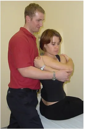
Figure 2. ROM TESTING WITH THE ARMDno2. The examiner reading the
ROM values stood behind the volunteer to ensure accurate readings were taken. Another examiner (not pictured) stabilised the participant’s pelvis to eliminate
pelvic rotation.
Muscle Energy Technique
five-second isometric contraction of side bending by the participant. After each isometric effort, a new rotation barrier was engaged and the participant repeated the isometric contraction. Four repetitions were completed on each volunteer.
Immediately following treatment post test ROM measures were recorded.
Figure 3. General Thoracic MET
Statistical Methods
ARMDno2 Reliability Study
All data was collated and analysed using Microsoft Excel. The test-retest reliability of the ARMDno2 device was determined using the "Pearson’s r correlation coefficient", T tests were used to determine whether a true linear relationship existed between the
axial rotation measures, furthermore 95% confidence intervals were calculated around the correlation coefficient to validate the findings.
Finally, 95% confidence intervals were also calculated to determine quantitatively how accurate the ARMDno2 device was for both right and left axial rotation.
Pre and Post MET Intervention
Pre and post-intervention ROM measurements were analysed for both the control and MET testing groups using independent T-tests and statistical significance was set at the p < .05 level, the effect size was calculated for all treatment and control group calculations.
RESULTS
ARMDno2 Reliability Pilot Study
The test-retest reliability of the ARMDno2 device was determined using the Pearsons r correlation coefficient for two continuous variables. The correlation coefficient for right axial rotation (r=0.8173) revealed a positive linear relationship between the participants first and last right rotation measures, the coefficient of determination (r²=0.668) explaining 66.8 percent of the variation between thesemeasures. With the level of significance set at 0.05, t tests demonstrated that the linear relationship between the right axial rotation measures was statistically significant (t(16) = 5.67>2.1199), this was confirmed by the 95% confidence interval calculations
(0.5972 - 1.3095). Similarily, the correlation coefficient for left axial rotation
Quantitatively the ARMDno2 reliability pilot study revealed with a 95% confidence level that all left rotation measurements were accurate within1.52 degrees (variance
0.10° - 1.52°), whereas the ARMDno2 right rotation measurements proved with 95%
confidence that they were accurate within 6.15 degrees (variance 0.40° - 6.15°).
Several outliers appeared responsible for the decreased accuracy for the measurement of right rotation.
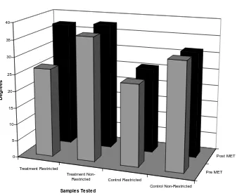
Effect of Thoracic MET
The mean trunk ROM measurements of the control and treatment groups in both restricted and non-restricted directions are presented in Table 1 and in Figure 1. The range of active trunk rotation was increased post-MET intervention for the treatment
group in the restricted direction (10.66°, SD 9.80°), whereas the untreated
non-restricted direction remained relatively unchanged (1.02°, SD 4.88°). The control
groups mean change in ROM following the ten minute latent period revealed a
minimal increase in trunk rotation (1.19°, SD 4.31°) for the restricted direction and
minimal decrease in trunk rotation (-0.5°, SD 2.59°) in the non-restricted direction.
Gross Trunk Range of Motion Mean Scores.
Treatment Treatment Control Control
Restricted Non-Restricted Restricted Non-Restricted
Pre MET 26.43 36.73 24.35
32.07
Post MET 37.09 37.76 25.54
31.57
Difference 10.66° 1.03° 1.19° -0.50°
Group Means for Gross Trunk Range of Motion
Treatment Restricted
Treatment
Non-Restricted Control Restricted
Control Non-Restricted
Pre MET Post MET 0
5 10 15 20 25 30 35 40
Deg
rees
Samples Tested
Figure 4. Mean Scores for Gross Trunk Rotation Pre and Post Intervention.
Independent group t tests were used to compare the mean pre and post-treatment ROM-values for the control and treatment groups. To determine whether the
T-Test Summary Table
Measure M SD T
Value P Value CI Effect size Treatment Group
Degree of Asymmetry (Pre
Test)
10.3 7.36 7.660 <
0.000 5
(7.66,12.
94)
1.40
Change in Restricted ROM (MET)
10.6 6
9.8 5.954 < 0.000 5
(7.15,14. 16)
1.09
Change in Non-Restricted ROM
1.02 4.88 1.147 > 0.05
(-0.72,2.77 )
0.21
Degree of Asymmetry (Post test)
0.67 11.2 7
0.324 > 0.25
(-3.37,4.70 )
0.06
Control Group
Degree of Asymmetry (Pre test)
7.72 7.38 4.440 < 0.000 5
(4.31,11. 13)
1.05
Change in Restricted ROM 1.19 4.31 1.167 > 0.10
(-0.80,3.17 )
0.28
Change in Non-Restricted ROM
-0.5 2.59 -0.821 > 0.10
(-1.69,0.69 )
-0.19
Degree of Asymmetry (Post
test)
6.04 8.37 3.059 < 0.01 (2.17,9.1
0)
0.72
Table 2. T-test Summary Table for Gross Trunk Range of Motion Testing.
(d = 1.05) respectively. The treatment groups restricted ROM direction demonstrated a statistically significant increase in gross trunk rotation after MET intervention (t(29) = 5.954, p<0.0005) and a large effect sixe (d = 1.09). The treatment groups non-restricted side (t(29)=1.15, p>0.05) (d = 0.12)and the control groups restricted (t(17)=1.17, p>0.10) (d = 0.28) and non-restricted (t(17)=-0.82, p>0.10) (d =
-0.19)directions failed to reveal statistically significant differences in trunk rotation at the post-test ROM testing, confirmed by the small effect sizes.
Finally, comparing the post-treatment restricted direction with the post-non-restricted direction demonstrated that MET treatment was effective in restoring symmetry in gross thoracic rotation (t(29)= 0.32, p>0.25). The same test completed in the control sample verified that a significant difference in rotation still remained (t(17) = 3.06, p<0.01).
DISCUSSION
This study demonstrated an increase in active thoracic rotation for subjects treated with MET, but no significant change in the control group, and so compliments the findings of two previous studies that investigated the effect of MET on cervical3 and lumbar4 ROM. MET applied to the thoracic spine in the direction of restricted rotation significantly increased the range of active trunk rotation (p<0.0005), while trunk rotation on the non-restricted side and in the untreated controls remained
unchanged. The effect size produced by the MET intervention proved to be large (d = 1.09). The mean amount of trunk rotation to the restricted side pre-treatment was 26.43 degrees, and immediately following treatment this range had increased to 37.09 degrees. Furthermore, the ranges of rotation in the treatment group became
significantly more symmetrical after MET therapy. The control group ranges of
The current study followed the recommendations set out by the previous cervical3 and lumbar4 MET studies as a larger sample size of forty-eight (30 treatment, 18 control) was used, giving this study greater power. Although this study supports the claims of various authors1,2,5 that MET can restore restricted spinal ROM, it is yet to be
scientifically proven that restricted range in symptomatic individuals is necessarily a
feature of spinal pain or dysfunction. It is plausible that such asymmetries represent normal variation and require no treatment, although it is clear that acute episodes of spinal pain are often accompanied by limited range. This study supports the concept that MET may be effective in the treatment of spinal pain with restricted motion, but investigations using symptomatic individuals are warranted.
Furthermore, it should be noted the MET applied throughout this study aimed to increase gross motion, which it succeeded in doing. MET practitioners often advocate more specific and subtle techniques aimed to increase the range of one motion
segment. It is likely that techniques applied in this way may increase segmental motion but not have the same effect on gross rotation that was documented by the current study.
Recommendations for Future Research
Future research should be directed to the effects of a single thoracic MET treatment over time, monitoring the changes in axial rotation over several hours would
demonstrate the longevity of the observed ROM changes documented in the current study. By examining the effect of multiple MET treatments on thoracic ROM over several weeks future researchers could determine whether multiple treatments enhance the observed changes in ROM. Validity testing of the neurophysiological models used to explain the actual mechanism of MET treatment, such as the effects of
MET on segmentally contracted muscles, connective tissue changes and investigating the efficacy of MET on reducing passive congestion, may elucidate the exact
mechanism behind the therapeutic benefits of MET.
relevance in the symptomatic population also requires investigation. It needs to be established that symptomatic individuals display abnormally restricted ROM (relative to their pain-free state, rather than a “normal” value), and that MET can produce long-term increases in range and improvement of symmetry. Researchers should examine the effects of MET on the ROM of symptomatic subjects, as well as using pain and
disability indicators, and these areas should prove to be a fertile field for future research.
CONCLUSION
Muscle energy technique was demonstrated to be effective in increasing the restricted range of trunk rotation and ameliorating rotational asymmetry in asymptomatic subjects. The restricted direction in the treatment group demonstrated a significant increase in gross trunk rotation as compared to the non-restricted untreated direction, and the bilateral rotation ranges of the control groups demonstrated no significant change in ROM. In addition the range of restricted rotation in the treatment group was returned to relative symmetry with the contra-lateral non-restricted side after MET treatment. These results support the efficacy of MET in increasing spinal rotation in the thoracic region, supporting the findings of two previous cervical3 and lumbar4 MET studies.
ACKNOWLEDGMENTS
The authors acknowledge the contribution of Mr Alisdair Murray throughout data collection, and his assistance with the MET intervention and ROM testing procedures. The authors also acknowledge Mr Jason Cowan and Mr Vincent Lenehan for the construction of the ARMDno2, and Jason Cowan for his assistance with presentation
of statistical data.
REFERENCES
1
Chaitow.L, Liebenson C, Muscle Energy Techniques. Edinburgh, Churchill Livingstone. 1996:
2
3
Schenk R, Adelman K, Rousselle J, (1994). The Effects of Muscle Energy Technique on
Cervical Range of Motion. Journal of Manual and Manipulative Therapy. 1994; 2(4): 149 – 155.
4
Schenk R, MacDiarmid A, Rousselle J, The Effects of Muscle Energy Technique on Lumbar
Range of Motion. Journal of Manual and Manipulative Therapy. 1997; 5(4): 179 - 183.
5
Mitchell FL, Jr. The Muscle Energy Manual. Volume 1. East Lansing, Michigan: MET Press;
1995:
6Denslow JS, Korr IM, Krems AD. Quantitative Studies of Chronic Facilitation in Human
Motorneuron Pools. American Journal of Physiology. 1947. In: The Collected Works of JS
Denslow, 1993 Year Book, Indiana: American Academy of Osteopathy.
7
Lederman E. Facilitated Segments: a Critical Review. British Journal of Osteopathy. 2000;
22:7 - 10.
8
Lederman E. Fundamentals of Manual Therapy. London; Churchill Livingstone; 1997.
9
Fryer G. Muscle Energy Concepts – A Need for a Change. Journal of Osteopathic Medicine.
2000; 3(2):54 - 59
10
Taylor DC, Brooks DE, Ryan JB. Viscoelastic Characteristics of Muscle: Passive stretching
versus Muscular Contractions. Medicine & Science in Sport and Exercise. 1997; 29(12):1619 –
1624
11
Hides JA, Stokes MJ, Saide M, Jull GA, Cooper DH. 1994. Evidence of Lumbar Multifidus
Muscle Wasting Ipsilateral to Symptoms in Patients with Acute/Subacute Low Back Pain. Spine
19:165-172. 12
Flynn TW. The Thoracic Spine and Rib Cage – Musculoskeletal Evaluation and Treatment.
Boston: Butterworth-Heinmann; 1996.
13
Kapandji IA. The Physiology of the Joints. 2nd Ed. Volume 3 – The Trunk and Vertebral
Column. London: Churchill Livingstone; 1974.
14
Troke M, Moore AP, Maillardet FJ, Hough A, & Cheek E. A New, Comprehensive Normative
Database of Lumbar Spine Ranges of Motion. Clinical Rehabilitation. 2001;15: 371-379.
15
Sturges K. The Importance of Axial Rotation in the Golf Swing. (Unpublished Thesis).