67 | P a g e
BRAIN TUMOUR IDENTIFICATION BY PIXEL
BASED SEGMENTATION
R.Shankari
1, S.Sruthi
2, M.Rahhni
2, T.Vishali
2Assistant professor
1, Department of ECE, U.G. Scholar
2, Department of CSE,
Velammal Engineering College, Chennai.
ABSTRACT:
Excrescence of unusual cells appear in the brain is called as Brain tumour.For the recognition of brain tumour MRI acts as an effective medium.Primeval diagnosis is necessary else oppressive medical issues occur if the tumour is discovered at the later stage.In this paper we automatically locate the tumour in MRI images by pixel based segmentation method.It involves enrichment,segmentation and classification steps.To bring out characteristics the enhanced images are segmented and collocated by characterizing images into normal and abnormal.Itis applied for various MRI instances.If samples are large number,medic could save time.
Keywords: Identification; segmentation; classification.
I.INTRODUCTION
By applying pixel oriented segmentation method a precise proposal of intuitive identification of brain tumour is
interpreted .The interpretation of the brain can be proposed by the kinds of scans called as MRI scan or CT
scan. In this approach MRI scanning is used for the entire technique. It is more accessible than the CT scan for
diagnosis since it neither affects the human body and nor uses any dissemination. It is reliant on the radio waves
and magnetic field.Different kinds of methods were spotted for the identification of brain tumour. But there may
have few disadvantages in the recognisation and extraction section. And it can be overthrown by our
approached intuitive pixel based segmentation algorithm which gives the better product for segmentation
procedure.Brain Tumour is a syndrome which is currently present within the brain or can be in the central spinal
canal. Tumour is of two types primary or secondary type.If the tumour is positioned at its provenance,then it is
primary and if the tumour is proliferated over other areas then it is secondary .Normally tumour disturbs
cerebral section and may instigate stroke. Subsequently the medic can give medication for the strokes but not
for syndrome. Hence necessary steps are antecedent identification and conventional medication is important
steps to upgrade the disease from out coming and it is acquired based on detailed investigation. The senescence
of the person experienced by the brain tumour may upturn about 2-3 years if it is recognized at the current
stage. In this paper we focussed on detailed identification of tumour part by applying automatic pixel based
segmentation algorithm. The advanced platform for the identification is matlab becauseit is satisfying and
68 | P a g e
II.DIFFERENT CURRENT METHODS
In the last 2 decades, several techniques have been put forth by the analysts to recognize anatomical part of the
brain tumors. Some of them are connected on edge identification, cluster recognisation, velutinuous senses and
watershed transformation.The edge identification strategy works better on high contrast images. But this
method miscarries in identifying the edges in low contradiction and smoothening of the images due to the weak
gradient degree. Corresponding, the clustering based technique such as K-means technique has a fast speed
operation which permits it to run on large datasets[8-9]. But, its main disadvantage is that it will not be able to
give the same results with each iteration, because the resulting clusters highly works on the fundamental
random allotment. A basic and easy way was marked controllers watershed segmentation which required less
exertion time and decreases over segmentation complexities.
Fuzzy C Means technique is a logic for processing the data which is implemented by allotting the partial
membership degree to each pixel in the given input MRI.[7] For example let us consider that a certain data point
that stands close to the center of a cluster , has a larger grade of membership to that cluster and another data
point that stands far away from the center of a cluster has a less degree of membership to that cluster. The
membership value of the velutinuous set ranges from 0 to 1. And it followed by the following steps:
a. A starting guess for the centers of clusters is made which are allowed to point out the mean position of
each cluster. But the beginning guess for these cluster centers is atmost probably wrong.
b. In the second step , Fuzzy C Means allots every data point or cluster a membership degree .
c. Then for each data point or cluster haphazardly on iterative basis refreshing the cluster centers and the
membership degree has to be allocated , later Fuzzy technique by iteratively transmits the cluster centers to the
right position within a data set.[6]
69 | P a g e
Fig.2. Segmented ImageFigure 1 and figure 2 represents the input and output image of Fuzzy C Means Method which shows the
Original and its Segmented part of an image respectively.
III.
CALCULATION OF OTHER TERMS
The projected procedure is examined through the computation of the following terms :tumour area , specificity
Sp, sensitivity Sn and accuracy A. These parameters are defined applying the formula [4]:
Sn = nTP , Sp = nTN and
nTP + nFN nTN + nFP
A = nTP + nTN . 100 …..…… (1)
nTP +nTN + nFP + nFN
The notations in above formula indicates: nTP :Tumour is viewed as Tumour only
nTN : Tumour is found as Tumour only nFN : Tumour is identified as Tumour only nFP :
Non-Tumour is recognised as Non-Tumour only
IV.
BLOCK DIAGRAM OF PIXEL BASED
SEGMENTATION METHOD
The executed technique has necessarily four blocks. They are pre processing, segmentation, extraction of
feature and Separation. Preprocessing includes mainly enrichment and refining of image. Segmentation is
hovered out by Pixel based Segmentation method. In Feature recognisation, area is extorted by thresholding and
lastly Collocation involves characterizing the images into normal and abnormal.
Normal is termed as “N” and abnormal is termed as “Y”.
A. Preprocessing
70 | P a g e
MATLAB tool . The pre processing steps includes some alteration. Intially it follows grayscale, histogram andrefining operations. It involves average type of refiner for smoothening purpose . The opportunities of existence
of noise in MRI are very low which can be be even removed by using this filter
.
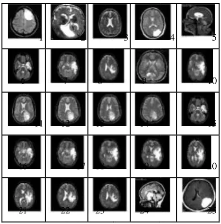
Figure 4 below shows some of the input original data sets of different types of brain MR Images . For deriving
the tumour part present in the Axial, saggital and coronal slices it is necessary initially to undergo the some
Preprocessing steps. In first step check wheather loaded input images in matlab environment are in MRI form
and in following step enriching the given image from any size into a particular size is done which is termed as
image enhancement . Here all the input images are enhanced to
particular size of 256 x256 .
1 2 3 4 5
6 7 8 9 10
11 12 13 14 15
16 17 18 19 20
21 22 23 24 25
Figure. 4: Original Data Sets.
B. Segmentation by Pixel Based Method
The valuable job is to recognise and segment the tumour portion. And it is accomplished favorably by applying
Pixel Based Segmentation algorithm. At first this method is subjected to some preprocessing steps. And coming
71 | P a g e
segmentation process. Graph resembling pie of an MRI resembles the allocation of pixels in the MRI imageagainst the grayscale is accomplished. Median filter is an kind of filter .This filter is of sliding-window type
which restores the intermediate value of the window with the middle of all the pixel values in the window [3].
Fundamental instrument in image processing is Histogram and popular as one of the excellent technique in
collecting facts about an image. It is basically beneficial in viewing the difference of an image . Grey-level
gives description high & low contrasts image established on there consolidation near a certain level .
1. Pixel based segmentation algorithm :
Last step is the thresholdingexamination , in this step the dual cover is applied to the entire image. It supports to
evolve into the white pixel to brighter and dim pixel to darker. Beginning portion of coding includes basically
correlating each transform coefficient with a threshold value. If the value is larger than the threshold, it will be
analysed as one and if it is less than the beginning then it is analysed as zero. This thresholding procedure is an
flexible method, here only those coefficients whose importance are above a threshold are kept inside each
block. Lastly the disjointed tumour part can be easily acquired by applying threshold analysis. Figure 5 below
shows the flowshart of pixel based segmentation algorithm.
Input Image
Preprocessing steps
Gray scale
Histogram of image
Apply Median Filter
Segmentation
Figure. 5: Pixel Based Segmentation Algorithm
2. Steps used for pixel based segmentation algorithm
The algorithm contains following steps:
STEP 1: First, the MR input images are captured and used for preprocessing steps.
STEP 2: In this step change of Input image to gray scale is executed .
72 | P a g e
STEP 4: Use median filtering for Image equalizing and getting rid of impulse noise.STEP 5: Pick the threshold value from the histogram. STEP 6: The disjointed tumour part is obtained by
picking the threshold value . It is done by applying the formula
1 if f(x,y) > T
g(x,y) = 0 if f(x,y)<=T ………. (2)
But for gaining the process accomplishment it needs the process of noise removal. For additional understanding
the task of this median filter, we finished the process of adding and removing the salt and pepper noise
artificially.
3. Output image of an mri sample by pixel based method :
Figure.6 Original Image
Figure.7 Segmented Image
73 | P a g e
No of white pixel (P) present in segmented tumour image is1611.8
Area in mm^2 is
10.598
Figure 6 and figure 7 shows the input and output image of Pixel Based Segmentation algorithm which
represents the Actual and its disjointed part of an image respectively.[6]
C. Feature Extraction
After the segmentation method, the next step is to extract the characteristics , here the characteristics which is to
be extracted in this paper is “Area” and it is examined by applying the binarization technique. Binary image has
only dual values either white or black .Since maximum input MRI size of an jpeg image taken here is 256x256.
The binary kind of image are represented as a total value of summation of all black and white pixels .
Equation for input MRI ,
= Σ 255 ……… (3)
I
Equation for Pixel,
……… (4) P= Height (H) *Width (W)
No of white pixel P = Σ 255 ………. (5) Where,
f (0) = white pixel (Consider as digit 0)
f (l) = black pixel (Consider as digit 1)
P = number of white pixels (width*height)
1 Pixel = 0.264 mm
The formula to calculate the area in mm^2 is
Size of tumour is ,
S = [(√ )*
0.264] mm2 …….. (6)
Where,
74 | P a g e
The tumour area can be obtained by applying simple matlabcommand . It is shown below:img = n; BW = img; bwarea(BW)
D. Classification:
The Classification involves labelling the images into normal and abnormal . The term „abnormal‟ indicates the presence of tumour and the term „normal‟ indicates the normal condition . The MATLAB functions such as
FIG-Files and M-Files of GUI is used for the classification of the Brain MR images into tumour and non tumour
parts. These two files are produced for the first time immediately after we save or run the GUI. A
FIGURE-HOLDING file with confine as .fig, contains of a detail explanation of the GUI with its outline & also the parts
of the GUI. An M- file with confine as .m, consists of the code that controls the GUI including the total flow for
its respective components.
These two files relates to the work of outlining and programming the GUI. When you have a outline or lay out
of the GUI in the Layout Editor section , your task will be saved in the FIG-file. And if u do the programming
of the GUI, your work will be saved in the M-file.
These functions are to be used to differentiate whether a given input MRI is normal or abnormal.
V.RESULT
The implemented technique have been tested fully on 25 different samples of Brain tumour only. Area present
in the segmented tumour is one of the feature we have extracted here. Segmentation method is done for input
image and its respective manually traced image so that some of the parameters such as Sensitivity Sn
,SpecificitySp and Accuracy A[%] can be calculated.The resultscalculated are tabulated in Figure 7 which
represents the segmentation quality in the tumour. This method is implemented on a personal computer
applying MATLAB software .
Table1. Segmentation Quality in the Tumour
Slice Tumour Sensitivity Specificity Accuracy
Area in Sn Sp A[%]
mm2
1 14.665 0.99 0.98 98.35
2 24.378 0.95 0.97 97.27
3 6.6722 0.93 0.98 98.22
75 | P a g e
5 13.830 0.98 0.98 98.67
6 5.716 0.92 0.98 98.2
7 8.128 0.98 0.97 97.4
8 7.082 0.98 0.97 97.7
9 7.323 0.98 0.99 99.40
10 10.679 0.99 0.97 97.8
11 6.432 0.91 0.99 99
12 7.051 0.93 0.99 98.96
13 4.739 0.922 0.99 99.05
14 2.488 0.96 0.995 99.5
15 5.808 0.95 0.982 98
16 5.825 0.99 0.97 97.5
17 8.039 0.96 0.973 97.26
18 10.679 0.99 0.97 97.7
19 10.999 0.97 0.97 97.52
20 10.593 0.97 0.95 96.09
21 11.120 0.99 0.95 96.37
22 10.950 0.98 0.96 96.33
23 7.087 0.98 0.97 97.25
24 17.585 0.98 0.98 98.87
25 23.332 0.97 0.98 98.57
Segmentation approach and its area calculation is done by applying the proposed segmentation method and is
tabulated above in table1.
VI.CONCLUSION
Since it is concluded that the suggested technique implements better in embellishing, segmenting and extracting
the brain tumour characteristics from MR images.The calculation of brain tumour area can help for tumour
staging for persuasive treatment and surgical planning.Our goal is to examine different Brain MRI samples and
get the accuracy of segmentation . The proposed approach presented for the segmentation of Brain tumour
images overcomes the area limitations of the current solutions and provides the veracity of segmentation . So
we illustrated this method on brain tumour area cases applying MR imaging method. The automatic
segmentation technique is not monotonous, labor intensive and requires very less time to obtain tumour area
measurements as compared to clinical method . This method is applied to 25 different types and shapes of MRI
slices and cosiderably good segmentation results are achieved as compared to earlier methods . Also this
76 | P a g e
could apply his method.Some parameters such as Sensitivity,Specificity and Accuracy are calculated during the computation of brain
tumour area measuremets.
REFERENCES
[1] Monica Subashini.M , Sarat Kumar Sahoo,” Brain MR Image Segmentation for Tumour Detection
using Artificial Neural Networks,” International Journal of Engineering and Technology (IJET), ISSN :
0975-4024,Vol 5 No 2 Apr-May 2013, pp.925-933.
[2] K.S.A Vrji, Dr J. JayaKumari, "Automatic detection of brain tumour based on magnetic resonance
image using System with watershed Segmentation,” Proceedings of 2011 International Conference on Signal
Processing, Communication, Computing and Networking Technologies (ICSCCN),978-1-61284-653-8/11,IEEE
21-22 July 2011, pp.145-150.
[3] M. UsmanAkram, AnamUsman., “Computer Aided Systemfor Brain Tumour Detection and
Segmentation,” 978-1-61284-941-6/11, IEEE 2011,pp.299-302
[4] Mikulka J, Burget R, Riha K and Gescheidtova E, "Segmentation of brain tumour parts in magnetic
resonance images,"36t International Conference on , Telecommunications and Signal Processing (TSP),
978-1-4799-0404-4/13,IEEE 2-4 July 2013 , pp.565-568.
[5] R.B.Dubey, M.Hanmandlu and S.Vasikarla, “Evaluation of Three Methods for MRI Brain
Tumoursegmentation,"Eighth International Conference on Information Technology: New Generations (ITNG),
978-0-7695-4367-3/11 ,IEEE 11-13 April 2011, pp.494-499.
[6] Rohit S. Kabade, Dr. M. S. Gaikwad“Segmentation of Brain Tumour and Its Area Calculation in
Brain MR Images using K-Mean Clustering and Fuzzy C-Mean Algorithm,” International Journal of Computer
Science & Engineering Technology (IJCSET),2013, Vol 4 No 05 May 2013,ISSN : 2229-3345,pp.524-531.
[7] Hassan Khotanlou, Olivier Colliot, Jamal Atif and Isabelle Bloch,“3D brain tumoursegmentation in MRI
using fuzzy
classification, symmetry analysis and spatially constrain deformable models”, 0165-0114, 27November 2008 , pp.1457 – 1473.
[8] Dessai V.S, Arakeri M.P and Ram Mohana Reddy G, "A parallel segmentation of brain tumour from
magnetic resonance images," International Conference on Computing Communication & Networking
Technologies , ICCCNT'12,IEEE26-28July2012, pp.1-6.
[9] Deepak K.S, Gokul K, Hinduj R and Rajkumar S,“An Efficient approach to predict tumour in 2D Brain
Image using classification tecniques," International Conference on Information Communication and Embedded
Systems, ICICES21-22 Feb 2013,pp.559-564.
[10]V. Rajamani and S. Murugavalli”A High Speed Parallel Fuzzy C-means algorithm for Tumour
77 | P a g e
[11]J. S. ZulaikhaBeeviM, Mohamed Sathik(2010)”An Effective Approach for Segmentation of MRIImages: Combining Spatial Information with Fuzzy C-Means Clustering”European Journal of Scientific
Research, ISSN I450-2I6X Vol. 41 No.3 pp.437-451.
[12]VeenaKalasappanavar, Prof. Sujata. S. Kotabagi, Prof. Prakash. H.Unki,"Brain Tumor Segmentation and
Its Area Calculation in Brain MR Images Using Fuzzy C Mean and Pixel Based Segmentation Algorithms", 5th
National Conference on Electronic Technologies 2014, GCE 25-26 April 2014 pp:132-135.
[13]Yong.Y. Z. and Chongxun. L. Pan,”A Novel Fuzzy C-Means Clustering Algorithm for Image
Thresholding”Measurement Science Review, Volume 4(1), 2004.
[14]Jude hemanth.D, D.Selvathi and J.Anitha”Effective Fuzzy Clustering Algorithm for Abnormal MR
Brain Image Segmentation ”Page Number 609-614, International/Advance Computing Conference (IACC
2009), IEEE,2009.
[15]A. Bleau and L. J. Leon ”Watershed Based Segmentation and Region merging”Computer Vision and
Image Understanding, vol. 77, no. 3, pp.317-370, 2000.
[16]H.P. Ng, S.H. Ong, K.W.C. Foong, P.S. Goh and W.L. Nowinski. ”Medical Image Segmentation using
K-Means clustering andImproved Watershed Algorithm”IEEE 2006.
[17]Veena.R.Kalasappanavar, Dr.Basavaraj, S.Anami, Prof.Prakash.H.Unki and Prof.
Sujata.S.Kotabagi,"Brain Tumor Detection In MRI Images Using Automatic Segmentation Method", 2nd
International Conference on Current Trends in Engineering and Management ICCTEM-17 July 2014, Mysore,