_____________________________________________________________________________________________________
*Corresponding author: E-mail: ghiassik@gmail.com, Khashayar.Ghiassi@drsulaimanalhabib.com, ghiassik@googlemail.com; (Past name: British Journal of Medicine and Medical Research, Past ISSN: 2231-0614, NLM ID: 101570965)
Skin Cancer Screening Makes It Possible to Detect
Dysplastic Lesions and Significantly Reduce the
Progression to Malignant Melanoma
Khashayar Ghiassi
1*1
Dr. Sulaiman Al Habib Medical Group,Saudi Arabia.
Author’s contribution
The sole author designed, analysed, interpreted and prepared the manuscript.
Article Information
DOI: 10.9734/JAMMR/2019/v31i730307 Editor(s): (1) Chan-Min Liu, School of Life Science, Xuzhou Normal University, No.101,Shanghai Road, Tangshan New Area, Xuzhou City 221116, Xuzhou City, PR China. Reviewers: (1) Heba Gamal Abd El-Aziz Nasr, Al-Azhar University, Egypt. (2)Manas Bajpai, Nizam's Institute of Medical Sciences, India. (3)Deepak Sethi, Ravindra Nath Tagore Medical College, Udaipur, India. Complete Peer review History:http://www.sdiarticle4.com/review-history/52995
Received 04 October 2019 Accepted 14 December 2019 Published 16 December 2019
ABSTRACT
This study takes a look at cultural practices around the world in terms of how they relate to being a contributing cause of malignant melanoma due to perceptions of skin colour and accepted tanning practices. This paper examines the effects of UV radiation in great detail and reviews skin cancer as a serious health risk due to overexposure and other factors. As part of the review of this global health threat, the demographic distribution of people who suffer from melanoma is also discussed. Currently, women between 31 and 50 years old are at the highest risk of developing skin cancer and, therefore, must be screened to reduce that risk, particularly given that malignant melanoma is better managed with earlier screening practices in middle-aged adults.
Keywords: Skin cancer; dysplastic lesions; malignant melanoma.
ABBREVIATIONS
AJCC : American Joint Committee of Cancer ALM : Acral lentiginous melanoma
CPD : Cyclobutane pyrimidine dimers DNA : Deoxyribonucleic acid
HE : Haematoxylin-eosin Illustr. : Illustration(s)
LDH : Lactate dehydrogenase LMM : Lentigo-maligna-melanoma MIA : Melanoma-inhibiting activity MSH : Melanocyte-stimulating hormone NMM : Nodular malign melanoma PMOC : Proopiomelanocortin SPF : Sun protection factor UV : Ultraviolet radiation WHO : World Health Organization
1. INTRODUCTION
There are several parameters of social behaviour influenced by the society that define health, attractiveness, and social status. Because these ideas are defined diversely, there are different definitions in different parts of the world and in the various cultures regarding the perfect pigmentation or tan of the skin. Caucasians aspire to have golden tanned skin, whereas Asians prefer a light skin tone. Due to the industrialisation of the United States and Europe at the beginning of the 20th century, light skin became associated with factory work, where there was no direct sunlight, and so in those parts of the world, it tends to be associated with low socio-economic status. In Asian countries, on the other hand, tanned skin is linked with fieldwork and therefore in those countries, tanned skin is associated with a low social status. In the 1920s, Coco Chanel established a cultural norm that saw a “golden skin tone” as a chic sign of having excess time and money for leisure activities. Yet, this ideal would not be worth striving for in Asian countries, due to social conventions such as those already mentioned.
1.1 Sunbathing – Prevalence and Risks
In European countries, a great number of people expose themselves to unhealthy doses of UV light in the pursuit of tanning their skin. This focus on suntanning has led to the use of sunbeds and excessive sunbathing on vacation, resulting in burns in the form of dermatitis solaris have become a regularity. From the age of 18 up to 45. every second person uses a sunbed at least once at a certain point in their life, and every fifth person in this age group states that he/she uses sunbeds regularly [1]. In the majority of other countries, the number of excessive sunbaths is lower [2,3]. Especially for teenagers, the high levels of exposure to the sun and the lack of use of sun protecting cremes/clothes are all of high importance. An investigation from Germany and Italy showed that people with a
high socio-economic status took more sunbaths than people with a low social status [4].
Caucasians who easily get burned and tan only a very little (skin phototypes I and II) [5,6], who have a positive personal family anamnesis for skin cancer, a light eye colour (e.g. blue), ginger or blonde hair and lots of freckles [7] are considered the biggest risk factors for getting
skin cancer after chronic UV exposure.
Furthermore, suffering severe sunburns with blistering in childhood and early adolescence up to the age of 20 have been associated with a higher risk for getting skin cancer [8,9,10] and childhood and adolescence have been identified as key periods for getting melanoma as an adult. This may be caused by the sensitive reaction of adolescent skin to chronic high doses of UV light [11,12]. The risk of contracting melanoma is extremely high when sunburns were suffered during adolescence, between the ages of 13 and 19, where there was frequent exposure to UV radiation for long periods of time [13,14]. The risk of getting melanoma is extremely high for women under the age of 45 who exposed themselves carelessly to UV radiation in their adolescence. This has been shown in a recent study conducted by Ting et al. [14] among 1,518 dermatological patients, where 79 of those having a malignant melanoma stated having such an anamnesis [15].
1.2 Reasons for Sunbathing
Sun-seekers have been asked for their
motivation in several studies. Ranking first has been having an attractive appearance, joined by a positive psychological effect and relaxation [16-18]. Furthermore, another study by Banerjee and colleagues showed that men find women with tanned skin more attractive and healthier. This, again, is a motivation for women to regularly tan their skin to improve their appearance [19]. This study significantly showed that adults who use sunbeds on a regular basis struggle to give up this habit. What’s more, experiments have shown that UV light releases endorphins, which could also promote an addiction [20]. In addition, the effects of an improved mood and mental brightening through frequent sunbathing have been described, which is reflected in seasonal mental disorders [21].
1.3 Biological Effects of Ultraviolet Radiation
pathological ones. The UV spectrum is categorised by spectra as UV-C (100-290 nm), UV-B (290-320 nm), and UV-A (320-400 nm). More than 90% of UV-B radiation is absorbed by the epidermis, especially the stratum corneum, and this spectrum is mainly responsible for carcinogenic processes. In contrast, UV-A radiation reaches the reticular and papillary dermis and has harmful effects on connective tissue (photoaging and solar elastosis) and also has a further immunosuppressive effect [22]. The pathological effects include acute sunburn and an acute inflammatory reaction caused by too much exposure. The resulting erythema is caused mainly by the UV-B part.
Cyclobutane pyrimidine dimers (CPD), which create mutations through cytosine, are relevant on a molecular basis. These changes in the genetic code constitute the risk of propagation in daughter cells and an accumulation with regard
to carcinogenesis. Further, UV-B has
immunosuppressive effects that reduce antigen detection and antigen processing potency on, for
example, Langerhans cells [22,23].
Physiologically, there are DNA repair
mechanisms whereby mutations are detected by enzymes that block the DNA replication and cut out the mutations in order to incorporate the wild type sequence again. These complex processes are supervised by the phosphor protein53 (p53) that serves as a tumour suppressor gene and enables the repair of DNA [24,25]. In this case, p53 serves as an arrester of the G1 phase in order to allow the DNA sufficient repair time. The cell is subjected to apoptotic cell death if a repair is not possible [24]. Under the microscope, these apoptotic keratinocytes can be recognised in the epidermis as so-called sunburn cells, eosinophil rounded cells. The p53-protein also gets damaged more and more by the UV radiation, and the occurring mutations allow for, after appropriate accumulation, aberrant growth and, finally, the development of tumours [25].
Furthermore, the protein p53, as an
effector/sensor, is linked to tanning [26]. It has been proven that the pigmentation is significantly stimulated by p53 through increased synthesis of melanin after UV exposure via direct activation of proopiomelanocortin (PMOC) [26]. Tanned skin has an effective sun protection factor (SPF) of only 2 to 3 [27]. The melanin pigment absorbs UV radiation, and it also absorbs free oxygen radicals that occur through the inflammation of the skin. The PMOC gene encodes the melanocyte-stimulating hormone (MSH), the adrenocorticotropic hormone (ACTH) and beta
endorphins. The transcription of these three genes takes place in keratocytes exposed to sunlight, which leads to the synthesis of the according preproproteins. After appropriate processing, MSH binds to the melanocortin receptor one on melanocytes, which induces melanogenesis, melanocyte differentiation, and the melanosome transfer from melanocytes to keratinocytes [28].
1.4 Pigmented Nevi
A locally sharply determined malformation of the skin that is characterised by exuberant, or in rare cases reduced, development of one or more skin components. It may derive from parts of the skin appendage, vessels, nerves, connective tissue, or the epidermis. Combined malformations that are named after the predominant tissue are called organoid nevi. Nevi can have been in existence since birth or they can arise later in life. Melanocytic nevi are a special case. In some cases, they are a malformation, but many melanocytic nevi are benign tumours of the melanocytes.
Almost everyone has pigmented nevi
(colloquially called moles). These skin lesions are of great importance. There is a tremendous variety of different types of nevi, including the non-pigmented nevi, but it is the pigmented nevi that are significant. Included among these are both innate and acquired nevi. A part of these nevi may get atypical, which means that single nevus cells show characteristics of a malignant tumour. Mostly, these atypical nevi arise from the influence of exogenous toxicants, for example UV radiation, but they also arise from genetic predisposition (so-called atypical nevus families). Black skin cancer lesions (so-called melanomas) may arise from a part of the atypical nevi. Malignant melanoma is the tumour with the fastest rise in occurrence in the world, especially in western Europe, America, and Australia. It is estimated that 1 in 75 persons born in 2000 will suffer from malignant melanoma during their lifetime.
1.4.1 Melanocytic Nevi (Nevuscellnevi)
1.4.1.1 Definition
Skin changes that are skin-coloured, pigmented to different degrees, macular, popular, or papillomatous are the clinical result. A 30-year-old central European person has, on average, 20–30 melanocytic nevi on their body. The existence and number of melanocytic nevi is genetically disposed.
UV exposure is etiologically of great importance. In addition, immunosuppression and hormonal changes (during or after puberty and during pregnancy) are important for the appearance of nevi, as well. Melanocytic nevi become manifest either at birth (congenital) or they occur over the course of life until the age of approximately 40 years. After that, a regression often takes place.
1.4.1.2 Types of melanocytic nevi
Melanocytic nevi of the junction type are melanocytic cells located in the basal cell layer of the epidermis in the form of nests. They are clinically flat, light to dark brown patches.
Melanocytic nevi of the compound type are melanocytic cells in the junction zone as well as in the corium and are clinically elevated, light to dark brown nodules or knots.
Melanocytic nevi of the dermal type are melanocytic cells located almost exclusively in the corium.
Unna type are papillomatous knots with a bumpy, grooved surface and are light to dark brown, and the Miescher type are flat, elevated, less pigmented, and often skin-coloured.
It is important to differentiate the acquired nevi from congenital nevi.
Congenital melanocytic nevi are benign innate melanocytic nevi that appear on about 1% of new-borns. Congenital nevi are based on a developmental disorder of the melanoblasts from the neural crest. This type of nevi mostly imposes as a flat patch or knot with a light to
dark brown colour, and a sometimes
papillomatous surface or hair. Their size varies from 1 cm to 3 cm with a potential large expansion (“giant nevi”). Over time, the pigmentation and the appearance of hair increase. One with a diameter of over 20 cm is called a nevus giganteas, which can involve the multiplication of melanocytes (melanosis verticis gyrate) at the head and also possibly at the leptomeninges (neuro-cutaneous melanosis), in
which case epilepsy, neurological disorders, and hydrocephalus should be observed.
1.4.2 Atypical melanocytic nevi
1.4.2.1 Definition
Clinically and histologically atypical melanocytic melanocytes that, initially, have been described on patients from families with a familiar malignant melanoma [29]. They have subsequently been
observed on patients with non-familiar
melanoma. Dysplastic nevi have been debated to be a marker for an increased risk for melanoma and/or as a precursor of melanomas. It has been estimated that approximately 5% of the population has one or more dysplastic nevi.
1.4.2.2 Anamnesis and symptoms
These are mostly asymptomatic and rarely itch.
Atypical nevi can be detected using the ABCDE guidelines, which are the following criteria:
A – Asymmetrical, irregular B – Border is blurred
C – Colour is variable, with patterns from light to dark
D – Diameter is > 5 mm E – Evolving and palpable
The more factors that apply, the more likely the diagnosis is malignant melanoma. This easy guideline should help the dermatologist doing risk ratification as well as making the choice of excision-worthy moles [30].
In addition, classification of the phototypes according to Fitzpatrick can provide a clue as to the risk of developing a malignant tumour [5].
1.4.2.3 Histopathology
The term “dysplastic nevus” is very imprecise and is not used uniformly as there are no clearly
defined criteria for making a diagnosis.
Histologically, the most frequently used criteria
are the following: polymorphism of the
melanocytic cells, bridge building of single nests, and patchy lymphohistiocytic infiltrate.
1.4.2.4 Therapy
Close-meshed clinical and dermatoscopic
Table 1. Skin photo types according to Fitzpatrick [5]
Photo type Skin type Pigmentation behaviour
I White, very light, ginger-blonde hair; blue
eyes; freckles
Always burns, never gets tanned
II White, light, ginger or blonde hair; blue,
green, or light brown eyes
Often gets burned; tans rarely
III Creamy-white, light, every skin and eye
colour; frequent skin type
Occasionally mild sunburns; tans gradually
IV Brown, typically Mediterranean; Caucasian
skin
Burns seldomly; tans quickly and regularly
V Dark brown; Middle Eastern Burns very seldomly; tans very
quickly
VI Black Never burns; tans very easily and
quickly
1.4.2.5 Progress(ive forms) and prognosis
According to Kraemer and Greene [31], patients with dysplastic nevi occurring in their family have a higher risk for melanoma than patients with infrequent dysplastic nevi. The melanoma risk reaches up to 100% for patients with dysplastic nevi having at least two or more relatives suffering from malignant melanoma.
1.4.2 Hereditary dysplastic nevus syndrome
For the subtype with familiar melanoma, BK-mole syndrome, BK-nevus syndrome and familiar atypical multiple mole melanoma (FAMM) syndrome.
1.4.2.1 Definition
Hereditary syndrome with many dysplastic nevi and a high risk for melanoma.
1.4.2.2 Genetics
Possibly in the case of the subtype with familiar melanoma, mutations at the p16-gene on the short arm of chromosome 9 (9p21).
1.4.2.3 Clinic
Great amount of so-called dysplastic nevi, especially on the torso, along with many normal melanocytic nevi.
Special form: dysplastic nevus syndrome with only one quadrant.
1.4.2.4 Therapy and progress
Close-meshed clinical and dermatoscopic
controls every 3–6 months, if possible, with photo documentation. Excision of suspicious nevi.
Intensive sun exposure should be avoided during infancy, especially in the case of familiar disposition.
1.4.2.5 Differential diagnosis
Malignant melanoma.
1.4.3 Spitz nevus
Benign juvenile melanoma, Spitz nevus.
1.4.3.1 Definition
Benign melanocytic reformation with
polymorphous and cytoplasmic cells.
1.4.3.2 Epidemiology
Mostly adolescent, rarely adults.
1.4.3.3 Clinic
Hemispheric elevated, rapidly growing, reddish nodules, often on the face, and sometimes globular. Lupoid infiltrate with glass spatula pressure.
Rare entities that are not going to be considered in this work are the pigmented spindle cell Reed nevus and the nevus coeruleus. Also not considered are benign pigmented skin changes without the growth of the nevus cells and the café-au-lait patch and freckles are not going to be considered neither.
1.5 Malignant Melanoma
clinically unremarkable skin (70%) or in the area of pre-existing nevi (30%).
1.5.1 Epidemiology
The melanoma has the most rapid increase in occurrence worldwide, where the incidence in Europe and America increases by 8-10% per year. It is the most common malignant tumour in young women in the age group of 25-29 years in the United States. The incidence per 100,000 inhabitants is lowest in Sub-Saharan Africa, and the highest is in Australia, with 60 cases per 100,000 people. The lethality could be reduced to less than 20% through intensive educational programmes and screening. The estimated risk of suffering from melanoma once in their lifetime for someone born in the year 2000 is 1:75.
1.5.2 Aetiology and risk factors
Genetic disposition of so-called melanoma-families suffering from the FAMM syndrome (see
above), dysplastic nevus syndrome, or
xeroderma pigmentosum. Other than genetic disposition, the disposition of the melanoma depends next on the following factors:
– Number and size of congenital nevi
(lifetime risk of up to 10%)
– Light skin of phototype I or II according to
Fitzpatrick [5]
– Living at geographical latitudes near the
equator
– Earlier melanomas or first-degree relatives
suffering from melanoma
– Exposure to UVA (number and severity of
sunburns during infancy)
– CAVE: The association with UV radiation is
not as strong as the association of epithelial tumours and UV exposure!
1.5.3 Clinic
The cutaneous malignant melanoma may show a great variety of manifestations. Typically, the melanoma is deep brown to blackish, but there are also reddish, blueish and even non-pigmented (so-called amelanotic) melanoma. Special forms include melanomas at the nails or non-healing, ulcerated melanoma.
1.5.4 Superficial spreading malignant melanoma (SSM)
65–70%; light brown or brownish to blackish, rarely whiteish (regression zone). In the
beginning, always flat; pigment changes of varying sizes with a change into infiltrated nodules or knots and irregular borders.
1.5.5 Nodular malignant melanoma (NMM)
15–20%; mostly small, smooth, blackish knots as a primary manifestation, but also in the form of a secondary NMM on a pre-existing SSM.
1.5.6 Lentigo-maligna-melanoma (LMM)
10%; auburn, café-au-lait, or dark brown coloured blemishes in chronically light- exposed areas or actinically damaged skin.
1.5.7 Acrolentiginous malignant melanoma (ALM)
5%; a melanoma growing at palmar, plantae, or subungual, often on pre-existing lentigines.
In all forms of melanoma except NMM, a horizontal growth phase occurs first, followed by a vertical growth phase that is crucial for early metastasis.
NMM and ALM often occur as amelanotic, skin-coloured primary tumours.
ALM and mucosa or uvea melanoma (missing protective melanin pigment) are nearly the only types found in Sub-Saharan Africans, Asians, and Hispanics.
1.5.8 Localizations
Melanoma can be found on every part of the body, but 90% can be found on the skin (for men, often on the torso; for women, often on the lower limbs), with 6% at the eye (mostly the choroid, so-called uvea melanoma), and 4% on the mucosa.
A metastasis mostly takes place at the lymph nodes > skin > lungs > liver > bones > brain.
1.5.9 Diagnostics
1.5.9.1 Incident light microscopy
Simple instrumental limitation to
non-melanocytic diseases (e.g. hematoma,
haemangioma, seborrheic keratosis).
The high-resolution ultrasound is a move in the
field of luxury diagnostics. Furthermore,
laboratory changes can only be found in the case of advanced tumours. In this case, tumour anaemia, increased blood sedimentation rate,
liver enzymes, and increased alkaline
phosphatase, if the metastasis in liver and bones is increased, will take place. An increase in LDH is a seldom-seen specific indicator for remote metastasis in the following tumour markers: soluble S-100ß in the case of remote metastasis and MIA (melanoma-inhibiting activity).
Imaging techniques should be realised for advanced malignant melanoma. Among these
are lymph node sonographies, abdomen
sonographies, and thorax X-ray.
A sentinel-lymph-nodes-diagnostic of the
regional draining lymph node should be used for melanomas with a skin depth > 1 mm. A method that is gaining more and more importance, especially for the survival of the patient, is the selective resection of lymph nodes.
For metastasising melanomas: bone
scintigraphy, full-body CTs, skull NMR, and if
necessary, positron emission tomography
(method for recognising an increased tumour metabolism with a resolution capacity of at least 5 mm, to be indicated in the case of unclear CT results).
1.5.10 Histology
Criteria for malignancy:
– Asymmetric tumour structure, flattening of
the reticular structure
– Papagoite ascent of light cells in the whole
epithelial unit (“full sickness atypia”)
– Missing differentiation of cells towards the
skin depth
– Invasion of the tumour cells into the corium
– Core-plasma relationship with prominent
nucleus; nuclear polymorphisms;
increased mitoses
– Ulceration, regression, and sclerotic areas
on pre-existing nevus cell
– Lymphohistiocytic infiltrate
– Important data are the Clark level and the
Index according to Breslow.
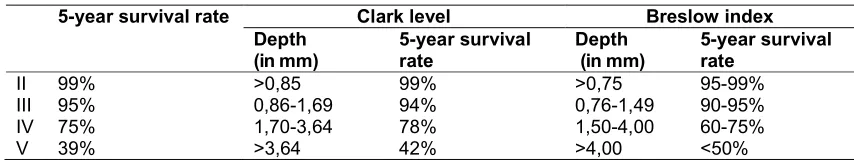
The so-called Breslow index provides information about the depth of the invasion in millimetres. This depth is determined with the help of an ocular micrometre and describes the invasion route that has been taken by the tumour cells. It covers the way from the granular cell layer until the deepest invasion of the tumour cells. It is easier to reproduce than the Clark Level.
The Clark Level, introduced by Dr. Wallace H. Clark Jr., has been defined as follows:
Level I Tumour cells exclusively in the
epidermis (melanoma in situ)
Level II Reaching up to the stratum papillary
Level III Filling the entire stratum papillary
Level IV Reaching up to the stratum reticular
Level V Reaching up to the subcutis
The Clark classification correlates with the five-year survival rate. Special forms include melanoma in situ, intraepidermal melanocytic hyperplasia with cell atypia, polypoid dysplastic desmoplastic neurotropic balloon cell-like or myoxid malignant melanoma.
1.5.11 Immunohistology
The antigens S-100, HMB-45, Melan A/MART-1
and the proliferation marker Ki-67/MIIB-1
associated with melanoma are used to
differentiate melanomas from undifferentiated pleomorphic spindle cell tumours of other entities or to determine high levels of proliferation activity. They are also used in sentinel lymph
node examinations to detect micro metastases.
Essentially, immunohistology is used to exactly determine the skin depth. A high mitosis index, ulceration, and regression zones are linked to a worse prognosis.
Table 2. Correlation of primary tumour thickness and survival rate
5-year survival rate Clark level Breslow index
Depth (in mm)
5-year survival rate
Depth (in mm)
5-year survival rate
II 99% >0,85 99% >0,75 95-99%
III 95% 0,86-1,69 94% 0,76-1,49 90-95%
IV 75% 1,70-3,64 78% 1,50-4,00 60-75%
1.5.12 Classification/prognosis
EORTC stadia classification:
I : Primary tumour without
metastases on the lymph nodes or remote metastases or satellite metastases (up to 3 cm around the primary tumour). Five-year survival rate, 80%
Na : In-transit metastases (< 3 cm
distance to the primary tumour) and not beyond the regionary lymph nodes. Five-year survival rate, 40%,
Mh : Regionary metastases on the
lymph nodes. Five-year survival rate, 20%.
II : Remote metastases
non-visceral : Five-year survival rate, 5%.
visceral : Five-year survival rate, 0%.
1.6 Special Forms of Malignant Melanoma
1.6.1 Melanomas during pregnancy
Melanocytic nevi get activated during pregnancy quite often. But it is not assured that it influences the progress of a malignant melanoma.
1.6.2 Malignant melanoma during infancy and adolescence (up to 18 years)
Children often get melanomas on the base of neuro-cutaneous melanosis. In this case, metastasising melanomas may occur during early infancy.
1.6.3 Congenital melanomas
They grow from transplacental metastasis of a motherly melanoma or from the area of congenital melanocytic giant nevi. Very poor prognosis.
1.6.4 Metastasising melanoma without known primaries (about 5% of all patients)
A detailed anamnesis (earlier excisions,
excisions of skin changes) is crucial in this case. During the inspection of the whole body and the mucosa, scars should also be taken into account. Signs of regression of earlier melanomas should be observed as well.
1.6.5 Familiar melanomas
Different candidate-genes (e.g. p16INK4a) on chromosome 9. The risk for melanomas is higher
the more family members suffer from
melanomas. Dermatological check-ups every 6 months.
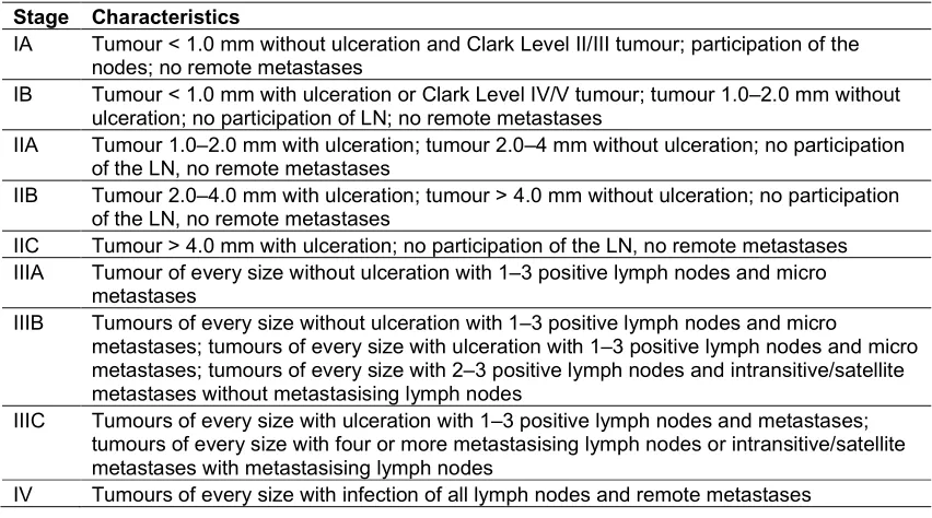
Table 3. The new American Joint Committee of Cancer (AJCC) classification
Stage Characteristics
IA Tumour < 1.0 mm without ulceration and Clark Level II/III tumour; participation of the
nodes; no remote metastases
IB Tumour < 1.0 mm with ulceration or Clark Level IV/V tumour; tumour 1.0–2.0 mm without
ulceration; no participation of LN; no remote metastases
IIA Tumour 1.0–2.0 mm with ulceration; tumour 2.0–4 mm without ulceration; no participation
of the LN, no remote metastases
IIB Tumour 2.0–4.0 mm with ulceration; tumour > 4.0 mm without ulceration; no participation
of the LN, no remote metastases
IIC Tumour > 4.0 mm with ulceration; no participation of the LN, no remote metastases
IIIA Tumour of every size without ulceration with 1–3 positive lymph nodes and micro
metastases
IIIB Tumours of every size without ulceration with 1–3 positive lymph nodes and micro
metastases; tumours of every size with ulceration with 1–3 positive lymph nodes and micro metastases; tumours of every size with 2–3 positive lymph nodes and intransitive/satellite metastases without metastasising lymph nodes
IIIC Tumours of every size with ulceration with 1–3 positive lymph nodes and metastases;
tumours of every size with four or more metastasising lymph nodes or intransitive/satellite metastases with metastasising lymph nodes
1.6.6 Multiple melanomas
Five percent of all melanoma patients get second or even more malignant melanomas from a primary tumour. Potential risk group is co-existence of xeroderma pigmentosum.
1.6.7 Neuro-cutaneous melanosis
Epidermal melanocytes on skin and meninges, sometimes on the galea (cutis verticis gyrate). Early malignant degeneration possible. This clinical picture often only gets discovered through neurological failures (e.g. attack of sudden cramps). Regular MRT-controls of the brain and further dermatological check-ups.
1.7 Skin Cancer as a Health Risk of UV Exposure
Freckles, mole formation, and scars are direct results of the acute erythematic consequences of inadequate UVB radiation. Chronical submaximal repeated doses of UVB (sunburn) include photoaging, the growth of actinic keratoses of non-melanocytic skin cancer (white skin cancer), as well as the risk of growing a melanoma [32,33]. It is estimated that about 65% of all skin cancer cases (melanoma and non-melanocytic tumours) are caused by inadequate sunlight exposure [33]. The special significance of UV exposure with regard to the growth of cutaneous malignant melanomas has been investigated narrowly by a Danish case study [34].
Basal cell carcinoma is by far the most common tumour induced by UV exposure (about one-quarter of all tumours). But the malignant melanoma, with about 4,000 new incidences in England, has a special importance as 75% of all skin cancer cases are melanomas [35].
Acute sunburns, photo-induced drug response, polymorphous light eruption, and atypical melanocytic lesions, as well as blistering and the suppression of DNA repair or suppression of immune functions, have been observed as dermatological consequences of using sunbeds [36]. Furthermore, these uses increase the risk for melanomas and plate-cell carcinoma [33].
1.8 Worldwide Incidence of Skin Cancer
Skin cancer is the most common tumour disease in Australia, with a significant mortality and morbidity [37] and a clear increase in incidences.
In addition, the increasing mortality rate due to melanoma is of high importance [8,38]. The increase in UV exposure is striking in Europe in the last half of the century as well. Sweden observed a yearly incidence of malignant melanoma of 8.2 cases in 1970 and an incidence of 27.3 cases in 100 inhabitants in 2006. In Greece, the incidence of skin cancer is increasing but not as dramatically as in northern
European countries [39,40]. Partly, these
campaigns should also be linked to the pursuit of a healthy lifestyle and organic nutrition, as many people can be reached through these channels. Furthermore, the significance of checking up on yourself or having regular dermatological check-ups should be imparted so that changes in moles can be detected. Skin screening programmes covered by public insurance should be especially emphasised. In addition, it would be desirable to change the ideal in the media of beautifully tanned skin to an elegant paleness to limit the incidences of photoaging and the growth of melanoma.
1.9 Early Detection of Skin Cancer with the Help of Screening Programmes
The German Dermatological Association, as well as associations of other countries, initiated intensive educational programmes because of the increasing incidence of melanoma (and non-melanocytic skin tumours). They made it,
together with the German professional
association of dermatologists, to initiate a skin cancer screening programme covered by the public and private insurances. Patients starting at the age of 25 or 35 are being checked for pigmented skin changes over the entire body. The number of insurances offering these screening programmes could be increased during the period of only a few years caused by the population accepting them. Dermatologists, having done a special formation, have biopsied the skin changes that showed clinical signs of an atypia. They used the ABCDE guidelines (see Section 1.4.2.2).
2. STUDY OBJECTIVES
With the help of this investigated collective, the following questions were to be answered:
– How often do pigmented skin changes
occur in the whole collective of a histological sending laboratory?
– What is the age distribution of patients with
pigmented nevi?
– What percentage of the pigmented lesions
are histologically atypical?
– Is there a special frequency for the sending
of pigmented samples according to the season (summer or winter)?
3. MATERIALS AND METHODS
The results were analysed with the help of descriptive statistics (average and standard deviation). The statistical evaluation was made with the programme SPPS version 06.0.1 (IBM Corp., Armonk, NY, USA). Every non-parametric test (Wilcoxon test) has been considered statistically significant with an error probability of
P < 0.05.
The histological diagnoses of the submitted histological material from the dermatological centre of Prof. Hengge were evaluated. The formal infected biopsies and excises were reprocessed histologically and analysed by fixation, paraffin embedding, and preparation of 4 nm slices as well as colourings (especially HE).
As a standard, special colourings by
immunohistochemistry (antibody against S100, HMB45, Melan A, and Ki-67) were used as proliferation markers for pigmented lesions, and they then were analysed by an expert. The expert, U.H., has professional experience of 17 years in the field of dermatohistopathology and was certified by the medical association Nordrhein in 2006. In the case of pigmented lesions that were hard to determine, the samples were presented to the special clinic Hornheide
(Münster) in the course of their
dermatohistopathological quality circle or they were resent to other special institutions (Prof. Kerl, Universitätsklinik Graz).
The histological diagnoses were populated to a Windows Access database and were browsed in the course of the doctoral thesis for the
corresponding diagnoses (dermal nevus,
compound nevus, junctional nevus, atypical compound nevus, atypical junctional nevus, lentigo malign, and malignant melanoma).
In total, all data sets from the 3rd quarter of 2009
(summer quarter) and the 3rd quarter of 2011
(summer quarter) or in the 1st quarter of 2010 (winter quarter) and the 1st quarter of 2011 (winter quarter) were analysed. A response to the question of whether there was any correlation between the season and the excision of moles or the probability of atypia was possible by choosing these specific periods.
4. RESULTS
4.1 Patient Characteristics
4.1.1 Demographical data
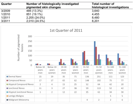
The number of excised pigmented lesions was determined for the different quarters (Table 4). The total number of histological investigations in the corresponding quarter is also shown in Table 4. This reveals that the percentage of investigated pigmented skin changes increased steadily from the 3rd quarter of 2009, starting with 13.3% (3/2019) and rising to 19.1% (1/2010), and in the year 2011, it rose to 24.0% (1/2011) and 24.5% in the 3rd quarter of 2011.
In the first quarter of 2011, for example, 2,205 pigmented skin changes were investigated, whereas there were only 2,010 investigations in the 3rd quarter of 2010. A part of the rising numbers can be explained by a general rise in sending to the laboratory; nonetheless, the relative changes of the individual forms of pigmented nevi have a significant importance.
In these model quarters (1/2011 and 3/2011), it is striking that 43.3% (1st quarter 2011) or 45.7% (3rd quarter of 2011) of the samples were taken from male patients. In total, the age distribution
(P < 0.05) shows that pigmented skin changes
were removed from more women than men in all four model quarters; only the first quarter of 2011 shows a slightly higher rate of men (321) than women (303) regarding the analysed pigmented skin lesions.
group of 16.9%, with 372 samples (3rd place). But in the 3rd quarter of 2011 (summer quarter), they resulted in being an important group with
21.0% and 423 samples. The different
histological entities in the corresponding gender and age distributions for both model quarters (1st and 3rd quarters of 2011) are shown in total numbers as well (Fig. 1 and Fig. 2). The fact that there were more dermal nevi being removed from women in all age distributions than from men (< 0.01) is striking. This may be caused by a higher aesthetical demand of women, who may be more affected by dermal nevi than men (Fig. 1 and Fig. 2).
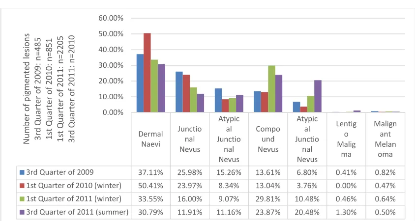
4.2 Types of Extracted Nevi
The next step was analysing the extracted pigmented moles. For that, the moment (quarter of extraction) was put into relation to the histological type of mole (Fig. 3). Next to the earlier described trend of dermal nevi being the most commonly extracted entity, the junctional
nevi along with the compound nevi are the second most commonly extracted types of skin changes. The junctional nevi dominated in the 3rd quarter of 2009 and in the 1st quarter of 2010 with 25.98% and 23.97%, respectively, whereas the compound nevi were dominant in the 1st quarter of 2011 and the 3rd quarter of 2011 with 29.8% and 23.9%, respectively. The rate of atypical nevi or pigmented malign biopsies is described in the next section.
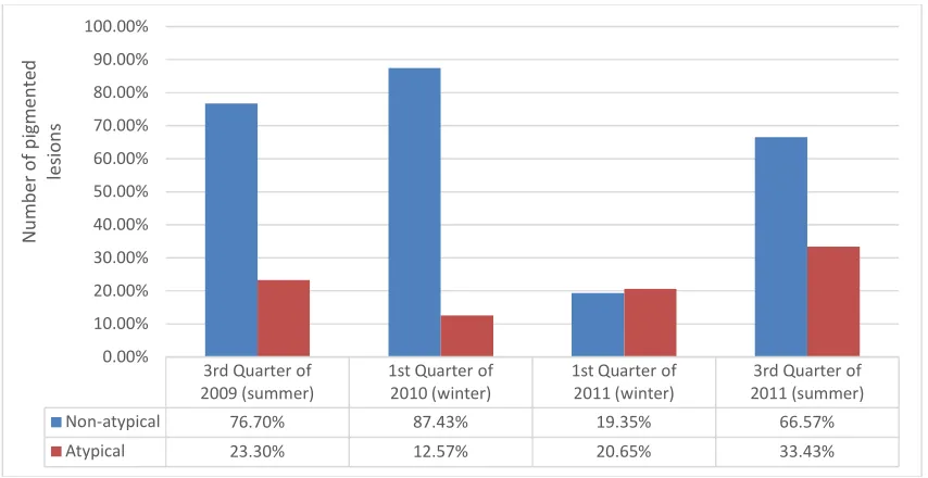
4.3 Dignity of the Extracted Pigmented Skin Changes
The percentage of atypical lesions in the total number of pigmented lesions were analysed for the earlier chosen quarters (Fig. 4). The percentage of atypical pigmented skin changes was between 12.57% and 33.43% (average: 22.5%) in the chosen quarters. The atypical junctional nevi, atypical compound nevi, and Lentigo malign and malignant melanoma shown in Fig. 3 were subsumed.
Table 4. List of histological investigation
Quarter Number of histologically investigated pigmented skin changes
Total number of
histological investigations
3/2009 485 (13.3%) 3,640
1/2010 851 (19.1%) 4,455
1/2011 2,205 (24.0%) 8,480
3/2011 2,010 (24.5%) 8,201
Fig. 1. Graphical representation showing pigmented lesions in 1st quarter of 2011
Below 18 years
men
Below 18 years women
19-30 years
men
19-30 years women
31-50 years
men
31-50 years women
51-100 years.
men
51-100 years women
Dermal Naevi 15 19 35 71 136 251 111 123
Compound Nevus 17 17 65 62 148 192 75 77
Atypical Compound Nevus 2 4 15 25 52 64 42 20
Junctional Nevus 11 3 27 38 59 113 52 42
Atypical Junctional Nevus 1 2 12 22 35 62 34 30
Lentigo Maligna 5 5
Malignant Melanoma 4 2 2 6
0 50 100 150 200 250 300
N
u
m
b
er
o
f
p
ig
m
en
te
d
le
si
o
n
s
Fig. 2. Graphical representation showing pigmented lesions in 3rd quarter of 2011
Fig. 3. Graphical representation showing pigmented lesions in all quarters
An important result of my work is the striking fact that the number of extracted atypical pigmented moles is higher in the summer months (July, August, September) than in the corresponding winter months (January, February, March;
P < 0.05%). The percentage of the Lentigo
maligna and the malignant melanomas were on the low side and did not show any significant fluctuation.
5. DISCUSSION
5.1 Discussion of the Method
From the retrospective analysis of the
histological evaluation of submissions to a
medium-sized histological laboratory, with
senders from all over Germany, a trend in the behaviour of dermatologists with regards to the
Below 18 years men Below 18 years women 19-30 years men 19-30 years women 31-50 years men 31-50 years women 51-100 years. men 51-100 years women
Dermal Naevi 13 16 53 90 127 142 98 73
Compound Nevus 9 20 34 62 119 140 54 39
Atypical Compound Nevus 16 8 32 61 102 124 59 37
Junctional Nevus 9 8 17 29 36 80 25 22
Atypical Junctional Nevus 3 13 32 43 67 32 30
Lentigo Maligna 17 9
Malignant Melanoma 7 3
0 20 40 60 80 100 120 140 160 N u m b er o f p ig m e n te d le si o n s
3rd Quarter of 2011
Dermal Naevi Junctio nal Nevus Atypic al Junctio nal Nevus Compo und Nevus Atypic al Junctio nal Nevus Lentig o Malig ma Malign ant Melan oma
3rd Quarter of 2009 37.11% 25.98% 15.26% 13.61% 6.80% 0.41% 0.82%
1st Quarter of 2010 (winter) 50.41% 23.97% 8.34% 13.04% 3.76% 0.00% 0.47%
1st Quarter of 2011 (winter) 33.55% 16.00% 9.07% 29.81% 10.48% 0.46% 0.64%
3rd Quarter of 2011 (summer) 30.79% 11.91% 11.16% 23.87% 20.48% 1.30% 0.50%
removal of pigmented skin lesions over the periods of the different quarters could be obtained. Of course, an investigation of that statistical power is not suitable for making generally valid statements. This is what the large-scale skin cancer screening programmes that are being analysed by the public health insurance and the associations for established dermatologists aim for. However, such a random sample shows that the number of samples of
pigmented skin lesions have significantly
increased over the last years. Therefore, it is up to the large-scale investigations by the health insurers to maybe quantify these trends on a large scale with the help of their billing data. At this time, it is uncertain at what point we will get such results. An obligation to report a tumour disease only exists in a small number of federal states. The register for cancer of the Saarland as well as the register for cancer of Schleswig-Holstein are to be mentioned. The register for cancer of dermatologists in North Rhine– Westphalia has collected the data for skin tumours for only a few years now, and atypical moles are not registered by the currently available registers for reporting cancer. This would be the only opportunity to make a comprehensive statement about the increase in reported atypical nevi in comparison to the total number of skin samples of all dermato-histopathologies. Unfortunately, an evaluation like this will not take place in the near future.
5.2 Reasons for the Rise in Dermato-histopathological Examinations of Pigmented Skin Lesions
There are two reasons, based on my work, that could possibly explain the observed increase in histological investigations of pigmented skin changes. On the one hand, the increased sensitivity of the affected patients should be mentioned here, which enables a dermatological examination of the entire intuition due to the
skin cancer screening that is available
nationwide and paid for from the 25th through the 35th year of life. Better education and more sensible treatment of patients with UV radiation has already had a positive effect— similar to smoking campaigns. However, a further improvement of this still relatively young area of medical education is necessary in order to reach as much of the population as possible.
On the other hand, the extra-budgetary remuneration of medical services within the framework of skin cancer screening, including the services of the dermatohistopathologist, is to be mentioned. This financial incentive also particularly motivates the treating physician to carry out such a skin cancer screening for all eligible patients because it represents a statutory insurance benefit every two years. This can— depending on the federal state-generate an
Fig. 4. Overall scenario of pigmented lesions 3rd Quarter of
2009 (summer)
1st Quarter of 2010 (winter)
1st Quarter of 2011 (winter)
3rd Quarter of 2011 (summer)
Non-atypical 76.70% 87.43% 19.35% 66.57%
Atypical 23.30% 12.57% 20.65% 33.43%
0.00% 10.00% 20.00% 30.00% 40.00% 50.00% 60.00% 70.00% 80.00% 90.00% 100.00%
N
u
m
b
er
o
f
p
ig
m
en
te
d
le
si
o
n
additional income of between 12 and 18 euros per insured person. However, these relatively
manageable additional costs are also
economically advantageous for the health insurance carriers in the context of early detection/prevention, since white skin tumours (plate epithelial carcinoma and basal cell carcinoma) as well as malignant melanomas are increasingly diagnosed in earlier stages, with less penetration depth. This is shown nicely in the publicly available cancer registers of Saarland and Schleswig-Holstein. Both changes are due to the fact that more and more patients
are having a dermatological screening
examination. The dermatologist also gives valuable advice on how to deal sensibly with the sun in the context of skin cancer screening and provides individual advice, which should increase the motivation to improve sun protection even further.
5.3 Rise in Histologically Examined Pigmented Skin Changes
Over the four different model quarters, it was shown that the proportion of pigmented lesions in the total test material of a medium-sized dermatohistopathological laboratory increased constantly over the two years tested. This share ultimately amounted to 25%.
5.4 Number of Pigmented Skin Changes in the Overall Collective of a Histological Laboratory Centre
The relative proportion of pigmented skin changes increased constantly over the analysed
two years, amounting to 13.3% in the third quarter of 2009 and rising to just under
25% in the other two quarters (1st and 3rd quarters of 2011). This underlines the importance of skin cancer screening and its use by the insured.
5.5 Gender and Age Distribution of the Removed Pigmented Skin Lesions
As in other screening programmes, the
proportion of women in such programmes is higher in skin cancer screening. The most frequently studied age groups were the middle-aged patients (31–50), followed by those over 50. There is a clear need for improvement in the age group of 19- to 30-year-old patients, whose percentage was between 16.9% and 21%.
5.6 Types of Extracted Pigmented Nevi
From the frequency of distribution of removed nevi, it became clear that many dermal nevi were removed. These skin changes, which can usually
be classified clinically flawless, possibly
represent a consequence of the relatively generous skin cancer screening, in which up to five moles may be taken within the framework of the programme. It is now up to the physician or the patient to strictly orient the number of removed moles according to the ABCDE criteria (suspicious pigmented skin changes) or whether aesthetically/cosmetically disturbing moles such as dermal nevi (between 30% and 50% of all removed pigmented lesions) were removed. This thesis may be stated according to the data from my work.
5.7 Dignity of the Extracted Pigmented Skin Lesions
The fact of whether more atypical moles are removed and whether this is reflected in a
reasonable cost–benefit ratio with the
expenditure incurred is essential for the success of an effective early detection programme. It can be seen from my work that in the hands of dermatologists the “hit rate” of atypical pigment lesions after histological processing lies between 12.6% and 33.4%. This is a significant proportion, which almost always allows a cure at this stage and thus improves the morbidity of the investigated patient collectives. This happens against the background that about 30% of the atypical nevi are precursors for malignant melanoma.
5.8 Seasonal Distribution of the Excision of Moles
screening week and the increasingly internalised early diagnosis by patients. Ultimately, the earlier detection of atypical moles that was described in the present study coincides with the prevalence data of malignant melanoma, according to which more and more melanomas are detected, but which, fortunately, show a decreasing depth of invasion (Breslow index) when diagnosed (cancer registries of Saarland and Schleswig- Holstein).
A similar study from 2007 is available for the federal state of North Rhine–Westphalia [41]. In this study, a network of physicians evaluated data from a population of about 75,000 individuals, 796 of whom were identified with a newly diagnosed skin cancer in a period from July 1998 to June 2006 (five-year period). In this study, 13.6 cases of melanoma were found in 75,000 individuals who were men and 18.5 who were women. This results in an incidence of malignant melanoma in North Rhine–Westphalia comparable to other incidence rates. For the Schleswig-Holstein cancer registry, age-related incidence rates of 12.3 for men and 14.8 per 100,000 women were each found in the observation period from 1998 to 2001 (three years) [42]. This earlier detection goes
hand-in-hand with a significantly improved prognosis and may be regarded as a success of the
educational and screening investigations. The fact that the lentigo maligna and melanoma entities had no seasonal frequency fluctuations may be due to the too small collective for this question.
6. CONCLUSION
Causes of skin cancer studied insufficiently. Some factors increase skin cancer risk; knowing them is possible to reduce the likelihood of this disease.
These results document clearly for the first time that after the end of the summer, when many patients expose themselves to the sun with their naked skin, conspicuous moles are recognized, and accordingly, after the end of the holiday, in autumn, the patients go to the dermatologist, who then excises suspicious moles. The project showed as well that large-scale systematic skin cancer screening is feasible and has the potential to reduce skin cancer burden, including mortality. Based on the results of SCREEN, a
national statutory skin cancer early
detection program was implemented in Germany in 2008.
Skin cancer screening made it possible to detect dysplastic lesions at an early stage and thus significantly reduce the progression to malignant melanoma.
In general, there are better survival prognoses through skin cancer screening for women and men with malignant melanoma.
There is a need for educational work with information about the early signs of malignant melanoma, about the dangers of intensive and long-term tanning, the identification of high-
risk groups subject to regular medical
examinations.
CONSENT AND ETHICAL APPROVAL
As per university standard guidelines, participant consent and ethical approval has been collected and preserved by the authors.
COMPETING INTERESTS
Author has declared that no competing interests exist.
REFERENCES
1. Schneider S, Zimmermann S, Diehl K,
Breitbart EW, Greinert R. Sunbed use in German adults: Risk awareness does not correlate with behaviour. Acta Derm Venereol. 2009;89:470-5.
2. Amir Z, Wright A, Kernohan EE, Hart G.
Attitudes, beliefs and behaviour regarding the use of sunbeds amongst healthcare workers in Bradford. Eur J Cancer Care (Engl). 2002;9(2):76-9.
3. World Health Organization (WHO)
Exposure to artificial UV radiation and skin cancer. In: IARC, Ed.Working Group Reports. Lyon; 2005
4. Diehl K, Litaker DG, Greinert R,
Zimmermann S, Breitbart EW, Schneider S. The prevalence of current sunbed use and user characteristics: The SUN-Study 2008. Int J Public Health. 2010;55(5):513-6.
DOI: 10.1007/s00038-009-0100-4
5. Fitzpatrick TB. The validity and practicality
of sun-reactive skin I trough VI. Arch Dermatol. 1988;124:869-71.
6. Armstrong, B. How sun exposure causes
skin cancer: An epidemiological
D., editors. Prevention of skin cancer.
Dordrecht, The Netherlands: Kluwer
Academic Publishers.
7. Carli P, Massi D, Santucci M, Biggeri A,
Giannotti B. Cutaneous melanoma
histologically associated with a nevus and melanoma de novo have a different profile of risk: Results from a case-control study. J Am Acad Dermatol. 1999;40(4):549-57.
8. Weinstock MA, Colditz GA, Willett WC,
Stampfer MJ, Bronstein BR, Mihm MC,
Speizer FE. Nonfamilial cutaneous
melanoma incidence in women associated with sun exposure before 20 years of age. Pediatr. 1989;84(2):199-204.
[PMID: 2748244]
9. Zanetti R, Franceschi S, Rosso S, Colonna
S, Bidoli E. Cutaneous melanoma and sunburns in childhood in a southern European population. Eur J Cancer. 1992; 28A(6-7):1172-6.
DOI: 10.1016/0959-8049(92)90480-p
10. Boldemann C, Breitner H, Jansson B,
Nilsson B, Ullén H. Sunbed use in relation to phenotype, erythema; sunscreen use and skin diseases. A questionnaire survey
among Swedish adolescents. Br J
Dermatol. 1996;135(5):712-6. [PMID: 8977669]
11. Whiteman DC, Whiteman CA, Green AC.
Childhood sun exposure as a risk factor for melanoma: A systematic review of epidemiologic studies. Cancer Causes Control. 2001;12(1):69-82.
DOI: 10.1023/a_1008980919928
12. Levine JA, Sorace M, Spencer J, Siegel
DM. The indoor UV tanning industry: A review of skin cancer risk, health benefit claims, and regulation. J Am Acad Dermatol. 2005;53(6):1038-44.
DOI: 10.1016/j.jaad.2005.07.066
13. English DR, Armstrong BK, Kricker A,
Winter MG, Heenan PJ, Randell PL. Case-control study of sun exposure and squamous cell carcinoma of the skin. Int J Cancer 1998;77(3):347-53.
DOI: 10.1002/(sici)1097-0215(19980729) 77:3<347::aid-ijc7>3.0.co;2-0
14. Ting W, Schultz K, Cac NN, Peterson M,
Walling HW. Tanning bed exposure increases the risk of malignant melanoma. Int J Dermatol. 2007;46:1253-7.
15. International Agency for Research on
Cancer Working Group on artificial
ultraviolet (UV) light and skin cancer; 2007.
16. Jones JL, Leary MR. Effects of
appearance-based admonitions against
sun exposure on tanning intentions in young adults. Health Psychol. 1994;13(1): 86-90.
DOI: 10.1037//0278-6133-13.1.86
17. Mathys P, Moser M, Bressoud D,
Ackerman-Liebrich U, Braun-Fahrlander C. Frequency, duration, and motivation of sun-bed use in Switzerland. 1999;S.117.
18. Geller AC, Brooks DR, Colditz GA, Koh
HK, Frazier AL. Sun protection practices among offspring of women with personal or family history of skin cancer. Pediatr. 2006; 117(4):e688-94.
DOI: 10.1542/peds.2005-1734
Ajzen I. The theory of planned behaviour.
Organization behaviour and human
decision process.1991;Y50:179-211.
19. Feldmann SR, Ligouri A, Kucenic M, Rapp
SR, Fleischer AB Jr, Lang W, Kaur M. Ultraviolet exposure is a reinforcing stimulus in frequent indoor tanners. J Am Acad Dermatol. 2004;51(1):45-51.
20. Hillhouse J, Stapleton J, Turrisi R.
Association of frequent indoor UV tanning with seasonal affective disorder. Arch Dermatol. 2005;141(11):1465.
DOI: 10.1001/archderm.141.11.1465
21. Berneburg M, Plettenberg H, Krutman J.
Photoaging of human skin. Photodermatol
Photoimmunol Photomed. 2000;16(6):
239-44.
22. Matsumura Y, Ananthaswamy HN.
Short-term and long-Short-term cellular and molecular events following UV irradiation of skin: implications for molecular medicine. Expert Rev. Mol. Med. 2004;4:1–22.
23. Tornaletti S, Pfeifer GP. UV damage and
repair mechanisms in mammalian cells. Bioessays. 1996;18(3):221-8.
DOI: 10.1002/bies.950180309
24. Brash DE. Roles of the transcription factor
p53 in keratinocyte carcinomas. Br J Dermatol. 2006;154(Suppl 1):8-10.
25. Cui R, Widlund HR, Feige E, Lin JY,
Wilensky DL, Igras VE, et al. Central role of p53 in the suntan response and pathologic hyperpigmentation. Cell. 2007; 128(5):853-64.
DOI: 10.1016/j.cell.2006.12.045
26. Agar N, Young AR. Melanogenesis: A
photo protective response to DNA
damage? Mutat Res. 2002;571(1-2):121-23.
27. Corre S, Primot A, Sviderskaya E, Bennett
MC1R genes, is dependent on the p-38-activated upstream stimulating factor-1 (USF-1). J Biol Chem. 2004;279(49): 51226-33.
DOI: 10.1074/jbc.M409768200
28. Clark WH Jr. 1978;114(5):732-8.
29. Lorentzen H, Weismann K, Kenet RO,
Secher L, Larsen FG. Comparison of
dermatoscopic ABCD rule and risk
stratification in the diagnosis of malignant
melanoma. Acta Derm Venereol.
2000;80(2):122-6. [PMID: 10877133]
30. Kraemer KH, Greene MH. Dysplastic
nevus syndrome: Familial and sporadic
precursor of cutaneous melanoma.
Dermatol Clin. 1985;3:225–37. DOI: 10.1016/0738-081X(85)90096-3
31. Nelemans PJ, Rampen FH, Ruiter DJ,
Verbeek AL. An addition to the controversy on sunlight exposure and melanoma risk: a
meta-analytical approach. J Clin
Epidemiol. 1995;48:1331-42.
32. Armstrong BK, Kricker A. The
epidemio-logy of UV induced skin cancer. J Photo-chem Photobiol B. 2001;63(1-3):8-18.
33. Osterlind A, Tucker MA, Stone BJ, Jensen
OM. The Danish case-control study of
cutaneous malignant melanoma. II.
Importance of UV-light exposure. Int J Cancer. 1988;42(3):319-24.
DOI: 10.1002/ijc.2910420303
34. Gloster HM, Brodland DG. The
epidemio-logy of skin cancer. Dermatol Surg. 1996; 22(3):217-26.
DOI: 10.1111/j.1524-4725.1996.tb00312.x
35. Swerdlow AJ, Weinstock MA. Do tanning
lamps cause melanoma? An epidemiologic assessment. J Am Acad Dermatol. 1998; 38: 89-98.
DOI: 1111/j.1600-0781.2009.00417.x
36. Giles GG, Marks R, Foley P. Incidence of
non-melanocytic skin cancer treated in Australia. Br Med J (Clin Res Ed). 1988; 296(6614):13-7.
DOI: 10.1136/bmj.296.6614.13
37. Cokkinides VE, Weinstock MA, O’Connell
MC, Thun MJ. Use of indoor tanning sunlamps by US youth, ages 11-18 years, and by their parent or guardian caregivers: prevalence and correlates. Pediatr. 2002; 109(6):1124-30.
DOI: 10.1542/peds.109.6.1124
38. Autier P, Doré JF, Lejeune F, Koelmel KF,
Geffeler O, Hille P, et al. Cutaneous malignant melanoma and exposure to sunlamps or sunbeds: An EORTC
multi-center case-control study in Belgium, France and Germany. EORTC Melanoma Cooperative Group. Int J Cancer. 1994; 58(6):809-13.
DOI: 10.1002/ijc.2910580610
39. Montague M, Borland R, Sinclair C. Slip!
Slop! Slap! And SunSmart, 1980-2000: Skin cancer control and 20 years of
population-based campaigning. Health
Educ Behav. 2001;28:290-305. DOI: 10.1177/109019810102800304
40. Stang A, Ziegler S, Büchner U, Ziegler B,
Jöckel KH, Ziegler V. Malignant melanoma and nonmelanoma skin cancers in North Rhine-Westphalia, Germany: A patient- vs. diagnosis-based incidence approach. Int J Dermatol. 2007;46:564-70.
41. Katalinic A, Kunze U, Schäfer T.
Epidemiology of cutaneous melanoma and non- melanoma skin cancer in Schleswig-Holstein, Germany: Incidence, clinical subtypes, tumour stages and localization (epidemiology of skin cancer). Br J Dermatol. 2003;149(6):1200-6.
DOI: 10.1111/j.1365-2133.2003.05554.x
42. Dissel M, Rotterdam S, Altmeyer P,
Gambichler T. Indoor tanning in North
Rhine-Westphalia Germany: A
self-reported survey. Photodermatol
Photoimmunol Photomed. 2009;25(2):94-100.
43. Marghoob AA. Congenital melanocytic
nevi. Evaluation and management.
Dermatol Clin 2002;20(4):607-16. DOI: 10.1016/s0733-8635(02)00030-x
44. Marghoob AA, Borrego JP, Halpern AC.
Congenital melanocytic nevi: Treatment
modalities and management options.
Semin Cutan Med Surg 2007;26(4):231-40.
DOI: 10.1016/j.sder.2008.03.007
45. Naeyaert JM, Brochez L. Clinical Practice.
Dysplastic nevi. N Engl J Med 2003; 349(23):2233-40.
DOI: 10.1056/NEJMcp023017
46. Stang A, Stausberg J, Boedeker W,
Kerek-Bodden H, Jöckel KH. Nationwide
hospitalization costs of skin melanoma and non-melanoma skin cancer in Germany. J Eur Acad Dermatol Venereol. 2008;22:65-72.
47. Stang A, Stausberg J. Inpatient
management of patients with skin cancer in Germany: An analysis of the nationwide DRG-statistic 2005-2006. Br J Dermatol. 2009;161(Suppl 3):99-106.
48. Valiukeviciene S, Gollnick H, Stang A.
Body-site distribution of common
acquired melanocytic nevi associated with severe sunburns among children in Lithuania. Int J Dermatol. 2007;46(12): 1242-9.
DOI: 10.1111/j.1365-4632.2007.03369.x
49. Zanetti R, Rosso S, Martinez C, Nieto A,
Miranda A, Mercier M, et al. Comparison of risk patterns in carcinoma and melanoma of the skin in men: A multi-centre case-control study. Br J Cancer. 2006;94(5): 743-51.
DOI: 10.1038/sj.bjc.6602982
_________________________________________________________________________________ © 2019 Ghiassi; This is an Open Access article distributed under the terms of the Creative Commons Attribution License (http://creativecommons.org/licenses/by/4.0), which permits unrestricted use, distribution, and reproduction in any medium, provided the original work is properly cited.
Peer-review history:
![Table 1. Skin photo types according to Fitzpatrick [5]](https://thumb-us.123doks.com/thumbv2/123dok_us/1709141.1217180/5.612.94.522.113.263/table-skin-photo-types-according-fitzpatrick.webp)