University of Windsor University of Windsor
Scholarship at UWindsor
Scholarship at UWindsor
Electronic Theses and Dissertations Theses, Dissertations, and Major Papers
1-1-2004
Size control and ligand exchange of gold and palladium
Size control and ligand exchange of gold and palladium
nanoparticles prepared by single phase method.
nanoparticles prepared by single phase method.
Weijuan Jia
University of Windsor
Follow this and additional works at: https://scholar.uwindsor.ca/etd
Recommended Citation Recommended Citation
Jia, Weijuan, "Size control and ligand exchange of gold and palladium nanoparticles prepared by single phase method." (2004). Electronic Theses and Dissertations. 7161.
https://scholar.uwindsor.ca/etd/7161
NOTE TO USERS
This reproduction is the best copy available.
SIZE CONTROL AND LIGAND EXCHANGE OF GOLD AND
PALLADIUM NANOPARTICLES PREPARED BY SINGLE
PHASE METHOD
by Weijuan Jia
A Thesis
Submitted to the Faculty of Graduate Studies and Research Through the Department of Chemistry and Biochemistry
in Partial Fulfillment of the Requirements for the Degree of Master of Science at the University of Windsor
Windsor, Ontario, Canada
2004
1*1
Library and Archives CanadaPublished Heritage Branch
395 Wellington Street OttawaONK1A0N4 Canada
Bibliotheque et Archives Canada
Direction du
Patrimoine de I'edition
395, rue Wellington OttawaONK1A0N4 Canada
Your file Votre reference ISBN: 978-0-494-57559-8 Our file Notre reference ISBN: 978-0-494-57559-8
NOTICE: AVIS:
The author has granted a
non-exclusive license allowing Library and Archives Canada to reproduce, publish, archive, preserve, conserve, communicate to the public by
telecommunication or on the Internet, loan, distribute and sell theses
worldwide, for commercial or non-commercial purposes, in microform, paper, electronic and/or any other formats.
L'auteur a accorde une licence non exclusive permettant a la Bibliotheque et Archives Canada de reproduire, publier, archiver, sauvegarder, conserver, transmettre au public par telecommunication ou par I'lnternet, prater, distribuer et vendre des theses partout dans le monde, a des fins commerciales ou autres, sur support microforme, papier, electronique et/ou autres formats.
The author retains copyright ownership and moral rights in this thesis. Neither the thesis nor substantial extracts from it may be printed or otherwise reproduced without the author's permission.
L'auteur conserve la propriete du droit d'auteur et des droits moraux qui protege cette these. Ni la these ni des extraits substantiels de celle-ci ne doivent etre imprimes ou autrement
reproduits sans son autorisation.
In compliance with the Canadian Privacy Act some supporting forms may have been removed from this thesis.
Conformement a la loi canadienne sur la protection de la vie privee, quelques
formulaires secondaires ont ete enleves de cette these.
While these forms may be included in the document page count, their removal does not represent any loss of content from the thesis.
Bien que ces formulaires aient inclus dans la pagination, il n'y aura aucun contenu manquant.
Abstract
Metal nanoparticles have become important building blocks for nano-structured materials
and nano-sized markers in biochemistry. This thesis focuses on the synthesis and purification of small gold and palladium nanoparticles with average diameters below 5 ran. All particles are prepared by the single-phase method without the presence of a surfactant, an
important advantage over the commonly used two-phase method.
Gold and Pd nanoparticles contained inorganic impurities, mostly lithium salts, which are
generated when the reaction mixture is quenched with ethanol. An acid wash procedure has been developed that successfully removes the impurities when particles are protected by stable thiol ligands. The removal of inorganic and organic impurities could be easily
monitored by TGA and XPS and is discussed in Chapter 2.
Chapter 3 describes the preparation of gold nanoparticles in the presence of bulky thiol
ligands. These particles have significantly smaller diameters than nanoparticles prepared in the presence of straight chain alkylthiols at otherwise identical reaction conditions. Highly stable and soluble nanoparticles are obtained with thiols based on dendrimer wedges. Their
structure, size, and size distribution was investigated by XRD, HR-TEM, and UV-VIS. Finally, first attempts to convert Pd nanoparticles into amphiphilic structures by ligand
exchange reactions are described in Chapter 4. Hydrophilic and hydrophobic ligands are based on thioctic acid and its ester, respectively, and exchange ratios are estimated from quantitative analysis of FT-IR spectra. Their amphiphilic character is demonstrated in
Acknowledgements
I am most grateful to my surpervisor, Dr.Holger.Eichhorn, for his guidance and help on my studies and research during the last two years. His wonderful idea and enthusiasm always encourage me. His kind advice and support have been greatly appreciated and will be remembered forever.
I deeply thank Dr. Fred Pearson for his help in HR-TEM; thanks Dr.Ramon A. Alvarez-puebla for his help in calibration FTIR; thanks DR. S. Tadayyon for his help in XPS; thank Mike for his help in NMR. I also thank Dr.Pandey, Dr.Li, Dr. Lectcher, and Dr.Fowel for providing centrifuge. I am also grateful to Dr. Antonelli's and Dr. MacDonald's group for distilled THF. Dr.Green's group is thanked for their generous help.
I am grateful to our group members, Scott, Nike, Bryant, Huisheng, Ami, Jessica, Alina for their help and advice.
Thank Ms. Aroca for lending some supplies and good advice. Many thanks are given to the faculty and staff of department of chemistry and Biochemistry, University of Windsor,
for their support and kind help during my study and research. Specially thanks Ms.Marlene for her warmheart and a lot of help in arranging my defence.
Thanks for all my friends in U. of Windsor for their enduring support.
Table of Contents
Abstract iii Acknowledgements iv
List of Figures ix List of Tables xii List of Scheme xiii List of Abbreviations xiv
Chapter 11 Introduction 1
1.1 General background 1
1.1.1 Nanoparticles - a general introduction 1
1.1.2 GoldNPs 3
1.2 Synthesis of gold NPs 5
1.2.1 Physical methods 5
1.2.2 Chemical methods 6
1.2.2.1 Two phase synthesis of gold NPs(the Brust-Schiffrin Method) 6
1.2.2.2 Single-phase method 7
1.3 Protecting ligands for gold NPs 8
1.3.1 Thiols and Other Sulfur Ligands 9
1.4 Characterizaton Techniques: 14
1.4.1 UV-VIS Spectroscopy 14
1.4.1.1 Size effect 14
1.4.1.2 The effect of the solvent refractive index 16
1.4.1.3 Core charge 16
1.4.2 X-ray diffraction (XRD) 16
1.4.3 High Resolution Transmission Electron Microscopy (HR-TEM) 21
1.4.4 Thermal analysis: Thermo Gravimetric Analysis (TGA) 23
1.4.5 Other characteristic technique 25
1.5 Palladium NPs 26
1.6 Applications: 27
1.7 Goal of this thesis 29
Reference 29
Chapter 2 Single-phase Synthesis of Gold NPs stabilized by straight 35 Chain Alkylthiols and Their Purification
2.1 Introduction 35
2.2 Results and discussion 36
2.2.1 TGA-MS and XPS investigations 37
2.2.2 HR-TEM 45
2.2.3 UV-VIS Spectroscopy in Solution 48
2.3 Conclusions 52
Reference 52
Chapter 3 Effect of Bulkiness of Thiols on Size and Size Distribution of 54
3.1. 3.2 3.3 3.3.1 3.3.2 3.3.3 3.3.4 3.3.5 3.3.6. 3.3.7 Reference Chapter 4 4.1 4.2 4.2.1 4.2.2 4.3
Au Nan op articles
Introduction Syntheses Characterization TGA measurements 54 56 57 57 High resolution of transmission electron micrograph (HR-TEM) 59 characterization
Ultraviolet- visible measurement
X-ray powder diffraction (XRD)
Gold NPs prepared in the presence of a polyether solvent
Exchange ligand for gold NPs
Conclusions
Ligand Exchange of Pd-NPs
Introduction
Results and discussion
Synthesis and characterization of parent Pd NPs (Pd-C12)
Ligand exchange of Pd-C12 NPs
Reference: 86
Chapter 5 Synthesis of Dendrimer Ligands 87
Reference 90
Chapter 6 Experiment Part 91
6.1 Measurements 91
6.2 Experiments 93
6.3 Synthesis of dendritic thiol ligands 97
List of Figures
Figure 1-1 Formation of a band structure 2
Figure 1-2 The Lycurgaus Cup 4
Figure 1-3 Absorption spectra of the gold NPs of different size 15
Figure 1-4 Upper frame contains spectra (normalized at 4ev) form 15 2.5nm diameter gold NPs. In the lower frame, several
spectra of differently obtained 1.7nm gold NPs are offset from each other
Figure 1-5 X-ray diffraction powder diffraction powder patterns for 20
four different sizes of gold nanocrystals
Figure 1-6 XRD patterns of gold NPs, decahedron, a three-shell 21
icosahedron, and a three-shell cuboctahefron, respectively.
Figure 1-7 Typical HRTEM micrographs of NPs stabilized by 23 dodecanehiol 4.1 nm sample
Figure 1-8 TGA of A crude gold NPs stabilized by dodecanethiol 24
Figure 2-1 TGA of Au-C12 before and after acid wash 38
Figure 2-2 Deconvoluted Au-signals of the XPS of the original Au-C 12 40 sample (before acid wash)
Figure 2-3 Deconvo luted C-signals of the XPS of the original Au-C8 40
sample (before acid wash)
Figure 2-4 HR-TEM of Au-C8 before (left) and after acid wash (middle 45
Figure 2-5 HR-TEM of Au-C12 before and after acid wash 46
Figure 2-6 HR-TEM of Au-C 18 before (left) and after acid wash 46 (middle and right was treated by plasma)
Figure 2-7 High resolution HR-TEM image of an octahedral Au-C 12 47 NP (before acid wash)
Figure 2-8 High resolution HR-TEM image of twinned Au-C 12 NPs 47 (before acid wash)
Figure 2-9 The size distribution of Au-C8, Au-C12, Au-C18, a, 48
beforeacid wash, b, after acid wash
Figure 2-10 UV-vis spectra of gold NPs in THF solution 49
Figure 2-11 XRD patterns diffraction patterns of Au-C8, -C12, -C18 51 Figure 3-1: HR-TEM images and size distribution of gold nanoparticles 60, 61
Figure 3-2. Ultraviolet-visible spectra of gold NPs 63
Figure 3-3. XRD of gold NPs a, J3, J4, J7, J9, J10, J12; (B=2sin Q/X 64, 65 nm"1) b, J3 before and after acid wash
Figure 3-4 HR-TEM in the presence of poly (ethyleneglycol) dimethyl 67 ether and THF
Figure 3-5. HR-TEM of gold NPs prepared by dodecanethiol in THF 67
Figure 3-6. UV-vis of gold nanoparticles prepared in THF and mixed 68 solvent a, dodecanthiol as ligand b, tert-butylthiol as ligand
mixed solvent a, dodecanthiol as ligand b, tert-butylthiol as ligand.
Figure 3-8. UV-vis spectra of gold NPs stabilized by tert-butylthoil 72 before and after exchange with octadecanethiol, and gold
NPs stabilized by octadecanthiol
Figure 3-9 XRD patterns of gold NPs stabilized by tert-butylthoil before 73
and after exchange with octadecanethiol, and gold NPs stabilized by octadecanthiol
Figure 3-10 HR-TEM of gold NPs stabilized by tert-butylthoil before and 74
after exchange with octadecanethiol
Figure 4-1 Palladium stabilized by dodecanehtiol a, TGA b, HR-TEM, 78
c,XRD
Figure 4-2 TGA of palladium stabilized by dodecanehtiol 79
Figure 4-3 TR-TEM images and size distribution of Pd-C 12 79
Figure 4-4 Pd-C 12 before and after partial ligand exchange by thioctic 81
acid dithiol in a biphasic diethylether water mixture
Figure 4-5 FT-IR a, dodecanthiol, b, thioctic acid dithiol, c, Pd 82, 83
exchanged NPs
Figure 4-6 FT-IR of thioctic acid dithiol, exchanged ligands, and the Pd 85
List of Tables
Table 2-1 TGA of Three Gold NPs Stabilized by C8, C12-, and C18- 41
Alkylthiols
Table 2-2 Calculated Size Related Values for Ideal Gold Clusters 42
Table2-3 Thiol Weight % Values for Au-C8, -CI2, and -CI 8 44 Assuming Surface Gold to Thiolate Ratios of 2:1 and 3:1
Table 2-4 XRD diffraction peaks for Au-C8, -C12, -C18 NPs 51
Table 3-1 Structures of Used Thiol Ligands and Their Acronym 54 Used Throughout This Thesis
Table 3-2 TGA Data of Gold NPs Stabilized by Bulky Thiols 58 Table 3-3 XRD Results of Gold NPs Stabilized by Bulky Ligand 65 Table 3-4 Table 3-4 Summarized data from TGA measurements for 70
selected samples
Table 3-5 TGA of before and after Exchange Gold NPs and Gold 74 NPs Stabilized by Octadecanethiol
List of Scheme
Scheme 1-1 Formation of gold NPs by two-phase method Scheme 1-2 Deriving Bragg's Law
Scheme 2-1 Lithium salts that might be generated in single-phase and contaminate Au NPs
Scheme 3-1 Exchange ligand scheme Scheme 4-1 Exchange for Palladium NPs Scheme 5-1 Synthesizing dendrimer ligand
List of Abbreviations
Nanoparticles X-ray Diffraction
X-ray Photoelectron Spectroscopy Ultra-Violt visible Spectroscopy
High Resolution Transmission Electron Microscopy Fourier Transfer Infra-Red Spectroscopy
Ttetrahydorfuran Dichloromethane
Nuclear Magnetic Resonance Mass spectroscopy
Surface plasmon band
Monolayer protected nanoparticles Decahedra
Icosahedra
Face centered cubic
Thermo Gravimetric Analysis Triethylene lithium boron hydride
NPs XRD
XPS UV-vis HR-TEM FT-IR THF DCM NMR MS SPband MNPs Dec Ico Fee TGA
Chapter 1. Introduction 1.1 General background
1.1.1 Nanoparticles - a general introduction
A nanoscale material is any material that has nanometer size in either three dimensions (particles), two dimensions (thin films), or one dimension (thin wires). Thus, a nanoparticle (NP) is a solid particle with a diameter between l-1000nm. Its structure might be a single crystal, a non-crystalline material, or an aggregate of smaller crystallites. A colloid is a stable distribution of NPs in another material, usually a liquid
or a glass phase.[1J Particles that only consist of few to about hundred atoms are often
called clusters.
NPs have attracted intense attention over the past decade because of their unique chemical and physical properties. At the beginning of the 20th century, Wilhelm Oswald
was the first person to put forward the concept that the surface atoms determine the properties of a NP and he also pointed out that NPs should exhibit novel properties in comparison to bulk materials. '
One obvious reason that the reduction of size might cause a major change in properties is the increased ratio of surface atoms to bulk atoms. A particle of 300 atoms has more than 50 % of its atoms sitting at surface sites. Surface atoms are not fully integrated into the lattice of the material but interact with other atoms of the surrounding. These atoms have different energy levels and, therefore, different reactivity. It was also predicted that
The full development of a metallic band structure, for example, requires a minimum number of electronic levels (energy gaps between levels become very small) and electrons can move inside the material by only thermal activation. All properties that we
know for a bulk metal derive from the existence of such a band: the electrical conductivity as well as the specific heat, the metallic luster, or the ductility, to cite just a
few typical characteristics.
The typical band structure of a bulk material is shown in Figure 1-1 (c) and originates from a non-differentiated infinite number of s and d electrons.
Most of the transition metals have unfilled d-orbitals EF (HOMO)
(a)
— DOS(E)
DOS(E)
and noble metals such as palladium, platinum, or gold have nearby s-band that can be used for electron transport. In bulk gold, the d shell is completely filled with 10 electrons, which are hidden below the Fermi level, whereas the valence shell contains a single s electron.
A particle of very few atoms has well-separated energy levels as shown in (a). Here, the highest occupied molecular orbital Figure l-l[ l ] Formation of a
band structure (a) form a molecular state, (b) from a nanosized particle with broadened energy states, and (c) the fully developed band structure consisting of s and d bands. EF=Fermi energy,
DOS=densityofstates.In(a) ( H O M O ) corresponds to the EF
EF corresponds at the highest
occupied molecular orbital ,„ . T ,
in (b) will occur, which is the typical band structure of a semi-conductor. It is the situation shown in (b), with a small gap between the occupied and un-occupied bands, for which new materials properties are predicted.
Metal particles of less than 2 nm lose the band structure to such an extent that the effects of the situation (b) can be observed. The unstructured s- and d-bands in (c) are split but without forming truly discrete levels as in (a). At this nm size range, quantum mechanical rules have to replace those of classical physics. Size dependent properties arise from a size and structure dependent derealization of valence electrons and might be extensive. Different chemical and physical properties, such as optical and magnetic properties, melting points, surface reactivity, surface energies, as well as surface morphologies arise from these changes of the electronic structure.[2] Due to these new
properties, nanomateirals have potential applications as advanced catalysts,[3] chemical
sensors/4-1 photochemical patterns/5-1 and lithographic materials/61 Applications of
emitting NPs, quantum dots,[7] and as inert tags in biochemistry181 are already well
developed and commercial products are about to hit the market.
1.1.2 Gold NPs
Figure 1-2,1 J The Lycurgaus Cup, dating from the 4 century A.D., is made form glass impregnated with gold NPs. a) transmitted light, b) reflected light,
Modern gold chemistry might date back to 1857, when Michael Faraday reported his famous experiment in which he reduced tetrachloroaurate with white phosphorus to yield a deep-red gold colloid.[10] The first well-defined ligand-stabilized gold NPs, [AU55
(PPh)3Cl6], were prepared by Guenter Schmid in 1981.[11] He used diborate (B2H6) as
reducing agent that was passed through a benzene solution of PPI13AUCI at 60 °C to give PPh3 protected gold NPs. Thirteen years later, Brust et al. reported a two-phase method
for the straightforward synthesis of gold NPs.[12] His paper had a considerable impact on
1.2 Synthesis of gold NPs
A number of different ways to generate gold NPs have been developed, which can be classified into two main groups, physical and chemical methods. The chemical method uses gold (I) or gold (III) salts as starting materials that are reduced to gold (0) by a reducing agent. This reaction has been performed in two-phase and single-phase systems. Physical methods generate gold NPs via the distribution of bulk gold into small gold aggregates by techniques such as laser ablation and radiolysis.
1.2.1 Physical methods
The simplest way is to generate metal atoms in the gas phase followed by their controlled condensation to NPs, which is often regarded as metal vapor synthesis/131
Numerous modifications of this technique have been successfully employed in the past/141
Laser ablation is the most advanced technique and has been used in the gas and liquid phase. It is based on the well expressed plasmon resonance of gold nanoparticles around 520 nm and was used to follow the size and geometry of the particles. Size distributions, however, are rather wide/151
Other methods for the preparation of NPs include in seed growth/161 photochemical
methods (UV, Near-IR)/171 sonochemistry/181 radiolysis/191 and thermolysis/201 Most
1.2.2 Chemical methods
1.2.2.1 Two phase synthesis of gold NPs (the Brust-Schiffrin method)
The two-phase synthesis is one of the most common methods for the preparation of
gold NPs. Typically, a gold salt (AuCLf) is first transferred from aqueous solution into the toluene phase by a phase transfer agent such as tetraoctylamrnonium bromide. It is
then reduced by NaBH4 in the presence of thiols (see Scheme 1-1).
Scheme 1-1 Formation of gold NPs by the two-phase method
AuCl4" (aq) + N(C8H17)4+ (toluene) • N(C8H17)4+AuCl4"(toluene)
mAuCl4"(toluene) + nC12H25SH(toluene) +3me" >- 4mCl"(aq) + [Aum(C12H25SH)n] (toluene)
The generated gold NPs are usually air- and thermal-stable and can be repeatedly precipitated from and dissolved in common organic solvents. Their particle size and size
distribution might be tuned by changing the reaction conditions such as thiol/gold ratio, the reductant addition rate, and the reaction temperature.^211
An important drawback of this method is the presence of an ammonium surfactant (phase-transfer reagent) as it can not be completely removed from the nanopartilces surface after the reaction was completed. A content of 1 w% phase-transfer reagent
remained in the samples even after repeated precipitations of the NPs from solution.[22]
This situation was even worse when dendrimeric thiols were used as ligands. Gopidas et
al. reported that 5 w% phase-transfer reagent was still trapped inside gold NPs stabilized by dendritic ligands after being purified for one week by selective precipitation.[23J The
phase-transfer reagent is difficult to remove as both, the quaternary ammonium cation
interactions with the quaternary ammonium cations. The alkyl chains of capping ligands
(thiols) and the phase-transfer reagent also interact with each other.
Cecilia A. Waters and coworkers employed Soxhlet extraction for the removal of the
phase-transfer reagent.[22] In this paper, the NPs were precipitated twice from solution,
transferred into a Soxhlet thimble, and then extracted with acetone. After 12 hours of extraction only 0.011 w% phase-transfer reagent was left behind. Therefore, this
technique removes most of the phase-transfer reagent but not all. However, a fair amount of the NPs is lost as they can not be easily recovered from the thimble.
1.2.2.2 Single-phase method
The first single phase synthesis of gold NPs was reported in 1951.[10] [AuCLt]- was
reduced by sodium citrate in hot aqueous solution to generate gold colloids of 15 nm to 20 nm diameter. In 1973, Frens reported the preparation of gold NPs with a wider size
range by tuning the ratio of citrate to gold salt.[25J
Citrate acts not only as reducing agent but also as protecting ligands. It is a much weaker ligand than phosphanes and thiols, which is why these particles are only stable in
solution. This method is still extensively used for the preparation of gold NP when a rather loose shell of ligands is required for easy ligand exchange reactions. An important shortcoming of this method is the low gold NPs concentration (<0.01M) solution that can
be achieved and the limitation to water as solvent.
M.P.Rowe et al.[26] recently reported a single-phase synthesis for stable gold NPs
time, equivalents of reducing agent, and concentration of reaction solution). They found that treatment of HAuCU and the thiol with an excess of IJBH4 in THF under ambient conditions produces gold nanoparticles in high yields. The advantage of this method is that the reducing agents are mild and do not react with ligands that contain ester and amide groups. The disadvantage is that the reducing agents have low solubilities in THF and ethers, which leads to NPs of wide size distributions.
Xiaogang Peng's group[27J reported a one-phase method with surfactant. AuCh,,
Ag(CH3COO), anhydrous Cu(CH3COO)2, and PtCLt were dissolved in toluene and reduced with either tetrabutylammonium borohydride (TBAB) or its mixture with hydrazine in toluene. Fatty acids or aliphatic amines were added as ligands, not thiols. These conditions are claimed to maintain a tunable activity of both the metal precursors and the reducing reagents. The weak ligands on the surface of the as-synthesized nanocrystals also allow for ligand exchange reactions with a variety of different functional groups.
Yee et alJ28-1 reported a surfactant-free single-phase method that used Superhydride
(lithium triethylborohydride) in THF as reducing agent. Thiol-functionalized gold and palladium NPs were prepared in only 2 h and the purification was much easier than for the two-phase method. Despite its obvious advantages, this method has been used by only few groups and will be studied in detail in this thesis.
1.3 Protecting Ligands for Gold NPs
then attach to the NP's surface as protecting monolayer. Ligands with other functional groups, however, also protect the gold NP sufficiently and might provide a wider range of properties. This section will exclusively discuss gold NPs and is subdivided into 3 parts,
thiols and other sulfur ligands, non-sulfur ligands, and ligand exchange reactions, respectively.
1.3.1 Thiols and other sulfur ligands
Mulvaney and Giersig first reported the stabilization of gold NPs with alkenethiols in 1993.[29] In their study, gold NPs were prepared from hydrosol and then capped by
alkanethiols. From then on, thiols became the most widely used ligands. The gold-sulfure bond is partially covalent and partially ionic and relatively strong for a self-assembling structure.
Most gold NPs stabilized by thiols are stable solid materials with reasonable solubility in common solvents. Gold nanopartilces stabilized by dodecanethiol can be separated and kept as solid to do characterization/30^ Gold NPs prepared with weak ligands such
as citrate do not form stable solids.
The stability of the Au-S bond is one of the reasons why gold NPs have become the pioneer in the research of NPs. Another outstanding feature of gold NPs stabilized by thiols is that they spontaneously form highly ordered super lattices if they have a narrow size distribution. J31J
Except that the strong Au-S bond, a number of thiols are important markers for diagnosing inherited and acquired metabolic disturbances and diseases/321 (For example:
HiNU /
found to be an independent risk factor for premature occlusive disease in the coronary, cerebral, and peripheral arteries, and for venous thrombosis.[34] Gold NPs stabilized by
this kind of ligands could be a good detector for thoils.
Except from simple alkyl thiols, a variety of structurally diverse organosulfur
compounds have been employed for stabilizing gold NPs (e.g. ro-functionalized alkanethiolates, arenethiolates, and dialkylsulfides). Porter's group described the preparation, isolation, and characterization of 3D SAMs generated by n-octadecyl
disulfide, (CigS)2 , on the surface of gold nanoparticle. These disulfides formed monolayers on the gold NPs that were higher ordered and more crystalline than those
obtained with n-octadecanethiol[35J P.D. Beer group, reported disulfide-functionalized
zinc metalloporphyrins self-assembled onto gold NPs exhibit remarkable enhanced anion binding affinities if compared to the free metalloporphyrin. The surface preorganization
of optical and electrochemical group functionalized host systems offers the opportunity to prepare highly sensitive sensor materials.[36]
Attachment of water soluble ligands might render the entire gold NP into a water soluble structure. The attachment of thiolated cyclodextrin, for example, was reported by Jian Liu group.t37] These modified gold NPs were soluble in water and behaved as hosts
towards conventional cyclodextrin guests. So, the NPs might be useful as multisided hosts in aqueous media.
Carboxylate modifed gold NPs have been synthesized by the Sihai Chen group and presents another water soluble example of a NP. These mercaptosuccinic acid stabilized gold NPs are easily dispersible in water and might have potential applications in cell
Thiol ligands based on arborols, starburst polymers, and dendrimers have attracted
attention recently.[39] Gold NPs are either prepared as guests in a dendrimer host or
dendrimer wedges with thiols at the focal point are used as large ligands. Gold NPs stabilized by dendrimer wedges show high stability and narrow size distributions.[40]
Polyphenylene dendrimers with 16 thiomethyl groups at the outside, on the other hand, gave bimodal size distributions.[41] The small particles were mostly single crystals and
the large particles consisted of multiple crystal domains. Dendritic polyaryl ether wedges with disulfide groups generated NPs that enclose large void spaces near the metal cluster. These might be used as cavities for guest molecules and could lead to NPs of high catalytic activity.[42]
1.3.2 Non-sulfur ligands Phosphine, Phosphine oxide
Phosphines bind to gold nearly as strongly as thiolates and were the ligand of choice for the early NP work conducted by Schmid et al. in the 1980th.[10] Triphenyl phosphine
in particular generated small gold NPs (<1 nm diameter) of very narrow size distributions. Phosphine stabilized NPs are also excellent precursors to functionalized NPs possessing well-defined metallic cores as they can be easily exchanged by thiols.[43]
The disadvantages for phosphines as ligands are the required strictly anaerobic conditions and the lower stability of solid NPs derived from them. This is why they lost their favor since the more stable and convenient thiol ligands were available as an alternative.
Ligands with bioactivity
very active field of research. Gold NPs of narrow size distribution and with high
dispersion stability, particularly in a physiological milieu, have been prepared with heterobifunctinal poly (ethylene glycol) derivatives containing both mercapto and acetal groups (a-acetal-coomercapto-PEG).[44] Denisov's[45] group reported the preparation of
soluble monodisperse discoidal lipid/protein particles of controlled size and composition. They obtained nanodiscs of average sizes form 9.5 to 12.8nm and with narrow size distributions (±3%) by varying the type of proteine. These kinds of gold NPs provided a versatile system to study lipid-protein interactions and a promising tool for solubilization and molecular studies of membrane components.
Nitrogen containing ligands and other ligands
Amines are rather weak ligands but are sufficient if incorporated in polymers or self-organized as surfactants. The reduction of a gold salt by NaBH4 in a mixture of tri-n-octylphosphine oxide and octadecylamine at 190 °C resulted in the controlled growth of spherical gold nanopaticles that are stable for months in toluene and formed 2D arrays.[46]
Microemulsions, copolymer micelles, reversed micelles, and membranes are other systems based on amine containing amphiphiles that have been used to stabilize gold NPs.[47]
Surfactants are good shape controller even though they are rather weak ligands and facile to aggregation. Leontidis[48] prepared threadlike gold NPs by reducing a gold salt
aggregation and specific crystal face stabilization and not through templating mechanisms.
In all the previously described cases the NPs were prepared in the presence of the ligand. The next section will describe ligand exchange reactions, an alternative approach to the attachment of ligands with different functionality.
1.3.3 Ligand exchange reactions on NPs
Monolayer protected NPs can also be prepared from monolayer protected NP precursors by ligand exchange reactions. Exchanging ligands at the NP's surface is a straightforward and widely employed approach for the introduction of novel functionalities. The most important advantage of this approach is that conventional reaction conditions could be used for the preparation of the parent NP and ligands that would otherwise interfere with the reaction conditions (e.g. are reduced by the reducing agent) are introduced afterwards. Weaker ligands might be easily replaced by stronger ligands but equally strong ligands also cause an exchange reaction as has been shown for different thiols. The final ratio between different ligands depends on the relative stabilities and concentrations of the different ligands. A (SN2) type associate mechanism has been suggested for thiol by thiol exchange reactions.[50]
The ligand exchange between triphenylphosphine stabilized gold NPs and 1-pentadeclamine has also been studied under a variety of conditions. The obtained NPs
1.4 Characterizaton Techniques:
Many techniques have been employed for the characterization of NPs but no single method provides a comprehensive analysis of NPs. The following parts give a brief introduction to the characterization methods that have been applied in this thesis.
1.4.1 UV-VIS Spectroscopy
Some metal NPs (colloids), such as gold, exhibit unusual optical properties that are conveniently elucidated by conventional optical spectroscopy. The use of colloidal gold as a colorant for glass dates back to the Roman's and still exhibits aesthetic, technological, and intellectual value. The color of gold NPs is generated by a phenomenon that is called surface plasmon (SP) band. It is a collective oscillation of the
electron gas at the surface of a NP that is correlated with the electromagnetic field of the incoming light, i.e., the excitation of the coherent oscillation of the conduction band. The nature of the SP band was first explained by Mie in 1908. According to the Mie theory, the total cross section composed of the SP absorption and scattering is the summation of electric and magnetic oscillations. The Mie theory ascribed the plasmon band to the dipole oscillations of the free electrons in the conduction bandJ51-1
Several parameters, such as particle size and shape, the dielectric constant of the medium, and the temperature, affect the UV-vis absorption of gold colloids.
1.4.1.1 Size effect
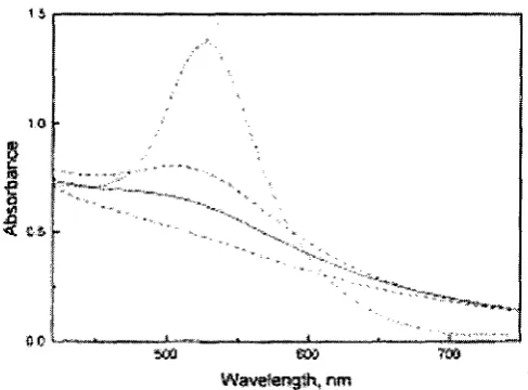
•60 *-»
Wavelength, nm
Figure 1-31521 Absorption spectra of the gold NPs of different size: 8 nm (dash-dot line), 3.2 nm (dash line), 2.6 nm (solid line), and 1.9 nm (lowest dash line).
Step-like spectral structures are indicative of transitions to the discrete lowest unoccupied levels of the conduction band and have been observed in spectra of solutions of monodisperse Au NPs with core diameters from 1.1 to 1.9 nm.[531
15 20 2J I,« 3J
1.4.1.2 The effect of the solvent refractive index
A change of the refractive index of a solvent induces a shift of the SP band. For instance, solutions of dodecanethiolate gold NPs (5.2 nm mean diameter) reveal a 8 nm shift of SP band maximum as the solvent index is varied from nd20 1.33 to 1.55. This can
be predicted by the Mie theory if the monolayer of alkanethiolate is included in the calculations.[53] Thiolate ligands generate a strong ligand field interaction with the
surface electron cloud.
1.4.1.3 Core charge
When electrons are transfered from a reducing agent to a metal particle, the electrons are given to the conduction band. This can cause a rise of the free electron density and, consequently, the metal plasma frequency also rises and generates a change in absorption. Shifts to higher energy are expected if there is excess electronic charge on the core, whereas electron deficiency causes shifts to lower energy.
The unique optical properties of metal NPs have found many applications, especially in biology and for sensors. Their use in optical filters, as labels for bio-macromolecules, in reversible photosensitive monochromatic glasses, for intensity enhancement in Raman spectroscopy (SER effect), for optical switching based on their large, ultrafast nonlinear optical response, and for optical trapping (or "tweezers") based on their high polarizability has been reported.[55] For their beauty and resilience, colloidal gold
suspensions have also found numerous decorative applications'
1.4.2 X-ray diffraction (XRD)
profiles. The distance between scattering contrasts (regions of higher e-density) is inversely proportional to the observed scattering angle.
This behaviour is quantified by Bragg's Law. Theta 0 is the diffraction angle, d is the
distance between atomic layers in a crystal, X is the wavelength of the incident X-ray
beam, and n is an integer.
Scheme 1-2 Deriving Bragg's Law
AB = d sinG (1).
Because AB = BC (2)
nX = 2AB= d sine (3);
(In order to produce a maximum, the path difference 2d sin6 must be equal to an integral
multiple of the wavelength.)
Substituting eq. (2) in eq. (3) we have,
Diffraction methods received particular attention for the examination of gold and other metal NPs because the sample preparation is simple and no artificial impurities are introduced. Besides, the sample volume is comparatively large, so that averaged properties of the material can be evaluated. X-ray diffraction can be used to estimate the average particle size and size distribution. It also can be employed to study the microstructural characteristics, phase composition as well as other features like texture and residual stresses.
It is possible to compare the XRD results with results obtained with other methods, in particular HR-TEM (see part 1.4.3). HR-TEM visualizes individual particles and can identify internal atomic arrangements, which might be helpful for the interpretation and analysis of the diffraction patterns. Although HR-TEM permits direct imaging of atomic positions, the image-contrast is strongly dependent on the NP orientation, which makes a
reliable statistical characterization of a complex NP system a difficult task. Determination of statistical bulk properties is the domain of XRD.
The first application of the line broadening techniques was performed by Scherrer. He put forward an equation for the calculation of the particle size:[56J
L=0.94A/pcos(9)
L: crystallite dimension, 0: the full width in radians subtended by the half maximum intensity width of the powder pattern peak. This method neglects the presence of crystal lattice defects, so that the entire broadening is attributed to a finite size of the crystalline domains.
As the average size and size distribution of NPs are important parameters, several other methods have been developed for and applied to the characterization of gold NPs.
The Debye function analysis (DFA) is based on the calculation of individual diffraction patterns for a set of model particles, covering the range of sizes and structures that may occur in the sample. The experimental data are simulated by finding the appropriate relative weightings for these different profiles.[57]
Gold NPs of sizes between 2-5 nm cause an evident line-broadening and possible crystal structures for these NPs are decahedra (dec), icosahedra (ico), and face centered cubic (fcc).t58] Daniel V. Leff et al.[59J normalized X-ray diffraction powder patterns for
(200) reflection is seen as merely a shoulder of the (111) peak. From this figure, we can see that the peak broaden with smaller particle sizes.
The reliability of crystal structure analysis of NPs depends strongly on the structural models must that are used for the simulation either of the reverse Fourier transform (radial electron density distribution function) or, more directly, of the measured intensity function. These structural models are adopted from theoretical predictions and probable cluster morphologies observed by HR-TEM. As an example, Figure l-6[60] shows three
of those model clusters of similar size (1.6 nm), namely a three-shell cuboctahedron, a three-shell icosahedron, and a Marks type decahedron. It is clear that even at these small sizes, X-ray diffraction can distinguish between these ideal alternative structures.
Figure l-5[59> X-ray
diffraction powder diffraction powder patterns
for four different sizes of gold nanocrystals. The sizes are (a) 201.6 ± 38.3 A, (b) 68.3 ± 3.8 A, (c) 29.5 + 2.0 A, and (d) 15.5 ± 3.0 A. Note that the resolving power of the syntheses to yield different size nanoparticles (as measured by XRD) is well within 15 A.
T
SO 60 70
Diffraction Angle (20)
the
by
UB-U» 4m^94tm
4 8 «
m
Figure 1-6 [60] XRD patterns
of gold NPs, decahedron, a three-shell icosahedron, and a three-shell cuboctahefron, respectively.
Interpretations must become complex when several different crystal structures are present in sample. Wide-angle XRD measurements are dominated larger coherent regions of structure in a sample and, therefore, an analysis of XRD
data actually yields information about the distribution of domain sizes. Domain sizes, however, are only indirectly related to the size distribution of the NPs.
1.4.3 High Resolution Transmission Electron Microscopy (HR-TEM)
A TEM works much like a slide projector. A projector shines a beam of light through (transmits) the slide, as the light passes through it is affected by the structures and objects on the slide. These effects result in only certain parts of the light beam being transmitted through certain parts of the slide. This transmitted beam is then projected onto the
TEM is definitely the most used and effective technique for the study of morphological features such as particle size, size distribution, and structure of NPs. TEM is rather suited to evaluate particle size because their determinations are not affected by the presence of structural defects. Histograms provide the size distribution of nanoparticle cores, which can give crucial information on the size dispersion of the sample.
Sample preparations are particularly important and might be difficult when nanomaterials are involved. In general, the preparation involves the distrinbution of very dilute particle suspensions onto carbon-coated copper grids. Another useful technique is the imbedding of NPs in a solid organic polymer, which is then sliced into very thin sections.
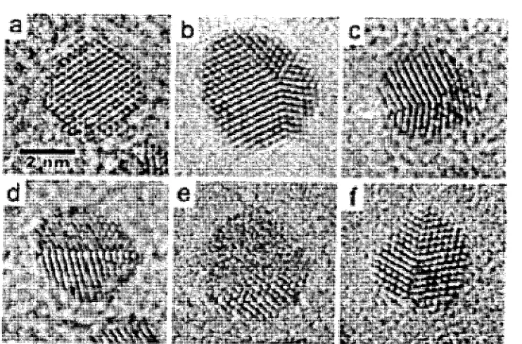
Figure 1-7 mi. Typical HRTEM micrographs of NPs stabilized by dodecanehiol 4.1
nm sample, (a) fee clusters, (b and c) decahedra, (d-f) multidomain particles. Considerable studies focused on the structure of gold NPs.[62] TEM analysis combined
with matrix-assisted laser desorption ionization (MALDI) MS and theoretical calculations, suggested that a more likely structure of the Au core is a truncated
octahedron[63] TEM can also provide a mean diameter, d, of the cores, which allows for
the determination of the mean number of gold atoms, NAU, in the cores: NAU = 59 nm"
(71/6) (DMs)3-[63] These data combined with elemental analysis data that provide a Au/S
ratio, allow for a calculation of the average number of S ligands. This number might also be deduced from thermogravimetric analysis (details see 1.4.4).[64]
1.4.4 Thermo Gravimetric Analysis (TGA)
TGA is the preferred technique for estimating the total organic content of NPs and their thermal stability. Generally, the TGA curve of a NP sample reveals several weight losses during a heating range from 30 °C to 1000 °C. In the temperature range from
30-105°C, the mass might decrease due to the evaporation of remaining organic solvent and moisture that is trapped in the NPs and might account for up to 3 w%. A substantial mass loss is observed between 100 °C and 500 °C, which is attributed to the decomposition and desorption of all organic ligands. TGA measurements that have previously published, to our best knowledge, have been limited to a maximum temperature of 550 °C. We found that the temperature range between 500 °C and 1000 °C often shows a third weight loss that is caused by the evaporation of inorganic impurities (e.g. lithium salts). What remains at last is assumed to be relatively pure gold.
100-
C?95-0s
_o £
90-5
858 0 -step 1|
step 2
i •
\ Dodecanethiol stabilized gold nanopa
\
1
^ — - _ _ _
step 3
^ X
\
v_
i •• ' i • • i ' i i
200 400 600 800 1000 Test temperature (°C)
Figure 1-8 TGA of a crude gold NPs stabilized by dodecanethiol, Step 1 is assumed to be caused by the evaporation of organic solvents and moisture trapped in the gold NPs, Step 2 is the loss of the thiol ligands, in this case dodecanethiol, Step 3 is mostly attributed to the loss of lithium salts which were trapped in the gold NPs as concluded from the increased C02 and H20 signals in MS and the reduction of
TGA results combined with HR-TEM results allow for the estimation of several average properties of the NP sample. TEM contributes numbers for the average size and shape of the NP polyhedra that allow for an estimation of the average surface area and the
number of gold atoms at surface sites. The ratio of organic ligands to total gold atoms is obtained from the TGA measurement. A combination of all sets of data then provides the average number of organic ligands per gold particle as well as the number of ligands per surface gold atoms.[65]
1.4.5 Other characteristic technique
Fourier-Transform Infrared Spectroscopy (FT-IR)
FT-IR is a powerful technique for studying the conformations and crystallinity of alkyl chains adsorbed to NPs surfaces.[66] In particular, the frequencies of the symmetric and
antisymmetric stretching vibrations of methylene groups are known to be related to the population of trans and gauche conformersJ ^
Long chain alkyl thiols attached to gold naoparticles have been shown to be essentially in an all-trans (zigzag) conformation at room temperature in the solid phase, with 5-25% of gauche defects at both inner and terminal locations. A comibination of IR, NMR, and DSC studies revealed the order-disorder transitions of the alkyl chains in gold NPs depending on the temperature and the length of the alkyl chains.[68] Shorter alkyl chains
(<Cg) tend to be more disordered with large amounts of gauche defects present (similar to alkanes in their liquid state), while longer alkane chains (>Cio) are predominantly in the all trans zigzag conformation and have a high degree of crystallinity. The degree of crystallinity increases towards the surface of the NP as the packing density increases.
XPS is a surface limited elemental analysis method with a penetration depth of 2-3 nm. Thus, entire particles must be of 1.5 nm core diameter or smaller to be fully measured. Larger particles do not give reliable ratios of Au to organic ligands. XPS is also sensitive to the chemical environment of the elements and has been used to study the oxidation of NPs (e.g. thiolates oxidize to sulfonates at the NP surface) and the presence of impurities.
XPS studies on small NPs to established that the Au-S bonding is comparable in 2D (Au(lll) surface) and 3D (gold NP) self-assembled monolayers, even though the bonding ratios are different as discussed before.[69J The high surface area of NP makes it
easy to probe surface properties by both bulk and surface sensitive spectroscopic techniques.
Brust et al. used the binding energies of the Au 4f7/2 (83.8 eV) and Au 4f5/2 (87.5 eV)
peaks to determine a ratio between Au° and Au1 species. Au1 (84.9eV), however, was
not found at all although about 1/3 of the gold atoms were located at the surface and bonded to thiols in these NPs. The study verifies that the gold-thiol(ate) bond does not have the character of a gold sulfide bond.[69]
1.5 Palladium NPs
In comparison to gold NPs, very few studies are concerned with palladium NPs. Wei Lu et al.170-1 synthesized thiol-stabilized palladium NPs of different sizes and with narrow
crystalline Pd NPs. The HR-TEM images and electron diffraction patterns clearly reveal the different structures of the crystalline and amorphous Pd particles.
Quiros et alJ ' synthesized palladium NPs in a range of 1.3-3.9 nm by using two-phase method. They claim that smaller particles are obtained when longer chain thiols are used.
1.6 Applications:
There are a lot of practical and potencial applications on gold NPs. Only the hot areas are introduced below.
Biology:
Gold NPs have already found commercial applications as markers for biochemical
investigations. Current areas of investigation include conjugates of gold NPs with oligonucleotides because they have the potential use of the programmability of DNA base-paring to organize nanocrystals in space and multiple ways of providing a signature for the detection of precise DNA sequence. The group of Mirkin reported extremely sensitive colorimetric methods for the DNA analysis capable of detecting trace amounts of a particular oligonucleotide sequence. It also allowed them to distinguish between perfectly complementary DNA sequences and those that exhibit different degrees of base pair mismatches.[12]
The recognition of proteins has been for some time the subject of research on bio-devices for diagnostics based on the interaction between gold NPs and antibody conjugates.^
Gold like most transition metals functions as a catalyst in different chemical reactions. It otherwise is chemically rather inert (e.g. resistant to oxidation), which is important for the long time stability of catalysts in commercial processes.
The first description the catalytic activity of bulk gold occurred at the beginning of the past century when Bone and Wheeler showed in 1906 that gold foils catalyze the
combustion of dihydrogen and dioxygen to give water. The catalytic activity of Au (0) was rediscovered in 1989 when Haruta et al. reported that the oxidation of CO to CO2, a technologically relevant reaction, could be effectively catalyzed by supported-Au (0).
Moreover, it was later observed that the reaction occurs even at 200 K. It soon became apparent that the key to this success was twofold: a) the presence of gold nanoparticles in
the gold catalysts and b) a strong co-catalytic role played by the support that had to be an inorganic oxide such as Fe203, Ti02, or C03O4. It also became apparent that the optimal
nanocluster diameter was about 3 nm, the size at which metal nanoclusters start to exhibit
quantum-size behavior.
Thin films of gold NPs have been shown to change their electrical conductivity rapidly
and reproducibly in the presence of organic vapors. This effect is based on the reversible swelling of the material upon gas absorption, which leads to an increase in the spacing between the metal cores. Since the typical electron hopping conductivity in these
materials depends very sensitively on this distance, the absorption of organic vapor leads to a strong decrease in electrical conductivity. This phenomenon has been exploited for
1.7 Goal of this thesis
The objective of this thesis was the development of a reliable preparation and characterization of gold and palladium NPs based on a single-phase method free of any surfactants. We expected the single-phase method to be superior to the two-phase method as it should produce cleaner NPs and might also allow for a better size control. We aim for NPs of average core diameters below 4 nm and of small size distribution.
Based on the optimized reaction conditions developed by a former PDF Dr. Xu for AU-C12 NPs, this thesis will focus on the investigation of different thiol ligands. Obtained NPs are planned to be investigated by a range of characterization methods, such as HR-TEM, XRD, TGA-MS, UV-VIS, FT-IR, NMR, and XPS. An assessment of the reliability and information content of each of the methods is expected to eventually provide a shorter list of essential characterization methods.
As long term objective, we aim for the preparation of NP surfactants by ligand exchange reactions and the investigation of their self-organizing (mesomorphism) and
self-assembling properties.
Reference:
(1) KJ. Klabunde, Nanoscale Materials in Chemistry, A John Wiley&Sons, Inc., Publication, 2001.
(3) a, Y. Lin, R.G. Finke, J. Am. Chem. Soc, 1994, 116, 8335. b, T.J. Schmid, M. Noeske, H.A. Gasteiger, R.J. Behm, P. Britz, W. Brijoux, H. Bonnemann, Langmuir, 1997,13,2591.
(4) R. Elghanian, J.J. Storhoff, R.C. Mucic, R.L. Letsinger, C.A. Mirkin, Science, 1997, 277, 1078.
(5) T. Vossmeyer, E. Delonno, J.R. Heath, Angew. Chem., Int. Ed. Engl, 1997, 36 (10),
1080.
(6) M.T. Reetz, M. Winter, G. Dumpich, J. Lohau, S. Friedrichowski, J. Am. Chem. Soc,
1997,119,4539.
(7) R. T. Senger, K. K. Bajaj, Phys. Stat. Sol. (b), 2003, No. 1, 82 (8) S.V. Sonti, A. Bose, J. Colloid Interface Sci., 1995, 170, 575.
(9) P. Mulvane, MRS Bulletin, 2001, December, 1009.
(10) G.Schmid, B.Corain, Eur. J. Inorg. Chem., 2003, 3081 and literature cited therein.
(11) M.C.Daniel, D.Astruc, Chem. Rev., 2004, 104, 293
(12) M. Brust, A. Walker, D. Bethell, D. J. Schiffrin, R. Whyman, J. Chem. Soc, Chem. Commun, 1994, 7, 801.
(13) J. R. Blackborow, D. Young, Metal Vapour Synthesis, Springer Verlag, New York 1979.
(14) A.Henglein, M. Giersig, J. Phys. Chem. B 1999,103, 9533.
(15) M. S. Sibbald, G. Chumanov, T. M. Cotton, J. Phys. Chem. 1996, 100, 4672
(16) F. Mafune, J. Kohno, Y. Takeda, T. Kondow, H. Sawabe, J. Phys. Chem. B , 2000,
104,9111.
(18) W. Chen, W. Cai., L.Zhang., G.Wang, L. Zhang, J. Colloid Surf. Set, 2001, 238, 291. (19) A. Dawson,, P.V. Kamat, J. Phys. Chem.B, 2000, 104, 11842
(20) M. Nakamoto, M.Yamamoto, M. Fukusumi, Chem. Commu., 2002, 1622-1623 (21) M. J. Hostetler, J. E. Wingate, C J . Zhong, J. E. Harris, R.W. Vachet, M. R. Clark, J.
D. Londono, S. J. Green, J.J. Stokes, G. D. Wignall, G. L. Glish, M. D. Porter, N. D. Evans, | R. W. Murray, Langmuir 1998, 14, 17.
(22) C. A. Waters, A. J. Mills, K. A. Johnson, D. J. Schiffrin, Chem.Commun., 2003, 4,
540.
(23) K. R. Gopidas, J. K. Whitesell, and M. A. Fox, J. Am. Chem. Soc, 2003, 125, 6491.
(24) T. Pajkossy, T. Wandlowski, D. Kolb, J. Electroanal. Chem., 1996, 414, 209. (25) Frens, G. Nature Phys. Sci. 1973, 241, 20
(26) M.P. Rowe, K.E. Plass, K. Kim, C. Kurdak, E. T. Zellers, A. J. Matzger, Chem.
Mater., 2004, 16, 3513.
(27) N. R. Jana, X. Peng, J. Am. Chem. Soc, 2003, 125, 14280.
(28) C. K. Yee, R. Jordan, A. Ulman, H White, A. King, M. Rafailovich, J. Sokolov, Langmuir, 1999,15, 3486-3491
(29) M. Giersig, P.Mulvaney, Langmuir, 1993, 9, 3408.
(30) A. Badia, L. Demers, L. Dickinson, F. G. Morin, R. B. Lennox, L. Reven, J. Am. Chem. Soc. 1997,119,11104
(31) S. Wang, H. Yao, S. Sato, K.Kimura, J. Am. Chem. Soc,. 2004, 126, 7438
(32) P. Lochman, T.Adam, D. Friedecky, Z., Syopkova , Electr-phoresis , 2003, 24, 1200.
(34) V.Cavalca, G.Cighetti, F.Bamonti, A.Loaldi, L.Bortone, C.Novembrino, M.De Franceschi, R.Belardinelli, M. D. Guazzi, Clin. Chem. 2001, 41, 887.
(35) L. A. Porter, Jr., David Ji, S. L. Westcott, M. Graupe, R. S. Czernuszewicz, N. J. Halas, T. R. Lee, Langmuir, 1998, 14, 7378.
(36) P.D.Beer, D. P. Cormode, J.J. Davis, Chem. Commun., 2004, 414.
(37) J. Liu,, W. Ong, E. Roman, M.J. Lynn, A.E. Kaifer, Langmuir, 2000, 16, 3000.
(38) S.Chen, K, Kimura, Langmuir, 1999, 15, 1075.
(39) J. C. Roberts, M. K. Bhalgat, R. T. Zera, J. of Biomedical Materials Research, 1996, Vol. 30, 53
(40) M. Kim,Y. Jeon, W. S.Jeon, H. Kim, S. G. Hong, C. G. Park, K. Kim, Chem. Commu., 2001, 667.
(41) A. Taubert, U. Wieler, K. Mullen, J. Mater. Chem., 2003, 13, 1090.
(42) K. R. Goidas, J. K, Whitesell, M.A. Fox, J. Am. Chem. Soc, 2003,125, 6491.
(43) R. P. Andres, T. Bein, M. Dorogi, S. Feng, J. I. Henderson, C. P. Kubiak, W. Mahoney, R. G. Osifchin, R. Reifenberger, Science, 1996, 272, 1323.
(44) H. Otsuka, Y. Akiyama, Y. Nagasaki, K.Kaaraoka, J. Am.Chem. Soc, 2001, 123, 8226.
(45) I.G. Denisov, Y.V. Grinkova, A.A.Lazarides, S.G.Sligar, J. Am.Chem Soc, 2004, 126,
3477.
(46) M.Green, P. O'Brien, Chem. Commun., 2000,3, 183.
(47) a, F.Chen, G.Q. Xu, T.S.A.Hor, Mater.Lett., 2003, 57, 3282 b, B.H. Sohn,, J.M. Choi, S. Yoo, S.H. Yun, W. C. Zin, J. C. Jung, M. Kanehara, T. Hirata,T. Teranishi,
(48) E.Leontidis, K. Kleitou, T K.Leodiou, V. Bekiari, P. Lianos, Langmuir, 2002, 18,
3659.
(49) L.O. Brown, J.E. Hutchison, /. Am.Chem. Soc, 1999, 121, 882.
(50) M.J. Hostetler, A.C.Templeton, R.W. Murray, Langmuir, 1999, 15, 3782. (51) M. A Zaitoun, W. R.Mason, C. T Lin, J. Phys. Chem. B ,2001,105, 6780.
(52) S. L. Logunov, T. S. Ahmadi, and M. A. El-Sayed, J. T. Khoury and R. L. Whetten, J. Phys. Chem. B , 1997, 101, 3713.
(53) T. G. Schaaf, M. N. Shafigullen, J. T. Khoury, I. Vezmar, R. L. Whetten, W.G. Cullen, P. N. First, C. Gutierrez-Wing, J. Ascensio, M. J. Jose-Yacamun, J. Phys. Chem. B
1997,101, 7885.
(54) M.M. Alvarez, J.T. Khoury, T. G. schaaff, M.N. Shafigullin, I.V., R.L. Whetten, J. Phys. Chem. B , 1997, 101, 3706.
(55) K. Fukumi, A. Chayahara, K. Kadono, T. Sakaguchi, Y. Horino, M. Miya, K. Fujii, J. Hayakawa, M. Satou, J. Appl. Phys., 1994, 75, 3075.
(56) B.E. Warren, X-ray diffraction, New York, 1990 (57) B. D. Hall, J. Appl. Phys., 2000, 87, 1666.
(58) S.I. Stoeva, B.L.V. Prasad, S. Uma, P.K. Stoimenov, V. Zaikovski, CM. Sorensen, K. J. Klabunde J. Phys. Chem. B, 2003,107, 7441-7448
(59) D.V. Leff, P.C. Ohara, J.R. Heath, W.M. Gelbart, /. Phys. Chem., 1995, 99, 7036.
(60) W. Vogel, J. Bradley, O. Vollmer, I. Abraham, J. Phys. Chem. B., 1998, 102, 10853. (61) D. Zanchet, B. D. Hall, D. Ugarte, J. Phys. Chem. B, 2000, 104, 11013
(63) R.L. Whetten, J.T. Khoury, M. M. Alvarez, S. Murthy, I. Vezmar, Z. L.Wang, P. W. Stephens, C.L. Cleveland, W.D. Luedtke, U. Landman, Adv. Mater. 1996, 8, 428.
(64) Templeton, A.C., Wuelfing, W.P., Murray, R. W. Ace. Chem. Res. 2000, 53, 27. (65) W.W. Weare, S.M.Reed, M.G. Warner, J.E. Hutchison, J. Am. Chem. Soc., 2000, 122, 12890
(66) M.D. Porter, T. B.Bright, D. L Allara, C. E. D.Chidsey, J. Am. Chem. Soc. 1987, 109, 3559.
(67) R. G. Nuzzo, L. H. Dubois, D. L. Allara,. J. Am. Chem. Soc, 1990,112, 558. (68) M.J.Hostetler, J.J.Stoke, R.W. Murray, Langmuir, 1996, 12, 3604.
(69) M. C. Bourg, A. Badia, R. B. Lennox, J. Phys. Chem. B, 2000,104, 6562 (70) W. Lu, B. K. Wang, X. Wang, J. G. Hou, Langmuir, 2003, 19, 5887.
(71) I. Quiros, M. Yamada, K. Kubo, J. Mizutani, M. Kurihara, H. Nishihara, Langmuir,
2002, 18, 1413.
(72) Hauat. M.A. Collidal gold, principles, methods and applications, Academic press , New York, 1989
(73) R. Elghanian, J. J. Storhoff, R. C. Mucic, R. L. Letsinger, C. A. Mirkin, Science,
1997, 277 ,1078.
(74) M. Haruta, M. Date, Appl. Catal. A: General, 2001, 222, 427
Chapter 2. Single-phase Synthesis of Gold NPs Stabilized by Straight Chain Alkylthiols and Their Purification.
2.1 Introduction
Au NPs stabilized by straight chain alkylthiols have been widely studied but the majority of the compounds were prepared by the two-phase method as described in chapter 1.2.1.1. Only few papers have described the synthesis of Au NPs by the single-phase method[1J (chapter 1.2.3) but no systematic study has been conducted on the purity
of these NPs.
We are interested in smaller than 5nm NPs for subsequent ligand exchange reactions and expected the single-phase method to be the better and cleaner approach. The avoidance of phase-transfer conditions, necessary for the two-phase method, should allow for a faster and more accurate control of the NP growth and also avoids the tedious removal of remaining phase-transfer agent from the NP's surface.^
It was known that the size of the NPs can be tuned by changing the reaction temperature, the relative amounts of reagents, as well as the addition rates of thiol and reducing agent to the gold salt solution in THF.[3] Optimized conditions for the
preparation of small Au NPs by the single-phase method were developed in our group by Dr. Xu for dodecanethiol ligands.
pump at a rate of 30 mL/h and the mixture was stirred for another 2 hours after the addition was completed. The reaction mixture was quenched with ethanol (dry 30 mL) and the precipitated NPs were filtered off and dried in house vacuum.
In this Chapter, we extended Dr. Xu's work and used alkylthiols of different chain length (octanethiol, dodecanethiol, and octadecanethiol) for the preparation of Au NPs and studied the properties of these particles in more detail.
2.2 Results and discussion
Au-C8 (Octanethiol), Au-C12 (dodecanethiol) and Au-C18 (octadecanethiol), respectively, were prepared following exactly the same procedure described above. Other work-up procedures than precipitation with ethanol were tested on two different batches of NPs but occurred to be inferior to the ethanol method because they are more time consuming and yield similar particles.
Particles were precipitated by ultracentrifugation and occurred to be purer according to TGA results, but the obtained NPs were larger than for the ethanol method. We assume the high RCF (gravitational acceleration 'g') necessary for their sedimentation caused coagulation, which might also account for the lower yields. Further tests, however, were not possible as the ultracentrifuge was no longer available. RCF can be caculated from the formula: speed (rpm) =946(RCF/R) (R is the diameter of centrifuge tube).
In another test, NPs were precipitated at low temperatures (4 °C), which resulted in particles of similar size as for the ethanol method. This precipitation, however, needed several days for completion and larger amounts of impurities were entrapped in NPs. Consequently, the precipitation of NPs by the addition of ethanol and subsequent
method. The obtained size distributions represent all NPs of each batch as no fractional precipitation was conducted. Isolated yields of dried Au NPs were typically around 60 % based on the initial amount of gold salt.
TGA and XPS measurements of these particles indicated a contamination with basic inorganic impurities (see part 2.2.1) and an acid wash procedure was developed. Typically, 50 mg of NPs were partially dissolved in 20 mL THF, 0.5 M HClaq were added
until a pH = 6 was reached, and add 30mL 50% ethanol to precipitate and then filter; dissolve the filtration in 5-10ml THF and then add 30mL 50% ethanol to precipitate and then filter to get powders. Repeat the process for three times. Acetic acid was tested as a milder replacement for 0.1 M HClaq but it did not always wash away all inorganic
impurities.
Most of these impurities were removed by the acid wash, as verified by TGA and XPS measurements, and from then on the acid wash was integrated in the quenching and work-up process. Typically, the reaction solution was first quenched with 0.5 M HClaq
until the pH = 6 and then fully precipitated by the addition of ethanol.
2.2.1 TGA-MS and XPS investigations
All 3 samples showed 3 distinct weight losses (Figure 2-1) before they were washed with dilute acid. Step 1 was attributed to the evaporation of remaining organic solvents and moisture and usually accounted for 1 w% or less. Step 2 covers the temperature range at which the thiol ligands desorb[4] and the third step above 600 °C is attributed to
impurities were generated by the reaction of excess LiBEtaH with water and ethanol
during the work-up.
100 J
95 J
CO o
I
9 085 A
80 A
Au-C12 original Au-C12 washed
0 200 400 600 800
Temperature (°)
1000
Figure 2-1 TGA of Au-C12 before and after acid wash
All TGA measurements on Au NPs described in literature have been limit to a temperature range of 30 °C to 600 °C and, consequently, the presence of inorganic
Scheme 2-1 Lithium salts that might be generated in single-phase method and contaminate Au NPs
H" + H20 *- OH" + H2 *
Li++ OH" • L i 0 H
L i O H + C 02 *- LiHC03
H" + CH3CH2OH *- CH3CH20- + H2j
Li+ + CH3CH20- *- LiOCH2CH3
TGA measurements on Au-C8, -CI2, and -CI8 after acid wash treatment revealed only one major weight loss between 150 °C and 500 °C, which is attributed to the desorption of thiols. Very little weight losses above 600 °C were detected and indicate the absence of most of the inorganic impurities (Figure 2-1 and Table 2-1).
XPS investigations were carried out to verify our hypothesis with regard to Li based inorganic impurities in the NPs. Measurements were conducted on original samples of Au-C12 and Au-C8 as well as samples that were washed with 0.1 M HClaq or heated to
500 °C in a stream of N2 for 30 min prior to the measurement. XPS measurements were performed by Seyed Tadayyon in Peter Norton's group at UWO. These measurements confirmed the presence of Li as well as CHO, CH (OH), and C02 species as well as the
XPS Au 4f peaks for J1 sample before
m 8S
Figure 2-2 Deconvoluted Au-signals of the XPS of the original Au-Cl2 sample
(before acid wash)
XPS carbon Peaks for JS sample before
23©
etreBsgGm»f$ffeV
Figure 2-3 Deconvoluted C-signals of the XPS of the original Au-C8 sample (before
Table 2-1 TGA of Three Gold NPs Stabilized by C8, Cu-, and C18- Alkylthiols
w% loss of step 1 (integration limits in °C)
w% loss of step 2 (integration limits in °C)
w% loss of step 3 (integration limits in °C)
Au:thiol ratio in (mol%) onset temperatures
for steps 1-3 (°C) Core diameter
(nm)
Atoms in ave NPs
No. ofthoilon
per NPs No. of thiol on
pernm2 Au-C8-b' 1.3% (30-120) 5.7% (120-500) 19.8% (500-1000) 9.80 80 125 640 4.0 1976 203 1.01 AuC12-b' 1.5 % (30-150) 10.5% (150-500) 8.6% (500-1000) 6.68 80 215 614 4.2 2287 346 1.55 Au-C18-b' 0% (30-200) 22.1% (200-500) 4.7% (500-1000) 2.50 250 635 3.9 1831 341 1.78 Au-C8-a' 0% (30-120) 7.0% (120-500) 0.04% (500-1000) 9.78 122 635 4.0 1976 200 1.00 AuC12-a' 1.2% (30-150) 13.8% (150-500) 0.21% (500-1000) 6.65 80 215 620 4.2 2287 342 1.55 Au-018-a1 0% (30-200) 22.1% (200-500) 0.09% (500-1000) 2.53 250 630 3.9 1831 341 1.78
b = original particles before acid wash, a = original particles after acid wash, 2these values are
Atoms in ave. NPs: NAu =59 rnn "3 (TI/6) ( DM S)3
No. of thoil on per NPs calculated from TGA and atoms in ave.[3] (TGA% of thiol/Mthioi)/(TGA% of
gold/ Mgoid)*Atoms in ave.NPs.
No. of thiol on per run2 calculated from TGA and diamters of NPs.[3) (No. of thiols on per NPsAfcir2).
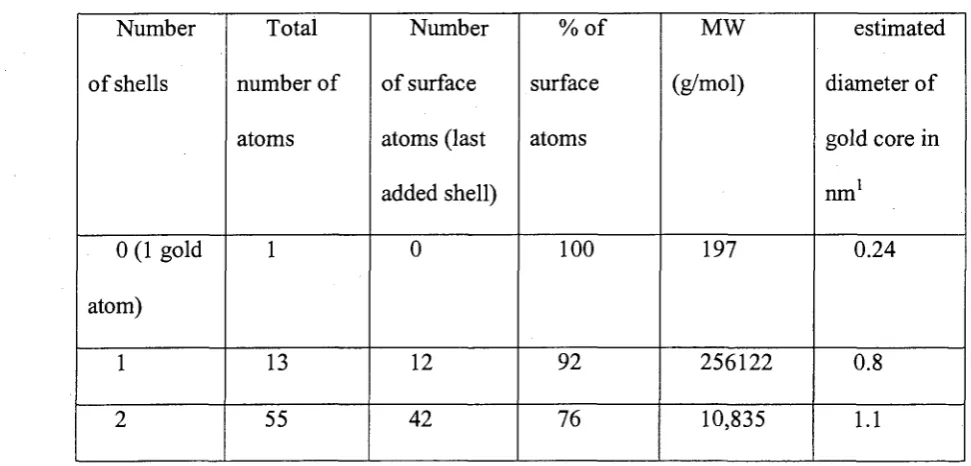
Thiol to gold ratios might be calculated from the TGA data (Table 2-1) and these ratios could be used to estimate the number of thiols that are attached to each Au-NP. This estimation, however, requires information on the average size, the crystal structure, and the shape of the NPs. From the HR-TEM and XRD data, we know that the prevalent structure of the NPs is truncated octahedron. Such a NP might be constructed by adding layer by layer to a first central gold atom assuming that incomplete shells are energetically disfavored. These numbers are known as Chini's Magic Numbers[5] and
obey the equation:
Number of atoms per shell = 10n2 + 2, with n = number of shell
Table 2-2 Calculated Size Related Values for Ideal Gold Clusters
Number
of shells
0 (1 gold
atom) 1 2 Total number of atoms 1 13 55 Number of surface atoms (last added shell) 0 12 42 %of surface atoms 100 92 76 MW (g/mol) 197 256122 10,835 estimated diameter of gold core in
ran1
0.24
0.8

![Figure 1-6 [60]three-shell cuboctahefron, XRD patterns of gold NPs, decahedron, a three-shell icosahedron, and a respectively](https://thumb-us.123doks.com/thumbv2/123dok_us/1487174.1181958/38.598.96.534.86.434/figure-shell-cuboctahefron-patterns-decahedron-shell-icosahedron-respectively.webp)