| INVESTIGATION
Wnt
Signaling in Sexual Dimorphism
Girish Deshpande,*,1Ali Nouri,* and Paul Schedl*,†,1 *Department of Molecular Biology, Princeton University, Princeton, New Jersey 08540, and†Laboratory of Gene Expression Regulation in Development, Institute of Gene Biology, Russian Academy of Sciences, 119334 Moscow, Russia
ABSTRACTThe embryonic gonad ofDrosophila melanogasterbegins to display sexually dimorphic traits soon after its formation. Here we demonstrate the involvement of awntfamily ligand,wnt-2, in the induction of these sex-specific differences. We show thatwnt-2
contributes to the survival of a male-specific population of somatic gonadal precursor cells (SGPs), the male-specific SGPs that are located at the posterior of the male gonad. We also show that the Wnt-2 ligand synergizes with the JAK-STAT ligand Upd, which is produced by SGPs at the anterior of the gonad to activate the STAT pathway in male germ cells. We suggest that the use of two spatially separated signaling systems to initiate the JAK-STAT stem cell maintenance pathway in germ cells provides a mechanism for increasing the pool of potential progenitors of the germline stem cells in the adult testes. Finally, we present evidence indicating that, like the JAK-STAT pathway,wnt-2stimulates germ cells in male embryos to re-enter the cell cycle.
KEYWORDSDrosophilasex determination; nonautonomous signaling; Wnt pathway; sexual dimorphism
T
HE primitive embryonic gonad inDrosophila melanogasteris composed of two distinct cell types, the germ cells and the somatic gonadal precursor cells (SGPs) (Santos and Lehmann 2004). These two cell types are formed at different locations in the embryo and are specified by distinct mecha-nisms. The germ cells arise as pole cells at the posterior end of the precellular blastoderm embryo, and their proper specifi -cation depends on maternal determinants that are assembled in the pole plasm during oogenesis. After cellularization of the blastoderm, the germ cells must make their way into the center of the embryo and then migrate toward the newly formed SGPs in parasegments (PS) 10–13. SGPs are derived from dorsolateral mesodermal tissue in these parasegments and are specified by the hierarchical action of zygotic pattern-ing genes. The dorsolateral mesoderm of PS 10–13 is formed under the control oftinman andzfh-1(Mooreet al.1998) while the eventual specification of SGPs from these cells de-pends upon the bifunctional transcription factoreyes absent (eya) (Boyleet al.1997; Mooreet al.1998). Althougheyais required for SGP identity, it is differentially expressed in
anterior and posterior SGPs. This difference depends upon the activity of the homeotic genesabdominal-a(abd-A) and
Abdominal-B(Abd-B). The specification of anterior SGPs de-pends uponabd-A, while the specification of posterior SGPs depends upon both abd-A and Abd-B (Boyle and DiNardo 1995; De Falcoet al.2004).
In addition to differences between anterior and posterior SGPs, the embryonic gonad is sexually dimorphic. One sex-specific difference is in the activity of signaling pathways that mediate communication between the SGPs and the primordial germ cells (PGCs). Wawersik et al. (2005) found that the ligand for the JAK-STAT pathway,unpaired(upd) (Harrison
et al.2003), is expressed in a small group of SGPs at that very anterior of the embryonic gonad in male but not female em-bryos. [The same sex-specific expression pattern is seen for the closely related upd-3 (Hombria et al. 2005).] The upd
ligand signals to the germ cells in male embryos upregulating the level and activity of the transcription factor STAT92E (Houet al.1996). By contrast, there is little, if any, STAT92E in the germ cells of female embryos. The activity of the JAK-STAT pathway in males and females is dependent upon the somatic sex determination pathway. The sex determination pathway can be bypassed in females by ectopic expression ofupd(orupd-2or
upd-3), which then activates STAT92E accumulation in their germ cells.
Another sex-specific difference is the presence of cell types in one sex but not the other. (De Falcoet al.2003, 2008). One example of a cell type found only in males is the pigment
Copyright © 2016 by the Genetics Society of America doi: 10.1534/genetics.115.177857
Manuscript received May 4, 2015; accepted for publication November 17, 2015; published Early Online November 24, 2015.
Supporting information is available online at www.genetics.org/lookup/suppl/ doi:10.1534/genetics.115.177857/-/DC1.
1Corresponding authors: Department of Molecular Biology, Princeton University, Lewis Thomas Labs, Washington Rd., Princeton, NJ 08544. E-mail: gdeshpan@princeton.edu; Institute of Gene Biology, Russian Academy of Sciences, Moscow, Russia.
precursor cell. These cells arise late in embryogenesis and are distributed around the outside of the embryonic gonad. Their specification depends upon thewinglessligandwnt-2, which is activated by thedsxgene. Another sex-specific cell type is the male-specific SGP (msSGP), which is clustered at the posterior end of the coalesced gonad. msSGPs are specified by a mechanism that seems to be independent oftinmanand
zfh-1, and they express three different molecular markers, namely Eya, the wnt-2ligand, and the transcription factor Sox100B. While Sox100B protein is detected only in the male gonads,wnt-2expression is observed in gonads of both sexes around the time that the germ cells and SGPs first make contact. Subsequently, at the gonad coalescence stage,wnt-2
is greatly enriched in the male gonads in the msSGPs (De Falco
et al.2003). At this stage another SGP-specific marker, Eya, is also enriched in msSGPs.
Although msSGPs are found only in the coalesced gonads of male embryos, their initial specification is not sex specific. Thus, Sox100B/Abd-B-positive cells are detected in PS 13 of both male and female stage 13 embryos. However, survival of msSGPs is controlled by thedoublesex (dsx) gene in a sex-specific manner. In female embryos, the female Doublesex protein activates a conserved cell death pathway, and the msSGPs are eliminated by stage 14–15. In contrast, msSGPs remain in the male embryos (De Falcoet al.2003).
In the studies reported here we have examined the role of
wnt-2in the development of the male gonad. We show that
wnt-2promotes survival of msSGPs in a sex-specific manner. In addition,wnt-2also plays an important role in the sex-specific development of the male germline. One of the instructive func-tions ofwnt-2is to potentiate the activation of the JAK-STAT pathway in male germ cells. As a consequence, there are two signaling centers: a group of SGPs at the anterior of the gonad, which express the JAK-STAT ligand Upd, and the msSGPs at the posterior, which express Wnt-2, that mediate the induction of STAT expression in male germ cells. We speculate that the use of this dual but spatially separated signaling system to initiate the JAK-STAT stem cell maintenance pathway provides a mechanism for specifying not only the anterior but also the posterior germ cells in the male embryonic gonad as potential progenitors of the germline stem cells (GSCs) in the adult testes. This would maximize the number of germ cells that can be incorporated into the stem cell niche when it is estab-lished at the anterior of the embryonic gonad during stage 17 of embryogenesis (Le Bras and Van Doren 2006). In addition to a role in upregulating the JAK-STAT pathway, wefind that
wnt-2induces male germ cells to re-enter the cell cycle and begin dividing. Like the activation of the JAK-STAT pathway, the early proliferation of germ cells is a male-specific trait.
Materials and Methods
Immunohistochemistry
The embryo stainings were performed essentially as described in Deshpandeet al.(1995). Embryos werefixed for 20 min in
3.7% formaldehyde/PBS:heptane and devitellinized in heptane: methanol for staining with the primary antibodies: conjugated secondary antibodies were obtained from Molecular Probes. Embryos were mounted in AquaPolymount (Polysciences, Inc.). Images were collected using a Zeiss LSM 510 confo-cal microscope, and Adobe Photoshop was used to generate the
finalfigures. The microscope settings including the gain and the laser intensity were maintained constantly for the duration of a complete experiment after the initial standardization to minimize the variability. In each imaging experiment, both experimental and control embryos were imaged under the same conditions.
To render older,i.e., stage 17, embryos accessible to immu-nostaining, sonication was used (for a detailed description, see Le Bras and Van Doren 2006). Embryos were rehydrated by washing several times in PBS containing 0.1% Triton (PBST), sonicated in 500ml of PBST with a 3-sec constant pulse using a Branson Sonifier 250 (set at 100% duty cycle and output setting 1), and washed several times with PBST. The embryos were subsequently stained with the primary antibodies as described above.
The following antibodies were used: Vasa (rabbit; 1:1000; from Paul Lasko), anti- Vasa (rat, 1:1000; from Paul Lasko), Sox100B (rabbit, 1:2000; from Steve Russell), STAT (rabbit, 1:1000; from Steven Hou), Eyes Absent (mouse, 1:15, from the Developmental Hybridoma Bank), Upd (1:500, from Doug Harrison), Sxl (mouse, 1:10, from the Developmental Hybridoma Bank),b-galactosidase (rabbit, 1:2000, purchased from Kappel; and mouse, 1:10 from the Developmental Hybridoma Bank).
Clonal analysis
Females carrying germline clones forsggwere produced as previously described (Chou and Perrimon 1992). Clones were induced in females homozygous for FRT insertion on the X chromosome (FRT 101) and heterozygous for the dom-inant female sterile mutation,ovoD1, andzw3(M11). Females of the genotypezw3,FRT 101/FM7were mated to males of the genotypeovoD FRT 101/Y;Flp/Flp. Progeny was heat-shocked at second and third instar larval stages, for 1 hr each. The virgin females recovered after the heat shock were crossed to OregonR males. The male progeny were not rescued whereas females were zygotically rescued.
Fly stocks
Two differentwnt-2null alleles Kozopas et al. 1998,wnt-2o andwnt-2L, were used for loss-of-function studies . They gave similar results. The following UAS and GAL4 stocks were used for the misexpression studies:UAS-wnt-2(three different transgene inserts were used), UAS-upd(two different trans-gene inserts were used), UASArm-S10(two different inserts were used),twist-GAL4,nanos-GAL4, and UAS-b-galactosidase. In most experiments, males carrying two copies of UAS-wnt-2
Results
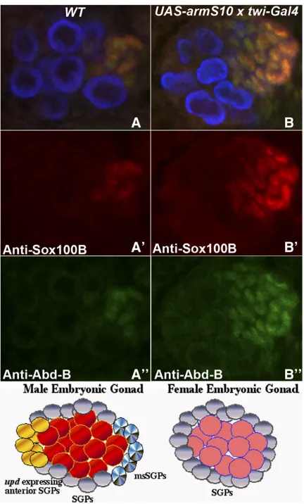
Hyperactivation of the canonical wnt-signaling pathway induces the formation of excess msSGPs
Previous work has shown thatwnt-2mutations cause sterility in males, but have no apparent effect on fertility in females. The testes ofwnt-2males lack the pigment cells of the testes sheath, are much smaller than wild type, and have an unusual oblong shape rather than the typical spiral (Kozopas et al.
1998; Kozopas and Nusse 2002; also see Linnemannstöns
et al. 2014). However, since many of the characteristic so-matic and germline cell types appear to be present, the pre-cise mechanism(s) responsible for the sterility ofwnt-2males has remained uncertain.
Studies by Boyleet al.(1997) indicate that the specification of the somatic component of the gonad, the SGPs, depends upon canonicalwntsignaling. They found that ectopic expres-sion of an activated form of the Armadillo protein, Arm-S10, in the mesoderm using atwist-GAL4driver orwgunder a heat-shock promoter, substantially enlarges the population of Eya-expressing SGPs in the coalesced gonads of stage 15 embryos. Since most of these extra SGPs appear to be clus-tered at the posterior of the coalesced gonad where the Wnt-2-expressing msSGPs are located, it is reasonable to suppose that they correspond to msSGPs. If this is correct, it raises the possibility that Wnt-2 and canonicalwnt signal-ing might be important in specifysignal-ing and/or maintainsignal-ing the msSGPs. On the other hand, sincewgis known to play a role in SGP specification, it is also possible thatwnt-2activity is not needed or thatwnt-2has some other function. Consis-tent with the latter possibility, Defalco et al. (2008) have shown that wnt-2 activity is necessary and sufficient for the specification of pigment precursor cells in male gonads (De Falcoet al.2008).
To explore the function ofwntsignaling in the male gonad further, wefirst re-examined the effects of ectopic ArmS10 on SGPs, focusing specifically on the msSGPs in the coalesced gonad of stage 15 embryos. While all SGPs are thought to express Eya, msSGPs can be differentiated from other SGPs in that they also express Sox100B and Abd-B (De Falcoet al.
2003, 2004). To determine if the population of msSGPs is expanded by activation of the canonicalwnt-signaling
path-way, we probed twist-GAL4/UAS-ArmS10 embryos with
Vasa antibodies to identify the gonad and with Sox100B and Abd-B antibodies to identify the somatic msSGPs. Wild-type, stage 15 male embryos have a cluster of 10 (n= 10; mean = 10; SD: 1.08) Abd-B/Sox100B-positive msSGPs at the posterior of the gonad, while there are no Abd-B/Sox100B-positive msSGPs in stage 15 female embryos (n= 10; mean = 0.091). Figure 1 shows that, as would be expected from the results of Boyle et al.(1997), there is a substantial increase in the number of Abd-B/Sox100B-positive msSGPs in the gonads of twist-GAL4/UAS-ArmS10embryos (n= 15; mean = 23; SD: 4.65;P-value,0.00001). There was also no obvious sex specificity in the inductive effects of ectopic ArmS10.
wnt-2 is required for the survival but not the specification of msSGP in male embryos
probed embryos produced bywnt-2/Cyo,en:LacZstocks for two different wnt-2 alleles, wnt-2° and wnt-2L, with Vasa antibodies to label the germ cells (imaged in blue) and with Abd-B and Sox100B antibodies to label the msSGPs in the embryonic gonad (see Figure 2 legend). To identify wnt-2
-males and their wnt-2+male sibs, we also probed the em-bryos with Sxl andb-galactosidase antibodies. Altogether we examined the coalesced gonads of 15wnt-2-and 15wnt-2+ stage 15 male embryos. The control (b-galactosidase-positive and Sxl-negative)wnt-2/+ stage 15 male embryos had be-tween 8 and 14 Abd-B-positive and Sox100B-positive msSGPs, with an average of 10.4 msSGPs. This is comparable to the number of msSGPs found in stage 15 wild-type male embryos. By contrast, as illustrated by several different ex-amples in Figure 2, there is a clear reduction in the number of Abd-B/Sox100B-positive msSGPs in thewnt-2mutant males (b-galactosidase and Sxl-negative). The number of msSGPs in the mutants ranged from 0 to 4 with an average of 2 Abd-B/Sox100B-positive cells (n = 10; mean = 2; SD: 1.63;
P-value,0.00001).
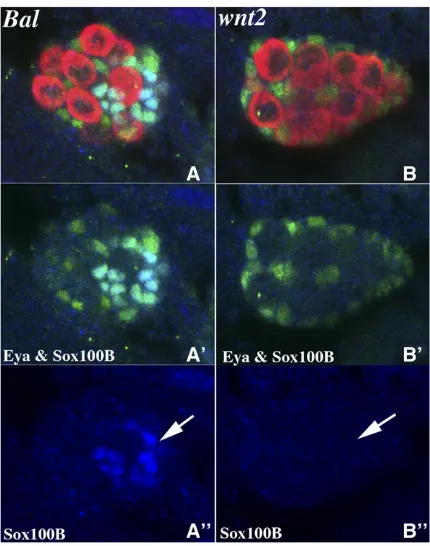
In the second set of experiments, we marked msSGPs in embryos from the twownt-2/Cyo en:LacZstocks using a com-bination of Sox100B and Eya antibodies. As before, we local-ized the gonad in stage 15 embryos with Vasa antibody and identified wild-type and wnt-2- male embryos by probing
with Sxl and b-galactosidase antibodies. As illustrated in Figure 3, while SGPs expressing Eya are observed in coa-lesced gonads ofwnt-2mutant male embryos, Eya/Sox100B-positive msSGPs could not be detected in almost half of the
wnt-2male embryos. In the remainingwnt-2embryos, one tofive msSGPs were observed (n= 10; mean 1.8; SD: 1.87;
P-value,0.00001).
One interpretation of these results is that msSGP specifi -cation depends uponwnt-2. However, this does not seem to be the case. When we examined the msSGPs in younger male
embryos (stages 12–13), we found that the number of Sox100B/Abd-B- or Sox100B/Eya-positive msSGPs in the
wnt-2mutant males (n= 12; mean = 8; SD: 1.73;P-value = 0.012) was close to that seen in wild type (n= 10; mean = 9.2; SD 1.3) (see Supporting Information, Figure S1). The presence of normal numbers of msSGPs at this earlier stage would argue thatwnt-2is not essential for the initial specifi -cation of msSGPs. On the other hand, since the number of msSGPs is substantially reduced in older stage 15wnt-2-male
embryos, it would appear thatwnt-2activity is important for the survival of these cells when the male gonad coalesces.
Ectopic wnt-2 can promote survival of msSGPs in female embryos
The findings in the previous section argue thatwnt-2is re-quired for the survival of msSGPs, but does not have an im-portant role in their specification. To test this further, we ectopically expressed Wnt-2 in the mesoderm using a twist-GAL4to drive expression of aUAS-wnt-2transgene. We used Sxl antibody to distinguish between male and female em-bryos, Vasa antibody to mark the embryonic PGCs, and Sox100B to identify the msSGPs. We found that there was a striking difference between the effects of ectopic ArmS10 and Wnt-2. While the former induced the formation of excess msSGPs, there was no obvious increase in the number of msSGPs with the latter (n= 15; mean = 11.6; SD: 3.44;
P-value = 0.051). In this context, it should be noted that the effects ofwgmisexpression using the sametwist-GAL4driver resembled that observed with ArmS10 (n = 10; mean = 26.4; SD: 6.1;P-value,0.00001). Thesefindings, together with the results described above, would be consistent with the conclusion thatwnt-2does not function in the specifi ca-tion of the msSGPs.
We next asked whether ectopic Wnt-2 could promote the survival of msSGPs in female embryos. As previously shown Figure 2 Wnt-2 is required for msSGP survival in male embryos. Embryos fromwnt-2/Cyo engrailed-LacZ stock were probed with Sxl (not shown),b-galactosidase (not shown), Vasa (imaged in blue), Sox100B (imaged in red), and Abd-B (imaged in green) antibodies simultaneously. A–D99show coalesced gonads from stage 15 embryos. (A–
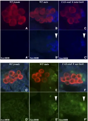
(De Falco et al. 2003), we found that Sox100B-positive msSGP cells are absent in the gonad of control stage 15 fe-male embryos (Figure 4, A and A9). In contrast, Sox100B-positive msSGPs were observed in the coalesced gonads of
twist-GAL4/UAS wnt-2female embryos (Figure 4, C and C9). While ectopic Wnt-2 clearly suppresses msSGP apoptosis in female embryos, it should be noted that msSGPs are found in the coalesced gonads of only 50–60% of the twist-GAL4: UAS wnt-2 female embryos, and the number of Sox100B-positive cells/gonad ranges from 1 to 6 (n= 11; mean = 2.8; SD: 2.04;P-value = 0.000267) as compared to10 in wild-type males (compare panels B’and C’). Taken together with the effects of wnt-2mutations in males (Figure 2 and Figure 3), this could mean thatwnt-2is important but not entirely sufficient to protect against apoptosis. Alternatively (or in addition), since the msSGPs themselves appear to be a source of Wnt-2 in male gonads, it is possible that the level of Wnt-2 accumulation in the gonad using thetwistdriver is not
equivalent to the autocrine signal produced by the male msSGPs themselves. Potentially consistent with this latter possibility, we found that the extent of rescue of msSGPs by ectopic Wnt-2 differs between differentUAS wnt-2transgenic lines (not shown).
To confirm that Wnt-2 promotes the survival of msSGPs in the coalesced gonad, we used ananos-GAL4driver to express Wnt-2 only in the germ cells. As shown in Figure 4, D–F, this localized source of Wnt-2 can act as a survival factor for msSGPs in female gonads and block apoptosis. Interestingly, expression of Wnt-2 in the germ cells is somewhat more effec-tive in protecting msSGPs from cell death than is ubiquitous expression in the mesoderm by thetwistdriver, and wefind that75% of the coalesced gonads innanos-GAL4:UAS-wnt-2
female embryos have Sox100B-positive msSGPs (n = 11; mean = 3.3; SD: 2.34;P-value = 0.000164). Thisfinding also indicates that germ cells in the embryonic gonad are able to signal to their somatic partners.
Does wnt-2 have functions in addition to sustaining msSGPs?
An important question is whetherwnt-2has other roles in the development of the male embryonic gonad. It has recently been shown that SGPs at the anterior of the male embryonic gonad express Upd and that this ligand activates the JAK-STAT-signaling pathway in male germ cells, leading to a sub-stantial upregulation of STAT protein expression. By contrast,
updis not expressed by anterior SGPs in female gonads, and the JAK-STAT-signaling pathway remains off in female germ cells. Since the Wnt/b-catenin pathway can up-regulate the expression ofStat3in mouse embryonic stem cells (Haoet al.
2006), we wondered whether the Wnt-2 produced by the msSGPs functions to synergize the activation of the JAK-STAT pathway in male germ cells.
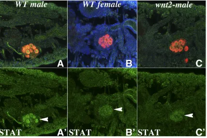
To investigate this possibility, wefirst examined the effects ofwnt-2mutations on STAT protein expression in the germ cells of male embryos. In wild type, high levels of STAT ac-cumulate in the germ cells of male embryos (Figure 5, A and A9) while the germ cells of female embryos have only low levels of STAT (Figure 5, B and B9). As can be seen in Figure 5, C and C9, STAT expression is appreciably reduced in the germ cells ofwnt-2mutant male embryos, and the level of STAT protein is only marginally greater than that seen in the germ cells of wild-type females. This result indicates that Wnt-2 is required to promote the accumulation of high levels of STAT protein in male embryonic germ cells.
We next tested whether ectopic expression of Wnt-2 can induce STAT protein accumulation in female embryonic germ cells. For this purpose, we used twist-GAL4 to drive wnt-2
expression in the mesoderm. Figure 6 shows that wild-type female embryos at stage 13–14 have barely detectable levels of STAT protein in their germ cells (see Figure 6, A–A99). In contrast, when wnt-2is ectopically expressed in the meso-derm of female embryos, it can promote the accumulation of STAT protein in the female germ cells (see Figure 6, C–C99). STAT protein also seems to persist in female PGCs of stage 15 Figure 3 wnt-2is required for survival of Eya/Sox100B-positive msSGPs
UAS-wnt-2/nos-Gal4embryos (Figure 6, D–D99). Note that the level of STAT protein observed in female germ cells in the presence of ectopic Wnt-2 is not equivalent to that seen in male germ cells (compare Figure 6, B99and D99). On the other hand, it is clearly elevated compared to that observed in the germ cells of wild-type female embryonic PGCs (Figure 6, A99; also see Figure 8B9). To confirm thisfinding, we tested whether ectopic expression of Wnt-2 using the germline-specificnos-GAL4driver induces STAT accumulation in female germ cells. We found that STAT accumulation could also be induced when Wnt-2 is expressed in female germ cells; how-ever, as was observed with thetwistdriver, the level of STAT protein is less than that in the germ cells of wild-type males (data not shown).
One likely reason why the level of STAT induced by ectopic Wnt-2 in female germ cells is less than in wild-type male germ cells is that the anterior SGPs in male gonads express high levels of the JAK-STAT ligand Upd while this ligand is not expressed by SGPs in female gonads. To better understand the respective functions of Wnt-2 and Upd in the germline and
soma of the embryonic gonad, we compared the effects of ectopic Upd and Wnt-2 in female gonads. In contrast to Wnt-2, expression of Upd using thenanos-GAL4driver did not pro-mote the survival of msSGPs in female embryonic gonads (Girish Deshpande data not shown). On the other hand, ec-topic Upd appeared to be more effective in inducing STAT protein accumulation in female germ cells than ectopic Wnt-2 (not shown). However, even in this case, the level of STAT accumulation induced in female germ cells by ectopic Upd was less than that observed in germ cells of wild-type male embryonic gonads.
How does wnt-2 upregulate STAT expression?
wnt-2-signaling pathway. Inwnt-2mutant males, these SGPs would fail to express the Upd ligands, and STAT expression would not be activated in the germ cells. In the second, Wnt-2 produced by msSGPs would signal directly to the germ cells to activate STAT expression. In this mechanism, Upd expres-sion by the SGPs at the anterior of the gonad would not in itself be sufficient to fully activate STAT expression in male germ cells in the absence of the synergistic activity of Wnt-2 produced by msSGPs at the posterior of the gonad.
Although we were unable to detect any obvious reduction in the level or distribution of the Upd ligand in the embryonic gonads ofwnt-2mutant males (data not shown),wnt-2could be required for expression of the Upd-3 or play some other role in the transmission of the Upd signal. For this reason, we focused on testing the second model. Wefirst asked whether the wnt-signaling pathway needs to be activated in male germ cells to turn on high levels of STAT expression in these cells. To inhibit the reception of thewnt-2 signal specifically in the germ cells, we overexpressedshaggy(sgg), which en-codes a negative regulator of the canonical wnt signaling pathway, Glycogen Synthase Kinase 3, in these cells using a
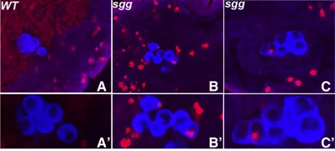
nanos-GAL4driver (Tolwinskiet al.2003). Overexpression of this kinase would be expected to prevent germ cells from responding fully to the Wnt-2 ligand because phosphoryla-tion of the co-activator Armadillo (Arm) by Sgg promotes degradation of the Arm protein. Figure 7 shows that STAT protein expression in germ cells ofUAS-shaggy/nanos-GAL4
stage 15 male embryonic gonads is reduced (Figure 7, B9)
compared to that in germ cells of similarly staged wild-type male gonads (Figure 7, A9). Figure 7, A99and B99, shows that the somatic msSGPs are not affected by downregulating the response towntsignaling specifically in male germ cells. An-other protein that cell-autonomously impedes the response to wntsignaling when overexpressed is Axin (Willertet al.
1999). Axin interacts with both Sgg and Arm and is part of the Arm destruction complex (Willert and Jones 2006). Whennanos-GAL4is used to drive Axin expression, the accu-mulation of STAT protein in male germ cells is also reduced as is seen forsgg(Figure 7, C and C9).
The effects of excess Sgg (and Axin) on STAT accumulation in male germ cells would be consistent with the second mechanism—namely that Wnt-2 produced by somatic go-nadal cells synergizes with the Upd ligand to induce a high level of STAT expression in male germ cells. To provide fur-ther evidence that activation of the canonical Wnt pathway in germ cells can promote STAT accumulation, we generated germline clones of the negative regulatorsggand analyzed STAT expression in the germ cells of their female progeny (Figure 8). We found that, unlike wild-type females (Figure 8, B and B9), STAT protein accumulated in the germ cells of
sggM-Z+ females (compare Figure 8, C and C9). Thus, the effects of inappropriately activating the canonicalwnt-signaling pathway in female germ cells are also consistent with the idea that the Wnt-2 ligand functions to synergize the activa-tion of the STAT pathway in male germ cells by Upd. This suggestion is supported by thefinding that ectopic expression of the gain-of-function ArmS10 protein in the germline using a nanos-GAL4 driver also induced STAT accumulation in female germ cells (data not shown). Although these results are most easily explained by awnt-2-dependent activation of the canonicalwnt pathway in male germ cells, they do not exclude the possibility thatwnt-2activates the production of some otherwnt-like ligand that actually signals to the male germ cells.
Do msSGPs participate in the signaling process?
Since the msSGPs are a major source of Wnt-2 in the male embryonic gonad, we wondered whether one of the functions of this male-specific group of somatic cells is to send the Wnt-2 signal to the male germ cells. To investigate this possibility, we needed to protect the msSGPs in female gonads from apopto-sis by a mechanism that did not depend upon ectopic expres-sion of Wnt-2. Defalco et al.(2003) have shown that the baculovirus anti-apoptosis protein p35 can partially protect the msSGPs in female embryos from cell death and that, un-like wild-type females, the p35-expressing msSGPs are able to persist once the female gonad has coalesced. We used the
twist-GAL4driver to express p35, and after confirming that it promotes the survival of msSGPs in female embryonic go-nads, we examined the expression of STAT in the germ cells of these embryos (see Figure S2). As shown in Figure S2, STAT expression is upregulated in female embryonic gonads when msSGPs are protected from cell death by ectopic p35 (compareFigure S2, B9with C9and D9). Thisfinding indicates Figure 5 wnt-2activity is necessary for the accumulation of high levels of
that one function of the male-specific msSGPs is to signal germ cells to upregulate STAT accumulation.
Wnt signaling promotes cell division in the germline
When the pole cells are formed in the precellular blastoderm embryo, they exit the cell cycle and do not divide during their migration from the exterior of the embryo to the somatic gonad. After the coalescence of the embryonic gonad, the germ cells in males re-enter the cell cycle and begin dividing, slowing expanding the germline population in the gonad. By contrast, the germ cells in the female gonad do not start dividing until much later in development. Wawersik et al.
(2005) have shown that the difference between males and females depends in part upon the activation of the JAK-STAT pathway in the male embryonic gonad. If wnt-2 signaling functions to potentiate STAT activation in male germ cells, then it should also influence the sex-specific choice of whether to re-enter the cell cycle or not. To test this possibil-ity, we used antibodies against phosphohistone H3 (pH 3) to identify germ cells that are undergoing cell division in the embryonic gonads of wild-type andwnt-2mutant males and females. We observed at least one pH 3-positive germ cell in
40% of the wild-type male embryos, while pH 3-positive germ cells were never detected in wild-type female embryos (Table 1). Wawersiket al.(2005) also found that pH 3-positive germ cells are never detected in female embryonic gonads, while pH 3-positive cells are readily detected in male embry-onic gonads. As was seen for mutations in stat and upd
(Wawersik et al. 2005), the frequency of male embryos with pH 3-positive germ cells is reduced in wnt-2- males
(see Table 1); however, the effects ofwnt-2are not as strong
as those observed for JAK-STAT pathway mutations. To dem-onstrate that re-entering the cell cycle depends upon a response by the male germ cells to the Wnt-2 signal, we ectopically expressed the pathway inhibitor Axin in the germ cells using ananos-GAL4driver. As indicated in Table 1, excess Axin in the male germ cells attenuated cell cycle entry, and the frequency of gonads with pH 3-positive germ cells also dropped to15%.
To confirm thesefindings, we asked whether it was possible to induce female germ cells to begin dividing by inappropri-ately activating Wnt-2 signaling. Wefirst expressed Wnt-2 in female germ cells using thenanos-GAL4driver. As shown in Table 1, ectopic Wnt-2 can induce cell division in female germ cells, and a small (5%) but significant number of the
nanos-GAL4:UAS-wnt-2 female embryos had pH 3-positive germ cells. To show that activation of the canonical Wnt pathway in germ cells can induce cell division in females, we examined the gonads of the female progeny ofsgg-
germ-line clone mothers. As was observed for ectopicwnt-2, loss of maternal sgg activity induces the female germ cells to un-dergo premature cell division and pH 3-positive germ cells are observed (see Figure 9 and Table 1).
wnt-2 activity is required for proper germline function in the adult testes
While adultwnt-2males are sterile, the nature of the defect (s) has not yet been established. Since ourfindings indicate that communication between the soma and germline is
andwnt-2mutant testes with the Vasa antibody and the DNA dye Hoechst. We observed several potentially related pheno-typic abnormalities in the stem cell niche region of wnt-2
testes. In wild-type testes, the highest levels of Vasa protein are found in the stem cells that are associated with the hub. As shown inFigure S3, the levels of Vasa protein in the germ cells near the hub were reduced in the mutant testes com-pared to wild type. Moreover, instead of being tightly associated with the hub as in wild type, the Vasa-positive cells were often displaced from the hub. This lack of tight association with the hub does not appear to be due to a defect in hub formation as the morphology and arrangement of the hub cells in the mutant testes is the same as in wild type (data not shown).
To assess if there were any other defects, we also probed the testes with antibodies against a component of the fusome,i.e., Spectrin. The fusome is a germline-specific organelle. In germ-line stem cells and their daughter gonialblasts, the fusome is a small spherical structure. When the gonialblasts divide, the fusome maintains connections between the two daughters cells and begins to enlarge to form a more branched-like struc-ture. In wild-type gonads, the cluster of GSCs and newly formed gonialblasts at the tip of the testes have small spherical fusomes, while branched and more heavily stained fusome structures characteristic of differentiating cysts are evident
only in germ cells that are displaced from the tip of testes. Unlike wild type, cells with branched and more heavily stained fusome structures characteristic of differentiating cysts can be seen close to the tip of thewnt-2mutant testes (not shown). These cells also show reduced levels of Vasa protein compared to wild type. In wild type, there are typically11 cells at the apical tip of the testes that have the small spherical fusomes characteristic of GCSs and gonialblasts. In contrast, we found that the average number of cells with spherical fusomes in
wnt-2mutant testes (n= 30) was only7.
Discussion
The primitive embryonic gonad inD. melanogasterconsists of the primordial germ cells and somatic gonadal precursor cells. Recent studies have shown that both the somatic and Figure 8 Female germ9cells compromised forsggfunction show ele-vated STAT expression. Wild-type embryos derived from females carrying sgggermline clones were probed with Sxl (blue), Vasa (red), and STAT (green) antibodies. Embryos in A–C show composite images while A9–C9 show only the STAT antibody staining. (A and A9) Gonad from a wild-type stage 15 male embryo. Note the absence of Sxl protein (blue) and the high level of STAT (green) in the germ cells. (B and B9) Gonad from a wild-type female (n= 6). Note the presence of Sxl and the background level of STAT protein. (C and C9) Stage 15 female embryo derived from sgg germline clone carrying female (n= 8). As in the case of the wild-type female embryo, high levels of Sxl are present in this embryo (C). How-ever, unlike wild type, thesggm-female germ cells have elevated levels of
STAT (C9). Figure 7 Germ-cell-specific overexpression of eithersggoraxinleads to
the germline components of the embryonic gonad display sex-specific differences (Casper and Van Doren 2006). One difference is the presence of msSGPs at the posterior end of the male embryonic gonad. The msSGPs are derived from parasegment 13 and, unlike other SGPs, they are Sox100B-and Abd-B-positive Sox100B-and express high levels of the Wnt-2 li-gand (De Falcoet al.2003). msSGPs are initially formed in both sexes, but subsequently undergo apoptosis in females during gonad coalescence and disappear by stage 15 (De Falcoet al.2003). Ectopic expression experiments by Boyle
et al.(1997) more than a decade ago showed that the pop-ulation of SGPs at the posterior of the gonad could be substantially increased by inappropriately activating the canonicalwntpathway. We have confirmed Boyleet al.’sfi nd-ings and shown that these extra SGPs correspond to msSGPs. However, while the canonicalwntpathway induces the for-mation of msSGPs, this does not seem to be the role ofwnt-2. Two observations argue against such a function. First, we found that ectopic expression ofwnt-2using either mesoder-mal or germline drivers does not induce the formation of excess msSGP (or other SGPs). Second, there does not appear to be any defect in the specification of msSGPs in wnt-2
mutant embryos.
Instead, our results suggest thatwnt-2is one of the factors responsible for the survival of msSGPs in the male embryonic gonad. Thus, although the msSGPs appear to be properly specified in the absence ofwnt-2activity, these cells do not persist as they should in males, and by the time the gonad has fully coalesced in stage 15 male embryos the number of msSGPs in wnt-2- gonads is greatly reduced compared to
the wild-type control. Conversely, ectopic expression of Wnt-2 in females can protect against apoptosis and msSGPs can be found in female stage 15 gonads. Since the msSGPs express Wnt-2, it would appear that this Wnt ligand normally functions as an autocrine survival factor. However, it can also promote survival of the msSGPs in females when expressed throughout much of the mesoderm or even when it is expressed in the germ cells. For reasons that we do not fully understand, the number of msSGPs that survive in female embryos when Wnt-2 is ectopically expressed is less than that in wild-type males. One likely problem is that the level of the
ectopic Wnt-2 signal produced by either the mesodermal twist driver or the germline nanos driver is not equivalent to the autocrine signal produced by the male msSGPs them-selves. Two observations are consistent with this idea. First, the number of surviving msSGPs differed with differenttwist drivers. Second, we found that the number of surviving msSGPs induced by ectopic Wnt-2 was roughly equivalent to that observed when the anti-apoptosis factor p35 was expressed in females using thetwistdriver (not shown). On the other hand, it is possible that other autonomous or non-autonomous factors in addition to Wnt-2 also contribute to the survival of the msSGPs in males. This suggestion is sup-ported by thefinding that a few surviving msSGPs are often observed inwnt-2mutant males. One factor that could sub-stitute forwnt-2would bewg. An alternative, and more likely explanation, is that other signals, probably from anterior SGPs, could promote survival (De Falcoet al.2008).
wnt-2 signaling from the msSGPs upregulates the JAK-STAT pathway in germ cells and helps induce cell division
Previous studies have shown that the JAK-STAT pathway is activated in male, but not female, germ cells (Hombriaet al.
2005; Wawersiket al. 2005). Activation of this pathway in the primordial germ cells depends upon SGPs at the anterior of the male embryonic gonad, which, unlike their female counterparts, express two of thefly JAK-STAT ligands, Upd and Upd-3. Our results indicate that the initial upregulation of the JAK-STAT pathway in male germ cells also depends upon a second signaling molecule, Wnt-2, which is expressed by the msSGPs at the posterior of the gonad (see diagram in Figure 1). Several lines of evidence support this conclusion. First, upregulation of the JAK-STAT pathway in male germ cells requireswnt-2, and inwnt-2mutant males the level of STAT accumulation is only marginally higher than in the germ cells of wild-type females. Second, it is possible to induce STAT accumulation in female germ cells by ectopically expressing Wnt-2 in either the mesoderm or the germ cells. Not surpris-ingly, the level of STAT induced in female germ cells by Wnt-2 expression is much less than that induced by ectopic Upd; however, Upd itself is also not sufficient to generate the same level of STAT accumulation in female germ cells as in wild-type males. Third, inhibition of the canonicalwntpathway in male germ cells by overexpression of Sgg (or Axin) downregulates STAT expression. Fourth, activation of the wnt pathway in female germ cells by mechanisms that bypass the Wnt-2 signal (sgggermline clones) upregulates STAT accumulation.
Although our results indicate that Wnt-2 promotes the activation of the JAK-STAT pathway in male germ cells by the Upd ligands, the mechanism for this synergistic activity of the Wnt-signaling pathway is unknown. One plausible idea is that the nuclear Arm-dTCF complexes formed after reception of the Wnt-2 signal activate the expression of some factor(s) that is needed to turn on STAT transcription. Alternatively, it is possible that dTCF interacts directly with regulatory elements in the STAT promoter. In the absence of the Wnt-2 signal, dTCF Table 1 Influence of the wnt-signaling pathway on sex-specific
germ cell division
Genotype
Percentage (and number) of pH 3+ embryos
WT male 40 (8/20)
WT female 0 (0/20)
wnt-2male 20 (5/25;P-value = 0.0337)
UAS axin X nos GAL4male 13 (2/15;P-value = 0.0443) UAS wnt-2 X nos GAL4female 5 (2/40;P-value = 0.15)
sgg glcfemale 13 (4/30;P-value = 0.0460)
would negatively regulate the STAT gene, opposing activation by Upd, while it would positively regulate the STAT gene when associated with nuclear-localized Arm. In either case, our results indicate that the induction of high levels of STAT accumulation in embryonic male germ cells depends upon input from both Upd and Wnt-2 signaling.
One of the early functions of the JAK-STAT pathway in male gonads is to induce the germ cells to re-enter the cell cycle and begin dividing again. This is a sexually dimorphic trait, as the germ cells in female embryos do not divide until much later in development. We found thatwnt-2also contributes to this sex-specific difference in the embryonic gonad. Whenwnt-2
signaling is inappropriately activated in females, it induces female germ cells to start dividing prematurely. Conversely, loss ofwnt-2signaling interferes with the induction of germ cell division in males. Moreover, as is the case for the poten-tiation of STAT accumulation, the induction of germ cell di-vision depends upon the activation of the canonical Wnt pathway in the germ cells. While the effects of gain and loss ofwnt-2signaling on cell cycle re-entry are not as strong as those reported for components of the JAK-STAT pathway, they closely parallel those seen when the activity of the JAK-STAT pathway is manipulated in males and females. At this point it seems likely that the induction of cell division by
wnt-2is mediated, at least in part, by its role in potentiating STAT accumulation in male germ cells. The JAK-STAT path-way has already been implicated in cell cycle re-entry, and factors that facilitate the activation of this pathway in male germ cells would indirectly promote cell division. On the other hand, it is possible thatwnt-2signaling simultaneously promotes cell division by a mechanism that is independent of its role in the JAK-STAT pathway. Potentially supporting this later possibility is the finding that mutations in the STAT pathway do not appear to completely eliminate germ cell division in male embryos.
Does bipolar signaling promote the formation of a more symmetric niche environment in the male
embryonic gonad?
From studies on the adult testes it is clear that the Upd-JAK-STAT pathway, together with Dpp signaling, is required to maintain GSC identity. When the Upd-JAK-STAT signaling pathway is compromised, the GCSs fail to self-renew and instead undergo differentiation. Conversely, when Upd-JAK-STAT signaling is inappropriately upregulated, GSCs prolif-erate without differentiating. The fate of the germ cells in the testes depends upon their proximity to the somatic cells, the terminal hub cells, which express the Upd ligand. The germ cells in close proximity to the hub retain GSC identity, while cells that are displaced from the hub as the GSCs divide are reprogrammed and undergo differentiation (Kigeret al.2001; Tulina and Matunis 2001). While it was initially thought that STAT regulates self-renewal of GSCs in a cell-autonomous man-ner, more recent analysis has suggested that it likely controls GSC adhesion to hub cells by maintaining the somatic cyst stem cell population possibly via BMP signaling (Leatherman and Dinardo 2008, 2010). Given the apparent connection betweenwnt-2signaling and the upregulation of JAK-STAT signaling in the embryonic gonad, it is interesting that a some-what similar phenotype is observed in the testes ofwnt-2 mu-tant males. Wefind that the tight association of germline stem cells with hub is not properly maintained in thewnt-2mutant and that the number of germline stem cells surrounding the hub is reduced.
In this context, it is interesting to note that, when the male embryonic gonad coalesces in stage 14–15 embryos, there is a similar spatial arrangement of primordial germ cells and the source of Upd signaling. Germ cells at the anterior are in close proximity to the Upd source and thus would be in an envi-ronment conducive to inducing association with anterior SGPs and assuming GSC identity. In contrast, cells in the mid-dle and at the posterior are displaced from the Upd source and would be in a much less favorable environment for estab-lishing appropriate contacts. However, this deficiency would be counteracted because the male embryonic gonad has a second somatic-signaling source, the msSGPs, at the poste-rior, which produces a ligand, Wnt-2, that functions in the initial upregulation of the JAK-STAT pathway. The presence of two distinct signaling centers at the opposite ends of the embryonic male gonad that synergistically activate the JAK-STAT pathway would tend to favor an initial equivalence in the potential of germ cells located at different positions along the anterior–posterior axis of the gonad. This would be im-portant because it would effectively increase the pool of that which can be drawn upon when the stem cell niche is assem-bled at the anterior of the male embryonic gonad during stage 17 (Shenget al.2009). If GSC fate is determined during this stage as has been suggested by the work of Le Bras and Van Doren (2006), a gonad populated exclusively by germ cells with the capacity to adhere to the somatic cells in the niche and thus develop as GSCs would ensure that the niche Figure 9 Female germ cells compromised for sgg function undergo
pre-mature mitosis. Both wild type and embryos derived from females carry-ingsgggermline clones were probed with Sxl (not shown), Vasa (blue) and Phospho-histone H-3 (red) antibodies. Embryos in A–C show female embryos of the denoted genotype. Wild-type stage 14 embryo in A is devoid of pH 3 specific staining (magnified view of just the PGCs in A9) whereassggm-germ cells shown in panels B (stage 14) and C (stage 14)
is fully occupied when it is formed. In this case, the number of GSCs that are ultimately specified by incorporation into the niche would be limited by the capacity of the niche in the stage 17 gonad and not by the developmental potential of the em-bryonic germ cells. Consistent with this model, Asaoka and Lin (2004) found that both anterior and posterior germ cells in the male embryonic gonad could give rise to GSCs in the adult testes. As would be expected from the fact that there is a small excess of germ cells in the embryonic gonad, the frequency of GSC identity is not identical for the two populations. While all anterior cells formed GSCs, this was true for only about half of the posterior cells. This difference could be explained by the proximity of the anterior germ cells to the nascent stem cell niche (Le Bras and Van Doren 2006). Interestingly, the female embryonic gonad is much more asymmetric: germ cells in the anterior become GSCs, while cells in the posterior differentiate directly into cystoblasts and do not become GSCs (Zhu and Xie 2003; Asaoka and Lin 2004). This asymmetry would suggest that the female embryonic gonad has a signaling center at the anterior that promotes GSC identity, while it does not have a signaling center at the posterior equivalent to that provided by the msSGPs in the stage 14–15 male gonad.
Acknowledgments
Doug Harrison, Steven Hou, Paul Lasko, Roel Nusse, Norbert Perrimon, Steve Russell, Mark Van Doren, and Eric Wieschaus kindly provided various reagents including fly strains and antibodies. We are grateful to Yashi Ahmed and Eric Wieschaus for stimulating discussions. A.N. thanks Eric Wieschaus for
financial support (Howard Hughes Medical Institute). We acknowledge J. Goodhouse and Gary Laevsky for help with confocal microscopy, Gretchen Calhoun for technical assistance, and Gordon Grey for fly food. This work is supported by a National Institutes of Health grant (no. RO1GM110015). P.S. also acknowledges support of the Ministry of Education and Science of the Russian Federation (project no. 14.B25.31.0022)
Literature Cited
Asaoka, M., and H. Lin, 2004 Germline stem cells in the Drosophila ovary descend from pole cells in the anterior region of the embry-onic gonad. Development 131: 5079–5089.
Boyle, M., and S. DiNardo, 1995 Specification, migration and as-sembly of the somatic cells of the Drosophila gonad. Develop-ment 121: 1815–1825.
Boyle, M., N. Bonini, and S. DiNardo, 1997 Expression and func-tion of clift in the development of somatic gonadal precursors within the Drosophila mesoderm. Development 124: 971–982. Brand, A. H., and N. Perrimon, 1993 Targeted gene expression as a means of altering cell fates and generating dominant pheno-types. Development 118: 401–415.
Casper, A., and M. Van Doren, 2006 The control of sexual identity in the Drosophila germline. Development 133: 2783–2791. Chou, T. B., and N. Perrimon, 1992 Use of a yeast site-specific
recombinase to produce female germline chimeras in Drosoph-ila. Genetics 131: 643–653.
DeFalco, T. J., G. Verney, A. B. Jenkins, J. M. McCaffery, S. Russell et al., 2003 Sex-specific apoptosis regulates sexual dimor-phism in the Drosophila embryonic gonad. Dev. Cell 5: 205–216. DeFalco, T., S. Le Bras, and M. Van Doren, 2004 Abdominal-B is essential for proper sexually dimorphic development of the Dro-sophila gonad. Mech. Dev. 121: 1323–1333.
DeFalco, T., N. Camara, S. Le Bras, and M. Van Doren, 2008 Nonautonomous sex determination controls sexually di-morphic development of the Drosophila gonad. Dev. Cell 14: 275–286.
Deshpande, G., J. Stukey, and P. Schedl, 1995 scute (sis-b) func-tion inDrosophilasex determination. Mol. Cell. Biol. 15: 4430– 4440.
Hao, J., T. G. Li, X. Qi, D. F. Zhao, and G. Q. Zhao, 2006 WNT/ beta-catenin pathway up-regulates Stat3 and converges on LIF to prevent differentiation of mouse embryonic stem cells. Dev. Biol. 290: 81–91.
Harrison, D. A., P. E. McCoon, R. Binari, M. Gilman, and N. Perrimon, 2003 Drosophila unpaired encodes a secreted protein that activates the JAK signaling pathway. Genes Dev. 12: 3252–3263.
Hombria, J. C., S. Brown, S. Hader, and M. P. Zeidler, 2005 Characterization of Upd-2, a Drosophila JAK-STAT path-way ligand. Dev. Biol. 288: 420–433.
Hou, X. S., M. B. Melnick, and N. Perrimon, 1996 Marelle acts downstream of the Drosophila HOP/JAK kinase and encodes a protein similar to the mammalian STATs. Cell 84: 411–419. Kiger, A. A., D. L. Jones, C. Schultz, M. B. Rogers, and M. T. Fuller,
2001 Stem cell self-renewal specified by JAK-STAT activation. Science 294: 2542–2545.
Kozopas, K. M., and R. Nusse, 2002 Directflight muscles in Dro-sophila develop from cells with characteristics of founders and depend on DWnt-2 for their correct patterning. Dev. Biol. 243: 312–325.
Kozopas, K. M., C. H. Samos, and R. Nusse, 1998 DWnt-2, a Drosophila Wnt gene required for the development of the male reproductive tract, specifies a sexually dimorphic cell fate. Genes Dev. 12: 1155–1165.
Leatherman, J. L., and S. Dinardo, 2008 Zfh-1 controls somatic stem cell self-renewal in the Drosophila testis and nonautono-mously influences germline stem cell self-renewal. Cell Stem Cell 3(1): 44–54.
Leatherman, J. L., and S. Dinardo, 2010 Germline self-renewal requires cyst stem cells and stat regulates niche adhesion in Drosophila testes. Nat. Cell Biol. 12: 806–811.
Le Bras, S., and M. Van Doren, 2006 Development of the male germline stem cell niche in Drosophila. Dev. Biol. 294: 92–103. Linnemannstöns, K., C. Ripp, M. Honemann-Capito, K. Brechtel-Curth, M. Hedderichet al., 2014 The PTK7-related transmembrane proteins off-track and off-track 2 are co-receptors for Dro-sophila Wnt2 required for male fertility. PLoS Genet. 10(7): e1004443.
Moore, L. A., H. T. Broihier, M. Van Doren, and R. Lehmann, 1998 Gonadal mesoderm and fat body initially follow a com-mon developmental path in Drosophila. Development 125: 837– 844.
Nanda, S., T. J. DeFalco, S. H. Loh, N. Phochanukul, N. Carmara et al., 2009 Sox100B, a Drosophila group E Sox-domain gene, is required for somatic testis differentiation. Sex Dev. 3(1): 26–37. Santos, A. C., and R. Lehmann, 2004 Germ cell specification and migration in Drosophila and beyond. Curr. Biol. 14: R578–R589. Sheng, X. R., T. Posenau, J. J. Gumulak-Smith, E. Matunis, M. Van Dorenet al., 2009 Jak-STAT regulation of male germline stem cell establishment during Drosophila embryogenesis. Dev. Biol. 334: 335–344.
Tulina, N., and E. Matunis, 2001 Control of stem cell self-renewal inDrosophilaspermatogenesis by JAK-STAT signaling. Science 294: 2546–2549.
Wawersik, M., A. MilutinovichA. L. Casper, E. Matunis, B. Williams, and et al., 2005 Somatic control of germline sexual develop-ment is mediated by the JAK-STAT pathway. Nature 436: 563–567. Willert, K., and K. A. Jones, 2006 Wnt signaling: Is the party in
the nucleus? Genes Dev. 20: 1394–1404.
Willert, K., C. Y. Logan, A. Arora, M. Fish, and R. Nusse, 1999 A Drosophila Axin homolog, Daxin, inhibits Wnt signaling. Devel-opment 126: 4165–4173.
Zhu, C. H., and T. Xie, 2003 Clonal expansion of ovarian germline stem cells during niche formation in Drosophila. Development 130: 2579–2588.
GENETICS
Supporting Information
www.genetics.org/lookup/suppl/doi:10.1534/genetics.115.177857/-/DC1
Wnt
Signaling in Sexual Dimorphism
Girish Deshpande, Ali Nouri, and Paul Schedl