| INVESTIGATION
Growth Coordination During
Drosophila melanogaster
Imaginal Disc Regeneration Is Mediated by Signaling
Through the Relaxin Receptor Lgr3 in the
Prothoracic Gland
Jacob S. Jaszczak, Jacob B. Wolpe, Rajan Bhandari, Rebecca G. Jaszczak, and Adrian Halme1 Department of Cell Biology, University of Virginia School of Medicine, Charlottesville, Virginia 22908 ORCID ID: 0000-0002-6608-5305 (A.H.)ABSTRACTDamage toDrosophila melanogasterimaginal discs activates a regeneration checkpoint that (1) extends larval development and (2) coordinates the regeneration of the damaged disc with the growth of undamaged discs. These two systemic responses to damage are both mediated by Dilp8, a member of the insulin/insulin-like growth factor/relaxin family of peptide hormones, which is released by regenerating imaginal discs. Growth coordination between regenerating and undamaged imaginal discs is dependent on Dilp8 activation of nitric oxide synthase (NOS) in the prothoracic gland (PG), which slows the growth of undamaged discs by limiting ecdysone synthesis. Here we demonstrate that theDrosophilarelaxin receptor homolog Lgr3, a leucine-rich repeat-containing G-protein-coupled receptor, is required for Dilp8-dependent growth coordination and developmental delay during the regeneration checkpoint. Lgr3 regulates these responses to damage via distinct mechanisms in different tissues. Using tissue-specific RNA-interference disruption ofLgr3expression, we show that Lgr3 functions in the PG upstream of NOS, and is necessary for NOS activation and growth coordination during the re-generation checkpoint. When Lgr3 is depleted from neurons, imaginal disc damage no longer produces either developmental delay or growth inhibition. To reconcile these discrete tissue requirements for Lgr3 during regenerative growth coordination, we demonstrate that Lgr3 activity in both the CNS and PG is necessary for NOS activation in the PG following damage. Together, these results identify new roles for a relaxin receptor in mediating damage signaling to regulate growth and developmental timing.
KEYWORDSLgr3; checkpoint; growth coordination; regeneration
G
ROWTH rate and developmental time must be regulatedin concert to ensure that organs develop to the correct size and proportion. Following damage to imaginal discs, Drosoph-ilalarvae activate a regeneration checkpoint that delays devel-opment and slows the growth of undamaged imaginal discs. These systemic responses to damage may function to coordi-nate regeneration with the growth and development of un-damaged tissues (Stieper et al. 2008; Halme et al. 2010; Parker and Shingleton 2011; Jaszczaket al.2015). The pep-tide Dilp8 is required for both delay and growth coordination
and is secreted by regenerating imaginal discs to activate the regeneration checkpoint (Colombaniet al.2012; Garelliet al. 2012). Dilp8 induces developmental delay by inhibiting pro-duction of the neuropeptide prothoracicotropic hormone (PTTH) in the central nervous system (CNS) (Halme et al. 2010; Colombaniet al.2012), whereas Dilp8 inhibits growth of the undamaged imaginal discs by reducing biosynthesis of the steroid hormone ecdysone through activation of nitric ox-ide synthase (NOS) in the prothoracic gland (PG) (Jaszczak et al.2015).
Dilp8 has been classified as a member of the insulin/ insulin-like growth factor/relaxin family of peptide hormones (Garelliet al.2012). Relaxin receptors in mammals belong to a larger family ofleucine-rich repeat-containingG -protein-coupledreceptors (LGRs), which are subdivided into type A vertebrate gonadotropin receptors; type B Wnt agonist R-spondin receptors Lgr4/5/6, which also includes theDrosophilabursicon receptor (Lgr2/rickets); and type C relaxin receptors (Barkeret al. Copyright © 2016 by the Genetics Society of America
doi: 10.1534/genetics.116.193706
Manuscript received July 8, 2016; accepted for publication August 18, 2016; published Early Online August 24, 2016.
Supplemental material is available online atwww.genetics.org/lookup/suppl/doi:10. 1534/genetics.116.193706/-/DC1.
1Corresponding author: Department of Cell Biology, University of Virginia School of
2013). The different classes of LGR receptors are distinguished by different numbers of extracellular leucine-rich repeats (LRRs), the presence of a low-density lipoprotein receptor class A domain, and the structure of the hinge region connecting the transmem-brane region to the LRR domain. Here we demonstrate that the relaxin receptor Lgr3 mediates Dilp8 signaling during the regen-eration checkpoint developmental delay and growth coordina-tion. Wefind that Lgr3 functions in the PG in addition to the CNS to regulate the coordination of growth and that these two Lgr3 pathways converge on the regulation of NOS activation in the PG.
Materials and Methods
Drosophila stocks
Stocks were obtained from the BloomingtonDrosophilaStock Center or the Vienna DrosophilaRNA interference (RNAi) Center, unless otherwise noted. Identifying stock numbers are referenced in thefigure legends. Upstream activation se-quence (UAS)-NOS was provided by Pat O’Farrell (Yakubovich et al.2010). y,w; phm-GAL4{51A2} was provided by Alexander Shingleton (Mirthet al.2005). elav-Gal80 was provided by Yuh Nung and Lilly Jan. hs-NOS Macand UAS-NOSIR-Xwas provided by Henry Krause (Cácereset al.2011). PTTH-GAL4 was provided by Michael O’Connor (McBrayeret al.2007; Halmeet al.2010). UAS-dilp8::3xFLAG was provided by Maria Dominguez (Garelliet al.2012). sfGFP::Lgr3 was gen-erated by Alisson Gontijo (Garelliet al.2015). For genotypes see Supplemental Material,File S1.
Drosophila culture and media
Larvae were reared at 25°on standard Bloomington Cornmeal, Molasses, and Yeast Medium supplemented with live baker’s yeast granules. Developmental timing was synchronized through the collection of eggs during a 4-hr interval on grape agar plates. A total of 20first-instar larvae were transferred to vials containing media 24 hr after egg deposition (AED).
Targeted irradiation damage
Targeted irradiation experiments were conducted as previously described (Jaszczaket al.2015). At 80 hr AED, shielded and unirradiated control larvae were immobilized on chilled glass coverslips and kept on ice during the duration of the irradia-tion. Ionizing irradiation was targeted to posterior portions of the larvae by placing a 0.5-cm2strip of lead tape (Gamma) over the estimated anterior third of the larval body. Larvae were exposed to 25 Gy X-irradiation generated from a Faxitron RX-650 operating at 130 kV and 5.0 mA. Irradiated and con-trol larvae were returned to cornmeal-molasses food and raised at 25°until dissection at 104 hr AED. Developmental delay after irradiation was assessed as previously described (Halmeet al.2010). Staged larvae were raised in petri dishes on standard media and irradiated in the food at 80 hr AED.
4,5-Diaminofluorescein diacetate assay
NO production was detected by 4,5-Diaminofluorescein diacetate (DAF2-DA) (Sigma Chemical, St. Louis, MO). Brain
complexes were dissected at 92–94 hr AED in phosphate-buffered saline (PBS), incubated in 10 mM DAF2-DA for 10 min at 28°, rinsed in PBS,fixed with 2–4% paraformalde-hyde along with DAPI stain at 1:1000, rinsed in PBS, and imaged by confocal microscopy. DAF2-DA fluorescence was quantified in ImageJ (National Institutes of Health) by mea-suring the mean gray value of each lobe of the PG normalized to the backgroundfluorescence of the adjacent brain hemi-sphere. Fold change was calculated relative to the mean of the control for each genotype.
Measurement of growth parameters
Time to pupariation was calculated by recording the number of pupariated individuals every 12 hr and using linear in-terpolation between 12 hr time points to estimate the median time of pupation for the population of larvae in each individual vial. Developmental delay was calculated as the median time to pupariation of the experimental larvae minus the me-dian time to pupariation of control larvae. Imaginal tissue area was measured using ImageJ on tissues dissected in PBS, fixed in 4% paraformaldehyde, mounted in glycerol, and viewed by DIC on a Carl Zeiss (Thornwood, NY) Axioplan2 microscope.
In situ expression analysis
PGs were dissected andfixed in 4% paraformaldehyde and then RNAin situhybridization was performed following estab-lished methods for dig-labeled probe detection using alkaline phosphatase. Briefly, following paraformaldehydefixation, larval tissues were stored in methanol at220°untilin situ analysis. Tissues were rehydrated in PBS + 0.1% Tween-20, treated with proteinase K,fixed again with 4% paraformalde-hyde, and treated with acetic anhydride prior to hybridization. Lgr3 targeting probes were generated from amplification of Lgr3 messenger RNA-specific sequences fromDrosophila Genomics Resource Center clone RE38148 using primers: forward 59-GCACAACCTCATAACGCACA-39 and reverse- 59 -GCTATTGTCAACGTGGCCAT-39. The amplified complemen-tary DNA sequence was then cloned into pCRII-TOPO vector (Invitrogen, Carlsbad, CA). Sense and antisense probes were generated from this construct byin vitrotranscription using T7 and Sp6 promoters, respectively. Hybridization was performed under conditions to produce maximal signal specificity. PG tissues were stained for the same duration and treated in parallel before mounting. Stained PG tissues were mounted in glycerol and imaged on a Carl Zeiss Axio Zoom microscope.
X-gal staining
Tissues were dissected in PBS and fixed for 15 min in 1% glutaraldehyde, incubated at 4° overnight in 0.25% X-gal in standard staining buffer, rinsed in PBS, and mounted in glycerol.
Data availability
Stocks and reagents described in this study are available upon request.
Results and Discussion
The Drosophila relaxin receptor homolog, Lgr3, is required for growth coordination and delay during the regeneration checkpoint
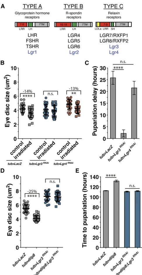
Based on the structural similarities between Dilp8 and relaxin proteins, we sought to determine whether Dilp8 activity is dependent on a Drosophilarelaxin receptor homolog. Dro-sophilahas four LGR proteins, of which only Lgr3 and Lgr4 share structural homology with the type C relaxin receptors (Figure 1A) (Van Hiel et al.2014). Lgr3 and Lgr4 have re-cently been shown to be expressed in many tissues through-out larval development (Van Hiel et al. 2014). To test whether these Drosophila relaxin homologs are necessary for growth coordination or developmental delay during the regeneration checkpoint, we ubiquitously expressed UAS-driven RNAi transgenes against each of the two receptors using tubulin-Gal4. We then activated the regeneration checkpoint in these larvae through targeted irradiation, ducing damage in posterior tissues of the larvae while pro-tecting anterior tissues like the eye imaginal discs and the PG (seeMaterials and Methodsand Jaszczaket al.2015). Follow-ing posterior irradiation, the growth of anterior tissues is normally reduced due to Dilp8-dependent growth coordina-tion (Jaszczaket al.2015). RNAi inhibition ofLgr3, but not Lgr4, reduces checkpoint growth inhibition, restoring the growth of undamaged tissues in larvae with targeted irradi-ation (Figure 1B), and also reduces checkpoint delay (Figure 1C). This was confirmed with a secondLgr3-targeting RNAi transgene (JF03217), as well a third RNAi-expressing line that targets distinct sequences inLgr3(HMC04196) (Figure S1, A and B). Additionally, we tested RNAi targeted to the otherDrosophilaLGR genes. We found that neitherLgr1nor Lgr2depletion reduced damage-induced growth inhibition or developmental delay (Figure S1, C and D), suggesting that they do not mediate Dilp8 activity. However, we did observe that expression of eitherLgr1orLgr2RNAi produced a sig-nificantly longer delay following irradiation than in control larvae (Figure S1D). Therefore, these genes may play other roles in the regulation of developmental timing.
Expression of Dilp8 alone, in the absence of damage, is sufficient to induce growth restriction and developmental delay (Figure 1, D and E) (Colombani et al. 2012; Garelli et al.2012; Jaszczaket al.2015). To test whether Dilp8 de-pends on Lgr3 for these activities, we coexpressed Dilp8 and an RNAi targetingLgr3using thetubulin-Gal4driver. In lar-vae depleted of Lgr3, Dilp8-induced growth inhibition and developmental delay were both rescued (Figure 1, D and E). In this experiment we observed thatLgr3depletion alone increased growth (Figure 1D) in contrast to the controls from the irradiation experiment (Figure 1B). This difference may
Figure 1 TheDrosophilarelaxin receptor homolog Lgr3 regulates Dilp8-mediated growth coordination and developmental delay during the re-generation checkpoint. (A) Comparison of the mammalian (black) andD. melanogaster(blue) LGR protein types. The number above LRR denotes the number of repeats typically found among receptors of that LGR type. 7TM, seven transmembrane domain; LH, long-hinge domain; SH, short-hinge domain. (B) Targeted irradiation to the posterior of the larva inhibits growth of the anterior-undamaged eye imaginal discs (tub.LacZ, irra-diatedvs. control). Systemic expression of Lgr3-RNAi (tub. Lgr3RNAi) rescues growth restriction. Systemic expression of Lgr4-RNAi does not rescue growth restriction. (C) Full irradiation induces a developmental delay (tub.LacZ), which is rescued by systemic expression of Lgr3-RNAi. (D and E) Systemic expression of Dilp8 is sufficient to inhibit imaginal disc growth and developmental delay (tub.dilp8). Systemic expression of Lgr3-RNAi simultaneously with Dilp8 blocks both growth inhibition and Dilp8-induced delay (tub. dilp8; Lgr3RNAi). Growth was measured by mean imaginal disc size from multiple repeated experiments6SD. Ima-ginal disc sample size, left to right: (B)n= 13, 17, 22, 14, 19, 20; (D)
be due to the short periods of chilling used to immobilize larvae during irradiation, which we observed produced some variation in measured growth between experiments. There-fore, comparisons were only made between larvae within individual experimental treatments. Together, these data demonstrate that of theDrosophilaLGR proteins, Lgr3 alone is required for Dilp8-dependent coordination of growth and developmental delay during the regeneration checkpoint.
Lgr3 mediates Dilp8 activation of NOS in the PG and is necessary for growth coordination during the
regeneration checkpoint
To identify tissues where Lgr3 is expressed and thus may re-spond to Dilp8 signaling, we initially examined a collection of Lgr3 enhancer-Gal4 transgenes (Figure S2A) (Pfeiffer et al. 2008). These transgenes allow us to express nuclear-localized b-galactosidase in tissues where Lgr3 regulatory regions are transcriptionally active. Following staining, we observed that these enhancer-Gal4 transgenes express predominantly in the CNS (Figure S2, B–F). Additionally, the enhancer-Gal4 trans-gene 18A01 consistently expresses in both the CNS and PG (Figure 2A;Figure S2, E and G). All PGs analyzed expressed the 18A01 transgene, however the expression was often only observed in a subset of PG cells. An overlapping enhancer re-gion, 17H01, also produced a minority of PG tissues where expression could be observed in a single cell (Figure S2G). Since none of the other transgenes tested produced any detect-able expression in the PG, we concluded that the PG expression observed in 18A01 and 17H01 was specific to these enhancer elements. To determine whether the PG expression of the 18A10 and 17H10 enhancer transgenes reflected expression of endogenous Lgr3 in the PG, we performedin situ hybridiza-tion using a probe that hybridizes to the Lgr3transcript and were able to observe a specific signal in the PG that was not detected with a probe targeted to the sense strand (Figure S2H). Moreover, we observed expression in the PG and the CNS of GFP-tagged Lgr3 (sfGFP::Lgr3) expressed from the na-tive Lgr3 promoter (Figure 2B). Based on these observations, we conclude that Lgr3 is expressed in both the brain and the PG. We have previously reported that Dilp8 coordinates growth through the activation of NOS in the PG (Jaszczaket al.2015), therefore we tested whether Lgr3 is required for growth reg-ulation in the cells that express the 18A01 enhancer-Gal4 transgene. When an Lgr3-targeting RNAi was expressed using the 18A01 enhancer Gal4, growth inhibition of the undamaged imaginal discs does not occur (Figure 2C); suggesting that the 18A01 enhancer expresses in cells that require Lgr3 to produce growth coordination following dam-age. To determine whether Lgr3 activity was specifically required in the PG for growth coordination following dam-age, we examined growth coordination in larvae expressing Lgr3RNAi using the PG-specificphantom-Gal4(Mirthet al. 2005) driver. To ensure that we were exclusively assessing the role of Lgr3 in the PG, we also included a neuron-expressed Gal4 repressor (elav-Gal80). In these larvae, we observed that growth inhibition of undamaged imaginal discs
was substantially reduced when compared to control larvae (Figure 2D). These results demonstrate that Lgr3 activity in the PG is necessary for growth coordination following regen-eration checkpoint activation. Our observation of functional Lgr3 expression in the PG is somewhat surprising given that two recent papers identifying Lgr3 did not observe any ex-pression of Lgr3 in the PG using either a Gal4 exon replace-ment line (Colombaniet al.2015) or a GFP-protein tagged line (Garelliet al.2015). Whereas, we have observed Lgr3-specific transcript in the PG, and observe a loss of growth coordination upon specific knockdown of Lgr3 in the PG. Why these other methods did not detectLgr3expression in the PG is unclear to us. However, it is possible that the alter-ations at the Lgr3locus required to make both of these re-porter constructs may abrogate endogenous expression in some tissues.
We also observed that RNAi depletion ofLgr3in the PG has no effect on the developmental delay produced by activation of the regeneration checkpoint (Figure S3A). This observation is consistent with what we have reported for NOS activity, where
Figure 2 Lgr3 in the PG regulates growth coordination during the regen-eration checkpoint. (A) Expression of nuclear-localizedb-galactosidase in the PG visualized with X-gal staining in 104-hr-AED larva driven by en-hancer 18A01 (18A01.LacZ). PG outlined by red dashes. Bar, 50mm. (B)Lgr3expression is detected in the PG (arrow) and CNS (*) of late third-instar larva. GFP is detected using an anti-GFP antibody (Hoffman La Roche, Nutley, NJ) targeting an N-terminal superfolder GFP-tagged Lgr3 (sfGFP:: Lgr3). Bar, 100mm. (C) Expression of Lgr3-RNAi with the Lgr3-enhancer Gal4 (18A01.Lgr3RNAi) reduces growth inhibition induced by targeted irradiation. (D) Expression of Lgr3-RNAi in the PG while also expressing the Gal4 inhibitor Gal80 in neurons (elav-Gal80, phm. Lgr3RNAi) rescues growth inhibition induced by targeted irradiation. (E) Expression of Lgr3-RNAi in the PG does not significantly affect developmental delay induced by irradiation. Growth was measured as mean imaginal disc size from multiple repeated experiments6SD. Imaginal disc sample size, left to right: (C)n= 35, 23, 27, 26; (D)n= 25, 18, 23, 15. Time was measured as mean of triplicate experiments6SEM.****P,0.001, calculated by two-tailed Student’st-test. See alsoFigure S2.
NOS activation in the PG is necessary for damage and Dilp8-mediated growth inhibition, but not developmental delay (Jaszczak et al. 2015). Therefore, we speculated that Lgr3 might be regulating NOS activity in the PG during the regen-eration checkpoint. To determine whether PG expression of Lgr3 is required for the damage-induced NOS activity, we used thefluorescent reporter molecule DAF2-DA to measure NOS activity through NO production in the PG. Using this assay, we have previously shown that Dilp8 expression is sufficient to induce NOS activation in the PG (Jaszczaket al.2015). After posterior irradiation of larvae, NO production increases in the PG in a Dilp8-dependent manner (Figure 3, A and B). When we express an Lgr3-targeting RNAi in the PG with the phantom-Gal4driver, activation of NOS is no longer detected in the PG following irradiation (Figure 3C). These data demon-strate that Lgr3 activity in the PG is required for NOS activation during the regeneration checkpoint. We have previously shown that NOS is required for Dilp8-mediated growth inhi-bition (Jaszczaket al.2015). To establish that NOS functions downstream of Lgr3, we determined whether artificially in-creasing NOS activity could restrict growth independently of Lgr3 function in the PG. To do this, we overexpressed NOS along with theLgr3-targeting RNAi in the PG using phantom-Gal4. We found that even when Lgr3 is depleted from the PG, NOS is still able to inhibit imaginal disc growth (Figure 3D). Together, these data demonstrate that Lgr3 in the PG functions upstream of NOS, is necessary for NOS activation, and is re-quired for Dilp8-mediated growth control through NOS.
Neuronal Lgr3 activity regulates regeneration checkpoint delay and growth coordination
Since all the Lgr3 enhancer-Gal4 transgenes analyzed express in the CNS (Figure S2, B–F), we wanted to determine if Lgr3 activity in neurons is important for regulating systemic re-sponses to damage during the regeneration checkpoint. In particular, Lgr3 function is essential for developmental delay in response to imaginal disc damage (Figure 1C), but not through its activity in the PG (Figure 2D). To test the neuronal function of Lgr3, we examined larvae that expressed Lgr3 un-der the control of the neuron-specific elav-Gal4 driver. In elav . Lgr3RNAilarvae, irradiation damage produced essen-tially no delay in development (Figure 4A), demonstrating that damage-induced Dilp8 requires Lgr3 function in the brain to regulate developmental timing. Unexpectedly, depletion of Lgr3in neurons also completely eliminated growth coordina-tion following targeted irradiacoordina-tion (Figure 4B). To confirm that the disruption of growth coordination fromelav-Gal4 ex-pression ofLgr3RNAiwas not due to additional expression in the PG, we examined the pattern of elav-Gal4 expression using UAS-GFP and observed no evident expression of GFP in the PG (Figure S3A). This suggests that Lgr3 activity in the brain may function in a separate pathway that is necessary for growth coordination during regeneration. We confirmed this observation using the neuron-specific synaptobrevin-Gal4 (Pauliet al.2008) to expressLgr3-targeted RNAi, which also eliminated growth coordination following targeted irradiation
(Figure S3B). In contrast, Lgr3-targeted RNAi in glial cells usingrepo-Gal4did not rescue growth inhibition or develop-mental delay (Figure S3, C and D), demonstrating that Lgr3 function is required specifically in neurons for growth coordi-nation during the regeneration checkpoint.
Regeneration checkpoint delay is the result of delayed expression of the neuropeptide PTTH (Halme et al.2010), therefore we tested whether Lgr3 might be acting in the PTTH-expressing neurons (McBrayeret al.2007) to directly regulate delay or growth inhibition. However, neither growth nor delay was affected by Lgr3-targeted RNAi expression
Figure 3 Lgr3 in the PG regulates NOS activity during the regeneration checkpoint. (A) Targeted irradiation increases NO production in the PG (lobes of PG outlined in white). Gray, DAPI; green, DAF2-DA. Bar, 100mm. (B) Activation of NO production in the PG after targeted irradiation is lost in larva mutant for Dilp8 (n= 5–10 PGs for each genotype and treatment). (C) Expression of Lgr3-RNAi in the PG blocks activation of NO production after targeted irradiation (n= 5–10 PGs for each genotype and treatment). (D) Overexpression of NOS in the PG (phm. NOS) inhibits imaginal disc growth even when Lgr3-RNAi is also expressed (phm .
specifically in the PTTH-expressing neurons (Figure S3, E and F). Therefore, other neurons expressing Lgr3 are likely com-municating regeneration checkpoint activation to the PTTH-expressing neurons.
Since theLgr3-dependent activation of NOS in the PG is required for growth coordination, we also tested whether NOS is required in the neurons for regulatingLgr3-dependent growth coordination and developmental delay during the re-generation checkpoint. Using aNOS-directed RNAi (Jaszczak et al.2015) expressed in neurons (elav .NOSRNAi) during targeted irradiation, we found that neuronal depletion of NOS did not restore growth to undamaged tissues (Figure S3G) or reduce developmental delay (Figure S3H). This sug-gests that Lgr3 in neurons regulates growth through distinct cellular pathways from Lgr3 in the PG.
Together, these data indicate that Lgr3 is required: (1) in the CNS to mediate the effect of Dilp8 on developmental timing, and (2) inboththe CNS and the PG to mediate Dilp8 effects on imaginal disc growth. To understand the relationship between these two roles for Lgr3 in regulating growth, wefirst sought to determine whether Lgr3 in the CNS is required for growth in-hibition by NOS activation. To do this, we used the heat shock promoter to overexpress NOS, which inhibits imaginal disc
growth by reducing ecdysone production from the PG (Jaszczak et al.2015), while also targeting expression of the Lgr3RNAi to neurons. We found that Lgr3 depletion from neu-rons has no effect on the ability of NOS to inhibit imaginal disc growth (Figure 4C), demonstrating that NOS functions down-stream of Lgr3 in the CNS to regulate imaginal disc growth. We then tested whether CNS Lgr3 functions upstream of NOS to regulate growth. We could determine this by examining the activation of NOS following damage in larvae where CNS ex-pression of Lgr3 is depleted. To do this, we measured NO pro-duction in the PG with the fluorescent reporter DAF2-DA following irradiation damage in control and elav. Lgr3RNAi larvae. After targeted irradiation of larvae, we found that NO production did not increase in the PG whenLgr3-RNAi expres-sion is targeted to the neurons (Figure 4D). This demonstrates that neuronal Lgr3 functions upstream of NOS and regulates the ability of NOS to be activated in the PG. Therefore, Lgr3 in the CNS and in the PG are both required for the activation of NOS to mediate Dilp8 regulation of imaginal disc growth.
Our observations demonstrate that theDrosophilarelaxin receptor Lgr3 mediates the effect of Dilp8 on developmental timing and growth coordination duringDrosophilaimaginal disc regeneration (Figure 4E). In three recently published
Figure 4Lgr3 in neurons regulates developmental delay and also regulates growth coordination during the regen-eration checkpoint through NOS activity. (A) Expression of Lgr3-RNAi in neurons (elav.Lgr3RNAi) largely abrogates developmental delay induced by irradiation. (B) Targeted irradiation of larvae expressing Lgr3-RNAi in neurons (elav.Lgr3RNAi) increases imaginal disc growth in con-trast to the growth inhibition in the control (elav.LacZ). (C) Expression of Lgr3-RNAi in neurons (elav.Lgr3RNAi) does not block NOS inhibition of imaginal disc growth. NOSMac was misexpressed by heat shock activation at 80 hr AED for 40 min in a 37°water bath. (D) Expres-sion of Lgr3-RNAi in neurons blocks activation of NO production after targeted irradiation (n= 5–10 PGs for each genotype and treatment). (E) Lgr3 mediates growth coordination and developmental delay during the regen-eration checkpoint through distinct tissues. Lgr3 in the PG regulates growth coordination, but not delay, through ac-tivation of NOS, which reduces ecdysone production. Lgr3 in the neurons mediates Dilp8 activation of developmental delay and also regulates growth coordination through reg-ulation of NOS activity in the PG. Growth was measured by mean imaginal disc size from multiple repeated exper-iments6SD. Imaginal disc sample size, left to right: (B) n= 44, 39, 37, 32; (C)n= 32, 14, 46, 25. Time was measured as mean of duplicate experiments with 5–10 larvae each6SD.**P,0.01,****P,0.001, calculated by two-tailed Student’st-test. See alsoFigure S3.
studies, researchers have demonstrated that Lgr3 is required in a specific subsets of neurons in the CNS to coordinate the effects of Dilp8 on growth and developmental timing (Colombaniet al.2015; Garelliet al.2015; Vallejoet al.2015). This published work is consistent with ourfindings that neu-ronal disruption of Lgr3 expression is required for growth reg-ulation and developmental delay. Our study here complements and extends thesefindings by demonstrating that (1) the role of Lgr3 in growth regulation and developmental delay are separable through Lgr3 function in the PG; (2) growth regu-lation depends on both Lgr3 activity in the CNSandthe PG; and (3) Lgr3 function in the CNS and in the PG is required for damage-induced NOS activation in the PG, explaining how Lgr3 function in both of these two tissues is necessary for growth coordination.
How Lgr3 activity in both the CNS and the PG coordinate to regulate NOS function is not yet clear from these studies. However, since Lgr3 activity in the CNS is important for extend-ing the regenerative period followextend-ing damage, it is possible that loss of Lgr3 may reduce the capacity to activate the regenerative checkpoint in response to damage, similarly to how damage induced late in larval development no longer elicits regenerative checkpoint delay (Halmeet al.2010). Thus, Lgr3 activity in the CNS may be necessary to maintain the capacity of the PG to respond to damage. This may be mediated by regulation of PTTH neurons or Dilps produced by the insulin-producing cells. Lgr3-positive neurons have been observed to connect to cells expressing both of these PG-regulating signals (Colombani et al. 2015; Garelli et al.2015; Vallejo et al. 2015). Future experiments examining the regulation of these signals should help to determine how Lgr3 activity in the brain and PG is integrated to coordinate growth during regeneration.
Previous understanding of the biological activities of re-laxins and their receptors have been largely restricted to their roles in sexual development and the function of the reproduc-tive organs (Bathgateet al.2013). We demonstrate that Dro-sophila relaxin receptor Lgr3 is necessary for coordinating growth between tissues during a regeneration checkpoint. Re-cently, allele polymorphisms at Lgr8/RXFP2 (the mammalian homolog of theDrosophilaLgr3) has been demonstrated to be an important genetic determinant of relative horn size within a population of wild Soay sheep (Johnstonet al. 2013). This suggests a role for relaxin receptors in regulating growth and organ allometry is likely to be conserved in mammals.
Acknowledgments
We thank M. Dominguez, A. Shingleton, P. O’Farrell, H. Krause, P. Leopold, and M. O’Connor forDrosophilastocks. We thank P. Leopold for helpful conversations on the work in this manuscript; and D. Castle, D. DeSimone, and D.G. Halme for suggestions on the manuscript. This work was funded in part by March of Dimes Basil O’Connor Starter Scholar award to A.H. (#5-FY12-60), and the National In-stitutes of Health to A.H. (R01 GM-099803) and to J.S.J. (T32 GM-008136).
Literature Cited
Barker, N., S. Tan, and H. Clevers, 2013 Lgr proteins in epithelial stem cell biology. Development 140: 2484–2494.
Bathgate, R. A, M. L. Halls, E. T. van der Westhuizen, G. E. Callander, M. Kocanet al., 2013 Relaxin family peptides and their receptors. Physiol. Rev. 93: 405–480.
Cáceres, L., A. S. Necakov, C. Schwartz, S. Kimber, I. J. H. Roberts
et al., 2011 Nitric oxide coordinates metabolism, growth, and development via the nuclear receptor E75. Genes Dev. 25: 1476–1485.
Colombani, J., D. S. Andersen, and P. Leopold, 2012 Secreted peptide Dilp8 coordinates Drosophila tissue growth with devel-opmental timing. Science 336: 582–585.
Colombani, J., D. S. Andersen, L. Boulan, E. Boone, N. Romero
et al., 2015 Drosophila Lgr3 couples organ growth with mat-uration and ensures developmental stability. Curr. Biol. 25: 2723–2729.
Garelli, A., A. M. Gontijo, V. Miguela, E. Caparros, and M. Dominguez, 2012 Imaginal discs secrete insulin-like peptide 8 to medi-ate plasticity of growth and maturation. Science 336: 579– 582.
Garelli, A., F. Heredia, A. P. Casimiro, A. Macedo, C. Nuneset al., 2015 Dilp8 requires the neuronal relaxin receptor Lgr3 to cou-ple growth to developmental timing. Nat. Commun. 6: 1–14. Halme, A., M. Cheng, and I. K. Hariharan, 2010 Retinoids
regu-late a developmental checkpoint for tissue regeneration in Dro-sophila. Curr. Biol. 20: 458–463.
Jaszczak, J. S., J. B. Wolpe, A. Q. Dao, and A. Halme, 2015 Nitric oxide synthase regulates growth coordination during Drosophila melanogaster imaginal disc regeneration. Genetics 200: 1219– 1228.
Johnston, S. E., J. Gratten, C. Berenos, J. G. Pilkington, T. H. Clutton-Brocket al., 2013 Life history trade-offs at a single locus main-tain sexually selected genetic variation. Nature 502: 93–95. McBrayer, Z., H. Ono, M. Shimell, J.-P. Parvy, R. B. Becksteadet al.,
2007 Prothoracicotropic hormone regulates developmental timing and body size in Drosophila. Dev. Cell 13: 857–871. Mirth, C., J. W. Truman, and L. M. Riddiford, 2005 The role of the
prothoracic gland in determining critical weight for metamor-phosis in Drosophila melanogaster. Curr. Biol. 15: 1796–1807. Parker, N. F., and A. W. Shingleton, 2011 The coordination of
growth among Drosophila organs in response to localized growth-perturbation. Dev. Biol. 357: 318–325.
Pauli, A., F. Althoff, R. A. Oliveira, S. Heidmann, O. Schuldiner
et al., 2008 Cell-type-specific TEV protease cleavage reveals cohesin functions in Drosophila neurons. Dev. Cell 14: 239– 251.
Pfeiffer, B. D., A. Jenett, A. S. Hammonds, T.-T. B. Ngo, S. Misra
et al., 2008 Tools for neuroanatomy and neurogenetics in Dro-sophila. Proc. Natl. Acad. Sci. USA 105: 9715–9720.
Stieper, B. C., M. Kupershtok, M. V. Driscoll, and A. W. Shingleton, 2008 Imaginal discs regulate developmental timing in Dro-sophila melanogaster. Dev. Biol. 321: 18–26.
Vallejo, D. M., S. Juarez-Carreño, J. Bolivar, J. Morante, and M. Dominguez, 2015 A brain circuit that synchronizes growth and maturation revealed through Dilp8 binding to Lgr3. Science 350: aac6767.
Van Hiel, M. B., H. P. Vandersmissen, P. Proost, and J. Vanden Broeck, 2014 Cloning, constitutive activity and expression pro-filing of two receptors related to relaxin receptors in Drosophila melanogaster. Peptides 68: 83–90.
Yakubovich, N., E. A. Silva, and P. H. O’Farrell, 2010 Nitric oxide synthase is not essential for Drosophila development. Curr. Biol. 20: R141–R142.