ADRIAN, DEREK EVAN. Maladaptive Pain in the Cat. (Under the direction of Dr. B. Duncan
X. Lascelles and Dr. Mark G. Papich).
Chronic pain affects over 40% of adult humans in the United States. Few new and
effective treatment options are available for these chronic pain sufferers because of a high failure
rate (90%) of new therapeutics, perhaps partly stemming from the use of inappropriate, induced
animal models. Degenerative joint disease (DJD), a chronic inflammatory and painful condition
of the joints, may affect 90% of the 94+ million pet cats in the United States. An estimated 40%
of those cats with radiographic evidence of DJD experience pain and disability secondary to the
condition. This high prevalence of a naturally occurring painful disease, combined with the cat’s
shared environment with humans, makes the cat an attractive model for analgesic research.
Developing the cat as a model requires filling gaps in our knowledge of effective analgesics and
their pharmacokinetics relevant to clinical use, and developing and refining tools for measuring
chronic pain in cats.
First, we identified these gaps in our knowledge by reviewing the current status of tools
to measure chronic (or maladaptive) pain, and pharmacological data surrounding potential
analgesics. These gaps include our ability to detect or monitor pain and disability, in our
knowledge regarding analgesic medications, and an inconsistent application of the currently
available tools in clinical research.
Next, we assessed out ability to detect and measure treatment-associated improvements
in pain and disability via a randomized, double-masked, clinical trial evaluating the efficacy of a
COX-2 selective non-steroidal anti-inflammatory drug (NSAID), robenacoxib, for the treatment
of DJD-associated pain in cats. Our aim was to identify gaps in the translational cat model, and
improve treatment options available to veterinarians. We detected modest improvements in both
activity and owner assessments of pain and disability. Given that NSAIDs are the mainstay of
treatment for DJD across species, we expected more significant results. Although several
explanations for our results exist, this study reinforced our observations that the current
approaches to measuring chronic pain in cats require refinement and improvement, which
analgesics of both veterinary and translational interest. A veterinarian-distributed survey
collected data on respondent demographics, medications prescribed, and typical dosing
regimens. Gabapentin, a medication without either evidence of analgesic efficacy in veterinary
species or pharmacokinetic data relevant to the typical dosing regimen, was prescribed by 71.0%
of respondents. Because gabapentin is used for maladaptive pain conditions in people,
understanding its efficacy in the naturally occurring chronic pain cat model would benefit
veterinary medicine, and would increase our understanding of the translational utility of the cat
model.
We then investigated the pharmacokinetics of gabapentin in cats because data for
long-term dosing was needed. We delong-termined the effects of twice daily oral dosing for two weeks on
the drug’s pharmacokinetics, and evaluated the systemic absorption of a transdermal preparation.
Repeated oral dosing did not significantly affect drug kinetics; therefore, adjustments are not
necessary for chronic administration. We also determined that the transdermal preparation used
was poorly absorbed and therefore not a viable dosing strategy.
Our final study aimed to develop and evaluate the feasibility, repeatability, and
sensitivity of a novel, objective measure of nociception in cats. The nociceptive withdrawal
reflex (NWR) test is used in human analgesic research to investigate both central mechanisms of
nociception, as well as medications affecting these central processes. We found testing was
feasible, and occasionally repeatable; however, we were unable to detect differences between
© Copyright 2019 by Derek Evan Adrian
by
Derek Evan Adrian
A dissertation submitted to the Graduate Faculty of North Carolina State University
in partial fulfillment of the requirements for the degree of
Doctor of Philosophy
Comparative Biomedical Sciences
Raleigh, North Carolina
2019
APPROVED BY:
_______________________________ _______________________________ Dr. B. Duncan X. Lascelles Dr. Mark G. Papich
Committee Co-Chair Committee Co-Chair
_______________________________ _______________________________ Dr. Samuel Jones Dr. Ronald Baynes
_______________________________ _______________________________ Dr. John Harris Dr. Margaret Gruen
DEDICATION
This dissertation is devoted to my wife, my family, and my friends, for their continued
BIOGRAPHY
Derek Adrian is originally from Durham, North Carolina. He earned a Bachelor of
Science in Zoology from North Carolina State University in Raleigh, NC to prepare for
veterinary school. He also obtained his Doctorate in Veterinary Medicine from North Carolina
State University College of Veterinary Medicine (NCSU CVM). Following this, he worked as
both a general practitioner in veterinary private practice and as a clinical trial investigator at the
NCSU CVM. He remained at the NCSU CVM for his graduate studies. His academic interests
include pharmacology, chronic pain and degenerative joint disease in companion animals,
ACKNOWLEDGMENTS
I would like to thank my committee co-chairs, Duncan Lascelles and Mark Papich, for
their continued mentoring, guidance, teaching, and time during my research. Without their
dedication to me, I would not have been able to complete these works. My other committee
members, Ronald Baynes, Sam Jones, and John Harris all heavily contributed to my research,
representing the diversity of backgrounds, experience, and knowledge I found necessary to draw
on during these years. Their willingness to read numerous drafts, analyze unending amounts of
data, and direct me both personally and educationally were vital to my progress.
Robert Reed and Jane Owens provided mentoring and teaching throughout my training
and career, which I am thankful for.
I would also like to thank both Andrea Thomson and Lyndy Harden-Plumley for their
repeated assistance during these projects- their skills and knowledge were invaluable. I must also
thank the entirety of the Translational Research in Pain lab, including Morika Williams,
Constanza Meneses-Evans, Bea Belda-Lopez, Masataka Enomoto, Carrie Muller, Margaret
Gruen, Samuel Chiu, Laura Minema, Jonathan Hash, Rachel Meyers, Lauryn Braxton, and Kayla
Freeman for their collaboration, feedback, and comradery.
I must also thank those that assisted in my various projects, including Delta Dise, Jim
Yeatts and Danielle Mzyk for their expertise and guidance with pharmacokinetic analysis and
mass spectronomy. I must also thank the entirety of the Central Procedures Lab and Laboratory
Animal Research for their care and protection of the cats that participated in my studies, and for
their assistance coordinating my research. I thank Janice Harvey and Brigid Troan for lending
me their histopathology expertise. I am also thankful for Jonathan King, Rudolph Parrish,
Stephen King, Steven Budsberg, and Gabriella Sandberg, who were all instrumental in our
clinical trial. I also thank Jo Murrell and Vicki Simmonds for their training and guidance
regarding the NWR test. This research also would not have occurred without all of the cats, cat
owners, and veterinarians that participated- Thank you.
Lastly, I must thank all of my friends and family who supported me throughout these
endeavors. I must especially thank Mandesa, who pushed me to be my best both personally and
professionally all of these years.
TABLE OF CONTENTS
LIST OF TABLES ... ix
LIST OF FIGURES ... xi
Chapter 1 Foreword ... 1
The Unmet Need of Chronic or Maladaptive Pain ... 1
The Spontaneous Feline DJD-Associated Pain Model ... 2
Subjective Assessment Tools ... 2
The Placebo Response in Feline DJD ... 7
Objective Assessment Tools ... 7
Measures of Somatosensory System Function ... 9
Treatment of Chronic Maladaptive Pain in Cats with DJD-Associated Pain ... 12
Chapter 1: Introduction - Chronic maladaptive pain in cats: A review of current and future drug treatment options ... 13
Abstract ... 14
Introduction ... 15
Methods... 15
Chronic Maladaptive Pain... 15
Assessment of Chronic Pain in Cats ... 23
Treatment of Maladaptive Pain in Cats ... 25
Gabapentin ... 26
Tramadol ... 28
Amantadine ... 29
Amitriptyline ... 31
Flupirtine ... 32
Tapentadol... 33
Maropitant ... 33
Future Medications in Development ... 34
Grapiprant ... 34
Anti-Nerve Growth Factor Antibodies ... 35
Conclusion ... 36
Declaration of Interest... 37
Acknowledgements ... 37
Chapter 1 Afterword ... 38
References ... 40
Chapter 2: Evaluation of the efficacy and safety of robenacoxib for the treatment of degenerative joint disease-associated pain and inflammation in cats: A randomized and masked clinical trial ... 53
Introduction ... 54
Materials and Methods ... 55
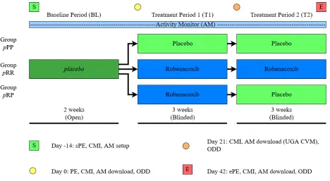
Study Design ... 55
Inclusion/exclusion criteria: ... 57
Outcome Measures and Statistical Model... 58
Results ... 62
Primary Outcome ... 67
Secondary Outcomes ... 71
Safety Measures ... 75
Discussion ... 76
References ... 81
Chapter 3 Foreword ... 84
Chapter 3: Prescribing practices of veterinarians in the treatment of chronic musculoskeletal pain in cats... 85
Abstract ... 87
Objectives ... 87
Methods... 87
Results ... 87
Conclusions and relevance ... 87
Introduction ... 88
Materials and Methods ... 89
Results ... 90
Discussion ... 110
Conclusions ... 113
Acknowledgements ... 113
Author note ... 114
Funding ... 114
Conflict of Interest ... 114
Chapter 3 Afterword ... 115
References ... 116
Chapter 4: The pharmacokinetics of gabapentin in cats ... 120
Abstract ... 125
Background ... 125
Objectives: ... 125
Animals: ... 125
Methods: ... 125
Results: ... 125
Importance: ... 126
Introduction ... 127
Materials and Methods ... 127
Animals ... 127
Instrumentation and Drug Administration ... 128
Gabapentin Analysis ... 130
Pharmacokinetic Analysis ... 132
Results ... 133
Discussion ... 141
Chapter 4 Afterword ... 144
References 145 Chapter 5: Pilot studies developing and evaluating the Nociceptive Withdrawal Reflex test in normal and degenerative joint disease-affected cats ... 148
Introduction ... 149
Materials and Methods ... 150
Control Animals ... 150
Client-Owned Animals DJD-Associated Pain ... 151
Testing... 151
Protocol Development ... 156
Threshold determination ... 157
Temporal Summation (TS) ... 158
Conditioned Pain Modulation (CPM) ... 158
Stimulus Response Curve (SRC) ... 158
Effects of Gabapentin ... 159
Data Extraction ... 159
Data Analysis ... 160
Results ... 162
Temporal summation ... 165
Conditioned Pain Modulation ... 165
Stimulus Response Curve ... 169
Client Specific Outcome Measures ... 169
Effects of Gabapentin ... 170
Repeatability ... 173
Discussion ... 178
References ... 184
Chapter 6: Future Directions ... 189
References ... 194
APPENDICES ... 196
Appendix A: Feline Musculoskeletal Pain Index ... 197
Appendix B: Client Specific Outcome Measures - feline ... 202
Appendix C: Quality of Life, Temperament, and Happiness Assessments ... 209
Appendix D: Robenacoxib Safety Outcome Measures ... 210
LIST OF TABLES Chapter 1
Table 1F.01: Studies validating CMIs in feline analgesic research. ... 5
Table 1F.02: Feline Outcome Measure Cross Validation. ... 6
Table 1.01: Potential therapeutics and their mechanisms for the treatment of maladaptive pain in cats ... 26
Chapter 2 Table 2.01: Treatment group designations and treatments by period. ... 56
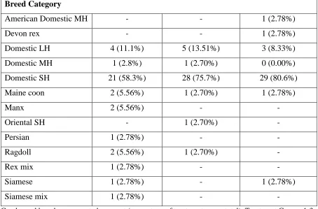
Table 2.02: Patient demographic data. ... 65
Table 2.03: List of early study withdrawals... 67
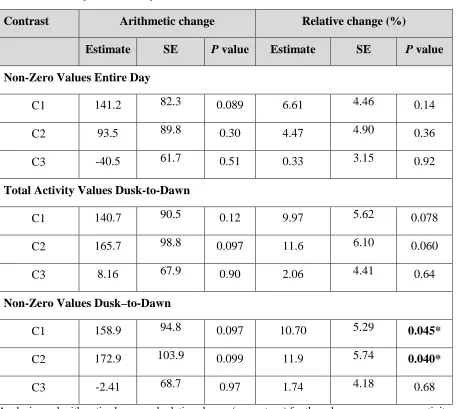
Table 2.04: Contrasts of total activity data between treatment groups. ... 68
Table 2.05: Analysis of activity data subsets. ... 69
Table 2.06: Analysis of within-cat activity data (N=35). ... 71
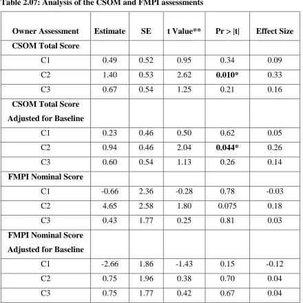
Table 2.07: Analysis of the CSOM and FMPI assessments ... 73
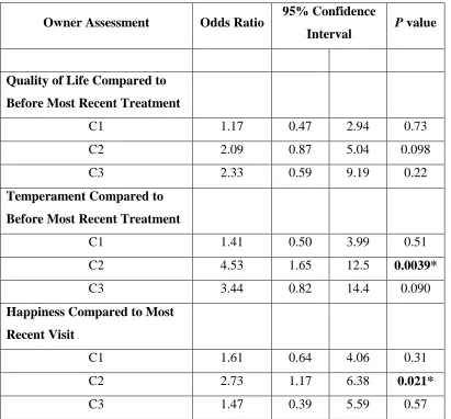
Table 2.08: Analysis of owner based assessments of quality of life (QoL), temperament, and happiness. ... 74
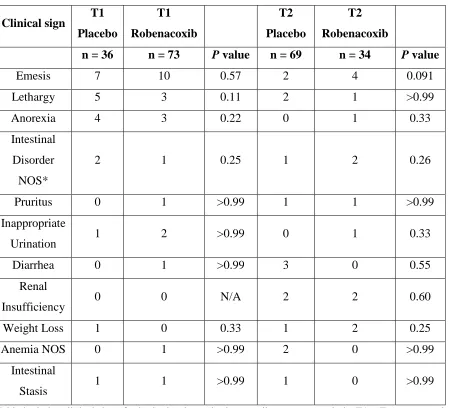
Table 2.09: Summary of adverse event frequencies ... 76
Table 2.10: Comparison of placebo effect sizes across feline CMSD/DJD studies. ... 78
Chapter 3 Table 3.01: Respondent demographic information including gender, nationality, years of experience and feline portion of caseload. ... 92
Table 3.02: Additional respondent demographic information. ... 94
Table 3.03: Rank order percentages of respondents indicating that they prescribed a medication for the treatment of chronic musculoskeletal pain/arthritis in cats. ... 97
Table 3.04: χ2 test for independence, showing where prescription of therapeutics varied significantly with years in practice ... 98
Table 3.05: Prescribing rates of therapeutics by years in practice. ... 99
Table 3.06: Therapeutic prescription frequency by percentage of feline patients. ... 100
Table 3.07: χ2 test for independence showing where prescription of therapeutics varied significantly with percentage of feline patients ... 101
Table 3.08: χ2 test for independence showing where prescription of therapeutics varied significantly between feline-only and all other practitioners. ... 103
Table 3.09: Fisher’s Exact Test of effect of a therapeutic’s prescription on gabapentin prescription frequency. ... 104
Table 3.10: Practitioner therapeutic dose calculation methods. ... 105
Table 3.11: Summary data of typical dosing regimen (including dose, frequency, duration and formulation), as well as percentage of patients prescribed the therapeutic. ... 106
Table 3.12: Ideal and acceptable therapeutic formulations and dosing frequencies, as indicated by respondents. ... 108
Chapter 4
Table 4.01: Summary pharmacokinetics statistics after administration of gabapentin as a single IV bolus (5 mg/kg; n=8), a single oral dose (10 mg/kg; n=7) and repeated oral doses (10mg/kg; n=7). ... 136
Chapter 5
Table 5.01: Baseline demographic information for normal and DJD-pain cats completing the study. ... 163
Table 5.02: Summary of results of NWR testing in both normal (n=6) and DJD-pain cats (n=6). ... 164
Table 5.03: Summary baseline data and results from DJD-pain cats that completed the optional gabapentin phase of the study. ... 170
Table 5.04: Summary of results of NWR testing in DJD-pain cats (n=6), including results for cats completing the optional gabapentin portion of the study (n=3). ... 171
Appendices
LIST OF FIGURES Chapter 1
Figure 1.01: Schematic illustration of Adaptive Pain. ... 19 Figure 1.02: Schematic illustration of Maladaptive Pain... 21
Chapter 2
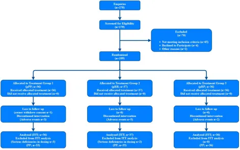
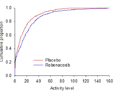
Figure 2.01: Study design and timeline. ... 57 Figure 2.02: Patient recruitment and enrollment flowchart (CONSORT flowchart). ... 64 Figure 2.03: Example Cumulative Distribution Function... 70
Chapter 3
Figure 3.01: Flow diagram of survey logic. ... 90
Chapter 4
Figure 4.01: Gabapentin plasma concentrations after administration as an IV bolus (5 mg/kg). ... 134
Figure 4.02: Gabapentin plasma concentrations after a single oral dose (10 mg/kg) ... 138 Figure 4.03: Gabapentin plasma concentrations after repeated oral dosing (10 mg/kg) ... 139 Figure 4.04: Gabapentin plasma concentrations after single (10 mg/kg; closed circles) and repeated oral dosing (10 mg/kg; open circles). ... 140
Chapter 5
Figure 5.01: Schematic of patient and equipment setup for Nociceptive Withdrawal Reflex recording. ... 154 Figure 5.02: Study Timeline of NWR Testing Sessions. ... 157 Figure 5.03: Example EMG tracing. ... 160 Figure 5.04: Comparison of Normal and DJD-Pain Cat Nociceptive Withdrawal Reflex Test Outcome Measures. ... 167 Figure 5.05: Stimulus Response Curves from normal and DJD-pain cats. ... 169 Figure 5.06: Treatment Effect of Gabapentin on NWR Test Outcome Measures. ... 174
Chapter 1 Foreword The Unmet Need of Chronic or Maladaptive Pain
Chronic pain is a major economic and public health crisis in the United States, affecting
over 40% of adults and costing more than $600 billion annually.1,2 Unfortunately, new and effective treatment options for chronic pain in humans are lacking because of a high failure rate
(90%) among therapeutics entering Phase 1 studies.3 This includes medications such as
Substance P/NK-1 antagonists that were promising in their originating animal models, but failed
to translate into efficacy in humans.4 Poor fidelity or face validity of the animal models and pain assessment tools used during research and drug development may contribute to this failure rate. 5-7 For example, peripheral nerve ligation in the rodent and its resulting hypersensitivity to evoked pain is used to simulate neuropathic pain conditions. Yet the majority of neuropathic pain
patients suffer from spontaneous pain,8 and the major sensory change is hypoesthesia.9 Similarly, acute animal models of osteoarthritis (OA) often include injection of an inflammatory or toxic
substance into the joint space, whereas chronic, traumatic, or idiopathic etiologies are more
common in human patients.10-12 Therefore, to evaluate treatment for chronic pain, a high fidelity model for both drug development and mechanistic exploration of the condition is needed.
Patients with chronic pain may develop secondary central changes in nociception that
lead to facilitated or enhanced pain processing, termed central sensitization (CS).13 Central sensitization is caused by increased synaptic efficiency or decreased inhibition of primary
afferent neurons in the dorsal root ganglion or dorsal horn neurons of the spinal cord, effected by
cytokines (e.g., prostaglandins), neurotransmitters (e.g., Substance P) and glial cells.13 Other patients with chronic pain may have deficient Endogenous Analgesic Systems (EAS).14,15 The EAS sends descending signals that tonically or dynamically dampen noxious input, controlling
the amount or intensity of pain signals reaching the brain. Identification and treatment of these
central nociceptive changes is therefore important for clinical management of chronic pain in
these subpopulations.
Translational chronic pain research could be improved with spontaneous, or non-induced,
Degenerative Joint Disease (DJD), a chronic inflammatory disease of the joints that is
inclusive of OA, affects approximately 90% of all 94+ million pet cats in the United States.21,22 Of these cats, an estimated 40% experience secondary pain and disability (unpublished findings
from 21). Because DJD and chronic pain are both common and spontaneous in cats, the use of this model reduces risks inherent to induced models. Furthermore, people share their
environment with their pet cats, and both species are self-motivated in their activities. There is
extensive research that has developed tools assessing pain and disability secondary to DJD in the
cat.23-25 These tools include owner-based assessments termed clinical metrology instruments (CMIs),23,24,26 and collar-mounted activity monitors that record acceleration and movement data (“accelerometers”; AMs).23,27 Novel study designs have also been implemented to improve the ability to separate treatment and placebo responses.26 Recently, research has aimed to detect signs of CS or altered nociception in cats, including the use of Quantitative Sensory Testing
(QST). 25,28-30
The Spontaneous Feline DJD-Associated Pain Model
Subjective Assessment Tools
Questionnaires termed Clinical Metrology Instruments (CMIs) have become heavily used
in veterinary chronic pain research.31-33 These questionnaires are completed by owners to evaluate the patient’s perceived pain and disability, or impact of disease (e.g., DJD) on the
patient’s daily activities. Two questionnaires have been developed and used in multiple
placebo-controlled clinical study in client-owned cats – the Feline Musculoskeletal Pain Index (FMPI)
and the Client Specific Outcomes Measure (CSOM).
The FMPI is comprised of 17 standardized activities, and asks the owner to rate their
cat’s ability to perform each on a 5-point Likert-type scale with options from “Normal” to “Not
at all.”34 An option for “Don’t know or not applicable” also exists for activities that the owner is unable to assess, or for activities that are not relevant to the cat (e.g., Climbing stairs for a cat
housed in a single-level residence). The CMI includes two questions for the owner to rate their
cat’s preceding and current pain levels. Whereas the FMPI activities are standardized and
unchanging, the CSOM is tailored to each individual cat/owner situation.23 The CSOM requires owners to select three activities that are important for their cat’s quality of life. The cat’s ability
to “Impossible.” These two CMIs may be complementary or even synergistic in a study, by
providing both global (FMPI) and individualized (CSOM) assessments of the patient’s pain or
disability.
A tool focused on determining changed activities or behaviors noticed by owners with
DJD-affected cats, and categorized the findings into four domains: mobility, activity, grooming,
and temperament.35 This tool asked owners to rate any abnormalities on a severity scale of 1 (mild) to 10 (severe), and provided examples of activities for owners to consider during the
assessment. The Montreal Instrument for Cat Arthritis Testing, for use by caretaker/owner
(MI-CAT[C]) requires further refinement and validation.36
Most feline DJD research has included global assessments of quality of life (QoL)
following treatment phases.23,37,38 These global measurements of QoL are included because pain impacts quality of life in addition to disease-specific metrics (e.g., activity counts, jumping) in
humans,39 and therefore should also be measured in analgesic trials for a complete assessment of treatment effects. There is a strong association between unfriendly temperaments and the
presence of DJD in cats.40 Treatment with an analgesic drug is associated with improved temperament.35 The description of “Grumpy on contact with [“other cats” or “other animals including owner”]” was included in its questionnaire,35 leading to the speculation that it may be possible to simplify the measure into patient happiness, and maintain responsiveness. However,
these QoL outcome measures are not validated in the cat.
Assessment of treatment effects using CMIs is especially affected by the high placebo
response in feline analgesic research, highlighting the importance of tool validation with placebo
controlled studies.41 Only the FMPI24,26,34 and CSOM23 have been validated and used in multiple studies.
Validation is especially important for these subjective tools, to ensure that the
questionnaires are reliable and measure the appropriate parameters. Table 1F.01 below
summarizes placebo-controlled, blinded studies that have been used to assess both CMI and
therapeutic. Validation of a CMI does not make it infallible, nor does it convert a subjective
measure into an objective one. Indeed, the FMPI and CSOM are still prone to the significant
placebo responses observed in feline arthritis research.41 One method to combat this risk of placebo response is to take each outcome measure into consideration as part of a whole picture-
increase/improvement in activity measures, the researcher may be more confident that the effect
seen is a true treatment effect. This has been termed “criterion validation,” assuming that
activity, being an objective measure, is the “gold standard.” This is probably not true, and this
approach is better termed cross-validation. The current status of cross-validation of feline
chronic pain CMIs is summarized in Table 1F.02, indicating which studies have shown a
5 CMI = Clinical Metrology Instrument; CSOM = Client Specific Outcomes Measure; FMPI = Feline Musculoskeletal Pain Index;
MI-CAT (C) = Montreal Instrument for Cat Arthritis Testing, for use by Caretaker/owner. CMI
Face Validity and Item Generation
Cronbach’s α Concurrent Validity
Test-Retest Repeatability
Responsiveness Validity
Extreme Group Validation
Criterion Validity Lascelles23
CSOM +
Zamprogno42 FMPI +
Benito24
CSOM +
FMPI + + + +
Gruen31
CSOM +
(FMPI) +
FMPI +
(CSOM) + + +
Gruen43 CSOM + +
FMPI + +
Klinck36
6
Lascelles23 Meloxicam + N/A + + Serial
Lascelles44 Diet + N/A - - Parallel
Gruen31
Meloxicam + - - - Parallel
+ + - - Crossover
Gruen43 Anti-NGF Ab + + + N/A Parallel
Adrian Robenacoxib + - + + Parallel
Guedes37 Tramadol + N/A +* ** Crossover
Guedes38
Gabapentin +
(decreased) N/A +* ** Crossover
Table includes previously reported studies utilizing multiple outcome measures. Activity = Activity Monitors. FMPI = Feline Musculoskeletal Pain Index. CSOM = Client Specific Outcome Measures. QOL = Quality of Life. Design = Study design employed.
+ = significant treatment effects observed. - = significant treatment effects not observed.
The Placebo Response in Feline DJD
While placebo-controlled double-blinded studies have become standard practice, feline
DJD research has experienced a dramatic placebo response rate.41 This meta-analysis found placebo effect sizes as high as 1.93 (95% CI 1.16-2.7), with a corresponding treatment over
placebo effect size of -0.35 (-0.98 to 0.28). There are several potential explanations for this
placebo effect. A caregiver placebo response can occur when an owner reports improvement
following a placebo intervention. Suggested causes for this caregiver placebo response include
the “better care effect,” where caregiver ratings on subjective measures are improved by better
access to healthcare and more frequent follow-up.41 The owner’s desires to please the
investigator or for a trial to be successful can also cause the caregiver placebo response. Subjects
may also experience apparent improvement because of a “regression towards the mean,” or the
natural waxing (when owners seek enrollment) and later waning of clinical signs of pain and
disability. An owner’s expectations of improvement following administration of a treatment
(active drug or placebo), and increased observations of the cat and subsequent altered
interactions can also result in the placebo response.41
Alternative study designs have emerged as one way to diminish the placebo response.
Inclusion of a baseline period between study screening and enrollment may reduce the impact of
regression towards the mean. One alternative to the classic parallel group design is inclusion of a
masked washout period. It was hypothesized if both the treatment and placebo responses were
similar with initiation of a medication or placebo, that owners would be more sensitive to
deterioration following masked withdrawal of the investigational drug. One feline analgesic
study was able to demonstrate this significant deterioration response in cats previously receiving
the NSAID meloxicam.26 This is similar to the RW portion of the Enriched Enrollment Randomized Washout (EERW) study design used in human medicine.45
Objective Assessment Tools
Activity is affected by chronic pain in humans,46 and activity monitors (AMs) have been used and reported in veterinary osteoarthritis research in recent years and offer an objective
comparison of outputs between studies using different monitors. However, several early studies
have evaluated the correlation between AM output and actual patient movement or behaviors,
generally showing AMs as an acceptable proxy for movement.47,50 Although inter-device
variability ,50 and effects of patient conformation51 have been observed in dogs, this has not been reported for cats. Investigators have hypothesized and tested the theory that DJD-associated pain
and disability produces decreased activity in cats as detected by AMs, and activity can be
improved or restored to normal levels with an effective analgesic.23,26,37,43 While these studies have used changes in activity counts as an outcome measure, it is not known what changes are
anticipated after administration of an effective analgesic. It is also possible that effective
analgesics could decrease activity, or result in increased activity via other drug effects (e.g.,
opioid-associated increases in activity in cats). For example, in the aforementioned studies with
gabapentin and tramadol in cats with chronic osteoarthritis,37,38treatment with tramadol
increased activity and gabapentin decreased activity, yet both manuscripts reported effectiveness.
Determining the optimal methods to use to analyze activity data has been hampered by a
relative lack of understanding of the factors influencing activity in cats. Cat activity can be
affected by human presence/interaction, as demonstrated in both research-housed and
privately-housed cats.47,48,52 Variation in owners’ schedules can result in day to day and weekday to weekend effects on cat activity in both normal and DJD-affected cats.48 Cats show a more
pronounced crepuscular pattern of activity during weekdays correlating with owner departures or
arrivals, which becomes muted during weekends when owner presence is more constant.48 Because of high inter-cat variability in activity, some studies have focused on the individual’s
percentage change in activity compared to baseline, instead of using summary values (e.g., mean
activity counts/period time), or absolute value changes.26,43,53 Cats spend a significant portion (up to 70-80%; observation) of the day inactive, suggesting that perhaps analysis should focus on
periods of activity, instead of analyzing total daily activity. Research-purpose cats with naturally
occurring OA (OA-research) were reported to be less active than healthy cats during the
nighttime period,54 with significant increases in activity in these OA-research cats following administration of meloxicam or tramadol alone, but not in combination.28,29
Another approach to data analysis is functional data analysis, which compares patterns of
and DJD-affected cats, healthy cats exhibited more pronounced peaks of activity at times,
compared to a smoother pattern in DJD-affected cats. Future methods of activity data analysis
may focus on time spent in specific “states” of activity (e.g., high vs low intensity).55 Finally, there is interest in developing algorithms or devices that can detect specific activities (e.g.,
climbing stairs, jumping onto furniture, or sleeping) or differences in activity signatures between
normal and DJD-affected cat. Some manufacturer’s websites claim the ability to perform this
analysis, without evidence or validation to support the claim.
Measures of Somatosensory System Function
Quantitative Sensory Testing
In people, QST has demonstrated utility for detecting alterations in pain processing and
sensitivity that can accompany knee and hip osteoarthritis.56,57 This modality applies a thermal (hot or cold), mechanical, chemical, or electrical stimulation to a patient, recording outcomes
such as the threshold for pain detection, or the stimulus intensity or duration required to elicit
withdrawal of the tested limb. The underlying presumption is that central sensitization, or
maladaptive pain, results in lowered thresholds or reflex latencies, and higher sensitivity.
Phenomena such as facilitated temporal summation can also be investigated, by delivery of repeated instead of single stimuli. However, QST is ultimately subjective, as its outcome
measures require either subject-reported pain, or observer-determined withdrawal response.
Patients can be categorized, or phenotyped, based on their responses to the various QST stimuli.
A study evaluating the NSAID etoricoxib in patients with OA found treatment-related effects on
QST outcomes.58
The growing evidence and use of QST in human medicine has led to an interest in the
modality in veterinary species, including cats. Early work in OA-research cats showed reduced
mechanical thresholds when compared to normal cats, as well as shorter latencies of response to
mechanical temporal summation (RMTS).25 Two additional studies in OA-research cats have evaluated RMTS, motor activity, and gait analysis, focusing on treatment effects of tramadol
alone or administered with meloxicam.28,29 While meloxicam treatment increased motor activity (including nighttime activity) and gait metrics, treatment with tramadol (alone or in combination
with meloxicam) resulted in increased nighttime activity and reduced sensitivity to RMTS. It is
results, though mydriasis and euphoria (common opioidergic adverse effects) were noted in
several cats. Finally, repeatability and discriminability of both thermal (hot and cold) and
mechanical threshold testing in client-owned cats with OA compared against normal controls has
been reported.30 While the group found only moderate repeatability between testing sessions, the results did indicate lower mechanical thresholds in OA-affected cats. The results also
demonstrated a lower frequency and shorter duration of paw lifts to cold stimulation in cats with
forelimb OA. This effect was not seen in cats with hind limb OA, nor were there any differences
seen with hot plate stimulation.
The Nociceptive Withdrawal Reflex Test
The Nociceptive Withdrawal Reflex (NWR) test is similar to QST, in that the response to
a noxious stimulus is measured. However, the response of interest with NWR is an
electromyogram (EMG) recorded from a withdrawing muscle, making the modality truly
objective.59 Limb withdrawal is elicited in response to a stimulus, which is typically direct electrical activation of local nerves.59 The withdrawal reflex was first described as a flexion reflex to noxious stimuli in decerebrate cats.59 The test/reflex includes both flexion and extension reflexes occurring to effect limb withdrawal.60,61 The movement produced can be described as the result of recruitment of a collection of reflexes required to withdraw a limb from a noxious
stimulus, dependent on the site of the insult. The NWR test measures the EMG response of one
or more of these reflexes. Typically, the area under the curve (AUC) or integral of the rectified
(absolute value) response is used to measure the EMG response. The distinct conduction
velocities and subsequent separation of motor responses of the myelinated A and unmyelinated C
neurons also allows focused evaluation of differential effects of treatments or pathologies on
either neuron population.62-64 The “early” response seen on EMG tracings (latency varies by species) correlates with Aβ (non-noxious and noxious) and Aδ (noxious) fiber stimulation, while the “late” or second response is produced by C fiber stimulation.
NWR was first described over a century ago in spinalized or decerebrate dogs and cats. It
is used in CNS-intact animals and humans, whether awake, sedated, or anesthetized.15,65-69 The NWR can diagnose and measure CS.68,70,71 Increased synaptic efficiency or altered neuronal excitability seen with CS facilitates the reflex arc, and can result in decreased response
(TS), or the phenomenon where an increasing response is observed when a stimulus of
unchanging intensity is repeatedly delivered, can be facilitated with CS, resulting in a greater
total EMG area to fewer stimuli in affected subjects,72 and increased pain in response to repeated stimuli, compared to the non-CS state.13
The modality can also explore the functionality of the endogenous analgesic system via
Conditioned Pain Modulation (CPM) or Diffuse Noxious Inhibitory Control (DNIC) testing,
where a “conditioning” stimulus is delivered to a remote limb prior to or during delivery of the
original NWR testing stimulus. 15,68,69,73,74 Both DNIC and CPM include a testing stimulus (e.g., noxious heat, cold, chemical, electrical, or mechanical stimuli) measured before and after
application of a conditioning stimulus (any of the above stimuli) at a distant body site/limb from
the testing stimulus.75 Human and animal testing uses different terminology because of the previous research and available information. DNIC was used in both animals and people
originally because of the similarity of testing paradigms, despite the differences in recording sites
and patient manipulations (e.g., spinal transection was only performed in animal subjects).
Separate terms are now used because animal-based research explored and elucidated specific
mechanisms and pathways, whereas human research evaluated the net clinical effects of
numerous pathways.75 The term CPM will be used in this thesis because of the clinical and translational nature of the research in spinally-intact cats. With an intact EAS, the conditioning
stimulus is expected to result in a reduced or ablated response to the NWR testing stimulus due
to conditioning stimulus-activated descending analgesic mechanisms projecting to the level of
the spinal cord.
The NWR test has explored differences between normal and affected individuals,
including subjects with acute induced pain and inflammation, those with chronic OA, and even
human patients with cluster headaches, by measuring CS and EAS.15,69,71,73,76-79 People with pain (acute or chronic) have facilitated withdrawal reflexes or facilitated nociceptive processing (CS),
in addition to less functional EAS. NWR testing may be more invasive or require more advanced
equipment than QST, but it objectively evaluates both spinal (CS) and supraspinal (EAS)
mechanisms that contribute to nociception. NWR testing examines the pathophysiology
associated with pain conditions, and can help define the pain phenotype of the individual.
An individual’s “pain phenotype,” or the underlying pathophysiology of their chronic
pregabalin, are effective for treating some patients with CS, such as those with fibromyalgia.13,80 The gabapentinoids exert their action via binding to and inhibiting of trafficking of the α2δ-1 subunit of voltage-gated calcium channels. These channels become upregulated in the dorsal root
ganglion and dorsal horn of the spinal cord in models of neuropathic pain, which coincides with
facilitated pain transmission and central sensitization. 81,82 Increased activity of α2δ-1 subunits on Ca2+ channels produces greater calcium influx in response to an action potential, leading to greater neurotransmitter release and increased synaptic transmission of signals.83
Patients with a dysfunctional EAS may be less capable of muting or blunting nociceptive
signals. The EAS consists of projections from the rostral ventral medulla and dorsolateral pons,
descending to the dorsal horn of the spinal cord, exerting its actions via α2 adrenoreceptors and 5-HT-ergic receptors in the superficial layers of the dorsal horn.84 Duloxetine, a serotonin and norepinephrine reuptake inhibitor, has been successfully used for treatment of chronic pain
conditions because it potentiates this EAS system.85 EAS efficiency and response to duloxetine is correlated as measured using the NWR test, with EAS-deficient patients experiencing a greater
improvement in their reported pain.86
Thus, it becomes fully apparent that a patient’s “pain phenotype” should be considered
when treating chronic or maladaptive pain, because the underlying phenotype may determine
which analgesic drugs will be most effective. NWR testing has been proposed as one method to
define the individual patient phenotype, and hence was an important focus of our work.
Treatment of Chronic Maladaptive Pain in Cats with DJD-Associated Pain
The following manuscript served as a literature review of chronic or maladaptive pain in
the cat. The manuscript focused on tools for the condition’s detection and measurement, and
Chapter 1: Introduction - Chronic maladaptive pain in cats: A review of current and future drug treatment options
Published (in final format) in The Veterinary Journal As submitted to The Veterinary Journal
Original Citation: The Veterinary Journal. Dec 2017; 230:52-61. DOI: 10.1016/j.tvjl.2017.08.006. Epub 2017 Aug 20
Chronic maladaptive pain in cats: A review of current and future drug treatment options Derek Adriana, Mark Papichb, Ron Baynesc, Jo Murrelld, B. Duncan X. Lascellesa,e,f,*
a Comparative Pain Research and Education Centre, Department of Clinical Sciences, College of Veterinary Medicine, North Carolina State University, Raleigh,
NC, USA
b Molecular and Biomedical Sciences, College of Veterinary Medicine, North Carolina State University, Raleigh, NC, USA
c Population Health and Pathobiology, College of Veterinary Medicine, North Carolina State University, Raleigh, NC, USA
d School of Veterinary Sciences, University of Bristol, Bristol, UK
e Center for Pain Research and Innovation, UNC School of Dentistry, Chapel Hill, NC, USA f Center for Translational Pain Research, Department of Anesthesiology, Duke University, Durham, NC, USA
* Corresponding author at: Comparative Pain Research and Education Centre, Department of
Clinical Sciences, College of Veterinary Medicine, North Carolina State University, Raleigh,
NC, USA. E-mail addresses: [email protected], [email protected] (B. D.X.
Lascelles).
Abstract
Despite our increasing understanding of the pathophysiology underlying chronic or
maladaptive pain, there is a significant gap in our ability to diagnose and treat the condition in
domestic cats. Newer techniques being used to identify abnormalities in pain processing in the
cat include validated owner questionnaires, measurement of movement and activity, and
measurement of sensory thresholds and somatomotor responses. While some data are available
evaluating possible therapeutics for the treatment of chronic pain in the cat, most data are limited
to normal cats. This review details our current understanding of chronic or maladaptive pain,
techniques for the detection and measurement of the condition and the associated central nervous
changes, as well as an overview of the data evaluating potential therapeutics in cats.
Introduction
While cats have become a very popular pet worldwide- with an estimated 75+ million in
the US alone, the assessment and treatment of pain in cats has lagged behind that of dogs 87. Though this knowledge gap is diminishing, most information on pain control in cats exists
regarding peri-operative analgesic use, 88-91 with chronic pain conditions still being undiagnosed and under-treated 87,92,93. Chronic pain situations typically don’t have easily identifiable inciting incidents and the behavioral changes develop slowly and are often subtle. This makes
measurement of chronic or long-standing pain conditions difficult, and although recent progress
has been made in the development of tools to assess chronic pain 24,31,42,94 our ability to measure chronic pain lags behind that of acute pain in veterinary species. The relative lack of validated
methods of chronic pain assessment contributes to our inability to assess efficacy of analgesics
for the alleviation of such pain in cats. This review details our current understanding of chronic
or maladaptive pain, techniques for the detection and measurement of the condition and the
associated central nervous changes, as well as an overview of the data evaluating potential
therapeutics in the cat.
Methods
The literature review was performed by searching on several databases, including
PubMed, CAB Abstracts, and Google Scholar. Specific search for medications of interest were
based on: personal experience, use, or knowledge, anecdotal reports of use or efficacy,
recommendations and guidelines for the treatment of pain in cats, medications being currently
researched, etc. Keywords used included: pain, chronic pain, maladaptive pain, feline, feline
pain, osteoarthritis, degenerative joint disease, analgesics, pharmacokinetics, efficacy, etc.
Chronic Maladaptive Pain
Chronic pain has been defined in human medicine as any pain that lasts more than 3-6
months 95, but the relevance of this timeline to veterinary species with considerably shorter lifespans should also be considered. Different disease conditions like cancer may also affect the
timeline, as it may not be prudent to “delay” treatment, or pathologies where the normal healing
and recovery period is expected to be much shorter. This difficulty in clearly demarcating the
acute and chronic pain are actually on a continuum, and alternative definitions may be more
useful in the context of understanding pain and how to treat it 96. Recently, the terms ‘adaptive’ and ‘maladaptive’ have been suggested as terms that better describe pain (Figures 1 and 2).
Adaptive pain encompasses both nociceptive and inflammatory pain 96. Nociceptive pain is only activated by high-threshold noxious stimuli, including stimuli that cause tissue injury.
Inflammatory pain occurs after tissue damage and produces heightened sensitivity of the tissue
associated with a classical inflammatory response. Both of these types of pain are considered
protective, or ‘adaptive’ pain in that they serve to sense and/or avoid actual or potential tissue
damage. These typically have an easily identifiable cause (surgery, injury, etc.), and are
reversible. Maladaptive pain, on the other hand, is not protective, and is primarily due to plastic
changes in the pain processing system. It can be further divided into neuropathic pain, which is
pain resulting from direct damage to neural tissue, and functional pain, where there are no neural
lesions or inflammation, and pain is driven by dysfunction or malfunction of the nociceptive
system. Classically, neuropathic pain is thought of as resulting from gross, obvious damage to
the spinal cord, or obvious damage to peripheral nerves such as with peripheral nerve sheath
tumors or surgical trauma. However, increasingly it is recognized that many diseases, such as
osteoarthritis (OA) and cancers, may involve a degree of peripheral neuropathy via either direct
damage to nerve endings present in the tissues, or via increased innervation that accompanies
joint remodeling and angiogenesis 97-100. This explains the neuropathic pain-like symptoms reported in many human patients with OA. Similarly, the obvious example of functional pain is
phantom limb pain or fibromyalgia– there is no evidence of a peripheral lesion or inflammation,
yet there is increased sensitivity to stimuli and spontaneous pain. Yet increasingly, it is
recognized that many conditions, such as OA, have a component of functional pain – changes in
the central nervous system function that heightens sensitivity or results in spontaneous pain. It
has been previously suggested that there is a central or maladaptive drive to pain in a significant
portion (20-40%) of human patients suffering from osteoarthritis-associated pain 100,101. This underscores the importance of understanding the driving factors of a patient’s pain, as one
patient may suffer from multiple types.
Central to the concept of maladaptive pain is the phenomenon of central plasticity (also
is the global response that lasts autonomously after the conditioning (original) stimulus has been
discontinued, or is sustained with low level nociceptor input from the periphery 13. This results in a stronger painful reaction to a less intense (hyperalgesia) or previously innocuous stimulus
(allodynia), and increases in the receptive fields of neurons, or the region of tissue that a neuron
functionally innervates and responds to stimuli in. Central plasticity is driven by changes at
various levels of the sensory transmission axis – primary afferent fiber, spinal cord and higher
centers. In general, the processes driving central plasticity are a combination of increased
neuronal excitability, facilitated synaptic transmission and decreased inhibitory influences 13. However, as well as a gain in function, some processes are down-regulated (loss of gain) and so
the term central plasticity is preferred over central sensitization.
Clinical long-standing pain (chronic pain) is a complex mixture of adaptive
(inflammatory) and maladaptive (neuropathic, functional) pain. It is likely that different
neurobiological processes are responsible for the different components of long-standing pain, but
it is also likely that there is tremendous overlap. Most information about the processes involved
in the maladaptive component of long-standing pain have been derived from work in rodents,
using models of neuropathic pain. A multitude of mechanisms play varying roles in maladaptive
pain states, and a laudable clinical goal would be to be able to understand the mechanisms
responsible for pain in an individual, and so make informed choices about analgesics. Currently
it is impossible to predict the mechanisms responsible for the pain state in individual patients,
however, progress is being made in this area, with recent studies in humans testing the function
of the endogenous analgesic mechanisms to predict response to analgesics 86,102.
Most chronic diseases that are associated with pain consist of several different pain
components, including both an active, sustained inflammatory component (as in degenerative
joint disease, gingivostomatitis, and others) as well as the maladaptive pain with associated
neuronal changes and sensitization 13,103,104. Although is it not easy to clinically recognize inflammatory versus maladaptive pain states, there is increasing recognition that many common
long-standing diseases are associated with central plasticity, and so maladaptive pain. Indeed, it
was recently shown that dogs with OA have measureable central sensitization indicative of
maladaptive pain 105. Commonly occurring diseases that are possibly associated with a
component of maladaptive pain in the cat include osteoarthritis and degenerative joint disease,
glaucoma, chronic anterior uveitis), dermatological conditions (including chronic infections,
Figure 1.01: Schematic illustration of Adaptive Pain.
Nociceptive Pain - A noxious stimulus (red starburst) activates high-threshold nociceptive primary
afferent sensory neurons (red/yellow line) with cell bodies in the dorsal root ganglion (DRG), and
termination in the dorsal horn (DH). Here, the afferent signal is transmitted to the second order neuron via mono- or multi-synaptic processes, and crosses over to the other side of the spinal cord, then transmitted to the brain via ascending tracts in the spinal cord (red arrow), where it is interpreted as a warning of actual or potential tissue damage. There is tonically active descending inhibition (green line) from the CNS (channeled via the rostro-ventromedulla) that helps control whether the information from the primary afferent neuron is blocked at the level of entry into the DH of the spinal cord.
Inflammatory Pain - Local tissue damage results in release of inflammatory mediators, recruitment of
inflammatory cells and further release of inflammatory mediators. These mediators either sensitize sensory nerves, or directly stimulate them, resulting in a lowering of thresholds in sensory nerves and generation of action potentials (nociceptive signals). These signals are carried by afferent neurons (red line) with cell bodies in the DRG and terminals in the DH. As before, ascending fibers carry the
Figure 1.02: Schematic illustration of Maladaptive Pain.
Neuropathic Pain–Physical damage to nervous system tissue (e.g. in this case, a tumor - yellow circle)
results in very abnormal activation of nociceptor sensory neurons – they become activated in response to previously sub-threshold stimuli (blue circle). The subsequent pathway is as described in ‘Adaptive’ pain, but at the level of the Dorsal Root Ganglion (DRG) and the dorsal horn of the spinal cord there are changes (nervous system plasticity) resulting in amplification of the signals and facilitation of throughput of the signals. Additionally, the tonically active descending inhibition is less effective (illustrated as a dashed green line), which again facilitates the signals being transmitted from the periphery to higher centers. Hypersensitivity (increased pain from a stimulus that normally provokes pain) and allodynia (pain due to a stimulus that does not normally provoke pain) occur as a result of these changes, and in addition, spontaneous pain can occur due to abnormal activity in the nervous system (e.g. generated at the site of nervous system injury). A hallmark of ‘neuropathic pain’ is the presence of actual physical damage to part of the nervous system and it is this that drives the changes in the way the system functions. Functional Pain–In Functional Maladaptive pain, the nervous system is grossly normal – there is no
physical damage of the system. However, the functioning of the system is abnormal. This abnormal central processing results from repeated input to the system, causing nervous system plasticity (changes in neurons and changes in the way supporting elements [e.g. microglia] communicate with neurons) and thus amplification and facilitation of the processing of nociceptive information. Under these conditions, a previously sub-threshold stimulus (blue circle) activates a physically normal nociceptor (red line) but abnormal central processing in the spinal cord or brain (inset) results in the stimulus being interpreted as painful. As with neuropathic pain, descending inhibition may be defective (dashed green line).
It is important to clarify that chronic pain can exist on a continuum, and can in fact be
comprised of multiple driving mechanisms. In some chronically painful conditions, the driving
condition may start and remain as inflammatory pain, with an easily understood coupling of
peripheral disease with degree of pain. In these painful conditions, nonsteroidal
anti-inflammatory drugs (NSAIDs) are expected to be effective. However, it is likely that in many
cases, the ongoing nociceptive input into the nervous system, along with damage to nerve
endings as a result of the peripheral disease process can cause changes in the central nervous
system and therefore produce maladaptive pain. It is this maladaptive component that makes
chronic pain difficult to treat. Hence the search for novel, non-NSAID therapies that can be used
along with, or in place of, NSAIDs. At this moment, we are limited in that we cannot clinically
differentiate between maladaptive pain, and pain with a purely inflammatory drive.
Assessment of Chronic Pain in Cats
Recently, progress has been made in the assessment of chronic pain in the cat using
owner questionnaires, called Clinical Metrology Instruments (CMIs). The two most studied
CMIs are the Client Specific Outcome Measures (CSOM) and Feline Musculoskeletal Pain Index
(FMPI) 23,24,31-33,44.
The objective measurement of movement or activity also have been developed as
methods to assess the impact of chronic/maladaptive pain and its treatment. Recently, activity
monitors that record changes in acceleration associated with movement have been used as an
objective outcome measure of mobility in cats 26,44,54. Cats are fitted with a small accelerometer on a collar or harness, and allowed to move about normally in their home environment. This the
tool can discriminate between normal and affected research cats 54, and can even be used to show treatment effects in client-owned affected cats 23,26,27,43,44.
To understand the mal-function of the somatosensory system present with maladaptive
pain, methods evaluating sensorimotor function are needed. Quantitative sensory testing (QST)
involves the measurement of the stimulus (mechanical, thermal hot/cold, etc.) strength or
frequency of application required to elicit a withdrawal or response (e.g. head turn, limb
withdrawal) by the patient, with the end of the test usually determined by observation of the
increasing response to repetitive stimuli), etc. 25. While QST is in its early development in cats, it can discriminate between healthy, non-affected cats, and those with OA 25. Other methods such as the measurement of Nociceptive Withdrawal Reflexes (NWR) have been explored in dogs, but
not yet in cats 65,107. NWR Testing measures the magnitude of the withdrawal responses to various stimuli using EMG. This modality can evaluate the threshold to elicit withdrawal, in
addition to the effect on withdrawal latency and magnitude after delivering repeated stimuli
(temporal summation). Data produced are objective, as opposed to the semi-objective QST
methodology. NWR testing is proposed to measure central plasticity and associated changes in
pain processing, and affected patients are expected to have lower thresholds and higher or
stronger EMG responses.
Overall, our ability to accurately measure chronic pain is limited, and our ability to
measure the maladaptive component of this pain is even more restricted. As a result, diagnosis
Treatment of Maladaptive Pain in Cats
In North America, there are no drugs approved for long-term use in cats with maladaptive
pain, and only one NSAID (meloxicam) is approved for long-term use in some parts of the
world. Despite recent information suggesting that NSAID therapy can partly reverse central
plasticity 58, it is generally accepted that the maladaptive component of pain conditions is poorly responsive to NSAIDs 102. Because there are also concerns around the potential for adverse effects from NSAIDs, interest in alternative drug therapy has emerged. Currently, drug choices
are based on experience in people, or because of their activity on mechanisms shown to be
important in rodent models of maladaptive pain. Medications that have been suggested for use in
cats for the treatment of maladaptive pain are gabapentin, tramadol, amantadine, amitriptyline,
tapentadol, flupirtine and anti-nerve growth factor antibodies (Table 1). This review outlines
what is currently known about non-NSAID drug treatments that may be effective for chronic or
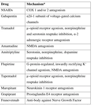
Table 1.01: Potential therapeutics and their mechanisms for the treatment of maladaptive pain in cats
Drug Mechanism*
NSAIDs COX 1 and/or 2 antagonism
Gabapentin α2δ-1 subunit of voltage-gated calcium channels
Tramadol µ-opioid receptor agonism, norepinephrine and serotonin reuptake inhibition, α-2 adrenergic receptor antagonism
Amantadine NMDA antagonism
Amitriptyline Serotonin, norepinephrine, dopamine
reuptake inhibition
Flupirtine G-protein-regulated, inwardly rectifying K+ channel agonism, NMDA antagonism
Tapentadol µ-opioid receptor agonism, norepinephrine
reuptake inhibition
Maropitant Neurokinin 1 receptor antagonism
Grapiprant Prostaglandin E4 receptor antagonism
Frunevetmab Anti-body against Nerve Growth Factor
* Generally accepted mechanism of action. There may be differences in the cat for some of the drugs dependent on metabolism
Gabapentin
Gabapentin is an analogue of the neurotransmitter γ-Aminobutyric acid 108,109.
Gabapentin exerts its effects on voltage-gated calcium channels, which are found on excitatory
cells such as neurons. These channels respond to depolarization currents by allowing the influx
of calcium ions 83. Four subunits of calcium channels have been identified, the main pore-forming α1 subunit, and the accessory α2δ, β, and γ subunits 83. Models of neuropathic pain have demonstrated an increase in the α2δ-1 subunit in dorsal root ganglion (DRG) and dorsal horn neurons 81,82. This subunit and binding target for gabapentin is responsible for guiding or
in altered neuronal excitability and pain processing 81-83,109. This binding results in a decrease in the influx of calcium ions in response to an action potential, and therefore decreased
neurotransmitter release or neuronal excitability. Gabapentin has been advocated for the
treatment of neuropathic pain in veterinary species because of experience treating neuropathic
pain in humans 80,108,110,111. In people it is only approved for post herpetic neuralgia, and as an adjunctive therapy for partial onset seizures, which are undocumented syndromes in animals.
The pharmacokinetics of oral (10 mg/kg) and intravenous (4 mg/kg) gabapentin in 6 adult
spayed female cats has been described 112. While bioavailability varied greatly (range: 49.6 – 118.3%), potentially partially due to ad libitum feeding, the half-life after oral administration was
approximately 3 hours (177 + 25 min), with peak concentrations (Cmax) values ranging from 4.6– 10.6 µg/mL 112. Previously reported data and modeling suggests a half maximal effective concentration (EC50) ranging from 1.4 and 16.7 µg/mL for treatment of hyperalgesia in the rat 110,113,114 and an EC50 of 5.4 µg/mL was estimated for humans with neuropathic pain 115. The authors then suggested a dosing regimen of 8 mg/kg every 6 hours for an antihyperalgesia effect
in the cat 112. However, caution is urged when extrapolating effective concentrations of the drug in cats based on pharmacokinetic-pharmacodynamic data from other species. There is also a
current lack of information on pharmacokinetics after repeated dosing. Minimal or no plasma
protein binding has been reported in other species, however this should be confirmed in the
cat116.
Currently, there are no clinical studies evaluating the efficacy of gabapentin in chronic
pain conditions in cats. In a study evaluating the effects of gabapentin on nociceptive thermal
thresholds in research cats 117, six female spayed adult cats received four dosages of oral
gabapentin: 0 (placebo), 5, 10, and 30 mg/kg in a crossover design. Peak plasma concentrations
ranged from 6.3 ± 1.3 µg/mL for the 5 mg/kg dosage, to 25.5 ± 8.6 µg/mL after administration of
30 mg/kg. Despite these plasma concentrations, there was no significant effect on thermal
thresholds. This is not unexpected as the mechanism of action gabapentin suggests it would only show efficacy when α2δ-1 subunits are expressed in an abnormal, hyperalgesic state.
Several case studies describing the use of gabapentin exist 118,119. One report details chronic use of gabapentin in three cats, following road trauma (two patients) or for
response. These individual uncontrolled case reports may not be helpful because of the high
placebo effects in owner reports 26,41. Additional research evaluating safety and efficacy treating chronic or maladaptive pain is necessary before treatment recommendations should be made.
Tramadol
Tramadol is an opioid-like drug that exerts its effects via many different mechanisms of
action including very weak µ-opioid effects, norepinephrine and serotonin reuptake inhibition, and binding of α2 adrenergic receptors in the pain pathway 120,121. The drug is formulated with mixed enantiomers, each with slightly different effects. The first metabolite, M1
(o-desmethyltramadol), may be responsible for the majority of the analgesic effect in humans
through opioidergic actions 122,123.
The pharmacokinetics of oral (5mg/kg) and intravenous (2mg/kg) tramadol in cats has
been described 124,125. Oral bioavailability was reported as high, at 93% ± 7, with a terminal half-life of4.82 +0.32hr for M1 124. The mean M1 CMAX values after IV dosing were 0.37 and
0.81µg/mL 124,125. Both studies found a ratio of tramadol: M1 of > 1, which contrasts with dogs which do not appear to produce the M1 metabolite 126. While more data needs to be collected about minimum effective concentration, the pharmacokinetic data collected so far is promising.
There have been two studies evaluating the efficacy of tramadol either alone, or in
combination with meloxicam, in research cats with naturally occurring chronic OA-associated
pain 28,29. In the first study, tramadol (3 mg/kg orally every 12 hours) was compared against placebo in fifteen meloxicam-treated cats (oral transmucosal preparation, 0.05 mg/kg every 24
hours) with radiographically confirmed OA 28. Peak vertical force (PVF, expressed as % bodyweight), accelerometer-based motor activity (MA), and response to mechanical temporal
summation (RMTS- determined by the number of subthreshold stimuli required for response)
were measured at baseline, and after 21-25 days of treatment. The group found that while both
cohorts showed improvement in PVF, cats receiving only meloxicam showed improvement in
motor activity, and only cats receiving both meloxicam and tramadol showed improvement
(increase) in RMTS.
In the second study, fifteen cats with radiographically confirmed OA, and five cats
included PVF, MA, and RMTS, though the PVF data set was incomplete due to technical
problems. The group found that both PVF and RMTS were able to discriminate between normal
and affected cats at baseline. They also found significant within and between-group increases in
all outcome measures in OA-affected cats after treatment with tramadol 29. Mydriasis, sedation, hypersalivation, vomiting, and stomatorrhagia were observed in cats receiving tramadol 28,29. It is suspected that the reported bitter taste of the medication is responsible for the latter observations.
While additional research in a larger cohort of client-owned cats would be ideal, the
pharmacokinetic data, and recent work suggesting that tramadol may help with maladaptive
components of chronic pain is encouraging. Aversion to administration of medication may
present a problem with clinical use, and may require compounding or reformulation.
Amantadine
Amantadine is used both as an antiviral medication (via unknown mechanism) in human
medicine, as well as for treatment of Parkinson’s, due to its modulatory effects on CNS
dopamine concentrations 127. Amantadine has also been described as an N-methyl-D- Aspartate (NMDA) antagonist 128, resulting in its evaluation as an analgesic 129. The NMDA receptor, and its ligand, glutamate, have long been implicated in the development and maintenance of central
plasticity, via increased and sustained excitation of neurons and subsequent alterations of gene
and receptor expression 103,130. Blockade of these receptors with NMDA antagonists has been shown to both prevent the development of central plasticity, as well as treat the condition in
affected animals 131,132.
Amantadine’s use in cats stems from anecdotal reports of efficacy 133, or from
demonstrated efficacy in dogs when used in conjunction with the NSAID meloxicam 33. In this latter study, amantadine was evaluated in dogs with OA that were not fully responsive to NSAID
therapy (maladaptive pain was suspected, though not specifically assessed for), and found to be
beneficial 33. While not indicative of amantadine’s efficacy as a sole analgesic, these data suggested promise when used as a part of multi-drug therapy, or in NSAID refractory cases.
The pharmacokinetics of amantadine in six healthy adult female spayed cats has been
maximal concentration (Tmax) after oral administration ranged between 1.5 and 5 hours, with a CMAX of 1.1 ± 0.1 µg/mL. Subsequent research aimed to evaluate amantadine’s effect on oxymorphone-induced thermal antinociception 135. A constant rate infusion (CRI) targeting the 1100 ng/mL Cmax or an equivalent volume of saline were administered in combination with increasing oxymorphone CRI concentrations ranging from 0 to 0.4 µg/mL, chosen to
approximate clinically relevant doses/concentrations 135. Overall, there was no effect of amantadine on thermal thresholds, however, similar to gabapentin, amantadine may require
changes present in the maladaptive pain state to exert appreciable effects. As no data exist for
minimum effective concentrations, no dosing recommendations were made. The current
recommendation is 3-5 mg/kg PO once daily according to other sources, likely derived from the
work in dogs.
Amantadine’s mechanism of action makes it an attractive candidate for further evaluation