PERSPECTIVES
Mammalian Developmental Genetics
in the Twentieth Century
Karen Artzt1 Molecular Genetics and Microbiology, University of Texas, Austin, Texas 78712
ABSTRACTThis Perspectives is a review of the breathtaking history of mammalian genetics in the past century and, in particular, of the ways in which genetic thinking has illuminated aspects of mouse development. To illustrate the power of that thinking, selected hypothesis-driven experiments and technical advances are discussed. Also included in this account are the beginnings of mouse genetics at the Bussey Institute, Columbia University, and The Jackson Laboratory and a retrospective discussion of one of the classic problems in developmental genetics, theT/tcomplex and its genetic enigmas.
T
HEfield of genetics experienced explosive growth in the 20th century. It traveled from the rediscovery of Mendel in 1900, to the discovery of DNA as the heredity material, and, for afinale, to the sequencing of the human genome in 2001. In the last four decades of the 20th century, the pace of discovery was such that it seemed one could hardly catch one’s breath. In a parallel time frame, developmental biol-ogy morphed from a descriptive science into developmental genetics and molecular biology. A capsule history of the major discoveries in pure genetics and in mammalian/de-velopmental genetics is presented in Table 1. Table 1 is by no means complete but is meant to be a personal overview and comparison time line of pure genetics verses mamma-lian developmental genetics.The progress, however, was never steady; there were both stumbles and rapid accelerations. At midcentury, in particular, the separatefields of developmental genetics and embryology were running out of questions to ask that could be addressed with the tools at hand. And, by 1980, they had more new genetic markers and developmental mutations than this rather small and intellectually inbred group could handle. From roughly that point onward, geneticists had to absorb the thinking and master the techniques of the new molecular biology, a total change of paradigm.
This Perspectives is about 20th century genetics from the standpoint of mouse/mammalian genetics, a mouse
geneti-cist’s eye view. To bring the reader through this exciting time, following the changing thread of ideas, I will use examples of interesting genes and present some of the high points of mammalian genetics in this period. They all pre-sented puzzles that my generation had to cut their teeth on. There is no attempt to cover all of the relevant early experi-ments, and only a small selection of the high points will be discussed. The author apologizes to all whose discoveries are not included.
Why study the history of genetics? What seems so difficult for a modern student to envision is how each experimentfit into the framework of what was known at the time, given the inevitable gaps and errors in understanding and the seemingly limited nature of the techniques that were available. In retrospect, it all seems so obvious once there are no intellectual or technical barriers. Thus, if today’s geneticists engaged in data gathering and“mining” never grasp the ingenuity or technical fearlessness of the history of the field, they are doomed never to experience those qualities in a modern context. If we do not continue to employ hypothesis-driven research, genetics will lose much of its intellectual power. The other loss will be that of the courage to believe that proving a hypothesis is doable, even in the face of widespread disbelief.
My favorite example of hypothesis-driven research is an experiment of Susumo Ohno (Atkin et al. 1965). He was interested in the function and evolution of chromosomes, and thus of genetic material. In particular, he was fascinated by mammalian chromosomes. In the 1960s, after observing many mitoticfigures in different mammalian species, Ohno had the impression that, even though each species had
Copyright © 2012 by the Genetics Society of America doi: 10.1534/genetics.112.146191
This Perspectives is dedicated to the memory of my mentor, Dorothea Bennett (1929–1990) and my“grand-mentor”L. C. Dunn (1896–1975).
1Address for correspondence: 901 West 9th St., Apt. 704, Austin, TX 78703.
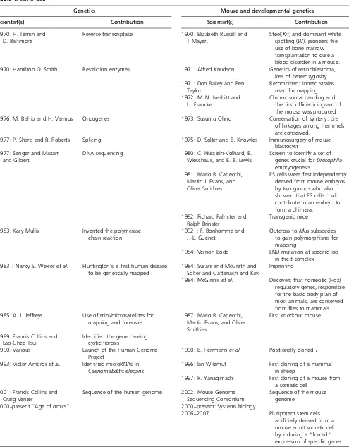
Table 1 Parallel time lines that trace the history of genetics and mammalian developmental genetics
Genetics Mouse and developmental genetics
Scientist(s) Contribution Scientist(s) Contribution
1850: Darwin Evolution 1850–1940s: Hans Spemann
and the German school
Long history of descriptive and experimental embryolgy 1866: Mendel Laws of inheritance
1900: Hugo de Vries, Carl Correns, and Erich von Tschermak
Rediscovery of Mendel 1900: Miss Abbie Lathrop Starts breeding“fancy mice” 1906: William Castle and
The Busey Institute
Founder of mouse genetics
1910s: Thomas Hunt Morgan Sex-linked inheritance, gene theory, principle of linkage, chromosome theory of inheritance
1908–1920: C. C. Little, Leonell Strong
Development of inbred stains
1927: H. J. Muller X rays are mutagenic inDrosophila 1927: Dobrovolskaia-Zavadskaia Discovery ofBrachury, the first developmental mutation in mice and its interaction withtalleles
1928: Griffith Transfer of DNA from one bacterium to another
1929: C. C. Little Founded The Jackson Laboratory
1931: Barbara McClintock Recombination is caused by a physical exchange of chromosomal pieces
1930: An academically inbred group of geneticists
31 loci and 7 linkage goups defined
1933: L. C. Dunn and Salome Glueckshon-Schoenheimer
Effectively found developmental genetics with the study of theT/t-complex
1935: Elizabeth Fekete First successful transfer of fertilized ova
1937: George Beadle and Boris Ephrussi
Foundation for one gene/one enzyme hypothesis
1936: The Jackson Laboratory researchers
First link between cancer and viruses in mammals 1941: George Beadle and
Edward Tatum
One gene/one enzyme hypothesis 1942–1948: George Snell Develops congenic strains of mice—identical but for a small chromosomal segment—by breeding for differences only at the H2 locus. This opens new areas of immunogenetics. 1944: Avery, McCloud,
and Macarty
Nucleic acid is the vehicle of heredity
1944: Barbara McClintock Hypothesis of transposable elements to explain color variations in corn. 1951: Rosalind Franklin Obtained sharp X-ray diffraction
photographs of DNA.
1950: Margaret Dickie Obese mouse is discovered. Thefirst animal model for obesity, the mouse later proves to have a mutation key in identifying theleptin gene.
1952: Alfred Hershey and Martha Chase
DNA rather than proteins carries genetic information
1953–1956: L. B. Russell Dominantwhitespotting andsteelloci identified 1953: Watson and Crick Resolved the structure of DNA 1954: Leroy Stevens Biology of stem cells in
teratocarcinomas in an inbred strain
1956: Vernon Ingram A single amino acid difference in a protein (hemoglobin) can cause a disease
1958: Margret Green Database of mouse linkages and loci, which forms the foundation of the Mouse Genome Database 1960s: Susumu Ohno The total amount of
chromosomal material in mammals was the same. Mammalian X chromosomes are conserved among species. 1961: F. Jacob and J. Monod The operon 1961: Mary Lyon X inactivation, the Lyon Hypothesis
1960s: Tarkowski and Mintz Mouse embryonic chimeras
Table 1,continued
Genetics Mouse and developmental genetics
Scientist(s) Contribution Scientist(s) Contribution
1970: H. Temin and D. Baltimore
Reverse transcriptase 1970: Elizabeth Russell and T Mayer.
Steel(Kit) and dominant white spotting (W). pioneers the use of bone marrow transplantation to cure a blood disorder in a mouse. 1970: Hamilton O. Smith Restriction enzymes 1971: Alfred Knudson Genetics of retinoblastoma,
loss of heterozygosity 1971: Don Bailey and Ben
Taylor
Recombinant inbred strains used for mapping 1972: M. N. Nesbitt and
U. Francke
Chromosomal banding and thefirst official idiogram of the mouse was produced 1976: M. Biship and H. Varmus Oncogenes 1973: Susumu Ohno Conservation of synteny; bits
of linkages among mammels are conserved.
1977: P. Sharp and R. Roberts Splicing 1975: D. Solter and B. Knowles Immunosurgery of mouse blastocyst
1977: Sanger and Maxam and Gilbert
DNA sequencing 1980: C. Nüsslein-Volhard, E. Wieschaus, and E. B. Lewis
Screen to identify a set of genes crucial forDrosophila embryogenesis
1981: Mario R. Capecchi, Martin J. Evans, and Oliver Smithies
ES cells werefirst independently derived from mouse embryos by two groups who also showed that ES cells could contribute to an embryo to form a chimera.
1982: Richard Palmiter and Ralph Brinster
Transgenic mice
1983: Kary Mullis Invented the polymerase chain reaction
1992 : F. Bonhomme and J.-L. Guénet
Outcross toMussubspecies to gain polymorphisms for mapping
1984: Vernon Bode ENU mutation at specific loci in thet-complex
1983 : Nancy S. Wexleret al. Huntington’s isfirst human disease to be genetically mapped
1984: Surani and McGrath and Solter and Cattanach and Kirk
Imprinting
1984: McGinniset al. Discovers that homeotic (Hox) regulatory genes, responsible for the basic body plan of most animals, are conserved fromflies to mammals 1985: A. J. Jeffreys Use of mini/microsatellites for
mapping and forensics
1987: Mario R. Capecchi, Martin Evans, and Oliver Smithies
First knockout mouse
1989: Francis Collins and Lap-Chee Tsui
Identified the gene causing cysticfibrosis
1990: Various Launch of the Human Genome Project
1990: B. Herrmannet al. Positionally clonedT
1993: Victor Ambroset al. Identified microRNAs in Caenorhabditis elegans
1996: Ian Wilemut First cloning of a mammal in sheep
1997: R. Yanagimachi First cloning of a mouse from a somatic cell
2001: Francis Collins and Craig Venter
Sequence of the human genome 2002: Mouse Genome Sequencing Consortium
Sequence of the mouse genome
2000–present“Age of omics” 2000–present: Systems biology
2006–2007 Pluripotent stem cells
artificially derived from a mouse adult somatic cell by inducing a“forced”
expression of specific genes
a different number of chromosomes and different chromo-some morphologies, he was looking at the same amount of genetic material in the different species. How could you test this idea before the advent of molecular biology? What Ohno did was to photograph the mitotic preparations from several species under very carefully controlled conditions, then cut out the chromosomes from the photographic print of single-cell chromosome spreads, and finally weigh each cell set on a balance, comparing the results for the different species. Thus he was able to demonstrate that all the species included had the same amount of genetic material. It would take another five decades—until the genomes of several mammalian species had been sequenced—to confirm that he was correct.
Thus, a history of genetics must be appreciated in the conceptual framework of the time. As this might seem obscure to a student of 21st century genetics, I will attempt to describe that context as we go along. Much of the history of early mammalian genetics, when one studied one gene at a time, was nevertheless grounded in a thorough knowledge of biology. This foundation seems in danger of being lost with today’s emphasis on molecular biology, the various “omics,” and systems biology approaches. Yet the knowl-edge of the actual biology of organisms remains just as es-sential today.
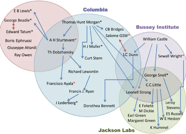
There were three great institutions that served as incubators of genetics and mammalian genetics in thefirst half of the 20th century: Columbia University, starting with Thomas Hunt Morgan’s fly genetics in the 1910s and con-tinuing with mice through the 1950s; The Bussey Institute at Harvard that existed from 1908 to 1936 started with William Castle and his many famous students; and finally, The Jackson Laboratory (http://www.jax.org/milestones/ researchhighlights.html) founded by C. C. Little in 1929.
Those institutions and their scientific personnel (see Figure 1) compose vital elements of the story. Figure 1 is an attempt to show the historical overlap between institutions, pioneer-ing researchers, and thus ideas. The arrows indicate mentors and students.
T. H. Morgan’s Fly Room: The Beginning of Modern Genetics
No history of mammalian genetics can be written without first paying homage to the early Drosophila geneticists at Columbia (Figure 1). Starting with Thomas Hunt Morgan and his student, A. H. Sturtevant, the Drosophilists first had to identify mutations. Morgan’s earliest mutant,white eye (now designated simplywhite), led him to hypothesize sex linkage. And from there and other sex-linked mutations, they determined that genes were arranged on chromosomes in a linear fashion, the so-called“beads on a string” hypoth-esis. They also showed that the gene for any specific trait was at a fixed locus and the important concept that the frequency of crossing-over between two genes could deter-mine their relative proximity on a linear genetic map.
Morgan’s fly room at Columbia became world famous, and the center of an informal exchange network, through which promising mutantDrosophilastrains were transferred freely from lab to lab. In 1928, however, Morgan’s group moved to the California Institute of Technology where they continued to be an epicenter of genetics, attracting young scientists such as George Beadle, Boris Ephrussi, and Edward L. Tatum, who would soon become leaders in thefield.
Columbia’s loss of the fly geneticists left a momentary vacuum, into which stepped the mouse geneticists, in par-ticular William Castle’s student L. C. Dunn. In 1914, Dunn had hoped to be an undergraduate researcher in Morgan’s
“fly room,”but, as he put it in 1965 in hisShort History of Genetics,“as I stood in the doorway. . .it was obvious that there was little room.”Later, in 1928, when Dunn returned to the Zoology Department at Columbia as a professor, it fell to him to convert the fly room to one suitable to working with mice. Yet, preparing for that task was instructive in itself. “The ingenuity with which simple homemade instal-lations had been made to serve as the basis for major scien-tific advances became apparent to me”(Dunn 1965).
William Castle and The Bussey Institute
After the rediscovery of Mendel’s work, William Castle be-came the father of mammalian genetics. Like thefly geneti-cists before him, however, he first had tofind and identify mutations. In 1903, Castle showed that the albino pheno-type was caused by a Mendelian recessive gene (see Morse 1978). Later, in 1909, with J. C. Phillips, he transplanted the ovaries of a black animal into an albino female and mated her to an albino male. All of the many offspring were black. Thus, in one ingenious experiment, two important ideas were proved: the germ cells are separate from the somatic cells, and genes are not contaminated by the environment (Morse 1985, page 113). How simple, how elegant a way this was of nailing two key principles at once.
In 1908, Castle became the director of The Bussey Institute at Harvard, the now famous incubator of mouse geneticists. Clarence C. Little, who was later to become the founder of The Jackson Laboratory, soon joined Castle in his studies. Together they established that thepink-eyed dilution
(p) locus as separate from dilute (d) and albino (c) and showed that dominant yellow (Ay) was lethal in
homozy-gotes (Russell 1978). This work served as the foundation for thefirst demonstration of linkage in the mouse.
By 1917, the groundwork for most subsequent studies in mammalian genetics had been laid through the work of Castle and his students. An important aspect of The Bussey Institute was Castle’s philosophy of collegial interaction.“ Ac-tive discussions of problems under study were greatly en-couraged in daily exchanges and in the weekly seminars in which both staff and students participated. Differences in opinion were freely aired but kept impersonal, and Castle was remarkably tolerant of students who voiced opinions contrary to his beliefs. In those instances when his own work or that of his students showed him to be wrong, he would publicly confess the error and provide the correct solution” (Morse 1985). Interestingly enough, these productive atti-tudes pervaded the laboratories of not only Castle’s students and his student’s students, but in some cases, their students, well into the 1950s–1970s.
Over a period of 28 years, The Bussey Institute trained 49 students, including L. C. Dunn, Clarence Little, Sewall Wright, Lionel Strong, and George Snell. Most students of mouse genetics can trace their scientific heritage back to Castle in one way or another (see figure 4 in Morse 1978).
Inbred Strains of Mice
A major contribution of Castle’s group, and of Clarence Little in particular, was the realization of the need for and devel-opment of inbred, genetically homogeneous lines of mice, a lesson well learned from Mendel. Thefirst mating to pro-duce an inbred line was begun by Little in 1909 and resulted in the DBA strain. By 1918, Little developed some of the most famous inbred lines that are still used today (Russell 1978).
An inbred strain is constructed by brother–sister or par-ent–offspring matings for a minimum of 20 generations. At that point, the mice are skin-transplant compatible and ge-netically identical. Inbred lines have played a crucial role in all areas of mouse genetics by allowing independent researchers to perform experiments on the same genetic material, which in turn allows the direct comparison of any results obtained in different laboratories around the world.
Curiously enough many of the inbred strains started not with professional researchers but with a hobbyist, Abbie Lathrop, who was the first American mouse “fancier.”She was a retired schoolteacher who around 1900 began to breed mice for sale as pets from her home. She reputedly began with a pair of waltzing mice but was so successful that she soon found herself in the small animal pet business. Her biggest clients, however, turned out to be the“new”mouse genetics researchers at the nearby Bussey Institute and other research institutions (Morse 1978). Together, they managed to turn curiosities into experimental material.
The Jackson Laboratory
Another crucial contribution of Little to mouse genetics was the role that he played in founding The Jackson Laboratory, also now known as JAX, in Bar Harbor, Maine. The laboratory was inaugurated in 1929 as the “natural heir” to The Bussey Institute (Snell and Reed 1993). Importantly, Little also served as itsfirst director for 27 years.
JAX was the site of many achievements in mammalian genetics. A list of the tip of the iceberg would include: George Snell’s discovery of the genetic factors (those of the major histocompatibility complex, Snell 1948) that deter-mine transplantation of tissue from one individual to an-other; the first evidence that viruses can cause cancer (Jackson and Little 1933); the discovery of the existence and nature of stem cells (Stevens 1958); thefirst successful mammalian bone marrow transplantation to cure a blood disorder (Lawson et al. 1956); and parabiosis experiments that led to the discovery of the hormone leptin (Coleman, and Hummel 1969). Beyond those discoveries, perhaps JAX’s great contribution was that, to many young investiga-tors and summer students, it was a magical place. What made it so was the combination of its isolation, its beautiful setting, the chance to work with an unlimited number of mice, the variety of mouse mutants, and its very special welcoming attitude for young investigators. It became a mecca for mouse geneticists and provided a model for the formation of research institutes elsewhere.
Developmental genetics: L. C. Dunn and the Tt complex
A sea change in thinking occurred with the marriage of development biology and genetics. Many people today regard this as something that occurred in the 1980s, but it was, in fact, an intellectual fusion of disciplines that had started in the early 1930s at Columbia with L. C. Dunn and his collaborator Salome Glueckshon-Schoenheimer, a Ger-man–Jewish refugee who had trained in Hans Spemann’s embryology laboratory. Later in this endeavor, they were joined by Dorothea Bennett. These investigators, as shall be described in a moment, had the perfect genetic source materials at hand: mouse mutations at the so called“t com-plex.”Although the synthesis of developmental biology and genetics seems natural, indeed inevitable, today, it was cer-tainly not always the case. Well into the 1980s there were university courses taught in developmental biology that em-phasized descriptive events and embryo manipulations but ignored genetics. And certain high-profile molecular biolo-gists, well into the 1970s, expressed skepticism about de-velopmental genetics ever contributing anything.
Brachyury: The genetics of what came to be called the “t
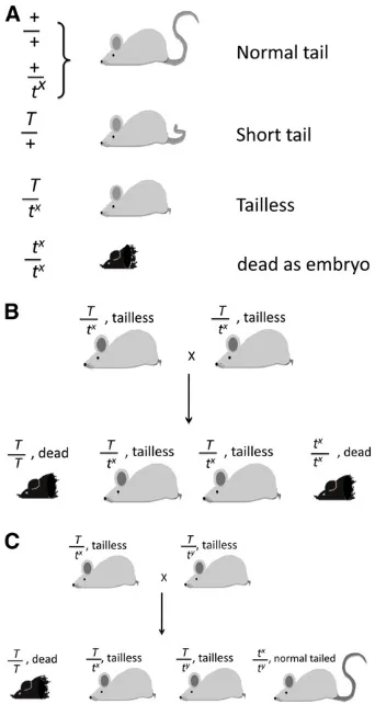
complex,” located on chromosome 17, presented several violations of Mendel’s rules and was, in fact, the starting point of L. C. Dunn’s studies. The Brachyury (T) mutation was discovered in 1927 in a radiation experiment in the lab of Dobrovolskaia-Zavadskaia in Paris (Dobrovolskaia-Zavadskaia 1927). The original mutation was a substantial deletion.Tis one of the veryfirst developmental mutations known in mammals. It is semidominant, causing a short tail in heterozygotes and lethality in homozygotes. In the embryo, it is essential for the emergence and maintenance of mesoderm. T/T mice die shortly after gastrulation, dis-playing a complete loss of the posterior mesoderm due to primitive streak defects and the absence of a notochord.
Ironically, half the amount of the wild-type protein made by this gene causes a seemingly trivial short tail, while ab-sence of the protein, as occurs in homozygotes, produces embryo lethality for lack of an axis. Dobrovolskaia-Zavad-skaia found that some wild-trapped mice carried a factor that interacted withTto cause taillessness. Thefirst puzzle here was that a gene that had no ostensible phenotype on its own had an interaction with T. Dobrovolskaia-Zavadskaia referred to these as recessive t’s. She failed to make head-way in dissecting the complexity of thetmutations that she found in wild mice, so she gave the mice to Dunn at Colum-bia University. “Dunny,”as he was called, and later Salome Glueckshon-Schoenheimer-Waelsch, were quick to latch on to the problem, but the secrets of this region of the genome were slow to reveal themselves. It posed several enigmas, which, in the end, took.70 years and the advent of molec-ular technology to sort out!
WhenTwasfinally cloned and identified in 1990 (Herr-mann 1990), it was found to encode a novel transcription factor that is essential for mesoderm development. In fact, it contains a conserved domain (T box) that defined a whole new class of developmentally important transcription fac-tors. There are presently 15 of them known to act at differ-ent points and in differdiffer-ent tissues in developmdiffer-ent (Bollag
et al.1994; Naicheet al.2005).
Tail interaction factor: All wild-trapped tmutant
chromo-somes carry the same tail interaction factor (tct). It is now thought thatTandtctare alleles, but the issue of allelism is still not entirely clear. Nevertheless,Tacts as a diagnostic tool to determine the presence oftchromosomes in wild-caught mice because compound heterozygotes for Tand tct (T/tct) cause taillessness.tct is a hypomorph because, on its own in homozygotes, it has no phenotype (see below). Allelism ofT
andtctwas certainly not evident in the 1930s, so to be safe Dunny called them “pseudoalleles.”Yet, whatever the termi-nology used, it was not understood how mutations affecting tail length could be allelic with a series of different lethal mutations (see below) that affected the embryo in different tissues at different times. Later, these wild-trapped mutant chromosomes were calledt-haplotypes, a term borrowed from immunogenetics and meaning a collection of linked genes that are inherited together as a group and symbolized by“tx,y.”
t-lethal mutations:With rare exceptions, everyt-haplotype
trapped in the wild contains at least one recessive mutation lethal to the embryo. Some carry more than one as a hidden lethal (Paterniti et al.1983). These would escape detection because, if homozygotes for one of the mutations died early, the death of a second one would be both undetectable and biologically irrelevant. The t-lethals were a series of differ-ent linked mutations defined by complemdiffer-entation between them. In this case, complementation meant that two tailless mice carrying the same mutation (T/tx) bred true for
tail-lessness because both homozygotes (T/T and tx/tx) died,
two different ones (tx and ty) made one class of normal
tailed progeny (Figure 2). In total, 16 different t comple-mentation groups were identified (Kleinet al.1984). Dor-othea Bennett studied many of the lethals extensively (Bennett 1964), finding that they affected different stages of development from morula to nearly completed fetus. These lethal or semilethal mutations were very informative for mammalian development. In fact, in the 1930s–1970s before large-scale mutagenesis and knockout technology were available, there were very few lethal mutations known. Virtually all of the then-known lethal mutations in the mouse were mapped to thet-complex.
The rare exception of at-haplotype carrying a gene that is not lethal is when some wild mice carry“semilethals.”This was a puzzling situation in which a proportion of homozy-gotes died as embryos, but the remainder of the litter de-veloped normally with normal tails (Figure 2C). The normal-tailed mice are once again tct/tctbut carrying two
copies of t-semilethals. The existence of semilethals was a very puzzling phenomenon in the 1950s; this was before there was awareness of the effects of genetic background in mammalian development, although such effects were well known inDrosophila. Now we know, especially from knock-out mice, that even a small amount of genetic background variation can make the difference between lethality and complete viability.
Recombination suppression: Recombination suppression,
as its name suggests, prevents genetic exchange. Its pres-ence was soon demonstrated in crosses between t -haplo-types and wild-type (+) chromosomes. Whereas in wild type, Tand an outside marker can be measured as 20 cM, the rate of recombination betweentand wild type is only an exceptional 1/1000 or 0.1 cM. This was a genetic puzzle for half a century and was not completely understood until 1982. We now know thatt-haplotypes contain four nonover-lapping inversions and behave like the balancer chromo-somes known in Drosophila that also lock up an entire region. Sequencing has shown that the length of thet -com-plex region is.37 Mb and contains well over 500 genes, so the idea that it is a region of some unique developmental importance is no longer tenable. After the 1980s screens in
Drosophilato identify genes crucial for embryogenesis (Jür-gens et al. 1984; Nüsslein-Volhard et al. 1984; Wieschaus
et al.1984), it was clear that, in a region of this size in the mouse genome, many loci causing recessive embryonic le-thality would be expected to occur. Nevertheless, as a result of this misconception of the unique importance of the t -complex region for development, and the additional fact that the major histocompatibility complex (H2) was in-cluded in the region, the consequence was that the proximal third of chromosome 17 became by far the best-mapped region of the mouse. This was true at a time when the rest of the mouse chromosome maps were still very sparsely populated with known mutations.
As noted above, we now know that the recombination suppression was due to four nonoverlapping inversions. In the mouse, these inversions, however, were cytologically invisible with the chromosome banding techniques available at the time. They all occurred between two distant light bands (Lyon, et al. 1988) (Figure 3). The 1/1000 excep-tional recombinants mentioned above retained a mutant portion of chromosome 17 and gained wild type at the other end. Thus, when particular recombinants are viable, they can be detected as either normal tails (tct/tct) or by the exchange of an outside marker on theTchromosome.
These features argue that all t-haplotypes must be descended from a common ancestor (Silver 1993). Thus, for the most part, the wholet-haplotype travels genetically as a single unit.
Recombination suppression had been a nasty barrier to genetic analysis and understanding the t-complex. When Lee Silver and I solved the problem, we did not really have inversions in mind since the cytogeneticists had assured us
that no inversions were involved. We just assumed that if“t -chromatin,”as Mary Lyon had dubbed it, was different from wild type and that if“good”chromatin would not recombine with “bad”(Lyonet al. 1979), then“bad”might recombine with “bad”!
In 1981 (Silver and Artzt 1981), the experiment to test this could be done for two reasons. First, at some point in time, a spontaneous mutation to tufted (tf) occurred in a com-pletet-haplotype containing a lethal.tfis a hair-growth-pat-tern mutation10 cM distal toTthat can be visibly scored at 28 days after birth. Second, theTmarker was available only in wild-type chromatin. However, in 1979, Mary Lyon showed that one of her t-haplotypes contained some “normal chro-matin” at its very proximal end. Thus we were able to get recombination between it andTon a wild-type chromosome, effectively putting Ttogether with distalt-chromatin. It was not until 1981 that Lee Silver and I configured the visible markers and two complementing lethals in a position where we could test recombination between twot-haplotypes.
The visible markers that we used were Tandtf(Figure 4). Ourfirst three litters contained 25% recombinant normal tailed (tufted) and short tailed (non-tufted). I remember returning to the mouse room several times that day to make sure that we had not mis-scored the mice fortfand thus our hypothesis was still true.
However, in 1981 before the inversions were known, this did not solve why t-chromatin could not recombine with wild type but, for the first time in 60 years, it gave us the tools to do recombinational genetics, map the different t -lethals, and solve the“pseudoallele problem.”Later studies showed that the recessivet-lethals were all non-allelic (Artzt
et al.1982).
Male germ-cell effects:Afinal and most remarkable feature
oft-haplotypes is transmission ratio distortion (TRD), which
is found in male heterozygotes and the sterility found viable t homozygotes. Surprisingly, 10% of all mice trapped throughout the world contain at-haplotype. This fascinated the population geneticists because, how could you explain the high frequency of such deleterious genes? TRD is com-mon to allt-haplotypes and makest’s look like a“viral ep-idemic”in terms of population genetics. In fact, it appeared that onet-haplotype (tw5) marched all the way from upper
New York state to South America. The explanation is that any wild-derived t-haplotype is transmitted from heterozy-gous males (but not females) to upward of 99% of the prog-eny. In addition, a t-haplotype double heterozygote, as in complementing t-lethals (tx/ty), and t semilethal
homozy-gotes are male sterile. In contrast, females of these geno-types transmit t-chromosomes in Mendelian ratios and are fertile.
The big question was, how do the TRD and male sterility of t-haplotypes work genetically? In 1979, Mary Lyon hy-pothesized that TRD involved interaction of three or more loci encoded in t-haplotypes. In her model, there were at least three or more segregation distorters and one responder element acting incis, and these were all locked together by recombination suppression (Lyonet al.1979). According to Lyon, the distorters were likely to be modules of dynein (Lyon 2003)
It became increasingly clear that Lyon’s model had been substantially correct. Nevertheless, the elements responsible for TRD were not unraveled for a decade or more, and there is still some disagreement about them. Although over time the number of putative distortersfluctuated, three of the distorter genes and the responder have been molecularly identified and all encode proteins that participate in RHO signaling pathways (Herrmannet al. 1999; Baueret al.2012).
In the 1970s, I wondered whether the genes for TRD were the same as those for sterility. Now it seems likely that they are not because, although many genes can cause
Figure 3 The cytologically invisible inversions in at-haplotype. The left chromosome is a normal one. The chromosome bands are indicated as they would have been seen with the techniques available in the 1970s. The inversions in at-haplotype occur between bands A2 and D as in-dicated by arrows, highlighted with a light gray box. Without modern cytological techniques, these would not have been detectable. (This dia-gram has been modified from Lyonet al.1988.)
sterility, even one of them locked up in recombination suppression and lethality can accumulate mutations without further consequences to the mouse (very much like the example of sterility andQkbelow.)
steeland DominantwhiteSpotting
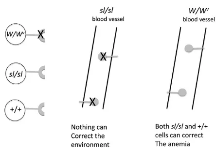
An intriguing biological puzzle for developmental geneticists in the 1960s was the steel (sl) and Dominant white (W) spotting mutations. Both are pleiotropic mutations with effects on pigment, hematopoiesis, and germ cells. In addi-tion, both mutations have differing degrees of severity in heterozygotes and homozygotes; i.e., they are semidomi-nant. The most interesting facet of their genetics, however, is that, although they reside on different chromosomes, they genetically interact.
Both mutations have been known for .60 years, and today there are a multitude of alleles at both loci, but the simple phenotypes that were known when both genes had just been identified from the initial alleles suffice to tell the tale. In most cases, surviving homozygotes forslandWare black-eyed whites, very anemic and sterile, while heterozy-gotes for alleles of either one show a variable degree of color dilution and/or white spotting, anemia, and sterility. Com-pound heterozygotes as in sl/Wessentially have the same phenotype as homozygotes for either gene. They are com-pletely white with black eyes and are severely anemic or dead. This non-allelic, noncomplementation indicated that the Wandsl gene products function in the same develop-mental pathway. What did these genes have in common except that all three affected cell types either had to migrate in the embryo or were derived from migrating cells?
If one gene caused a cell defect and the other an environmental defect, an elegant and simple transplantation experiment done at The Jackson Laboratory in the 1960s would demonstrate it (McCullochet al.1965). It was found thatsteelmice cannot be cured of their hemapoietic defects with bone marrow cells from either W or wild-type mice, implying thatsteel has a pervasive effect on the microenvi-ronment, preventing function or survival of the transplanted cells. In contrast, transplantation ofWmice with bone mar-row from either +/+ orsteelmice resulted in a hematologic cure of the recipients via function of the transplanted cells (Figure 5). This result indicates that the defect inWmice is a defect intrinsic to the cell. Twenty years later, when both genes were cloned, this hypothesis was validated. How sim-ple it is in retrospect: W, the first gene cloned, was the oncogenec-kit(also known askit), which encodes a trans-membrane tyrosine kinase receptor (Chabot et al. 1988; Geissler et al. 1988), and steel, the second to be cloned, was named its cognate ligand (kitl) (Andersonet al.1990; Copelandet al.1990).
The multitude of spontaneous alleles of both genes are an added lesson in how genetics can help dissect biological and biochemical function because different alleles can affect different parts of the molecule and its interactions and, thus,
have slightly different phenotypes. This was later shown to be of great usefulness when the mutagen N-ethyl-N -nitro-sourea (ENU) was used to induce specific mutations (Bode 1984) and later when multiple alleles of many genes be-came available.
X Inactivation: The Lyon Hypothesis
Mary Lyon, R. A. Fisher’s last Ph.D. student, was both an“at the bench”researcher and an inspired theoretical geneticist. Working at the Medical Research Council Harwell in Great Britain with colleagues such as Charles Ford, Ted Evans, and Tony Searle, she was particularly interested in radiation bi-ology and genetics, but some of her early work provided a landmark in the understanding of X chromosomes and mammalian sex differentiation. This work began with her interest in“calico”mice, which show a tricolor coat pattern much like the common calico cat. In 1960, she wondered why most X-linked mutants showed a variegated effect in heterozygous females and why the pattern of mottling re-sembled that seen in somatic mosaics. Her idea was that one X chromosome became inactive early in the development of a female embryo. She hypothesized that this happened at random so that either the maternally or the paternally de-rived X chromosome could be inactivated in different cells, but, once the choice was made, it was permanent. The resulting variegated coat color resulted from the mutant X chromosome that was active in some patches and the nor-mal X chromosome that was active in others. Since nor-males have only one X chromosome, it is always active. She further suggested that the inactive X chromosome was not only genetically inert but also heterochromatic. The latter idea would explain the long standing observation of the so-called
Barr body, found only in the nuclei of female cells (reviewed in Lyon (2002).
Another important contribution of Mary Lyon that might seem unimaginably trivial today but which had a major impact is that, in the 1950s, she took over, from Margret Green at The Jackson Laboratory, an index card file of all mouse gene loci and mappings and incorporated these data into the Mouse News Letter or MNL. MNL was a “ mimeo-graphed”private publication circulated biannually and free to the community where mouse geneticists reported all their findings. This was the first mammalian genetics“journal,” and it functioned as such untilMammalian Genome incorpo-rated MNL when it started publication in 1991!
Knockout Mice: Creation of Gene-Specific Mutations
Probably the most important technical innovation of genet-ics in the 20th century was the ability to “knock out”(KO) specific genes by homologous recombination. This revolu-tionized mammalian genetics and biology. The first step involved the ability to establish cell lines from the uncom-mitted cells of the mouse embryo, the so-called embryonal stem cells (ES cells) (Evans 1981). In the beginning, manip-ulating the early mouse embryo faced several problems. At first it was the inaccessibility of the embryo itself that was a major technical hurdle. Once that was overcome, it was the inaccessibility of the inner cell mass. After superovula-tion, 2-day mouse embryos can beflushed from the oviduct. They are surrounded by an acellular shell that is easily re-moved by a limited protease digestion. At that point, the embryo is a very simple structure called a blastocyst (Figure 6). It consists of only two cell types. The inner cell mass that will give rise to the entire future embryo (these are the ES cells). But, the ES cells are rendered inaccessible because they are encased in the trophoblastic cells that will contrib-ute to the extra-embryonic support tissues.
The breakthrough in isolating viable and all important inner-cell mass cells was another example of an obvious idea that left all the developmental biologists knocking their heads and saying, “why didn’t I think of that!” In 1975, Davor Solter and Barbara Knowles invented a technique called “immunosurgery.” The used a variation of comple-ment-dependent antibody cytotoxicity. Blastocysts were ex-posed to a rabbit anti-mouse antibody alone. After removing the antiserum, complement was added and only the tropho-blastic cells were killed. The inner cell mass cells, never having been exposed to the antiserum, is healthy and can then be grown in culture (Solter and Knowles 1975). This method is still used extensively for deriving ES cells.
Once stable ES cell lines were established (Evans 1981), it was discovered that a construct containing two regions of homologous sequence flanking a deleted part of the gene could be electroporated into the ES cells (Smithies et al.
1985; Wong and Capecchi 1986). First, since homologous recombination is rather inefficient, a selectable marker for incorporation is necessary. Second, clones of treated cells need to be validated by Southern blot to assure that the recombination has in fact been correct. The construct inserted also contains a marker such as b-galactosidase so that the treated cells and gene can be visually followed.
What follows next is to turn the cloned cells into mice! It had been demonstrated much earlier that if you aggregate the uncommitted cells from two embryos together, the cells would intermingle and form a“chimera”(Buehr and McLa-ren 1974). Thus, if the two embryos were genetically des-tined to be two different coat colors, for example, black and albino, then the chimera would have patches of each color. Therefore, if a clone of homologously recombined cells was injected into the blastocyst, the ES cells would naturally mix with the inner cell mass and create a chimera.
However, there are still some genetics needed before one has a KO mouse. The progenitor cell presumably had only one copy KO’ed and is thus heterozygous (+/2). First, the chimeric male is mated to an albino. All the white offspring can be discarded, but the black offspring need to be tested for carrying the KO’ed allele. Two carriers are mated to-gether to determine if2/2is viable or if there is a detectable mutant phenotype.
Homologous recombination, a classic genetic phenome-non, has thus proven to be part of the essential means by which genetic function in mice is currently studied. Indeed, there is a worldwide effort to knock out every mouse gene (Bradleyet al.2012).
quaking: How to Positionally Clone a“Deleted”Gene
In the 1980s when positional cloning in mammals was becoming possible, it was a long and tedious task involving either cosmids (holding 40 kb of DNA) and chromosomal walking or, worse yet, tiresome microdissection of chromo-somes. The latter procedure had been used for cloningT. My lab was interested in a pleiotropic mutation called quaking Figure 6 Making a knockout mouse. (A) Anatomy of the blastocyst. (B)
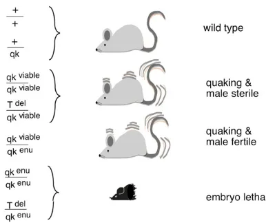
(Qk) that was included in thet-complex. Homozygosity for the originalquakingmutation (Qkv) permitted viability but
resulted in severe dysmyelination and male sterility. This phenotype contrasted with the other four existing, recessive, ENU-induced mutations that were all lethal at midgestation (Bode 1984; Justice and Bode 1988; Shedlovsky et al.
1988). The viable mutation was found to complement the lethality inQkv/QkENUwhile the lethal ENU mutations
com-plemented the Qkv male sterility. All of these compound
heterozygotes, however, exhibited the quaking phenotype (Figure 7).
My lab was doing a considerable amount of mapping in the chromosomal region of Qk. Tom Ebersole, a graduate student in my lab, had managed to use whole cosmids as probes on Southern blots by blocking the repetitive sequen-ces with total mouse DNA, a pretty gross technique in light of today’s sophisticated molecular protocols. To our surprise, a cosmid mapping to the Qk region and much of the sur-rounding DNA (at least 40 kb) was deleted (Ebersole et al.
1992)! This should have not been such a surprise because
THP, a known, very large deletion around the locus of T,
acted as a nullisomic for the nearbyQklocus. Thus, puzzling but forgotten by us was that THP/Qk was quaking. The
whole story left us with the genetic riddle of how one could delete a gene that was necessary for embryogenesis yet still have viability. However, the historical context was that the literature was taking first note of tissue-specific enhancers,
and this could in fact be part of the solution: a deleted tissue-specific enhancer preceding an intact gene.
The solution to identify a candidateQkvgene had to be
a purely genetic-based cloning strategy (Figure 8). As the four ENU-induced lethal alleles were presumably point mutations andQkvcomplements all of them for their
lethal-ity but not their defective myelination, a single gene for these two functions ofQkwas proposed. When the original
Qkvmutation was found to be associated with a deletion,
a model was introduced that predicted the location of the gene. Because the ENU mutations are recessive lethals, it is unlikely that a homozygous deletion of the Qk-coding sequences would be viable. The solution that we proposed was that two separate elements were necessary forQk func-tion and that the delefunc-tion interferes with a regulatory ele-ment necessary in the nervous system but not the embryo. Since regulatory regions were known to be generally close to coding sequences, the strategy was to clone the DNA flanking the deletion breakpoint. Theqkgene was expected to be located very close to the breakpoint on one or the other side. In addition, since the compounds Qkv/QkENU
were male fertile, a separate gene in the deletion must have been responsible for the male sterility (Figure 8).
As predicted, on one side of the breakpoint a candidate gene was found that was expressed in the earliest cells of the embryonic nervous system and in the myelinating tracts of prenatal brain. The hypothesis was correct (Ebersole et al.
1996)! TheQk gene turned out to be one of the founding members of a new family of RNA-binding proteins that act both in signal transduction and in RNA metabolism and that came to be called STAR proteins forsignaltransduction and
activation ofRNA (Vernet and Artzt 1997).
Conclusions
The beginnings of mouse genetics were strongly focused within a small group of scientists in the northeastern United States: first at The Bussey Institute, then at Columbia, and lastly at The Jackson Laboratory. At these institutions, the academic descendants of a very small number of key early figures in genetics were retesting Mendel’s principles in mice.
This article has tried to touch on examples of the several important paradigm changes that revolutionized mammalian
Figure 7 Phenotypes and genotypes ofQkmice.
genetics and some essential technical changes like the early advent of inbred strains of mice. Among the early experi-ments discussed was the dissection of development through the study of mouse mutations. A particular example was the work on the t-complex, a pioneering example of the use of genetic techniques to understand development. It thrust us into seeing the genetic underpinnings of mammalian devel-opment and develdevel-opmental pathways.
An obvious, essential change was the transition from genetics to molecular biology. Consequently, one of the most important concepts to come out of 20th-century mammalian genetics was the slow realization that by descent from common ancestry the essential genes had been conserved though millennia. Starting early in the 1970s, the sequenc-ing of genes from diverse species showed that parts of the essential genes were very similar. This meant that with some caveats, mice could be considered as a model organism to study the biology of these genes. It allowed developmental geneticists to do research on mice as a surrogate for humans and was responsible for a huge surge in mouse genetics.
Two final essential turning points that should be men-tioned here are, first, the ability to manipulate the mouse embryo. This ultimately led to knockouts, whose develop-ment I have briefly reviewed here, and to the understanding of gene function. Finally, of course, there was the sequencing of whole genomes that now allows the relatively easy identi-fication of genes and the hope of understanding complex bio-logical systems.
Acknowledgments
I am grateful to Jiang I. Wu and Adam Wilkins for comments on this manuscript and to the many members of my laboratory for their inspiration.
Literature Cited
Anderson, D. M., S. D. Lyman, A. Baird, J. M. Wignall, J. Eisenman
et al., 1990 Molecular cloning of mast cell growth factor, a hematopoietin that is active in both membrane bound and soluble forms. Cell 63: 235–243.
Artzt, K., P. McCormick, and D. Bennett, 1982 Gene mapping within the T/t-complex of the mouse. I. t-Lethal genes are non-allelic. Cell 28: 463–470.
Atkin, N. B., G. Mattinson, W. Beçak, and S. Ohno, 1965 The comparative DNA content of 19 species of placental mammals, reptiles and birds. Chromosoma 17: 1–10.
Bauer, H., S Schindler, Y. Charron, J. Willert, B. Kusecek et al., 2012 The nucleoside diphosphate kinase gene nme3 acts as quantitative trait locus promoting non-mendelian inheritance. PLoS Genet. 8: e100256.
Bennett, D., 1964 Abnormalities associated a chromosome region in the mouse. II. Embryological effects of lethal alleles in the T-region. Science 144: 263–267.
Bode, V. C., 1984 Ethylnitrosourea mutagenesis and the isolation of mutant alleles for specific genes located in the T region of mouse chromosome 17. Genetics 108: 457–470.
Bollag, R. J., Z. Siegfried, J. A. Cebra-Thomas, N. Garvey, E. M. Davison et al., 1994 An ancient family of embryonically
ex-pressed mouse genes sharing a conserved protein motif with the T locus. Nat. Genet. 7: 383–389.
Bradley, A., K. Anastassiadis, and A. Ayadi, J. F. Battey, C. Bellet al., 2012 The mammalian gene function resource: the interna-tional knockout mouse consortium. Mamm. Genome 23: 580– 586.
Buehr, M., and A. McLaren, 1974 Size regulation in chimaeric mouse embryos. Embryol. Exp. Morph. 31: 229–234.
Chabot, B., D. A. Stephenson, V. M. Chapman, P. Besmer, and A. Bernstein, 1988 The proto-oncogene c-kit encoding a trans-membrane tyrosine kinase receptor maps to the mouseWlocus. Nature 335: 88–89.
Coleman, D. L., and K. P. Hummel, 1969 Effects of parabiosis of normal with genetically diabetic mice. Am. J. Physiol. 217: 1298–1304.
Copeland, N. G., D. J. Gilbert, B. C. Cho, P. J. Donovan, N. A. Jenkins et al., 1990 Mast cell growth factor maps near the steel locus on mouse chromosome 10 and is deleted in a number of steel alleles. Cell 63: 175–183.
Dobrovolskaia-Zavadskaia, N., 1927 On the spontaneous death of the tail in newborn mice and the existence of a hereditary, non-viable character (factor). Crit. Rev. Soc. Biol. 97: 114–116 (in French).
Dunn, L. C., 1965 A Short History of Genetics. McGraw-Hill, New York.
Ebersole, T. A., O. Rho, and K. Artzt, 1992 The proximal end of mouse chromosome 17: new molecular markers identify a deletion associated with quakingviable. Genetics 131: 183– 190.
Ebersole, T. A., Q. Chen, M. J. Justice, and K. Artzt, 1996 The quaking gene product necessary in embryogenesis and myelina-tion combines features of RNA binding and signal transducmyelina-tion proteins. Nat. Genet. 12: 260–265.
Evans, M. J., 1981 Origin of mouse embryonal carcinoma cells and the possibility of their direct isolation into tissue culture. J. Reprod. Fertil. 62: 625–631.
Geissler, E. N., M. A. Ryan, and D. E. Housman, 1988 The dominant-white spotting (w) locus of the mouse encodes the c-kit proto-oncogene. Cell 55: 185–192.
Herrmann, B. G., S. Labeit, A. Poustka, T. King, and H. Lehrach, 1990 Cloning of T gene required in mesoderm formation in the mouse. Nature 343: 617–622.
Herrmann, B. G., B. Koschorz, K. Wertz, K. J. McLaughlin, and A. Kispert, 1999 A protein kinase encoded by the t complex re-sponder gene causes non-Mendelian inheritance. Nature 402: 141–146.
Jackson, R. B., and C. C. Little, 1933 The existence of non-chromosomal influence in the incidence of mammary tumors in mice. Science 78: 465–466.
Jürgens, G., E. Wieschaus, and C. Nüsslein-Volhard, 1984 Mutations affecting the pattern of the larval cuticle in Drosophila mela-nogaster. II. Zygotic loci on the third chromosome Roux’s Arch. Dev. Biol. 193: 283–295.
Justice, M. J., and V. C. Bode, 1988 Three ENU-induced alleles of the murine quaking locus are recessive embryonic lethal muta-tions. Genet. Res. 51: 95–102.
Klein, J., P. Sipos, and F. Figueroa, 1984 Polymorphism of t-complex genes in European wild mice. Genet. Res. 44: 39–46. Lawson, F. A., E. S. Russell, and L. J. Smith, 1956 Implantation of normal blood-forming tissue in radiated genetically anemic hosts. Science 124: 1076–1077.
Lorenzetti, D., C. E. Bishop, and M. J. Justice, 2004 Deletion of the Parkin coregulated gene causes male sterility in the quaking (viable) mouse mutant. Proc. Natl. Acad. Sci. USA 101: 8402– 8407.
Lyon, M. F., 2003 Transmission ratio distortion in mice. Annu. Rev. Genet. 37: 393–408.
Lyon, M. F., E. P. Evans, S. E. Jarvis, and I. Sayers, 1979 t-Haplotypes of the mouse may involve a change in intercalary DNA. Nature 279: 38–42.
Lyon, M. F., J. Zenthon, E. P. Evans, M. D. Burtenshaw, and K. R. Willison, 1988 Extent of the mouse t complex and its inversions shown by in situ hybridization. Immunogenetics 27: 375–382. McCulloch, E. A., L. Siminovitch, J. E. Till, E. S. Russell, and S. E.
Bernstein, 1965 The cellular basis of the genetically deter-mined hemopoietic defect in anemic mice of genotype Sl-Sld. Blood 26: 399–410.
Morse, H. C. (Editor), 1978 Historical perspective, Introduction inOrigins of Inbred Mice, edited by H. C. Morse. Academic Press, New York (http://www.informatics.jax.org/morsebook/). Morse, H. C., 1985 The Bussey Institute and the early days of
mammalian genetics. Immunogenetics 21: 109–116.
Naiche, L. A., Z. Harrelson, R. G. Kelly, and V. E. Papaioannou, 2005 T-box genes in vertebrate development. Annu. Rev. Genet. 39: 219–239. Review.
Nüsslein-Volhard, C., E. Wieschaus, and H. Kluding, 1984 Mutations affecting the pattern of the larval cuticle in Drosophila mela-nogaster. I. Zygotic loci on the second chromosome Roux’s Arch. Dev. Biol. 193: 267–282.
Paterniti, J. R. Jr., W. V. Brown, H. N. Ginsberg, and K. Artzt, 1983 Combined lipase deficiency (cld): a lethal mutation on chromosome 17 of the mouse. Science 221: 167–169.
Russell, E. S., 1978 Origins and history of mouse inbred strains: contributions of Clarence Cook Little, Chapter 5 in Origins of Inbred Mice, edited by H. C. Morse. Academic Press, New York (http://www.informatics.jax.org/morsebook/).
Shedlovsky, A., T. R. King, and W. F. Dove, 1988 Saturation germ line mutagenesis of the murine t region including
a lethal allele at the quaking locus. Proc. Natl. Acad. Sci. USA 85: 180–184.
Silver, L. M., 1993 The peculiar journey of a selfish chromosome: mouse t-haplotypes and meiotic drive. Trends Genet. 9: 250– 255.
Silver, L. M., and K. Artzt, 1981 Recombination suppression of mouse t-haplotypes due to chromatin mismatching. Nature 290: 68–70.
Solter, D., and B. B. Knowles, 1975 Immunosurgery of mouse blastocyst. Proc. Natl. Acad. Sci. USA 72: 5099–5102.
Smithies, O., R. G. Gregg, S. S. Boggs, M. A. Koralewski, and R. S. Kucherlapati, 1985 Insertion of DNA sequences into the hu-man chromosomal beta-globin locus by homologous recombina-tion. Nature 317: 230–234.
Snell, G. D., 1948 Methods for the study of histocompatibility genes. J. Genet. 49(2): 87–108.
Snell, G. D., and S. Reed, 1993 William Ernest Castle, pioneer mammalian geneticist. Genetics 133: 751–753.
Stevens, L. C., 1958 Studies on transplantable testicular terato-mas of strain 129 mice. J. Natl. Cancer Inst. 20: 1257–1275. Vernet, C., and K. Artzt, 1997 STAR, a gene family involved in
signal transduction and activation of RNA. Trends Genet. 13: 479–484.
Wieschaus, E., C. Nüsslein-Volhard, and G. Jürgens, 1984 Mutations affecting the pattern of the larval cuticle in Drosophila mela-nogaster. III. Zygotic loci on the X-chromosome and fourth chro-mosome Roux’s Arch. Dev. Biol. 193: 296–307.
Wong, E. A., and M. R. Capecchi, 1986 Analysis of homologous recombination in cultured mammalian cells in transient expres-sion and stable transformation assays. Somat. Cell Mol. Genet. 12: 63–72.