CSEIT17244 | Received : 01 July 2017 | Accepted : 07 July 2017 | July-August-2017 [(2)4: 39-45 ]
International Journal of Scientific Research in Computer Science, Engineering and Information Technology © 2017 IJSRCSEIT | Volume 2 | Issue 4 | ISSN : 2456-3307
39
Review on Automatic Segmentation Techniques in
Medical Images
Jithy P K, Philomina Simon
Department of Computer Science, University of Kerala, Kariavattom, Thiruvananthapuram, Kerala, India
ABSTRACT
Automatic image segmentation has a greater significance in medical imaging. Accurate segmentation poses a serious challenge in medical diagnosis. Manual detection and analysis of region of interest from medical images may lead to false positives thereby making the patient diagnosis difficult. This paper focuses on the segmentation techniques in medical imaging. This paper investigates different approaches and issues in automatic image segmentation in various types of medical images and comparative analysis is carried out.
Keywords: Medical Image Segmentation, Lung images, Fundus images, Liver MRI, Brain MRI, Cardiac MRI, Automatic Image Segmentation
I.
INTRODUCTION
The medical image analysis assist physicians in diagnosis and to help them in the follow-up of a patient; for detecting lesions, tumors or anatomical structures vary over size, position, composition etc. Now, manual analysis of medical images has become a tedious and challenging task. The objective of automatic image segmentation is to automatically detect regions of interest. Image segmentation is the process of partitioning an image into meaningful pieces together for the convenience of perceiving. Automatic image segmentation performs image segmentation without any human intervention. Segmentation techniques can be grouped into supervised and unsupervised segmentation. Supervised segmentation or model-based methods rely on the prior knowledge of object and the background. Prior information is used to determine if specific regions are present within the image or not. Unsupervised segmentation partitions an image into a set of regions which are distinct and uniform with respect to specific properties, such as grey-level, texture or color. In general, medical segmentation techniques are classified as edge based and region based. Region based [18] methods segment the image into homogeneous and spatially connected regions Edge based [18] segmentation is a popular method based on detection of edges and boundaries. Edge detection method is based on marking of discontinuities
in gray level, color. Contour-based methods rely on boundaries of regions. Clustering methods groups those pixels with same properties and might result in connected regions.The key objective of the medical image segmentation [18] is to investigate the anatomical structure, identify the ROI and locate the tumor areas, measure the growth of tumor and assist doctors in diagnosis and treatment. Section II discusses medical image segmentation. Section III explores different automatic image segmentation methods for various medical images. Section IV explains the application of image processing in medical images. Section V describes conclusion.
II.
Medical Imaging Modalities
has proven to be most effective in detecting and diagnosing by analyzing the difference between normal and malignant soft tissues of the body. Ultrasound machine sends sound waves into the body and convertst the returning sound echoes into a picture. Ultrasound technology produces audible sounds of blood flow, allowing medical professionals to use both sounds and visuals to assess a patient’s health.used in determining the foetus growth. X-ray technology is the oldest and popular form of medical imaging. X-ray images are used to evaluate bone, broken teeth, blood vessels.
III.
Review of Automatic Segmentation
Algorithms for various MRI images
Automatic image segmentation is very challenging task in medical imaging. Accurate boundaries are necessary in efficient decision making. Each method has its own advantages and limitations. In this paper, automatic segmentation methods for various medical images are discussed. Automatic segmentation of medical images is a complicated task as medical images are complex in nature and rarely have any simple linear feature. The factors affecting the results of segmentation algorithms [18] include partial volume effect, intensity in homogeneity and presence of artifacts. There are several imaging techniques that can be applied on different medical images. In this paper, different methods for analyzing and segmenting medical images are discussed. The segmentation techniques can be widely divided into supervised and unsupervised.
3.1 Brain MRI Images
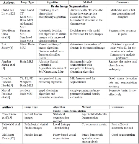
Chih-Chin Lai et.al proposed a clustering based hierarchical evolutionary algorithm [2] for MR Images. Hierarchical evolutionary algorithm (HEA) is used as a genetic algorithm. This approach automatically classifies the image into appropriate classes by means of a hierarchical structure in the chromosome. Wen-Hung Chao et.al developed a decision tree [3] to classify brain tissues in brain MR Images. The method is based on a decision tree with spatial information for MR Images. Spatial information on the general gray level, spatial gray level, and two dimensional wavelet transform is combined in-plane in two coordinate systems. The decision tree is constructed based on a binary tree with nodes created by splitting the distribution of input features of the tree. E A Zanaty [4]
proposed Kernelized Fuzzy C–means algorithm, which determines the number of clusters in the medical image. This optimal number of clusters helps in accurate segmentation in medical images. This algorithm checks the validity of the cluster analysis and number of clusters formed. In this method, Gaussian radial basis function classifier (GRBF) is used and the corresponding algorithms of Fuzzy C- means methods are derived. These derived algorithms are called as the kernelized fuzzy C-means (KFCM) and kernelized fuzzy C-means with spatial constraints (SKFCM). Jingdan Zhang et.al [1] proposed an algorithm for normal segmentation of brain MR images based on an Adaptive Spatial Clustering method. It is a competitive learning algorithm-Self Organizing Map. An adaptive spatial growing hierarchical is used for classification, fusing multi-scale segmentation with the competitive learning clustering algorithm which helps to overcome the problem of overlapping grey-scale intensities on boundary regions. An adaptive spatial distance is integrated with adaptive spatial growing hierarchical self organizing map. Lynn M. Fletcher-Heath [5] proposed automatic image segmentation of non-enhancing brain tumors in brain MR images. MR feature images used for the segmentation. Method is tested using three weighted images for each axial slice through the head. Segmentation is performed using an unsupervised fuzzy clustering algorithm. A knowledge-based system is constructed using domain knowledge. Image processing techniques combined with the knowledge based system helped in final tumor segmentation. Marcel Prastawa [6] et.al proposed a segmentation method for newborn brain analysis. Automatic segmentation of newborn MRI is challenging mainly due to the low intensity contrast and the growth process of the white matter tissue. In Newborn Brain MRI, it is necessary to identify the white matter tissue as myelinated or nonmyelinated regions. Robust graph clustering is used and parameter estimation is calculated to find the initial intensity distributions as the next step. Automatic segmentation is refined using training sample pruning and nonparametric kernel density estimation.
3.2 Liver Images
is the process of outlining the medical image manually, segmenting CT or MR images semi-automatically. It is because surface features of the liver and partial-volume effects make automatic discrimination from other adjacent organs or tissues very difficult. Oliver Gloger et.al [7] proposed an three step fully automatic liver segmentation based on LDA-based probability maps for multiple contrast MR images. Multiclass linear discriminant analysis is used for segmentation. The probability maps generated are then used for segmentation. They also proposed a fully automatic three-step 3D segmentation approach based upon a modified region growing approach and a further threshold technique. Seong-Jae Lim et.al [8] discussed automatic liver segmentation for volume measurement in CT Images. In this method, recursive morphological filter with regionlabeling and clustering is utilized to detect the search range and to generate the initial liver contour. In this search range, deform liver contour using the labeling-based search algorithm following pattern features of the liver contour. In the final step, volume measurement is automatically performed on the segmented liver regions. Hassan Masoumi et.al discusses automatic liver image segmentation based on an iterative watershed algorithm and artificial neural network [9]. In this method an edge preserved enhancement of the liver image is performed and it utilizes the properties of both Multilayer perceptron (MLP) neural networks and watershed algorithm. Trained neural network is used to extract the features of the liver region. They are used to monitor the quality of the segmentation using the watershed transform and adjust the required parameters automatically. This algorithm extracts liver region in one slice of the MRI images and the boundary tracking algorithm is suggested to extract the liver region in other slices. Laszlo Rusko et.al [10] proposed automatic image segmentation of the liver from multi- and single-phase contrast-enhanced CT images. Two methods are discussed to automatically segment abdominal CT images for segmenting liver region. The method uses region-growing facilitated by pre- and post-processing functions for multi-phase CT images and portal-venous phase for portal-venous CT images.
3.3 Lung Images
Some automatic image segmentation method are discussed for used for segmenting lung region in abdominal CT images. First method discuss is the
Fuzzy entropy based optimization of clusters for the segmentation of lungs in CT scanned images proposed by M. Arfan Jaffar , Ayyaz Hussain and Anwar Majid Mirza [11]. This method utilizes Fuzzy Entropy and Morphology segmentation methods. Fuzzy entropy is used for determining dynamic and adaptive optimal threshold. Histogram based background removal operator is proposed for removing the background areas. Xiangrong Zhou et.al [12] discussed automatic segmentation and recognition of anatomical lung structures from high-resolution chest CT images. This method recognize lung anatomical structures in chest by segmenting the different chest internal organ and tissue regions sequentially from high-resolution chest CT images. A sequential region-splitting process is used to segment lungs, airway of bronchus, lung lobes and fissures based on the anatomical structures and statistical intensity distributions in CT images. Jiantao Pu et.al [13] proposed technique for lung segmentation Adaptive border marching algorithm. It smoothes the lung border in a geometric way and can be used to reliably include juxtapleural nodules while minimizing over segmentation of adjacent regions such as the abdomen and mediastinum.
3.4 Cardiac Images
Here in this section we discuss one image segmentation methods used for segmenting cardiac images. First we discuss a comprehensive approach for segmenting left ventricle from cardiac short axis cine MR images [14]. This procedure first locates the region of interest from the image. ROI is extracted using the center point of the LV blood pool. Image in ROI is then transformed to a rectangular image by polar transformation. In the transformed image, canny edge detection and one-way region growing methods are applied iteratively to compute the endocardial boundary and epicardial boundary. After contour extraction, reverse polar transformation is applied on to the result. Thresholding is applied onto the image to find candidate regions of papillary muscles. Finally mathematical morphology is applied to pick up the two biggest regions as the papillary muscles.
3.5 Fundus Images
diseases .Fundus images help doctor in proper diagnosis of eye sight issues without any human intervention. Retinal vessel segmentation, Cataract detection, age related sight problems through these fundus images. Cemel Kose et.al discussed statistical segmentation method used for measuring age-related macular degeneration (ARMD) in retinal fundus images [15]. ARMD can be measured from retinal fundus images. The statistical properties of healthy textures around the macula are obtained and optic disc and macular area are detected. Apply statistical segmentation method for segmentation of healthy areas
in the macula. For highlighting the region of interest, inverse of the segmented image is obtained. Fabiola M et.al proposed a fast, efficient and automated method to extract vessels in fundus images[16]. In this technique an automatic threshold value is used for extracting vessels from fundus images. This threshold value is determined for each input image by the information contained in the image data. Local entropy acts as the threshold value. Here local entropy threshold is determined using the co-occurrence matrix.
IV.CONCLUSION
With the development of imaging technology, patients are getting utmost care in their disease diagnosis and treatment. Automatic image segmentation has been a popular research in medical imaging field. Manual identification of the diseases is difficult and sometimes it may lead to the wrong diagnosis of the disease. In this paper, automatic segmentation techniques im medical imaging such as Brain images, Lung images, Liver images, Fundus images, Cardiac images are discussed. This paper also investigated the advantages and limitations of each methods used in medical imaging.
V.
REFERENCES
[1]. Jingdan Zhang and Daoqing Dai, "An adaptive spatial clustering method for automatic brain MR Image segmentation”, Progress in Natural Science 19 (2009) 1373-1382, Elsevier.
[2]. Chih-Chin Lai and Chuan Yu Chang, "A hierarchical evolutionary algorithm for automatic medical image segmentation” Expert Systems with Applications 36 (2009) 248-259, Elsevier. [3]. Wen-Hung Chao et.al, "Automatic segmentation
of magnetic resonance images using a decision tree with spatial information” , Computerized Medical Imaging and Graphics 33 (2009) 111-121,elsevier
[4]. E A Zanaty, "Determining the number of clusters for kernelized fuzzy C-means algorithms for automatic medical image segmentation”, Egyptian Informatics Journal (2012) 13, 39-58
[5]. Lynn M. Fletcher-Heath et.al ",Automatic segmentation of non-enhancing brain tumors in magnetic resonance images” Artificial Intelligence in Medicine 21 (2001) 43-63.elsevier [6]. Marcel Prastawa, John H. Gilmore, Weili Lin and Guido Gerig,” Automatic segmentation of MR images of the developing newborn brain ”, Medical Image Analysis 9 (2005) 457-466, Elsevier
[7]. Gloger et.al, "A fully automatic three-step liver segmentation method on LDA-based probability maps for multiple contrast MR images”, Magnetic Resonance Imaging 28 (2010) 882-897, Elsevier.
[8]. Seong-Jae Lim, Yong-Yeon Jeong and Yo-Sung Ho, "Automatic liver segmentation for volume measurement in CT Images”, J. Vis. Commun. Image R. 17 (2006) 860-875,Elsevier.
[9]. Hassan Masoumi et.al, "Automatic liver segmentation in MRI images using an iterative watershed algorithm and artificial neural network”, Biomedical Signal Processing and Control 7 (2012) 429- 437, Elsevier.
[10]. Laszlo Rusko, Gyorgy Bekes and Marta Fidrich, "automatic image segmentation of the liver from multi- and singlephase contrast-enhanced CT images" , Medical Image Analysis 13 (2009) 871-882 ,Elsever.
[11]. M. Arfan Jaffar , Ayyaz Hussain and Anwar Majid Mirza, "Fuzzy entropy based optimization of clusters for the segmentation of lungs in CT scanned images”, Knowl Inf Syst (2010) 24:91-111 DOI 10.1007/s10115-009-0225-z
[13]. Jiantao Pu et.al, "Adaptive border marching algorithm: Automatic lung segmentation on chest CT images” Computerized Medical Imaging and Graphics 32 (2008) 452-462
[14]. Su Huang et.al, "An Image-Based Comprehensive Approach for Automatic Segmentation of Left Ventricle from Cardiac Short Axis Cine MR Images” Society for Imaging Informatics in Medicine Online publication 10 July 2010.
[15]. Cemel Kose et.al, "A Statistical Segmentation Method for Measuring Age-Related Macular Degeneration in Retinal Fundus Images”, J Med Syst (2010) 34:1-13 DOI 10.1007/s10916-008-9210-4 Springer.
[16]. Fabiola M et.al, "A fast, efficient and automated method to extract vessels from fundus images” J Vis (2010) 13:263 270 DOI 10.1007/s12650-010-0037-y
[17]. Giri Babu Kande , P. Venkata Subbaiah and T. Satya Savithri, "Unsupervised Fuzzy Based Vessel Segmentation In Pathological Digital Fundus Images” J Med Syst (2010) 34:849-858, Springer.
[18]. Sharma.N , Aggarwal LM. Automated medical image segmwntation techniques. J Med Phys 2010; 35(1),pp 3-14
[19]. Raj Acharya,et.al, BIOMEDICAL IMAGING MODALITIES: A TUTORIAL, Computerized Medical Imagmg and Graphics. Vol. 19, No. I. pp. 3-25, 1995
[20]. Sasa Galic; Sven Loncaric Cardiac image segmentation using spatiotemporal clustering Proc. SPIE 4322, Medical Imaging 2001: Image Processing, (3 July 2001)
[21]. M.F. Santarelli , Automated cardiac MR image segmentation: theory and measurement evaluation
[22]. Xiahai Zhuang; Rhode, K.S.; Razavi, R.S.; Hawkes, D.J.; Ourselin, S., "A Registration-Based Propagation Framework for Automatic Whole Heart Segmentation of Cardiac MRI," Medical Imaging, IEEE Transactions on, vol.29, no.9, pp.1612,1625, Sept. 2010
[23]. Dakua, S.P., "Annularcut: A graph-cut design for