Research Article
FORMULATION, DEVELOPMENT AND
IN-VITRO
RELEASE EFFECTS OF ETHYL CELLULOSE
COATED PECTIN MICROSPHERES FOR COLON TARGETING
Department of Pharmaceutical Technology, Calcutta Institute of Pharmaceutical Technology and Allied Health Sciences, Howrah-711316, West Bengal, India. E mail: [email protected]
Received: 14 August 2013, Revised and Accepted: 2 September 2013
ABSTRACT
Objective: The objective of present investigation is to design a colon targeted microspheres of 5-flourouracil by using natural polysaccharide based carrier which is inexpensive and naturally occurring and also having hydrophilic and swelling properties.
Methods: The pectin microspheres were prepared by ionotropic-external gelation technique and drug loaded pectin microspheres were coated with ethyl cellulose by co-acervation phase separation method.
Results and Conclusion: The in-vitro drug release effects behavior of 5-flourouracil microspheres done in various pH conditions for pectin
microspheres, ethyl cellulose coated pectin microspheres and ethyl cellulose coated pectin microspheres in presence of pectinase enzyme up to 12
hr. The prepared microspheres were characterized by entrapment efficiency, particle size, micromeritic properties, in-vitro release behavior,
scanning electron microscopy (SEM), fourier transform infrared spectroscopy (FTIR) etc. It was observed that increasing the polymer concentration along with the cross-linking time given the better affect of microspheres characteristic and percentage release of drug.
Keywords: 5-Flourouracil; Natural Polysaccharide; Ethyl cellulose; Ionotropic gelatination; Pectinase enzyme.
INTRODUCTION
Colon-specific drug-delivery systems offer several potential therapeutic advantages. In a number of colonic diseases such as colorectal cancer, crohn’s disease and spastic colon, it has been
shown that local is more effective than systemic delivery. Colonic
drug delivery can be achieved by oral or by rectal administration. The absorption and degradation of the active ingredient in the upper part of the gastrointestinal tract is the major obstacle with the delivery of drugs by the oral route and must be overcome for successful colonic drug delivery. The digestive enzymes of the gastrointestinal tract generally degrade these agents. However, these enzymes are present in significantly lower amounts in the colon compared with the upper portion of the gastrointestinal tract [1]. Colon-specific drug delivery has been attempted in a number of ways that primarily seek to exploit the changes in the physiological parameters along the gastrointestinal tract [1,2].
Pectins are a family of complex polysaccharides that contain 1,4-linked α-D galactosyluronicresidues. The ability of pectin to rapidly form viscous solutions and gels on contact with aqueous media has been exploited by the pharmaceutical industry in its wide application as a carrier in oral controlled release dosage forms.
Sodium alginate (NaAlg), [3] a water soluble salt of alginic acid, is a natural polysaccharide extracted from marine brown algae. Sodium alginate (NaAlg), [3] has been used as a matrix for entrapment of drugs and macromolecules. A mixture of pectins with other polysaccharides such as alginate has found that good gels are formed from high methoxy pectin and guluronic rich alginates. A pH above 4 also hinders the gel formation.
The selection of appropriate coating material decides the physical and chemical properties of the resultant microcapsules or microspheres. The polymer should be capable of forming a film that is cohesive with the core material. A number of coating materials have been used successfully examples of these include gelatin, polyvinyl alcohol, ethyl cellulose, cellulose acetate phthalate and styrene maleic anhydride.
Pectinex ultra SP-L (Pectinase) is a commercial enzyme preparation
from Aspergillus aculeatus used in the food industry for fruit juice
processing to reduce viscosity. It contains different pectinolytic and cellulolytic enzymes (endo-poly-galacturonase, endo-pectinylase
and pectin esterase),and other activities such as ß-galactosidase, [4]
chitinase, and transgalactosidase.
5-Fluorouracil [5] is an antineoplastic anti-metabolite.
Anti-metabolites masquerade as purine or pyrimidine which become the building blocks of DNA. Fluorouracil blocks an enzyme which converts the cytosine nucleotide into the deoxy derivative. It interferes with DNA synthesis by blocking the thymidylate synthetase conversion of deoxyuridylic acid to thymidylic acid. Ionotropic gelation [1] by divalent metal interaction was employed as an approach to design a modified release multiple-unit oral drug-delivery system. Ionic gelation involves cross-linking of polyelectrolytes in the presence of multivalent counter ions. Spraying a sodium alginate solution into calcium chloride solution produces rigid gel particles. The starting materials and stabilizing additives are dispersed in an aqueous solution.
The present study was to design a colon targeted system for site-specific delivery of 5-fluorouracil using by natural polysaccharides and pH-sensitive polymer for the treatment of colon cancer.
MATERIALS AND METHODS
Materials
5-Flourouracil was obtained as a gift sample from Get Well Life
Science Pvt. Ltd., Delhi, India and Pectin, Span 80 were purchased from Roehm Pharma, Germany and Sodium alginate, Ethyl cellulose, n-Hexane were purchased from Merck Specialities Pvt. Ltd., Mumbai, India and Pectinex Ultra SP-L was purchased from Novozyme, U.S.A and others reagents were of analytical grade.
Methods
Method of Preparation of Microspheres [6] The pectin
containing 6% w/v solution of calcium chloride with continuous stirring by magnetic stirrer. Then the solution containing the gel formed microspheres was filtered by using Whatman filter paper no-1. The microspheres were allowed to dry at about 30 to 40°C and stored in well closed container for further use.
Preparation of Ethyl Cellulose Coated Pectin Microspheres [7]
Drug loaded pectin microspheres were used as a core material for the preparation of double-coated system. A co-acervation phase separation method was applied for this step. A known amount of the microspheres having particle size of 100-250 µm was dispersed in an ethyl acetate (25ml) solution containing ethyl cellulose and containing 0.02% w/v span 80. This mixture was agitated for 5 min at 400 rpm. Subsequently 50 ml n-hexane was poured into the polymeric solution containing the core material with the rate of 1 ml/min and core: coat ratio is 1:3. The medium was stirred for 60 min to complete the process of microparticles coating. Coated microspheres were then washed with an excess of n-hexane, filtered and dried at room temperature.
Process Variables The following process variables were
investigated (Bore diameter of the needle; concentration of pectin and sodium alginate; concentration of calcium chloride; height of dropping stirring speed and stirring time) and the different batches thus produced were analyzed for size, shape, drug content and drug release.
Fourier Transform Infrared Spectroscopy (FTIR) Studies The
infrared (IR) spectra were recorded using an FTIR
spectrophotometer (Perkin Elmer Spectrum GX) by the KBr pellet
method in the wavelength region between 400 and 4000 cm-1. The
spectra obtained for flourouracil and physical mixtures of 5-flourouracil with polymers were compared to check compatibility of drug with polymers.
Differential Scanning Calorimeter (DSC) Studies [8]
Thermograms of the samples were obtained by a Perkin-Elmer differential scanning calorimeter (Pyris 6 DSC, software Pyris manager, Perkin-Elmer Schweiz AG, Hunenberg, Switzerland). Samples of 3 mg were accurately weighed into aluminum pans and then hermetically sealed with aluminum lids. The thermograms of samples were obtained at a scanning rate of 10°C/min over a temperature range of 50 to 350°C.
Drug Entrapment Efficiency, Drug Loading and Yield Percentage
[6]The amount of 5-FU present in the Pectin microspheres was
determined by taking the known amount of microspheres in which 10 mg of drug should be present theoretically. Then the microspheres ware crushed and the powdered microspheres was taken and dissolved in 10 ml of phosphate buffer (pH-7.4) solution and stirred for 15 minutes with an interval of 5 minutes and allowed to keep for 24 hours. The solution was filtered through Whatman No.1 filter paper. 0.1ml of this solution was diluted up to 10ml with phosphate buffer (pH-7.4) solution and the absorbance was measured spectrophotometrically at 267.5 nm against phosphate buffer (pH-7.4) solution as blank with the help of UV double beam spectrophotometer and concentrations were determined by employing simultaneous equation: Y= mx+c
Drug entrapment efficiency (DEE) percentage (%) = [Experimental
drug Content/ Initial Drug Content into the Formulation] 100
Drug Loading percentage (%) = [ Qm / Wm] 100 Where, Wm =
Weight of the microspheres; Qm = Quantity of the drug present in Wm
microspheres. microspheres was calculated by using the following formula:
Mean particle size = (mean particle size of the fraction weight
Carr’s Index [10]Compressibility index (Ci) or Carr’s index value of
microparticles was computed according to the following equation: Carr’s index (%) = [(Tapped density – Bulk Density) / Tapped Density] × 100.
Hausner’s Ratio [10] Hausner’s ratio of microparticles was
determined by comparing the tapped density to the bulk density using the equation: Hausner’s Ratio = Tapped density / Bulk Density.
Surface Topography [10]The samples for the scanning electron
microscope (SEM) analysis were prepared by sprinkling the microspheres on one side of an adhesive stub. Then the microspheres were coated with gold before microscopy. Finally the morphology and size of the microspheres were observed with the scanning electron microscope (FEI Quanta-200 MK2, Netherlands).
In-Vitro Drug Release Study in Stimulated Gastrointestinal
Fluids [11]Ethyl cellulose-coated pectin microspheres and uncoated
pectin microspheres were evaluated for the in vitro drug release in simulated GI fluids (SGF). The drug dissolution test of microspheres was performed by the paddle method specified in USP XXIII. Microspheres (100 mg) were weighed accurately and gently spread over the surface of 900 mL of dissolution medium (SGF). The content was rotated at 100 rpm at 37°C ± 0.5°C. The simulation of GI transit condition was achieved by altering the pH of dissolution medium at different time intervals. The pH of the dissolution medium was kept
1.2 for 2 hours using 0.1 N HCl. Then KH2PO4 (1.7 g) and
Na2HPO4.2H2O (2.25 g) were added to the dissolution medium
adjusting the pH to 4.5 with 1.0 M NaOH and the release rate study was continued for an additional 2 hours. After 4 hours, the pH of the dissolution medium was adjusted to 6.8 with 0.1 N NaOH and maintained up to 2 hours. After 6 hours, the pH of the dissolution medium was adjusted to 7.4 with 0.1 N NaOH and maintained up to 2 hours. The samples were withdrawn from the dissolution medium at various time intervals using a pipette fitted with a microfilter. The rate of drug release was analyzed by using UV -visible
spectrophotometry at λmaxof 267.5 nm.
In-Vitro Drug Release Study in the Presence of Pectinase
EnzymeEhyl cellulose-coated pectin microspheres were evaluated
for the in-vitro drug release in the presence of pectinase enzyme. Ethyl cellulose-coated pectin microspheres were weighed accurately and gently spread over the surface of 900 mL of dissolution medium (SGF). The content was rotated at 100 rpm at 37°C ± 0.5°C. The pH of the dissolution medium was kept 1.2 for 2 hours by using 0.1 N HCl.
Then KH2PO4 (1.7 g) and Na2HPO4.2H2O (2.25 g) were added to the
dissolution medium by adjusting the pH to 4.5 with 1.0 M NaOH and the release rate study was continued for an additional 2 hours. After 4 hours the pH of the dissolution medium was adjusted to 6.8 with 0.1 N NaOH and maintained up to 2 hours. After 6 hours, the pH of the dissolution medium was adjusted to 7.4 with 0.1 N NaOH, add 3 ml of pectinase enzyme (pectinex ultra spl) and maintained up to 2 hours. The samples were withdrawn from the dissolution medium at various time intervals using a pipette fitted with a microfilter. The
rate of 5-FU release was analyzed using UV -visible
spectrophotometry at λmaxof 267.5 nm.
Swelling Study [11] A known weight (100 mg) of various 5-flourouracil loaded pectin microspheres were placed in enzyme-free simulated intestinal fluid (pH 7.4) and allowed to swell for the required period of time at 37ºC ± 0.5ºC. The microspheres were periodically removed and blotted with filter paper then their change in weight was measured until attainment of equilibrium. The swelling ratio (SR) was then calculated using the following formula:
SR= (Wg – Wo) / Wo, where, Wg is final weight, Wo is initial weight
of formulation.
RESULTS AND DISCUSSION
In this project attempts have been made to prepare the pectin - alginate microspheres bearing 5- fluorouracil by ionotropic gelation technique. On characterizing the microspheres it was observed that the pectin concentration as well as cross-linking time affect the microspheres characteristics and the percentage release of drug. Better results were found by increasing the polymer concentration along with the cross-linking time.
The process variables were investigated and the different batches thus produced were analyzed for size, shape, drug content and drug release. Optimized process variables are described by Table 1.
Table 1: Process Variables Optimized Data
Table 2: In-vitro Release Kinetics Parameters for 5-Flourouracil Microspheres
Zero Order Model
First-Order
Model H-M Model Korsmeyer-Peppas Model
Fig 3: FTIR Spectra of Sodium Alginate. Fig 4:FTIR Spectra of 5-Flourouracil Microspheres with Pectin and
Sodium Alginate.
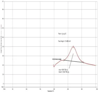
Fig 5: DSC Thermogram of 5-Flourouracil. Fig 6: DSC Thermogram of Pectin.
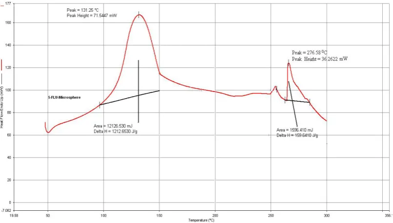
Fig 7: DSC Thermogram of Sodium Alginate. Fig 8: DSC Thermogram of Physical Mixture Containing Drug and Polymers
Fig 9: DSC Thermogram of the Formulation
Fig 10: Histogram Diagram of Percentage Fig 11: Mean Particle Size of F1 to F9 Drug Entrapment Efficiency
Fig 12: SEM of 5-Flourouracil Microspheres Fig 13: SEM of 5-Flourouracil Microspheres of EPMS-F8 after Dissolution of PMS-F8 before Dissolution
55
60
65
70
75
F1F2F3F4F5F6F7F8F9
%
D
R
U
G
E
N
TR
A
P
M
EN
T
FORMULATION CODE
% DRUG ENTRAPMENT EFFICIENCY
0
200
400
600
800
F1 F2 F3 F4 F5 F6 F7 F8 F9
M
EA
N
P
A
R
TI
C
LE
S
IZE
FORMULATION CODE
mixtures shows that there was no significant interaction between drug and polymers as shown in Fig 1 to 4. By DSC study there is no detectable endotherm if the drug is present in a molecular dispersion or solid solution state in the polymeric microspheres loaded with drug. In the present investigation, DSC thermograms of pure drug, pure pectin, pure sodium alginate, drug and polymer physical mixtures as shown in Fig. 5 to 9, prominent melting endotherms of pure 5- fluorouracil and a physical mixture of drug and polymer were found at 294.70°C and 279.39°C. Drug-loaded
Pectin microspheres showed a broad small peak at 276.58°C, indicating the presence of drug in crystalline form.
The drug loading ability increased from 11.53 ± 0.050% to 13.55 ± 0.008% of microsphere with increasing the amount of polymer as well as cross linking time..
Particle size can be determined by sieve analysis method. The mean diameter of pectin microspheres increased from 601± 0.81µm to 720 ± 0.68µm. The average particle size of microspheres increased with increasing the concentration of the polymer. The average size of pectin microspheres which can describe by Fig.11.
The angle of repose of formulation within the range of 30˚, indicating good flow properties for the microspheres. The tapped density 0.018 % which indicates good flow property of microspheres.
Scanning electron microscopy was used to observe the surface morphology of pectin–alginate microspheres by after dissolution study and before dissolution study which can describe by Fig. 12 to 13.
The Pectin- alginate microspheres were subjected to in-vitro drug
release rate by dissolution profiles of 5-flourouracil is shown in Fig. 14 to 16. Ethyl cellulose coated pectin- alginate microspheres were
subjected to in-vitro drug release rate by dissolution profiles of
flourouracil is shown in Fig. 17 to 19.The initial drug release of 5-flourouracil microsphere at 1hr is1.33% and then found 91.02 % at
the end of 12 hr. In-vitro drug release of ehyl cellulose-coated pectin
microspheres in the presence of pectinase enzyme found 94.21% at the end of 12 hr.
It was found that drug release rate decreased as the concentration of pectin increased. It was also concluded that the drug release rate was decreased as the cross-linking time increased. The results of dissolution data from dissolution profile fitted to various drug release kinetic equations of zero order, first order, Higuchi and korsemeyer‐peppas having r, n and k. r is value of correlation coefficient, k is a release rate constant and n is the diffusional release exponent. The drug release kinetic of different formulation can be described by Table 2.
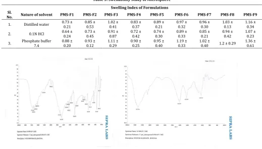
Pectin - alginate microspheres swell in water, 0.1N HCl and phosphate buffer 7.4.the result of swellability index can be described by Table 3. As a result of increasing the polymer concentration and cross- linking time the overall swelling of polymer increased significantly.
CONCLUSION
In the present study a formulation method was developed and in- vitro characterization of pectin - alginate based microspheres were prepared for colon specific drug delivery where pectin and alginate act as a natural Polysaccharides carrier which are inexpensive and naturally occurring and also having hydrophilic and swelling properties.The FTIR spectra’s and DSC study revealed that there were no interaction between polymers and drug. The Polysaccharides remain intact in the physiological environment of stomach and small intestine but degrade in colon by enzymatic
degradation. This natural polymer is also appealing for use in drug delivery for a wide range of molecular weights, varying chemical compositions, low toxicity and biodegradability. Therefore more research is necessary to be focused on the specificity of drug uptake at the colon site. It could be concluded that the ethyl cellulose coated pectin-alginate microspheres can be used for effective and controlled delivery of anticancer drug.
ACKNOWLEDGEMENTS
Authors wish to give thanks to Calcutta Institute of Pharmaceutical Technology and A.H.S. authority for constant support and given research laboratory to carry out this project work. We also thanks to Dr. Reddy’s Laboratories Ltd., Hyderabad, India for providing gift sample of diltiazem hydrochloride .We also acknowledge the help provided by our fellow colleagues in completion of the project.
REFERENCES
1. Vyas SP, Khar RK, Targeted and Controlled Drug Delivery
Novel Carrier System. 1st ed. New Delhi, India: CBS
Publishers; 2002: 38-40.
2. Fasano A, Novel approaches for oral delivery of
macromolecules. J Pharm Sci 1998; 87: 1351-1356.
3. Davis SS, Overcoming barriers to the oral administration
of peptide drugs. Trends Pharm Sci 1990; 11: 353-355.
4. DeLuca PP, Hickey AJ, Hazrati AM, Wedlund P, Rypacek F,
Kanke M, Topics in Pharmaceutical Sciences. In: Breimer DD, Speiser P. ed. New York: Elsevier; 1987: 429-442.
5. Hartalsky A, Salicylazobenzoic acid in ulcerative colitis.
Lancet 1982; 1: 960.
6. Mazumder R, Nath LK, Haque A, Maity T, Choudhury PK,
Shrestha B, Chakraborty M, Pal N, Formulation and in vitro evaluation of natural polymers based microspheres
for colonic drug delivery. International J Pharm
Pharmaceut Sci 2010; 2 (1): 211-219.
7. Atyabi F, Vahabzadeh R, Dinarvand R, Preparation of
Ethylcellulose coated gelatin microspheres as a
multiparticulate colonic delivery system for
5-aminosalicilic acid. Iranian J Pharmaceut Researc 2004; 2: 81-86.
8. Rahman Z, Kohli K, Khar RK, Ali M, Charoo NA, Shamsher
AA, Characterization of 5-fluorouracil microspheres for
colonic delivery.AAPS Pharm Sci Tech 2006; 7(2): 47.
9. Subrahmanyam CBS, Textbook of Physical Pharmaceutics.
2nd ed. New Delhi, India: Vallabh Prakashan; 2000: 85.
10. Michael EA, Pharmaceutics the Design and Manufacture of
Medicines. 3rd ed. New York: Churchill Livingstone
Elsevier; 2007: 109-168.
11. Paharia A, Yadav AK, Rai G, Jain SK, Pancholi SS, Agrawal