CSEIT184510 | Published - 14 April 2018 | March-April-2018 [ (4 ) 5 : 81-87 ]
81
Calculation of bone Disease using Image Processing
Dr. M. Yuvaraju1, R. Haripriya2
1 Assistant Professor, 2 PG Scholar
Electrical & Electronics Engineering, Anna University Regional Campus, Coimbatore, India
ABSTRACT
The designed system used to measure the bony fracture by using image processing and it also detects the bone tumor of the person. Every human has a variousossein length so by applying imageprocessing it also give the length of the bony. The bony tumor is not a cancer it just form like a tissue formed over the osseous matter it reduce the bony strength. It very useful in real time.This mainly contribute about the bone density and it also checkoutwhat are all the causes for bone weakness. It also gives the 3 dimensional view of the density distribution in the human body. And it gives the visage like height, weight of the human. The system contributes on estimation of lower limb kinematic analysis. It finds the GRF between the human body and ground
Keyword: GRF, Ossein Length, Kinematic Analysis, Osseous
I.INTRODUCTON
CSEIT184510 | Published - 14 April 2018 | March-April-2018 [ (4 ) 5 : 81-87 ]
82
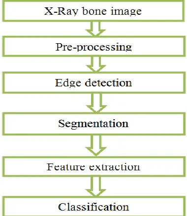
surgical resection and for defining the treatment modalities of patients who will undergo radiotherapy.The strength of a bone can be determined based on BMD. In addition, BMD measures the amount of bone mass, predicts fracture risks, observes the usefulness of treatment, and measures the amount of calcium in a specific region of the bone. This process used to detect the bone fracture in step by step process input as bone image, and the preprocessing helps to improve the image data, edge processing used to find the boundaries of an image, and the segmentation used for partitioning and then it classify the images
Fig 1 : The flow diagram of steps in detecting the bone fracture in X-ray/CT images
II.NOISE REMOVAL
In computer-aided analysis of the medical imagery, image processing tools for noise removal, image segmentation and characteristic removal play a important role in the achievement of such systems. The X-ray/CT images are obtained from the hospital that contains normal as well as fractured bones images. In the first step, apply preprocessing technique such as RGB to grayscale exchange and take out the noise from the image by via the median filter.
Noise can be defined as unwanted pixels present in the image that degrade the quality of the image. It can be written as:
f (x, y) = g (x, y) + η (x, y)
CSEIT184510 | Published - 14 April 2018 | March-April-2018 [ (4 ) 5 : 81-87 ]
83
black dots. It can be detached by apply mathematical conversion on the images. It conserve the boundaries while remove noise. The median filter is a nonlinear digital filtering technique, used to remove noise such as salt and pepper noise.
III. RELATED WORKS
In 2014 Elyse passmore and morgan sangeux proposedthe 3D gait analysis system that has been used to improving volume origin and coordinate system and 3D system improvise the system performance. In 2014 Gennady and yu.Kulikova has been Accurate Numerical implementation of continuous-discrete kalman filter that used to involve the activity of coordinate system performance and it efficiency level of an input. Here, the dicom used to view the 3 dimensional view of bone density it gives the exact level of a density in fig1 and it also finds the all supplements in bone. To find a bone cancer and fracture itgives high resolution to find level of a cancer and fracture in the bone. It is the combined process of a bone feature extraction.
Edge Detection
Edge detection issignificantprocess in image processing, that decrease the number of pixels and save the arrangement of the image by decisive the boundaries of objects in the image. Edge detection is the method of identifying points in a digital image at which the image brightness changes sharply or, more formally, has discontinuities. The points at which image brightness changes sharply are typically organized into a set of curved line segments termed edges. There are two general approaches to edge detection that are commonly used are: gradient and Laplacian. Gradient method use the first derivative of the image, and the Laplacian method use the second derivative of the image to find edges. In our method use sobel edge detector and it is a gradient family.
Segmentation
CSEIT184510 | Published - 14 April 2018 | March-April-2018 [ (4 ) 5 : 81-87 ]
84
algorithm distance is squared or absolute difference between a pixel and cluster center is calculated. The difference is typically based on pixel intensity, color, texture and location. The quality of the solution depends on the initial set of clusters and value of k. After the segmentation crop the image and the area of fracture with some limitation.
Feature Extraction
Feature extraction is the main step in various image processing applications. Gray-Level Co-occurrence Matrix is used for feature extraction and selection. GLCM was defined by Haralick et al. in 1973.GLCM is main tool used in image texture analysis. Textures of an image are complex visual patterns that are composed of entities or regions with sub-patterns with the characteristics of brightness, color, shape, size, etc. GLCM is a statistical way to indicate image texture structure by statistically sampling the pattern of the grey-levels occurs in relation to other grey levels. We use the Gray Level Co-occurrence Matrix (GLCM) method to extract textural features such as entropy, contrast, correlation, homogeneity.
V.BONE DENSITY
It finds by, The T-score is the standard deviation (SD) of the patient’s BMD above or below the mean for the Young-Adult normal reference population. The Z-score is the SD of thepatient’s BMD above or below the mean of the Age-Matched
T Score = (BMD of patient - Average BMD of Young-Adult) ______________________________
SD of Young-Adult
CSEIT184510 | Published - 14 April 2018 | March-April-2018 [ (4 ) 5 : 81-87 ]
85
V.RESULTS
The result of bone density in 3D view
Fig1: bone density
Fig2: bone fracture
CSEIT184510 | Published - 14 April 2018 | March-April-2018 [ (4 ) 5 : 81-87 ]
86
Fig4: bone cancer detection VII. CONCLUSION
Here, we can able to predict the disease of bone density ,bone cancer and other bone diseases, The given image processing increase the saturation level of an output. So we can able to identify the exact level of an disease. It increase the output efficiency of an given input.
VIII. FUTURE WORK
Hardware implementation for bone density calculation
REFERENCES
[1] Elyse Passmore, Morgan Sangeux,“Improving repeatability of setting volume origin and coordinate system for 3D gait analysis,” Gait & Posture, vol. 39, pp. 831-833, 2014.
[2] L. Tesio, V. Rota, C. Chessa, and L. Perucca, “The 3-D path of body center of mass during adult human walking on force treadmill,J. Biomechan., vol. 43, no. 5,
[3] Arnaud Faivre, Marc Dahan, Bernard Parratte, GuyMonnier, “Instrumented shoes for pathological gait assessment,” Mechanics Research Communications, vol. 31, pp. 627-632, 200[4] S.S. Galen, C.J. Clarke, D.B. Allan, B.A. Conway, “A portable gait assessment tool to record temporal gait parameters in SCI,” Medical Engineering & Physics, vol. 33, pp. 626-632, 2011.
[4] Ruth E. Mayagoitia, Anand V. Nene, Peter H. Veltink, “Accelerometer and rate gyroscope measurement of kinematics: an inexpensive alternative to optical motion analysis systems,” Journal of Biomechanics, vol. 35, pp. 537-542, 2002.
[5] E.C. Wentink, S.I. Beijen, H.J. Hermens, J.S. Rietman, using EMG and kinematic P.H. Veltink, “Intention detection of gait initiation data,” Gait & Posture, vol. 37, pp. 223-228, 2013.
CSEIT184510 | Published - 14 April 2018 | March-April-2018 [ (4 ) 5 : 81-87 ]
87
[7] Jan Rueterbories, Erika G. Spaich, Ole K. Andersen, “Characterization of gait pattern by 3D angular accelerations in hemiparetic and healthy gait,” Gait & Posture, vol. 37, pp. 183-189, 2013. [8] Cola G., Avvenuti M., Vecchio A., etc., “An On- Node Processing Approach for Anomaly Detection in Gait,” IEEE Sensors Journal, vol. 15, pp. 6640-6649, Aug. 2015.
[9] T. Liu, Y. Inoue, K. Shibata, K. Shiojima, M.M. Han, “Triaxial joint moment estimation using a wearable three-dimensional gait analysis system,” Measurement, vol. 47, pp. 125-129, 2014.
[10] Jingyuan Cheng, Amft O., Bahle G., Luckowicz P., “Designing Sensitive Wearable Capacitive Sensors for Activity Recognition,” IEEE Sensors Journal, vol. 13, pp. 3935-3947, April 2013.
[11] Orengo G., Lagati A., Saggio G., “Modeling Wearable Bend Sensor Behavior for Human Motion Capture,” IEEE Sensors Journal, vol. 14, pp. 2307-2316, March 2014.
[12] Tao Liu, Yoshio Inoue, Kyoko Shibata, “A wearable force plate system for the continuous measurement of triaxial ground reaction force in biomechanical applications,” Measurement Science and Technology, vol. 21, no. 8, pp. 085804, 2010.
[13] Tao Liu, Yoshio Inoue, Kyoko Shibata, K. Shiojima, “A mobile force plate and three-dimensional motion analysis system for three-three-dimensional gait assessment,” IEEE Sensors Journal, Vol.12, No. 5, pp. 1461-1467, May 2012.
[14] Guangyi Li, Tao Liu, LinyiGu, Yoshio Inoue, HaojieNing, and Meimei Han, “Wearable gait analysis system for ambulatory measurement of kinematics and kinetics,” in Proc. 2014 IEEE Sensors, Valencia, Spain, 2014, pp. 1316-1319.
[15] Nikhil Raveendranathan, Stefano Galzarano, VitaliLoseu, etc., “From Modeling to Implementation of Virtual Sensors in Body Sensor Networks,” IEEE Sensors Journal, vol. 12, no. 3, pp. 583-593, March 2012.
[16] Zhou H, Stone T, Hu H, Harris N, “Use of multiple wearable inertial sensors in upper limb motion tracking,” MedEngPhys, vol. 30, pp. 123-133, 2008.