Characterization of novel F-actin envelopes surrounding
nuclei during cleavage of a polychaete worm
SABINE JACOBSOHN*
Institut für Zoologie, Fachbereich Biologie, FU Berlin, Germany
ABSTRACT F-actin accumulations and their possible functions were investigated during cleavage of the polychaete Ophryotrocha puerilis. Unusual cytoplasmic accumulations of F-actin were detected which have never been described before in animal embryos. As shown by TRITC-phalloidin labeling, envelopes of F-actin surrounded late prophase nuclei for a short period of time. DTAF-immunofluorescence of β-tubulin showed that the F-actin envelope was closely associated with microtubules of the developing spindle apparatus. However, experimental disassembly of microtubules by nocodazole did not prevent the assembly of the F-actin envelope. Disturbance of F-actin envelope formation by cytochalasin B did not alter the course of mitotic events, i.e. position of the nuclei and orientation of the spindle apparatus were not affected, although the respective blastomeres remained uncleaved. However, disassembly of the F-actin envelope correlated tempo-rally with breakdown of the nuclear envelope. Therefore, it is suggested that this new structure plays a role in fragmentation of the nuclear envelope during cleavage of Ophryotrocha puerilis.
KEY WORDS:
F-actin, late prophase nucleus, cleavage
0214-6282/99/$10.00 © UBC Press
Printed in Spain www.lg.ehu.es/ijdb
*Address for reprints: Freie Universität Berlin, Institut für Zoologie, Königin-Luise-Str. 1-3, 14195 Berlin, Germany. FAX: ++ (0)30 838 3916. e-mail: [email protected]
Original Article
Introduction
In the course of fast embryonic cell divisions, proper functions of the cytoskeleton during mitosis and cytokinesis are essential (Strome, 1993). Microtubules are especially involved in mitosis, whereas microfilaments primarily contribute to cytokinesis. Much interest has been focused on the cortical distribution of microfilaments during cytokinesis, e.g., contractile ring formation (Schroeder, 1973; Mabuchi, 1990, 1994). In contrast, little is known about the cytoplasmic distribution of microfilaments during cleavage.
In this study, the cytoplasmic distribution of microfilaments and their possible functions during cleavage of the polychaete Ophryotrocha puerilis are investigated using fluorescence microscopy techniques. Whole-mount embryos of O. puerilis are very well suited for fluorescence microscopy studies because of their low background staining which results in transparent prepara-tions in which the mitotic stage of each blastomere can be easily determined. Moreover, laboratory cultures of O. puerilis are very easy to maintain and embryos can be obtained throughout the year. O. puerilis is a protandric hermaphrodite and much interest has been focused on its sex determination and differentiation (Pfannenstiel, 1973a; Grothe and Pfannenstiel, 1986; Pfannenstiel et al., 1990). Insemination occurs via simultaneous release of oocytes and sperm from the body cavities of females and males, respectively. Fertilization takes place in the egg mass in the
seawater. Newly shed oocytes are arrested in met-anaphase of the first meiotic division (Ruthmann, 1964). Embryos are deposited in egg masses each containing about 50 to 150 eggs. Spiral cleavage of O. puerilis is unequal, i.e., the blastomeres of the 4-cell-stage can be distinguished by their size, the D-quadrant containing most of the cytoplasm of the embryo (for review of spiral cleavage: Dorresteijn, 1998).
In polychaete oocytes actin was described to be a structural component of the egg cortex (Eckberg and Anderson, 1995). Furthermore, microfilaments were shown to contribute to the cortical reaction following fertilization (Kluge et al., 1995; Dondua et al., 1997). However, there does not exist any information on the distribution and function of cytoplasmic actin during the early development of polychaetes.
In the present study, the cytoplasmic distribution of F-actin was determined by specific labeling with tetramethylrhodamine B isothiocyanate-conjugated phalloidin (TRITC-Phall; Wulf et al.,
Abbreviations used in this paper: ASW, artificial seawater; BSA, bovine serum
1979; Faulstich et al., 1988). The spatial relationship between the detected F-actin structures and the developing spindle apparatus was studied by indirect immunofluorescence staining of microtubules (anti-β-tubulin/DTAF). The mitotic stages of the blastomeres were demonstrated using 4’6 diamidino-2-phenylindol (DAPI)-staining. Furthermore, experiments were performed to investigate the role of the detected F-actin structures during early development of O. puerilis by the use of the cytoskeleton inhibitors cytochalasin B (CB), cytochalasin D (CD) and nocodazole (Noc).
Results
Distribution of F-actin and microtubules in zygotes and early embryos of O. puerilis
After fertilization and completion of meiosis, male and female pronuclei meet in early prophase. In late prophase each pronu-cleus is surrounded by F-actin accumulations which form an envelope-like structure (Fig. 1A,B,D,E). Thin sections of these pronuclei stages show indentations of the nuclear envelope at the respective sides of the centrosomes (Fig. 1C). Usually fusion of the pronuclei does not occur before metaphase chromosomes have been formed.
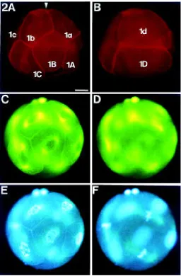
One embryonic cell cycle lasts about 90 min. Cleavage is unequal and results in blastomeres of different size (Fig. 2A,B). In one embryo, duration of the cell cycle differs among blastomeres of different size. The larger blastomeres exhibit relatively shorter
cell cycle durations, while the smaller blastomeres show relatively extended cell cycles. Therefore, the larger blastomeres of an embryo are always in advanced mitotic stage (Fig. 2C-F). Embryos of one egg mass do not always develop synchronously. Asynchro-nous development is thought to be due to asynchroAsynchro-nous fertiliza-tion of the shed eggs.
During cleavage, the spatial accumulations of F-actin are also formed around late prophase nuclei of the blastomeres. Figure 2 (A,B) shows an 8-cell-stage embryo with F-actin envelopes in the blastomeres 1a, 1b and 1C. In each cell cycle, the F-actin envelope exists only for a short period of time. It is present in late prophase (Figs. 2A,E; 3E-G) and exists for approximately 10 min before it suddenly disappears. Exact determination of the duration of this stage is not possible, because 1) the embryos of one egg mass do not always develop synchronously and 2) fixed material had to be used in this investigation. Consequently, the observed states only represent the appearance of an embryo at the time of fixation.
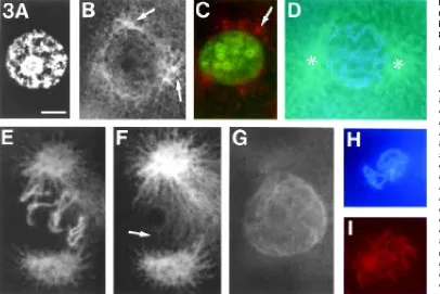
nucleus (Fig. 3C). In early prophase, when the chromatin was organized in thin threads, the centrosomes were located opposite to each other and lay in the respective mitotic plane (Fig. 3D). Each centrosome built up an aster with microtubules growing in the direction of the cortex. In late prophase, when the F-actin envelope surrounded the nucleus, polar microtubules extended from one spindle pole to the other forming a basket-like scaffold that enclosed the F-actin envelope (Fig. 3E-G). At this stage, there were more or less prominent protrusions of microfilaments extend-ing from either side of the envelope to the centrosomes (Figs. 1B,D; 2A,C,E). The F-actin envelope disappeared at prophase-metaphase transition, at the time when breakdown of the nuclear envelope normally occurs. Occasionally, some microfilaments could be detected in the vicinity of the chromosomes (Fig. 3H,I). They are thought to be remnants of the disrupted F-actin envelope.
Role of F-actin envelope formation in cleavage of O. puerilis
Embryos were incubated in artificial seawater (ASW) contain-ing either CB, CD or Noc. Cytochalasins are commonly used in investigating cellular actin dynamics. They act by either inhibiting actin polymerization or severing polymers (Cooper, 1987; Sampath and Pollard, 1991). Noc is known to inhibit microtubule polymeri-zation as described by Samson et al. (1979) and Vasquez et al. (1997).
After inhibitor-treatment, the embryos were fixed and stained. For each set of experiments a control group of embryos developed untreated. During the experiment, the mitotic stages of the control embryos and of the inhibitor treated embryos were determined in vivo using DAPI-staining. Embryos that were stained in vivo with DAPI were discarded after examination.
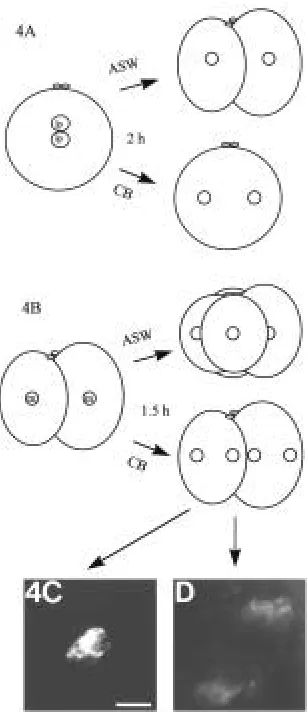
Incubation in 10 µg CB/ml ASW or 30 µg CD/ml ASW respec-tively resulted in disturbed formation of the F-actin envelope and in a total block of cytokinesis. In rare cases, the F-actin envelope did not form, but rather aberrant F-actin accumulations in the centrosomal regions of late prophase nuclei were seen (Table 1, Fig. 4C,D). Due to the comparatively high concentration of CD, CB was used for the following investigations. Two series of experi-ments were performed. In the first series, zygotes (pronuclei in late prophase) were placed in 10 µg CB/ml ASW for 2 h. Both controls and experimental embryos were fixed when the controls had reached the 2-cell-stage with mitotic phases ranging from prophase to metaphase (Fig. 4A). In all control embryos, F-actin envelopes developed around late prophase nuclei (Table 1). Embryos, treated with CB showed the same mitotic phases, but no formation of F-actin envelopes around their late prophase nuclei was observed. In the second series of experiments, 2-cell-stages were incubated in metaphase for 1.5 h in 10 µg CB/ml ASW. Both controls and experimental embryos were fixed when the controls had reached the 4-cell-stage (Fig. 4B). At that time the control embryos showed mitotic phases from prophase to metaphase. In all control embryos F-actin envelopes developed around late prophase nuclei (Table 1). Embryos, treated with CB showed the same mitotic stages, but again no F-actin envelopes were formed. However, in 3 cases aberrant F-actin accumulations were detected in the centrosomal regions (Table 1, Fig. 4C,D). In both sets of experiments, the expected F-actin envelopes normally developing round late prophase nuclei were not observed following CB-treatment.
As shown by immunofluorescence, the F-actin envelope is closely associated with microtubules of the developing spindle
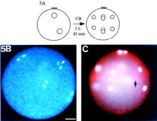
divisions had been completed in controls (Fig. 5A). Pronuclear migration and karyogamy occurred as in untreated zygotes. Cyto-kinesis was blocked totally, but mitoses occurred in regular mitotic planes. In the uncleaved embryos the four anaphase planes of the third cell cycle were oriented obliquely to the animal-vegetal axis as in untreated embryos (Fig. 5B). Moreover, after completion of these anaphases, the uncleaved embryos developed four nuclei in the animal half and four nuclei in the vegetal half (Fig. 5C). The position of the nuclei in the uncleaved embryos was similar to that
in the 8-cell-stage control group which had developed in ASW (Fig. 2). Comparable results were obtained in an experiment where zygotes were exposed to 20 µg CB/ml ASW. In summary, these results show that the F-actin envelope is neither necessary for positioning the nuclei in the prospective compartments of the embryo nor for maintaining the mitotic planes.
In order to test whether the microtubules are a prerequisite for formation of the F-actin envelope, embryos were incubated in 1 µg Noc/ml ASW. To this end, an egg mass containing 2-cell-stage
TABLE 1
EFFECTS OF CB AND NOC ON FORMATION OF THE F-ACTIN ENVELOPE AROUND LATE PROPHASE NUCLEI IN EMBRYOS OF O.PUERILIS
Set of experiment Embryos in late prophase Embryos in other Observed embryos
with F-actin without F-actin Aberrant F-actin mitotic stage
envelope envelope accumulations
Fig. 4A CB (10 µg/ml) - 6 - 22 28
controls 9 - - 17 26
Fig. 4B CB (10 µg/ml) - 8 3 25 36
controls 7 - - 15 22
Fig. 6A Noc (1 µg/ml, 45 min) 12 - - 8 20
Noc (1 µg/ml, 1.5 h) 12 - - 12 24
controls (1.5 h) 4-cell-stages with mitotic phases from ep to lp, lp with F-actin envelope 13
Fig. 3.Distribution of F-actin and microtubules around nuclei from interphase to pro-metaphase in 8-cell-stage embryos. (A,B) Inter-phase blastomere double-stained with DAPI for DNA (A) and anti-β -tubulin/DTAF for microtubules (B). The centrosomal region has divided. A small aster-like accumulation of β -tubulin (arrow) marks each single centrosomal area. (C) Interphase blastomere double-stained with DAPI to locate DNA and TRITC-Phall to locate F-actin. The nucleus is surrounded by focal accumulations of F-actin (arrow). (D)Early prophase blastomere double-stained with DAPI for DNA and anti-β-tubulin/ DTAF for microtubules. Around the nucleus the centrosomes are ori-ented opposite to each other on either side of the prospective mi-totic plane. The centrosomal regions (asterisks) are enriched with microtubules and building up aster-like structures. The chromatin has condensed into thin threads. (E-G) Late prophase blastomere with F-actin envelope formation, triple-stained with DAPI for DNA (E), anti-β-tubulin/DTAF for microtubules (F) and TRITC-Phall for F-actin (G). (F) and (G) are in the same focus plane. Extended centrosomal regions are oriented opposite to each other. Polar microtubules (arrow) seem to enclose the nucleus and thereby form a basket-like structure. The polar microtubules are closely associated with the F-actin envelope. Single chromosomal threads can be distinguished in the nucleus. Due to the strong immunostaining of β-tubulin, DTAF-fluorescence is also visible under UV-excitation for DAPI. (H,I) Pro-metaphase blastomere double-stained with DAPI for DNA (H) and TRITC-Phall for F-actin (I). Remnants of the F-actin envelope are located in the region of the developing metaphase plate. Bar, 10 µm.
Discussion
In this study, the formation of a novel envelope-like F-actin structure associated with the dividing nucleus and with the devel-oping spindle apparatus was shown in early embryos of the polychaete O. puerilis. Although there exist reports of both G-actin and F-actin being associated with the nucleus of diverse organ-isms, to the author’s knowledge, an F-actin envelope surrounding an animal nucleus has not yet been reported. Until know, spatial F-actin accumulations that resemble the F-F-actin envelope described here have only been shown in some plant cells (Seagull et al., 1987; Meindl et al., 1994).
In animals, Maro et al. (1984) detected F-actin as a perinuclear array during mouse pronuclear centration. Heil-Chapdelaine and Otto (1996) demonstrated F-actin accumulations in whole-mounts of maturating starfish oocytes around the germinal vesicle at the onset of germinal vesicle breakdown. In certain embryonic cells of the oligochaete Tubifex hattai, a diffuse accumulation of F-actin was also found to be associated with the nucleus (Shimizu, 1988). The function of all of these F-actin accumulations is still unclear.
In other animal cells actin was demonstrated to occur within the nucleus (Clark and Merriam, 1977; Clark and Rosenbaum, 1979, Milankov and De Boni, 1993). In addition, an isoform of actin has been described in nuclear fractions of interphase cells (Bremer et al., 1981). Finally, actin-binding proteins were also demonstrated to occur in nuclei (Ankenbauer et al., 1989). There is much speculation concerning the function of the nuclear actin, e.g., involvement in chromatin motion, nucleocytoplasmic transport, or gene expression (De Boni, 1994).
In O. puerilis embryos the F-actin accumulations form an envelope-like structure around late prophase nuclei. This F-actin envelope exists for a short period of time and disappears immedi-ately before the mitotic spindle is established. Thus, the first idea was that the F-actin envelope interacts with microtubules of the mitotic apparatus. It was assumed that the F-actin envelope was involved in mitotic plane formation during cleavage of O. puerilis. This view was supported by earlier observations in tobacco BY-2 cells, which showed, that cytoplasmic F-actin associated with the nucleus plays a role in positioning the nucleus in the prospective mitotic plane (Katsuta and Shibaoka, 1988; Katsuta et al., 1990). Moreover, in rat kangaroo cells actin was demonstrated to be closely associated with the mitotic spindle apparatus (Sanger, 1975).
In order to test this hypothesis, the formation of the F-actin envelope was blocked with CB. Although no F-actin envelope was formed after treatment with CB, in rare cases F-actin accumulated in the centrosomal regions (Fig. 4C,D). Similar accumulations of microfilaments have been observed after CD treatment of epider-mal cells in allium seedlings (Mineyuki and Palevitz, 1990). In the allium study, microfilaments that ramify throughout the cytoplasm of the epidermal cells fragmented and accumulated as clusters at each spindle pole similar to those which were observed here for O. puerilis. In O. puerilis embryos, as in allium cells, these accumula-tions of microfilaments are possibly due to interaction of microtubules and microfilaments, the latter being severed by cytochalasin.
However, disturbance of microfilament polymerization by CB in O. puerilis embryos, did not alter orientation of the mitotic spindle. Moreover, the orientation of the prospective mitotic plane is estab-lished in early prophase, i.e., before the F-actin envelope has been Fig. 4. Effects of CB on the F-actin envelope formation in zygotes and
2-cell-stages. (A,B) Schematic representation of the experiment. (A) Zygotes of one egg mass with juxtaposed late prophase pronuclei (lp) were incubated for 2 h in 10 µg CB/ml ASW. Controls developed for 2 h in ASW. Controls developed in 2-cell-stage embryos. CB-treated zygotes remained uncleaved, but mitoses occurred normal. (B) 2-cell-stages of one egg mass incubated in metaphase (m) for 1.5 h in 10 µg CB/ml ASW. Controls developed for 1.5 h in ASW. Controls developed in 4-cell-stages. CB-treated embryos remained uncleaved, but mitoses occurred normal. (C,D) Aberrant F-actin accumulations in the centrosomal regions of a blastomere of a 2-cell-stage embryo. The embryo was incubated in 10 µg CB/ml ASW, as described in (B). (C) DAPI-staining of DNA. (D) TRITC-Phall-staining of F-actin. Bar, 10 µm.
formed. Finally, inhibition of microtubule polymerization by Noc showed that the F-actin envelope develops independently of the spindle apparatus. In summary, the results show that the F-actin envelope is not involved in establishing the mitotic plane in O. puerilis embryos.
Conspicuously, disappearance of the F-actin envelope is cou-pled temporally with nuclear envelope breakdown. Recently, it was shown in mammalian cells, that cytoplasmic cytoskeletal proteins push on the nuclear surface and thereby deform the nuclear membrane during nuclear envelope breakdown (Georgatos et al., 1997). In the present study, indentations of the pronuclei were detected at the respective centrosomal side in O. puerilis zygotes (Fig. 1C). Additionally, Stricker and Schatten (1991) have shown, that inhibition of actin polymerization by CB results in a delay of the germinal vesicle breakdown in starfish oocytes. Remarkably, CB inhibited shape changes of the germinal vesicle normally occurring prior to its breakdown. Furthermore, this study shows, that the novel F-actin envelope is not a uniform structure (Fig. 1D). Protru-sions of F-actin extend from either side of the F-actin envelope to the centrosomes. Presumably, these protrusions are a result of shape alterations of the F-actin envelope during nuclear envelope breakdown.
In summary, it is likely that the novel F-actin envelope is involved in fragmentation of the nuclear envelope in embryos of O. puerilis, although its functional significance in this process remains to be elucidated.
Materials and Methods
Animals and recovery of egg masses
Mass cultures of the polychaete O. puerilis were maintained in ASW as described previously (Pfannenstiel, 1973b). For experimental use, 4 males and 4 females each were transferred to small polystyrol beakers filled with 18 ml ASW and maintained under mass culture conditions. Newly produced egg masses, which contained approximately 50 to 150 eggs, were col-lected daily. Synchronous development of embryos in each egg mass was determined by observation under the microscope and by in vivo DAPI-staining (Sigma, Deisenhofen). In order to determine the mitotic stages in an egg mass, 10 embryos of each egg mass were stained on a slide with 50 µl of 1 µg/ml DAPI in phosphate buffer (PB, 0.01 mol/l Na
2HPO4/ NaH
2PO4, pH 7.2) for about 15 sec. An egg mass was considered to be homogeneous when 1) all embryos of the egg mass were in the same cell-stage and when 2) the 10 embryos stained with DAPI were in the same mitotic stage.
Fluorescent labeling of DNA and F-actin
Embryos were fixed in 4% paraformaldehyd in phosphate buffer saline (PBS; 0.01 mol/l Na
2HPO4/NaH2PO4, 0.15 NaCl, pH 7.2) for 1.5 h at 4°C. After repeated rinsing in PBS, embryos were incubated in 400 mM Glycin (pH 7.2) for 1 h at room temperature, washed twice in PBS and then stained in DAPI solution (1 µg/ml PB) for 20 min. After DAPI-staining, embryos were washed briefly in PBS and stained with TRITC-Phall (Sigma, Deisenhofen) for 10 min. TRITC-Phall was prepared as a 0.1 mg/ml stock solution in methanol. Embryos were stained in 0.2 µg TRITC-Phall/ml PBS.
Immunofluorescence of microtubules
In order to visualize microtubules, embryos were fixed and washed as described above. After rinsing in 0.25 % Triton-X-100 in PBS for 1 h following the Glycin-treatment, they were incubated with monoclonal mouse antibodies against β-tubulin of bovine brain (courtesy of U. Euteneuer and M. Schliwa) diluted 1:30 in a solution of 0.25 % BSA in 0.25 % Triton-X-100 in PBS for 12 h at 4°C. After repeated rinsing in 0.25 % Triton-X-100 in PBS, embryos were incubated in DTAF-conjugated goat-anti-mouse IgG and IgM (Dianova, Hamburg) diluted 1:50 for 2 h. For triple staining of microtubules, F-actin, and DNA the embryos were stained afterwards with TRITC-Phall and DAPI. For triple staining, 0.1 µg DAPI/ml PB instead of 1
µg DAPI/ml PB was used to avoid quenching of the DTAF-fluorescence under UV-excitation.
Electron microscopy
Embryos were fixed as described by Palade (1952) and modified by Harris (1962). Then they were dehydrated in graded ethanol series, block stained in saturated uranyl acetate in 70 % alcoholic solution and embedded in araldite. Semithin sections and thin sections were cut on a Reichert OmU 3 microtome with a diamond knife. Semithin sections were stained with Mallory Azur II (1:1 mixture of 1% azur in A. dest. and 1% methylenblue in 1% borax) and sealed in araldite. Thin sections were stained with lead citrate.
Drug treatment
CB, CD and Noc were prepared as stock solutions (2 mg CB/ml DMSO, 3 mg CD/ml DMSO and 2 mg Noc/ml DMSO) and stored at - 20°C. Embryos were incubated in concentrations from 1 to 30 µg inhibitor/ml ASW. The highest concentration of DMSO was 1 % in ASW. Development of control embryos in 1 % DMSO in ASW was normal.
Observation and documentation
Observations were carried out with a Zeiss Axiophot and with a Zeiss IM 35 equipped with epifluorescence optics. Filter combinations being recom-mended by Zeiss for DAPI-, TRITC- and DTAF-fluorescence were used. Photographs were taken on Kodak (Ektachrome) 400 ASA and Pan Ilford 50 ASA films. In some cases, slides were exposed twice to document DAPI-and TRITC-Phall-fluorescence (Figs. 1E, 3C, 5C DAPI-and 6B).
Sections prepared for electron microscopy were examined with a Zeiss EM 10 transmission electron microscope.
Acknowledgments
The author thanks Dr. Ursula Euteneuer and Dr. Manfred Schliwa for the generous gift of the monoclonal anti-β-tubulin antibody. Special thanks to Dr. Hans-Dieter Pfannenstiel, Dr. Wendy Thuß-Patience and Dr. Jörg Willuhn for helpful discussions concerning this work. The technical help of Charlotte Schroer in preparing the figures is greatfully acknowledged.
References
ANKENBAUER, T., KLEINSCHMIDT, J.A., WALSH, M.J., WEINER, O.H. and FRANKE, W.W. (1989). Identification of a widespread nuclear actin binding protein. Nature 342: 822-825.
BREMER, J.W., BUSCH, H. and YEOMAN, L.C. (1981). Evidence for a species of nuclear actin distinct from cytoplasmic and muscles actins. Biochemistry 20: 2013-2017.
CLARK, T.G. and MERRIAM, R.W. (1977). Diffusible and bound actin nuclei of Xenopus laevis oocytes. Cell 12: 883-891.
CLARK, T.G. and ROSENBAUM, J.L. (1979). An actin filament matrix in hand-isolated nuclei of X. laevis oocytes. Cell 18: 1101-1108.
COOPER, J.A. (1987). Effects of cytochalasin and phalloidin on actin. J. Cell Biol. 105: 1473-1478.
DE BONI, U. (1994). The interphase nucleus as a dynamic structure. Int. Rev. Cytol. 150: 149-171.
DONDUA, A.K., KOSTYUCHENKO, R.P. and FEDOROVA, Z.E. (1997). Effects of some cytoskeleton inhibitors on ooplasmic segregation in the Nereis virens egg. Int. J. Dev. Biol. 41: 853-858.
DORRESTEIJN, A.W.C. (1998). How do spiralian embryos accomplish cell diversity? Zoology 100: 307-319.
ECKBERG, W.R. and ANDERSON, W.A. (1995). Cytoskeleton, cellular signals, and cytoplasmic localization in Chaetopterus embryos. In Current Topics in Develop-mental Biology (Ed. D.G. Capco). Vol. 31. Academic Press, pp. 5-39.
FAULSTICH, H., ZOBELEY, S., RINNERTHALER, G. and SMALL, J.V. (1988). Fluorescent phallotoxins as probes for filamentous actin. J. Muscle Res. Cell Motil. 9: 370-383.
GEORGATOS, S.D., PYRPASOPOULOU, A. and THEODOROPOULOS, P.A. (1997). Nuclear envelope breakdown in mammalian cells involves stepwise lamina disassembly and microtubule-driven deformation of the nuclear membrane. J. Cell Sci. 110: 2129-2140.
GROTHE, C. and PFANNENSTIEL, H.D. (1986). Cytophysiological study of neurose-cretory and pheromonal influences on sexual development in Ophryotrocha puerilis (Polychaeta, Dorvilleidae). Int. J. Invert. Reprod. Dev. 10: 227-239.
HARRIS, P. (1962). Some structural and functional aspects of the mitotic apparatus in sea urchin embryos. J. Cell Biol. 14: 475-487.
HEIL-CHAPDELAINE, R.A. and OTTO, J.J. (1996). Characterization of Changes in F-actin during Maturation of Starfish Oocytes. Dev. Biol. 177: 204-216.
KATSUTA, J. and SHIBAOKA, H. (1988). The roles of the cytoskeleton and the cell wall in nuclear positioning in tobacco BY-2 cells. Plant Cell Physiol. 29: 403-413.
KATSUTA, J., HASHIGUCHI, Y. and SHIBAOKA, H. (1990). The role of the cytoskel-eton in positioning of the nucleus in premitotic tobacco BY-2 cells. J. Cell Sci. 95: 413-422.
KLUGE, B., LEHMANN-GREIF, M. and FISCHER, A. (1995). Long-lasting exocytosis and massive structural reorganisation in the egg periphery during cortical reaction in Platynereis dumerilii (Annelida, Polychaeta). Zygote 3: 141-156.
MABUCHI, I. (1990). Cleavage furrow formation and actin-modulating proteins. In Cytokinesis. Mechanisms of furrow formation during cell division. (Eds. G.W. Conrad and T.E. Schroeder). Annals of the New York Academy of Sciences, Vol. 582. The New York Acadamy of Sciences, pp. 131-146.
MABUCHI, I. (1994). Cleavage furrow: timing of emergence of contractile ring actin filaments and establishment of the contractile ring by filament bundling in sea urchin eggs. J. Cell Sci. 107: 1853-1862.
MARO, B., JOHNSON, M.H., PICKERING, S.J. and FLACH, G. (1984). Changes in actin distribution during fertilization of the mouse egg. J. Embryol. Exp. Morphol. 81: 211-237.
MEINDL, U., ZHANG, D. and HEPLER P.K. (1994). Actin microfilaments are associ-ated with the migrating nucleus and the cell cortex in the green alga Micrasterias. Studies on living cells. J. Cell Sci. 107: 1929-1934.
MILANKOV, K. and DE BONI, U. (1993). Cytochemical localization of actin and myosin aggregates in interphase nuclei in situ. Exp. Cell Res. 209: 189-199.
MINEYUKI, Y. and PALEVITZ, B.A. (1990). Relationship between preprophase band organization, F-actin and the division site in Allium. J. Cell Sci. 97: 283-295.
PALADE, G.E. (1952). A study of fixation for electron microscopy. J. Exp. Med. 95: 285-294.
PFANNENSTIEL, H.D. (1973a). Zur sexuellen Differenzierung von Ophryotrocha puerilis (Polychaeta: Eunicidae). Mar. Biol. 20: 245-258.
PFANNENSTIEL, H.D. (1973b). Der Meeresborstenwurm Ophryotrocha puerilis. Ein ideales Labortier. Mikrokosmos 62: 97-100.
PFANNENSTIEL, H.D., SCHLAWNY, A., HAMANN, T., MÜLLER, M., RHODE, B.
and SPIEHL, D. (1990). Dopamine and male/female differentiation in a hermaph-roditic polychaete (Ophryotrocha puerilis). In Progress in comparative endocrinology. (Eds. A. Epple, C.G. Scanes and M.H. Stetson), Wiley-Liss, Inc., pp. 219-225.
PLICKERT, G. and KROIHER, M. (1988). Proliferation kinetics and cell lineages can be studied in whole mounts and macerates by means of BrdU/anti-BrdU tech-nique. Development 103: 791-794.
RUTHMANN, A. (1964). Zellwachstum und RNS-Synthese im Ei-Nährzellverband von Ophryotrocha puerilis. Z. Zellforsch. 63: 816-829.
SAMPATH, P. and POLLARD, T.D. (1991). Effects of cytochalasin, phalloidin and pH on the elongation of actin filaments. Biochemistry 30: 1973-1980.
SAMSON, F., DONOSO J.A., HELLER-BETTINGER, I., WATSON, D. and HIMES, R.H. (1979). Nocodazole action on tubulin assembly, axonal ultrastructure and fast axoplasmic transport. J. Pharmacol. Exp. Ther. 208: 411-417.
SANGER, J.W. (1975). Presence of actin during chromosomal movement. Proc. Natl. Acad. Sci. USA 72: 2451-2455.
SCHROEDER, T.E. (1973). Actin in dividing cells: contractile ring filaments bind heavy meromyosin. Proc. Natl. Acad. Sci USA 70: 1688-1692.
SEAGULL, R.W., FALCONER, M.M. and WEERDENBURG, C.A. (1987). Microfilaments: Dynamic Arrays in Higher Plant Cells. J. Cell Biol. 104: 995-1004.
SHIMIZU, T. (1988). Localization of actin networks during early development of Tubifex embryos. Dev. Biol. 125: 321-331.
STRICKER, S.A. and SCHATTEN, G. (1991). The Cytoskeleton and Nuclear Disas-sembly during Germinal Vesicle Breakdown in Starfish Oocytes. Dev. Growth Differ. 33: 163-171.
STROME, S. (1993). Determination of Cleavage Planes. Cell 72: 3-6.
VASQUEZ, R.J., HOWELL, B., YVON, A.M., WADSWORTH, P. and CASSIMERIS, L. (1997). Nanomolar concentrations of nocodazole alter microtubule dynamic instability in vivo and in vitro. Mol. Biol. Cell 8: 973-985.
WULF, E., DEBOBEN, A., BAUTZ, F.A., FAULSTICH,H. and WIELAND, T. (1979). Fluorescent phallotoxin, a tool for the visualization of cellular actin. Proc. Natl. Acad. Sci. USA 76: 4498-4502.