Conserved modularity and potential for alternate splicing in
mouse and human Slit genes
MELISSA LITTLE*, BREE RUMBALLE
1, KYLIE GEORGAS, TOSHIYA YAMADA and ROHAN D. TEASDALE
Institute for Molecular Bioscience and Centre for Functional and Applied Genomics, University of Queensland, St. Lucia, Australia and 1Department of Biochemistry, University of Queensland, St. Lucia, Australia
ABSTRACT The vertebrate Slit gene family currently consists of three members; Slit1, Slit2 and Slit3. Each gene encodes a protein containing multiple epidermal growth factor and leucine rich repeat motifs, which are likely to have importance in cell-cell interactions. In this study, we sought to fully define and characterise the vertebrate Slit gene family. Using long distance PCR coupled with in silico mapping, we determined the genomic structure of all three Slit genes in mouse and man. Analysis of EST and genomic databases revealed no evidence of further Slit family members in either organism. All three Slit genes were encoded by 36 (Slit3) or 37 (Slit1 and Slit2) exons covering at least 143 kb or 183 kb of mouse or human genomic DNA respectively. Two additional potential leucine-rich repeat encoding exons were identified within intron 12 of Slit2. These could be inserted in frame, suggesting that alternate splicing may occur in Slit2. A search for STS sequences within human Slit3 anchored this gene to D5S2075 at the 5’ end (exon 4) and SGC32449 within the 3’ UTR, suggesting that Slit3 may cover greater than 693 kb. The genomic structure of all Slit genes demonstrated considerable modularity in the placement of exon-intron boundaries such that individual leucine-rich repeat motifs were encoded by individual 72 bp exons. This further implies the potential generation of multiple Slit protein isoforms varying in their number of repeat units. cDNA library screening and EST database searching verified that such alternate splicing does occur.
KEY WORDS:
leucine-rich repeat, epidermal growth factor repeat, in silico mapping, Slit genes
0214-6282/2002/$25.00 © UBC Press
Printed in Spain
www.ijdb.ehu.es
*Address correspondence to: Dr. M. Little. Institute for Molecular Bioscience, University of Queensland, St. Lucia, 4072, Australia. Fax: +61-7-3365-4388. e-mail: [email protected]
Electronic Supplementary Material for this paper, entitled "Intron-exons boundary sequences for each of the three vertebrate Slit genes" is available at the following addresses: http://www.ijdb.ehu.es/abstract.0204/esmX.htm or http://www.imb.uq.edu.au/groups/little/SLIT/
Introduction
The Drosophila slit gene encodes a large extracellular protein comprising seven epidermal growth factor motifs (EGF1-7), four N-terminal regions of leucine-rich repeat regions (LRR1-4) and a C-terminal cysteine-rich knot. Each of these motifs has been shown to play a role in protein-protein interactions (Rothberg et al., 1990, 1992). Three members of a vertebrate Slit gene family have been isolated in human and mouse (Itoh et al., 1998; Holmes et al., 1998; Nakayama et al., 1998). The encoded vertebrate proteins contain the same groups and arrangement of structural motifs as the fly, al-though they contain two additional EGF motifs. Drosophila slit is expressed in the midline glial cells of the developing fly neural tube and acts as a midline chemorepellant thereby facilitating the proper development of axonal scaffolds and commissural events (Rothberg et al., 1990; Kidd et al., 1999). This role appears to have been conserved in vertebrates (Brose et al., 1999; Li et al., 1999). All three of the Slit genes discovered to date are expressed in the developing central nervous system in the floor plate, considered to be the
vertebrate equivalent of the fly midline glial cells (Itoh et al., 1998; Holmes et al., 1998; Nakayama et al., 1998; Yuan et al., 1999). Vertebrate Slit2 protein has been shown to act as a chemorepellant, as does slit in Drosophila (Brose et al., 1999). As in fly, the vertebrate Slit proteins interact with the product of the vertebrate orthologs of the fly roundabout (robo) gene to elicit this chemorepulsive cue (Brose et al., 1999, Li et al., 1999). Together with defects in neural patterning,
Abbreviations used in this paper: AMCN, Arthrogryposis Multiplex Congenita,
Drosophila slit mutants also display defects in mesoderm migration and muscle attachment (Rothberg et al., 1990, 1992). In support of additional non-neuronal functions for vertebrate Slits, we and others have shown distinct expression patterns for the vertebrate Slit genes in non-neuronal tissues, including the limbs, skeletal muscle and developing urogenital tract (Itoh et al., 1998; Holmes et al., 1998; Piper et al., 2000). Functional evidence for non-neuronal Slit2 activities have also been demonstrated and include regulation of prechordal mesoderm migration in zebrafish (Yeo et al., 2001), and inhibition of leucocyte chemotaxis in mammals (Wu et al., 2001).
Slit proteins are large, highly modular proteins containing both LRR and EGF repeats. In many other protein families in which multiple extracellular protein-protein interacting motifs are present, alternate splicing results in the generation of protein isoforms with distinct activities upon ligand binding. This has been exemplified by the neural cell adhesion molecules of the immunoglobulin superfam-ily (Walsh and Doherty, 2001). The aim of this study was to fully characterise the genomic structure of each of the Slit genes in human and mouse using long distance PCR and in silico mapping, search for additional members of the vertebrate Slit gene family and search for evidence of alternate splicing of Slit genes. Analysis of the exon-intron boundaries in both mouse and human has revealed that all three vertebrate Slit genes are highly conserved and highly modular, indicating considerable potential for alternate splicing resulting in the generation of multiple protein isoforms. In silico analysis has pro-vided support for this possibility by revealing the existence of Slit transcripts which vary in their motif complement.
Results
In Silico Genomic Mapping of Human Slit Genes
Initial characterisation of human Slit3 was commenced using long distance reverse transcription-polymerase chain reaction (RT-PCR) on either genomic DNA or Slit3-containing P1-artificial chromosome (PAC) sequences. This indicated that this gene was likely to have many exons with very large introns (data unpublished). Hence, as the
human genome sequence became available, an in silico approach was adopted for all three known human Slit genes. The in silico approach involved identifying a number of overlapping genomic sequences for each Slit gene (see Electronic Supplementary Mate-rial 1) using the sequences described by Itoh et al. (1998) for each gene. These were NM_003061 for Slit1, NM_004787 for Slit2 and NM_003062 for Slit3. Each unordered contig was broken down into its derivative fragments for recompilation in order according to the exons detected within each fragment.
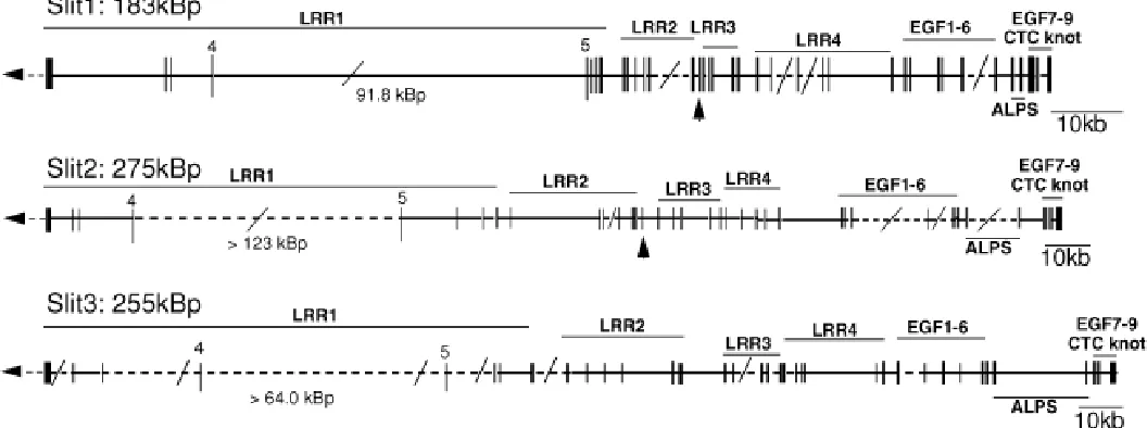
Slit3 (NM_003062) (Itoh et al., 1998) was found to be encoded by a total of 36 exons encompassing at least 255 kb (see ESM. 1; Fig. 1; Tables 1,2). While the sequence of exon 5 and exon 18 were not encoded by the genomic sequences within the public genome databases they were defined based on the flanking exons. The largest gap was between exons 4 and 5, suggesting an intron of >64kb. While in silico mapping predicted the presence of all intron-exon boundaries, the nature of the unordered contigs was such that not all intron lengths were predicted. In the case of introns 16 and 18, the intron length was actually clarified using long distance-PCR (LD-PCR) as 821bp and 3.2 kb respectively. In addition, this confirmed the precise nature of the exon 18 sequence that was not contained in the in silico map.
In silico mapping was subsequently performed for Slit1 (NM_003061) and Slit2 (NM_004787) (Itoh et al., 1998) as for Slit3. This process predicts total genomic lengths of at least 183 kb and 275 kb for Slit1 and Slit2 respectively, with each gene consisting of 37 exons. There is remarkable uniformity of exon-intron boundary placement and intron length between all three Slit genes (Figs. 1,2; Tables 1,2). The single additional exon present in Slit1 and Slit2 is exon 15 (24bp), which encodes in frame 8 amino acids, these being inserted between two adjacent LRR repeats (see ESM. 1 and Fig. 1). We were unable to detect any sequences equivalent to exon 15 in the Slit3 genomic contigs. As in Slit3, both Slit1 and Slit2 genes contain a large gap, 92 kb and >123 kb respectively, within the LRR1-encoding region (between exons 4 and 5) indicating the existence of a very large intronic region (Fig. 1, Table 1). Curiously, within intron
12 of Slit2, there are two additional exons very similar to exons 13 and 14. We have termed these ‘exon13like’ and ‘exon14like’. If these sequences were included in a Slit2 mRNA, they would encode an additional two LRR repeats within LRR2 in frame (ESM. 1).
The Slit1 positive genomic sequences (AL442123, AL356126 and AL442123) are annotated as being localised to Chromosome 10 which is consistent with the published chromosomal localisation for Slit1 (Nakayama et al., 1998). Likewise, Slit2 positive genomic sequences (AC046194, AC012142 and AC02118) annotated to be located on Chromosome 4 correlates to the published chromo-somal localisation of Slit2, which characterised a localisation of human 4p15.2 (Georgas et al., 1999). It was of note that the genomic sequences AL024551 (Slit1 5’ exons) and AL136447 (Slit2 5’ exons) appear to have incorrect annotation of their chro-mosomal localisation. For example, AL024551 is annotated as been localised on chromosome 4 but large segments (>10 kb) are greater than 99% identical to the sequence AL512424 which is annotated to be located on Chromosome 10 suggesting that AL024551 has been incorrectly assigned.
Comparison with Gene Structure Predictions from the Draft Human Genome
Next we compared the assembled SLIT genes with those generated as part of the analysis of the human genome project.
We identified the genes annotated as SLIT1 (ENSG00000052758); SLIT2 (ENSG00000067745); SLIT3 (ENSG00000094749) in Ensembl v 1.0 (http://www.ensembl.org/ June 2001). As antici-pated, comparison of our SLIT gene contigs to those within Ensembl revealed numerous discrepancies. Our assembled genes contained more sequence detail, particularly within the larger introns. Surprisingly, given that the cDNA for each SLIT is known, each of the Ensembl predicted transcripts contained inaccuracies that resulted in the disruption of the protein open-reading frame. The following discrepancies were identified within the predicted exons. SLIT1:- 1) Incorrect splice donor sequence for exon 29; 2) Incorrect splice acceptor sequence for exon 30. SLIT2:- 1) Incorrect splice donor sequence for exon 14; 2) Incorrect splice donor sequence for exon 33; 3) Exclusion of exon 34; 4) Inclusion of unrelated sequence as exon 33; 5) Fragmented exon 35 into two separated exons. SLIT3:- 1) Fragmented exon 1 into three separated exons with gaps; 2) Inclusion of unrelated sequence as exon 4; 3) Incorrect splice acceptor sequence for exon 2; 4) Incorrect splice acceptor sequence for exon 6; 5) Incorrect splice acceptor sequence for exon 19. This would suggest that gene structure prediction algorithms are flawed without parallel analysis of known cDNA and protein information and that focussed in silico analyses such as ours continue to more accurately predict gene structure.
Intron placement, exon length and the genomic contigs (shaded) used for the in silico mapping are indicated. The total length of each gene includes the length of the open reading frame (ORF).
TABLE 1
A SUMMARY OF THE GENE STRUCTURE FOR THE THREE VERTEBRATE SLIT GENES
Analysis of the Human Genome for Additional Slit Protein Family Members
Bioinformatics was also used to investigate the possibility that there are further undescribed Slit family members. Initially, the public human genome database was analysed for sequences with significant relationship to the three human Slit proteins. This failed
to detect any additional predicted gene or ge-nomic sequence related to the currently defined human Slit proteins other than those already known. In addition, analysis of the Celera Genomics and public human expressed se-quence tag (EST) databases failed to detect any further mouse or human family members. There-fore, within the currently available human and mouse DNA sequences, the Slit family appears to only include the three previously described human members.
In Silico Genomic Mapping of Murine Slit Genes
To determine if the genomic structure of the murine SLIT genes showed the same conserved modularity as the human genes, these genes were identified in the assembled mouse genome generated by Celera Genomics (www.celera.com). The genomic lengths of these genes, based on the Celera assembly, is pre-dicted to be at least 143kb, 330 kb and 530 kb for murine Slit1, Slit2 and Slit3 respectively. Each gene consisted of the same number of exons as the human counterpart and each contained a large intron between exons 4 and 5. With the exception of exon 32 from murine Slit1, which contained an additional nine nucleotides, each exon in the three murine SLIT genes was iden-tical in length and position relative to the coding sequence as the human Slit genes. While the region is incomplete, analysis of the intronic region between exon 12 and exon 13 of murine Slit2 genomic sequences failed to detect the two putative exons present in the human gene.
Modularity of Genomic Structure within the LRR-Encoding Regions
The leucine rich repeat regions of the three human Slit genes contain a large number of very small exons. In most instances, individual leu-cine rich repeats of 24 amino acids were en-coded by individual exons (72bp) (ESM. 1, Fig. 2A, Table 1). This highly modular structure would allow individual repeat motifs to be alternately spliced in or out within altering the frame. For example, each of the four blocks of leucine-rich repeats could be inserted or removed without altering the rest of the amino acid sequence. Indeed, in most instances the individual leucine-rich motifs within each block of repeats have the potential to be removed or inserted without alter-ing frame. This implies that these genes may be
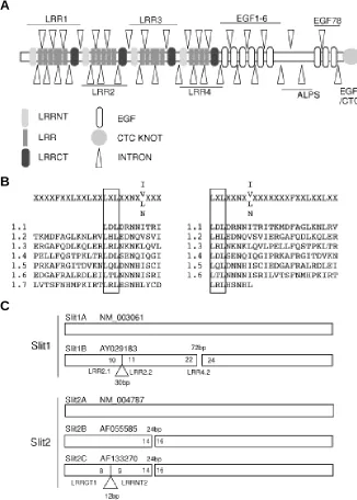
Fig. 2. Modularity within human Slit genes. (A) Generic ideogram of a Slit protein, based upon Slit3, demonstrating the relationship between each structural motif and placement of introns. (B) Redefinition of the LRR motif based upon intron placement in Slit genes. The LRR motifs from within the first group of LRRs in human Slit3 is shown. The left alignment is according to that previously used by Taguchi et al. (1996). The right alignment reflects a redefinition of the LRR based upon the observation of uniform intron placement at the LXL of each repeat. (C) Novel Slit1 and Slit2 gene alternately spliced transcripts determined via cDNA library screening or analysis of EST databases. Triangles refer to insertions and gaps refer to deletions with respect to the A transcript. Size of inserts and deletions is indicated in bp with the flanking exons marked within the ideogram and the motifs they encode indicated below.
highly alternately spliced. LRRs are structurally composed of a beta-alpha unit (Kobe and Deisenhofer, 1994) forming an elon-gated non-globular structure classically involved in protein-protein interactions. The Pfam website would identify the protein se-quence XLXXLXLXXN Xn I Xn FXXL as an LRR. However, Taguchi et al. (1996), when comparing comparing novel LRR
A
B
proteins with those previously isolated, aligned the leucine-rich repeat motif as XXXX(F/L)XXLXXLXXLXXLXLXXNX(IVLN)XX(I/ M) (Fig. 2B). Here we demonstrate that LRR-encoding exons always started on the second position base of the codon encoding the first leucine of the LXL (as underlined). We have therefore redefined the LRRs as a repeat unit of LXLXXNXXIX7FXXLXXLXXLXX (see Fig. 2B). In contrast to the LRRs, there is approximately 1 exon encoding each EGF repeat. However, these exons vary in length such that loss of one such exon would usually shift the frame resulting in premature termination of the protein.
Evidence for Alternate Splicing resulting in Multiple Slit Pro-tein Isoforms
Given the provocative intron placement in the 5’ end of these genes and the existence of additional putative exons within intron 12 of Slit2, we searched for evidence that alternate splicing does
occur. This was approached by screening a human fetal brain cDNA library using a combination of human Slit gene cDNA probes. cDNA library screening resulted in the recloning of multiple partial transcripts, with most sequences been identical to the original either Slit1, Slit2 or Slit3 mRNAs. However, one clone, which we term Slit1B, aligned with Slit1 except for the deletion of exon 23 encoding LRR4.2 (Fig. 2C). Additionally, Slit1B contained a 30bp insertion between Slit1 exons 10 and 11 which is present within the genomic sequence of Slit1 (NM_003061). This inserted 10 amino acids in frame between LRR2.1 and LRR2.2, according to our revised description of an LRR. This insertion was generated by the use of an alternate splice acceptor site within the intron preceding exon 11. Normally, the intron/exon junction at the 5’ end of exon 11 is ccaccttgcag/AGACCTG, demonstrating a character-istic splice acceptor site of 6Pyncag/N (Lewin, 2000). In this clone
cDNA, an alternate site of agggattcag/ACG was employed
insert-TABLE 2
DEFINITION OF SLIT PROTEIN STRUCTURE
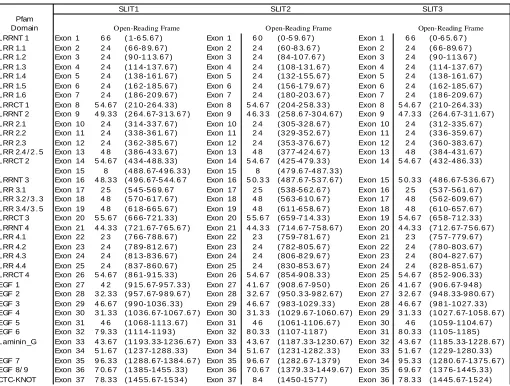
The relationship between each exon and the amino acids and structural motifs encoded for each of the three vertebrate Slit genes is tabulated. LRR, leucine-rich repeat; LRRNT, N-terminal conserved region from within a leucine rich repeat domain; LRRCT, C-terminal conserved region from within a leucine rich repeat domain; laminin_G, agrin-laminin-parlecan-slit domain; EGF, epidermal growth factor repeat; CTC-KNOT, cysteine rich knot motif. LRR domains were detected using the criteria defined in this manuscipt (see text for details). Other structural motifs were as predicted by Pfam (http://pfam.wustl.edu).
SLIT1 SLIT2 SLIT3
Pfam
Domain Open-Reading Frame Open-Reading Frame Open-Reading Frame
LRRNT 1 Exon 1 6 6 ( 1-6 5. 6 7 ) Exon 1 6 0 ( 0 -5 9. 6 7 ) Exon 1 6 6 ( 0 -6 5. 6 7 )
LRR 1.1 Exon 2 2 4 ( 6 6-8 9. 6 7 ) Exon 2 2 4 ( 6 0 -8 3. 6 7 ) Exon 2 2 4 ( 6 6- 8 9. 6 7 )
LRR 1.2 Exon 3 2 4 ( 9 0-1 1 3. 6 7 ) Exon 3 2 4 ( 8 4 -1 0 7. 6 7 ) Exon 3 2 4 ( 9 0- 1 1 3. 6 7 )
LRR 1.3 Exon 4 2 4 ( 1 1 4-1 3 7. 6 7 ) Exon 4 2 4 ( 1 0 8 -1 3 1. 6 7 ) Exon 4 2 4 ( 1 1 4- 1 3 7. 6 7 )
LRR 1.4 Exon 5 2 4 ( 1 3 8-1 6 1. 6 7 ) Exon 5 2 4 ( 1 3 2 -1 5 5. 6 7 ) Exon 5 2 4 ( 1 3 8- 1 6 1. 6 7 )
LRR 1.5 Exon 6 2 4 ( 1 6 2-1 8 5. 6 7 ) Exon 6 2 4 ( 1 5 6 -1 7 9. 6 7 ) Exon 6 2 4 ( 1 6 2- 1 8 5. 6 7 )
LRR 1.6 Exon 7 2 4 ( 1 8 6-2 0 9. 6 7 ) Exon 7 2 4 ( 1 8 0 -2 0 3. 6 7 ) Exon 7 2 4 ( 1 8 6- 2 0 9. 6 7 )
LRRCT 1 Exon 8 5 4. 6 7 ( 2 1 0-2 6 4. 3 3 ) Exon 8 5 4. 6 7 ( 2 0 4 -2 5 8. 3 3 ) Exon 8 5 4. 6 7 ( 2 1 0- 2 6 4. 3 3 )
LRRNT 2 Exon 9 4 9. 3 3 ( 2 6 4. 6 7-3 1 3. 6 7 ) Exon 9 4 6. 3 3 ( 2 5 8 .6 7-3 0 4. 6 7 ) Exon 9 4 7. 3 3 ( 2 6 4. 6 7-3 1 1. 6 7 )
LRR 2.1 Exon 10 2 4 ( 3 1 4-3 3 7. 6 7 ) Exon 10 2 4 ( 3 0 5 -3 2 8. 6 7 ) Exon 10 2 4 ( 3 1 2- 3 3 5. 6 7 )
LRR 2.2 Exon 11 2 4 ( 3 3 8-3 6 1. 6 7 ) Exon 11 2 4 ( 3 2 9 -3 5 2. 6 7 ) Exon 11 2 4 ( 3 3 6- 3 5 9. 6 7 )
LRR 2.3 Exon 12 2 4 ( 3 6 2-3 8 5. 6 7 ) Exon 12 2 4 ( 3 5 3 -3 7 6. 6 7 ) Exon 12 2 4 ( 3 6 0- 3 8 3. 6 7 )
LRR 2.4 / 2 . 5 Exon 13 4 8 ( 3 8 6-4 3 3. 6 7 ) Exon 13 4 8 ( 3 7 7 -4 2 4. 6 7 ) Exon 13 4 8 ( 3 8 4- 4 3 1. 6 7 )
LRRCT 2 Exon 14 5 4. 6 7 ( 4 3 4-4 8 8. 3 3 ) Exon 14 5 4. 6 7 ( 4 2 5 -4 7 9. 3 3 ) Exon 14 5 4. 6 7 ( 4 3 2- 4 8 6. 3 3 )
Exon 15 8 ( 4 8 8. 6 7-4 9 6. 3 3 ) Exon 15 8 ( 4 7 9 .6 7-4 8 7. 3 3 )
LRRNT 3 Exon 16 4 8. 3 3 ( 4 9 6. 6 7-5 4 4. 6 7 Exon 16 5 0. 3 3 ( 4 8 7 .6 7-5 3 7. 6 7 ) Exon 15 5 0. 3 3 ( 4 8 6. 6 7-5 3 6. 6 7 )
LRR 3.1 Exon 17 2 5 ( 5 4 5-5 6 9. 6 7 Exon 17 2 5 ( 5 3 8 -5 6 2. 6 7 ) Exon 16 2 5 ( 5 3 7- 5 6 1. 6 7 )
LRR 3.2 / 3 . 3 Exon 18 4 8 ( 5 7 0-6 1 7. 6 7 ) Exon 18 4 8 ( 5 6 3 -6 1 0. 6 7 ) Exon 17 4 8 ( 5 6 2- 6 0 9. 6 7 )
LRR 3.4 / 3 . 5 Exon 19 4 8 ( 6 1 8-6 6 5. 6 7 ) Exon 19 4 8 ( 6 1 1 -6 5 8. 6 7 ) Exon 18 4 8 ( 6 1 0- 6 5 7. 6 7 )
LRRCT 3 Exon 20 5 5. 6 7 ( 6 6 6-7 2 1. 3 3 ) Exon 20 5 5. 6 7 ( 6 5 9 -7 1 4. 3 3 ) Exon 19 5 4. 6 7 ( 6 5 8- 7 1 2. 3 3 )
LRRNT 4 Exon 21 4 4. 3 3 ( 7 2 1. 6 7-7 6 5. 6 7 ) Exon 21 4 4. 3 3 ( 7 1 4 .6 7-7 5 8. 6 7 ) Exon 20 4 4. 3 3 ( 7 1 2. 6 7-7 5 6. 6 7 )
LRR 4.1 Exon 22 2 3 ( 7 6 6-7 8 8. 6 7 ) Exon 22 2 3 ( 7 5 9 -7 8 1. 6 7 ) Exon 21 2 3 ( 7 5 7- 7 7 9. 6 7 )
LRR 4.2 Exon 23 2 4 ( 7 8 9-8 1 2. 6 7 ) Exon 23 2 4 ( 7 8 2 -8 0 5. 6 7 ) Exon 22 2 4 ( 7 8 0- 8 0 3. 6 7 )
LRR 4.3 Exon 24 2 4 ( 8 1 3-8 3 6. 6 7 ) Exon 24 2 4 ( 8 0 6 -8 2 9. 6 7 ) Exon 23 2 4 ( 8 0 4- 8 2 7. 6 7 )
LRR 4.4 Exon 25 2 4 ( 8 3 7-8 6 0. 6 7 ) Exon 25 2 4 ( 8 3 0 -8 5 3. 6 7 ) Exon 24 2 4 ( 8 2 8- 8 5 1. 6 7 )
LRRCT 4 Exon 26 5 4. 6 7 ( 8 6 1-9 1 5. 3 3 ) Exon 26 5 4. 6 7 ( 8 5 4 -9 0 8. 3 3 ) Exon 25 5 4. 6 7 ( 8 5 2- 9 0 6. 3 3 )
EGF 1 Exon 27 4 2 ( 9 1 5. 6 7-9 5 7. 3 3 ) Exon 27 4 1. 6 7 ( 9 0 8 .6 7-9 5 0 ) Exon 26 4 1. 6 7 ( 9 0 6. 6 7-9 4 8 )
EGF 2 Exon 28 3 2. 3 3 ( 9 5 7. 6 7-9 8 9. 6 7 ) Exon 28 3 2. 6 7 ( 9 5 0 .3 3-9 8 2. 6 7 ) Exon 27 3 2. 6 7 ( 9 4 8. 3 3-9 8 0. 6 7 ) EGF 3 Exon 29 4 6. 6 7 ( 9 9 0-1 0 3 6 . 3 3 ) Exon 29 4 6. 6 7 ( 9 8 3 -1 0 2 9 .3 3 ) Exon 28 4 6. 6 7 ( 9 8 1- 1 0 2 7. 3 3 ) EGF 4 Exon 30 3 1. 3 3 ( 1 0 3 6. 6 7-1 0 6 7 .6 7 ) Exon 30 3 1. 3 3 ( 1 0 2 9 .6 7-1 0 6 0 . 6 7 ) Exon 29 3 1. 3 3 ( 1 0 2 7. 6 7-1 0 5 8 . 6 7 )
EGF 5 Exon 31 4 6 ( 1 0 6 8-1 1 1 3 . 6 7 ) Exon 31 4 6 ( 1 0 6 1 -1 1 0 6 .6 7 ) Exon 30 4 6 ( 1 0 5 9- 1 1 0 4. 6 7 )
EGF 6 Exon 32 7 9. 3 3 ( 1 1 1 4-1 1 9 3 ) Exon 32 8 0. 3 3 ( 1 1 0 7 -1 1 8 7 ) Exon 31 8 0. 3 3 ( 1 1 0 5- 1 1 8 5 )
Laminin_G Exon 33 4 3. 6 7 ( 1 1 9 3. 3 3-1 2 3 6 .6 7 ) Exon 33 4 3. 6 7 ( 1 1 8 7 .3 3-1 2 3 0 . 6 7 ) Exon 32 4 3. 6 7 ( 1 1 8 5. 3 3-1 2 2 8 . 6 7 ) Exon 34 5 1. 6 7 ( 1 2 3 7-1 2 8 8 . 3 3 ) Exon 34 5 1. 6 7 ( 1 2 3 1 -1 2 8 2 .3 3 ) Exon 33 5 1. 6 7 ( 1 2 2 9- 1 2 8 0. 3 3 ) EGF 7 Exon 35 9 6. 3 3 ( 1 2 8 8. 6 7-1 3 8 4 .6 7 ) Exon 35 9 6. 6 7 ( 1 2 8 2 .6 7-1 3 7 9 ) Exon 34 9 5. 3 3 ( 1 2 8 0. 6 7-1 3 7 5 . 6 7 ) EGF 8/ 9 Exon 36 7 0. 6 7 ( 1 3 8 5-1 4 5 5 . 3 3 ) Exon 36 7 0. 6 7 ( 1 3 7 9 .3 3-1 4 4 9 . 6 7 ) Exon 35 6 9. 6 7 ( 1 3 7 6- 1 4 4 5. 3 3 )
ing the amino acids RPLSFCSPCR within the predicted protein. The function of this insertion is unknown. Slit1B has been submit-ted to Genbank (AY029183).
Nucleotide database searching was also employed in an attempt to identify additional alternatively spliced forms of the three Slit proteins. Searches were carried out on both the nr nucleotide database at NCBI and various EST databases located including dbEST, Unigene and TIGR Human and Mouse gene indexes. The majority of sequences identified matched with the original mRNAs. However, evidence for alternate splicing was found. The full-length cDNA for Slit2 characterised by Holmes et.al (1998) (AF055585) varies from that described by Itoh et al. (1998) (NM_004787) in that it does not contain the small exon 15 that encodes for eight amino acids of unknown function located between the second LRRCT and third LRRNT domains. There-fore the sequences at this position not only varies between the Slit proteins but can vary within the individual Slits. We redefine these alternative transcripts as Slit2A (NM_004787) and Slit2B (AF055585) (Fig. 2C). An additional alternative full-length cDNA for Slit2, which we term Slit2C, was described by Brose et al. (1999) (AF133270). Slit2C, like Slit2B, is missing exon 15, but also contains an additional 12bp exon located within the intron between exon 8 and exon 9 of Slit2A (Fig. 2C). This results in an insertion of the four amino acids between the first LRRCT domain and the second LRRNT. Analysis of all human Slit3 ESTs did not detect any alternatively spliced forms that varied from NM_003062. However, the partial Slit3 cDNA AF075240, representing the fully sequenced EST 21651 from human brain (Holmes et al., 1998), contains a 300bp deletion encompassing part of exons 31 and 32. This results in the in frame deletion of 100 amino acids, including one EGF repeat (EGF 6), corresponding to residues 1117-1217 of Slit3 (NM_003062). There are no apparent cryptic slice donor or acceptor sites to explain this event. Hence, this is unlikely to be an alternative splice variant, but may represent a recombination event either in the genomic context or during the plasmid replica-tion in the bacterium, between the sequence tggtc in each exon. Additional cDNAs containing variant nucleotide sequences that do not maintain frame were also detected for the three Slit genes. These included cDNAs that contained intact introns, presumably representing pre-mRNA. Several variant cDNAs were also de-tected that could not be readily explained by alternative splicing events. These included deletions within individual exons and the duplication of partial exons within the one cDNA. Further characterisation of these cDNAs would be required to confirm they occur in vivo. No additional alternative splice variants were detected in the mouse nucleotide databases.
Discussion
In this study we have described the genomic structure of all three human Slit genes, demonstrated that the conserved modu-larity can result in alternate splicing, have used this to redefine the LRR motif and have found no evidence for the existence of additional human Slit gene family members. This study empha-sizes the power of in silico mapping, even for genes of this size, and demonstrates that careful analysis of gene families such as this with knowledge about the predicted protein can generate more reliable information than that generated in exon prediction data-bases such as Ensembl.
Chromosomal Orientation and Implication for AMCN Nakayama et al. (1998) have demonstrated that Slit1 maps to human chromosome 10q24. We have previously reported the mapping of Slit2 to human chromosome 4p15.2, distal to the Msx1 gene (Georgas et al., 1999). Slit3 has been localized to human chromosome 5q35.5, distal to the Msx2 gene (Nakayama et al., 1998), implying an evolutionary relationship between chromo-some 4p and 5q. The localisation of Slit3 is also close to the minimal critical region for Arthrogryposis Multiplex Congenita, Neurogenic Type (AMCN), a mild motor neuron disorder characterised by rigidity of the joints with the basic defect being one of neuronal degeneration in the anterior horn (Shohat et al., 1997). During the characterisation of the human Slit3 gene, a search for sequence tagged sites (STS) anchored Slit3 to D5S2075 at the 5’ end (exon 4) and SGC32449 at the 3’ end (UTR), suggesting that Slit3 may cover greater than 693kBp. This also indicates that the Slit3 gene lies in a telomeric to centromeric direction (5’ to 3’). The minimal critical region for AMCN has only been roughly defined as lying between D5S1456 and D5S498 (Shohat et al., 1997), within which D5S2075 and SGC32449 lie. However, this MCR is estimated to cover 24cR or around 7.1Mbp (as indicated by the MIT chromo-some 5 map). Hence, further analysis will be required to define whether there is an involvement of Slit3 in this condition.
Materials and Methods
In Silico Mapping
Using full length Slit gene cDNA sequences (SLIT1 (NM_003061); SLIT2 (NM_004787); SLIT3 (NM_003062)), BlastN analyses were performed searching either the unfinished High Throughput Genomic Sequences (htgs) or the completed genomic sequences (McPherson et al., 2001) using the National Centre for Biotechnology Information website (http://www.ncbi.nlm.nih.gov/ BLAST/). Each human genomic BAC clone identified to encode for an exon from one of the SLIT genes was further analysed. Initially the unordered genomic sequences were fragmented into their constituent segments. Contiguous sequences from all the Slit positive BAC clones were then assembled using Sequencher™ 3.0 (Gene Codes Corporation, Ann Arbor, MI) and manually verified. Exact positioning of exon/intron boundaries was verified manually using the consensus splice donor and acceptor quences (Lewin, 2000). Identification of the murine genomic se-quences for each full length murine Slit gene cDNA sese-quences (SLIT1 (AF144627); SLIT2 (AF144628); SLIT3 (AF144629) was performed using BlastN analyses on the Mouse Genome database using the Celera Discovery System™ #.
an attempt to simultaneously isolate multiple Slit family members. All isolated cDNAs and in silico identified alternative splice events were compared to the parental Slit cDNA sequences (see above) using the Blast2sequences tool. The presence of genomic (intronic) sequences was detected by comparison to the assembled ge-nomic contigs generated above.
Chromosomal Orientation
To determine the orientation of Slit3 on human chromosome 5, each fragment from within the Slit3-containing genomic BACs was compared to the dbSTS using BlastN. The resulting STS se-quences from each end of Slit3 were searched for using Entrez / Gene Maps / Homo sapiens (http://www.ncbi.nlm.nih.gov/cgi-bin/ Entrez) and simultaneously viewing the STS, cytogenetic, radia-tion-hybrid and morbid maps.
Acknowledgements
This work was funded by the National Health and Medical Research Council of Australia (Grant ID. 990605). ML is a Sylvia and Charles Viertel Senior Research Fellow. The Centre for Functional and Applied Genomics is a Special Research Centre of the Australian Research Council. In memory of our colleague TY, 1960-2001.
Electronic Supplementary Material for this paper, entitled "In-tron-exons boundary sequences for each of the three vertebrate Slit genes" is available at the following address:
http://www.ijdb.ehu.es/abstract.0204/esmX.htm or http://www.imb.uq.edu.au/groups/little/SLIT/
# This data was generated through use of the Celera Discovery System and Celera’s associated databases.
References
BROSE, K., BLAND, K. S., WANG, K.H., ARNOTT, D., HENZEL, W., GOODMAN, C.S., TESSIER-LAVIGNE, M. and KIDD, T. (1999) Slit proteins bind Robo receptors and have an evolutionarily conserved role in repulsive axon guidance. Cell 96,795-806
GEORGAS, K., BURRIDGE, L., SMITH, K., HOLMES, G.P., CHENEVIX-TRENCH, G., IOANNOU, P.A. and LITTLE, M.H. (1999) Assignment of the human slit homologue SLIT2 to human chromosome band 4p15.2. Cytogenet. Cell Genet. 86: 246-7.
HOLMES, G.P., NEGUS, K., BURRIDGE, L., RAMAN, S., ALGAR, E., YAMADA, T. and LITTLE, M.H. (1998) Distinct but overlapping expression patterns of two vertebrate slit homologs implies functional roles in CNS development and organo-genesis. Mech. Dev. 79: 57-72.
ITOH, A., MIYABAYASHI, T., OHNO, M. and SAKANO, S. (1998) Cloning and expressions of three mammalian homologues of Drosophila slit suggest possible roles for Slit in the formation and maintenance of the nervous system. Mol Brain Res. 20: 175-86.
KIDD, T., BLAND, K.S. and GOODMAN, C.S. (1999) Slit is the midline repellent for the robo receptor in Drosophila. Cell 96: 785-94.
KOBE, B. and DEISENHOFER, J. (1994) The leucine-rich repeat: a versatile binding motif. Trends Biochem Sci 19: 415-421.
LEWIN, B. (2000) Genes VII. Oxford University Press.
LI, H.S., CHEN, J.H., WU, W., FAGALY, T., ZHOU, L., YUAN, W., DUPUIS, S., JIANG, Z.H., NASH, W., GICK, C., ORNITZ, D.M., WU, J.Y. and RAO, Y. (1999) Vertebrate slit, a secreted ligand for the transmembrane protein roundabout, is a repellent for olfactory bulb axons. Cell 96: 807-18.
MCPHERSON, J.D., MARRA, M., HILLIER, L., WATERSTON, R.H., CHINWALLA, A., WALLIS, J., et al. (2001) Nature. 409: 934-41.
NAKAYAMA, M., NAKAJIMA, D., NAGASE, T., NOMURA, N., SEKI, N., and OHARA, O. (1998) Identification of high-molecular-weight proteins with multiple EGF-like motifs by motif-trap screening. Genomics 51: 27-34.
PIPER, M., GEORGAS, K., YAMADA, T. and LITTLE, M. (2000) Expression of the vertebrate Slit Gene family and their putative receptors, the Robo genes, in the developing murine kidney. Mech Dev. 94: 213-217.
ROTHBERG, J.M., JACOBS, J.R., GOODMAN, C.S. and ARTAVANIS-TSAKONAS, S. (1990) slit: an extracellular protein necessary for develop-ment of midline glia and commissural axon pathways contains both EGF and LRR domains. Genes. Dev. 4: 2169-87.
ROTHBERG, J.M. and ARTAVANIS-TSAKONAS, S. (1992) Characterisation of a conserved carboxy-terminal sequence in secreted proteins and a motif implicated in extracellular protein interactions. J. Mol. Biol. 227: 367-370.
SHOHAT, M., LOTAN, R., MAGAL, N., SHOHAT, T., FISCHEL-GHODSIAN, N., ROTTER J.L. and JABER L. (1997) A gene for arthrogryposis multiplex congenita neuropathic type is linked to D5S394 on chromosome 5qter. Am J Hum Genet 61: 1139-43.
TAGUCHI, A., WANAKA, A., MORI, T., MATSUMOTO, K., IMAI, Y., TAGAKI, T and TOHYAMA, M. (1996) Molecular cloning of novel leucine-rich repeat proteins and their expression in the developing mouse nervous system. Mol. Brain Res. 35: 31-40.
WALSH, F.S. and DOHERTY, P. (2001) Neural cell adhesion molecules: role in axon growth and guidance. Ann. Rev. Cell Dev. Biol. 13: 425-456.
WU, J.Y., FENG, L., PARK, H.T., HAVLIOGLU, N., WEN, L., TANG, H., BACON, K.B., JIANG, Z.H., ZHANG, X.C. and RAO, Y. (2001) The neuronal repellent Slit inhibits leukocyte chemotaxis induced by chemotactic factors. Nature 410: 948-52.
YEO, S.Y., LITTLE, M.H., YAMADA, T., MIYASHITA, T., HALLORAN, M.C., KUWADA, J.Y., HUH, T.L. and OKAMOTO, H. (2001) Overexpression of a slit homologue impairs convergent extension of the mesoderm and causes cyclopia in embryonic zebrafish. Dev Biol. 230: 1-17.