DOI: 10.1534/genetics.106.057380
A Novel Gain-of-Function Mutant of the Cyclic GMP-Dependent
Protein Kinase
egl-4
Affects Multiple Physiological Processes
in
Caenorhabditis elegans
David M. Raizen,*
,†,1Kevin M. Cullison,
‡Allan I. Pack
§,†and Meera V. Sundaram
‡*Department of Neurology,‡Department of Genetics,§Department of Medicine and†Center for Sleep and Respiratory Neurobiology, University of Pennsylvania School of Medicine, Philadelphia, Pennsylvania 19104
Manuscript received February 18, 2006 Accepted for publication March 6, 2006
ABSTRACT
cGMP-dependent protein kinases are key intracellular transducers of cell signaling. We identified a novel dominant mutation in theC. elegans egl-4cGMP-dependent protein kinase (PKG) and show that this mutation causes increased normal gene activity although it is associated with a reduced EGL-4 protein level. Prior phenotypic analyses of this gain-of-function mutant demonstrated a reduced longevity and a reduced feeding behavior when the animals were left unperturbed. We characterize several additional phenotypes caused by increased gene activity ofegl-4. These phenotypes include a small body size, reduced locomotion in the presence of food, a pale intestine, increased intestinal fat storage, and a decreased propensity to form dauer larvae. The multiple phenotypes ofegl-4dominant mutants are consistent with an instructive signaling role of PKG to control many aspects of animal physiology. This is among the first reported gain-of-function mutations in this enzyme of central physiological importance. In a genetic screen we have identified extragenic suppressors of this gain-of-function mutant. Thus, this mutant prom-ises to be a useful tool for identifying downstream targets of PKG.
C
YCLIC GMP (cGMP) is an important second mes-senger that regulates diverse cellular processes. The levels of cGMP are controlled by activities of a family of several soluble and membrane-bound guany-late cyclases (Wedel and Garbers 2001) and by theopposing activities of phosphodiestereases (Rybalkin
et al.2003). In contrast to this rich regulation of cGMP levels, known effectors of this second messenger are few. One of these key effectors is the cGMP-dependent protein kinase (PKG). In mammals, there are three main isozymes of PKG, PKGIa, PKG1b, and PKGII, which are encoded by two different genes. PKGIa and PKG1b differ only in their N terminus, a protein domain that is thought to promote dimerization, provide autoinhibi-tion to the catalytic domain, and influence substrate specificity by conferring subcellular localization to the enzyme (Pfeiferet al.1999). Biochemical and cell
cul-ture studies of PKG have identified several potential targets, both direct and indirect for PKG (Pfeiferet al.
1999). However, the physiological relevance of many of these targets has yet to be demonstrated.
The study of the role of PKG in metazoan physiology has been greatly advanced with the use of invertebrate animal models. Levels of PKG activity in Drosophila determine the feeding strategy of fly larvae and adults in
natural isolates (Osborneet al.1997). Higher levels of
PKG result in the rover phenotype, where the animal travels far from the food source, whereas low levels result in the sitter phenotype, where the animal remains close to the food source. Loss-of-function mutations in the Caenorhabditis elegans egl-4 locus, which encodes a PKG with greater similarity to mammalian PKG1 than to PKGII (Hiroseet al.2003), has many physiological
consequences including a large body size (Danielset al.
2000; Fujiwaraet al.2002; Hiroseet al.2003), impaired
sensory adaptation (L’Etoile et al. 2002), increased
locomotion in the presence of food (Fujiwara et al.
2002), defective regulation of egg laying (Trentet al.
1983; Danielset al.2000), and an increased propensity
for dauer development (Danielset al.2000). Although
direct targets of PKG have not been identified in in-vertebrates, genetic studies suggest that the EGL-4 PKG may act through different signaling pathways to control different physiological processes.
To date, most studies of PKG physiology have relied on analysis of the consequences of reduced gene func-tion using either dominant negative constructs in cell culture or gene inactivation in mice, Drosophila, andC. elegans. Such studies would be greatly aided by a muta-tion that increases gene activity. In this study, we report the molecular and genetic characterization of a novel mutation in theC. elegans egl-4gene that confers increased gene activity. We use this mutant to study the physiological consequences of activation of PKG signaling in worms. 1Corresponding author: Center for Sleep, University of Pennsylvania
School of Medicine, TRL 2122, 125 S. 31st St., Philadelphia, PA 19104. E-mail: [email protected]
MATERIALS AND METHODS
General:The wild-type strain used was variety Bristol, strain N2 (Brenner1974). Worms were maintained on the surface of NGM agar media. Food source was OP50 (Brenner1974) and cultivation temperature was 15° for strains with an in-creased propensity to form dauers and 20°for all other strains. Strains used: Strains used were MT1074 egl-4(n479) IV (Trentet al.1983), DA521ad450sd IV(Avery1993), DA1113 eat-2(ad1113) II (Raizen et al. 1995), DR40 daf-1(m40) IV (Riddle 1977), CB1364 daf-4(e1364) III (Riddle 1977), CB1372daf-7(e1372) III(Riddle1977), CB1393daf-8(e1393) I(Riddle et al. 1981), JT195daf-11(sa195) V (Vowels and Thomas 1994), DR77 daf-14(m77) IV (Riddle et al. 1981), JT6130 daf-21(p678) V (Vowels and Thomas 1994), and JK2958 nT1 [qIs51] (IV; V)/dpy-11(e224) unc-42(e270) V (Siegfriedet al.2004).
Isolation of egl-4 loss-of-function mutations in cis to ad450sd and of extragenic suppressors of ad450sd: We screened the progeny of 1875 daughters of EMS-mutagenized ad450sdhermaphrodites for worms that were larger or darker thanad450sdworms. We then assessed identified suppressors for linkage to chromosome IV andad450sdby crossing the suppressor strain withnT1[qIS51]/1males and then examin-ing nongreen progeny ofnT1[qIS51]/sup. If linked, then all nongreen hermaphrodites would be suppressed whereas if unlinked or weakly linked, then some of the nongreen hermaphrodites would not be suppressed. Linked suppressors were then tested for complementation of the egl-4 loss-of-function allele n479 by examining the nongreen non-Dpy progeny ofad450 sup/nT1[qIS51]males mated withn479; dpy-11hermaphrodites. Using this approach, we identified four loss-of-functionegl-4alleles and three extragenic suppressors. Geneticcis–transtest:Males of genotypenT1[qIs51]/1were crossed byad450cs80doubly mutant worms and byad450sd singly mutant worms. Cross-progeny hermaphrodites of ge-notypenT1[qIs51]/ad450cs80andnT11[qIs51]/ad450, respec-tively, were identified as green worms. To make ad450/ ad450cs80heterozygote animals,ad450sdhomozygous males were mated with ad450cs80 hermaphrodites under condi-tions that favored efficient mating (Hodgkin 1983) and cross-progeny were identified as those that were not egg-laying defective.
Construction of double-mutant strains: To construct the ad450sddouble mutants with the various Daf-c mutants, we made the assumption that ad450sd would not completely suppress the dauer constitutive phenotype of these mutants. From the double-heterozygousdaf/1; ad450sd/ nT1[qIs51], we isolated nongreen dauer worms at 25°. We then recovered the double mutant at 15°. In the case ofdaf-1mutants, whose Daf-c phenotype is maternally rescued, we used the dark intestine and egg-laying-defective phenotypes to first identify daf-1 homozygotes and then identifiedad450sdhomozygotes in this background on the basis of their small appearance and segregation of 100% small progeny.
To construct the eat-2(ad1113) II; egl-4(ad45sd) IVdouble mutant, we picked several cross-progeny individually from a cross between eat-2/1; nT1[qIs51]/1 males and ad450sd hermaphrodites. We identifiedeat-2homozygous worms among the progeny of these worms on the basis of a pharyngeal pumping rate,60/min (Raizenet al.1995). Among these eat-2homozygous worms, animals that did not carry thenT1 translocation were also homozygous forad450sd.
Molecular biology and sequence analysis: First-strand cDNA was reverse transcribed from total RNA isolated from ad450sdmutants using a GIBCO (Grand Island, NY) BRL kit. The EGL-4A coding region was PCR amplified from this cDNA and subjected directly to sequencing. The sequence of the
mutated residue inad450sdwas confirmed by sequencing two separate cDNA PCR reactions as well as a genomic PCR fragment spanning the residue. For sequencing ofad450cs80 double mutants, we were unable to obtain a PCR product when using this mutant’s cDNA as template so we sequenced all 10 exons and intron–exon boundaries using PCR products amplified from genomic DNA.
Analysis of sequence chromatogram primary data was done using Sequencher 4.2 (Gene Codes, Ann Arbor, MI). The alignment of PKG and PKA proteins shown in Figure 1 was performed using MacVector version 7.2 (Accelrys) and the alignment with the Escherichia coli catabolite gene activator protein (CAP) was taken from Weberet al.(1989).
Protein level measurement: Total worm protein was pre-pared from mixed-stage populations of well-fed worms by sonication followed by centrifugation to remove worm debris. Total protein level was estimated using the Bradford method (Bradford 1976) and 20mg of total protein was loaded in each lane on the gel. Proteins were separated using SDS–PAGE and transferred to nitrocellulose membranes. Primary anti-bodies used to probe the blots were the anti-EGL-4 polyclonal antibody described by Hiroseet al.(2003) and a commercial anti-tubulin antibody [DM1A from Sigma (St. Louis)]. The EGL-4 antibody detects a doublet protein band at 89 kDa, which is absent inegl-4null mutants (Hiroseet al.2003) (and data not shown). Detection and quantification of the protein was performed using the Li-Cor Odyssey infrared protein detection system (Li-Cor Biosciences, Lincoln, NE).
Microscopy and photography: A Spot Insight B/W digital camera (Diagnostic Instruments) mounted on a Zeiss Stemi 2000 stereomicroscope was used to capture the images. Images were stored with 8-bit grayscale resolution. Illumination of the worms was provided by a Fostec DCRIII direct current light source. We found that the use of a direct current light source was far superior to that of an alternating current light source in providing consistent specimen illumination and allowing us to compare grayscale measurements quantitatively.
Body length measurements: Body length measurements were made on adult worms 2 days (Table 6) or 1 day (all other length data) after the L4 larval stage. Digital images of these adult worms were then subjected to analysis using IPP software (Media Cybernetics, Silver Springs, MD). The spine of the worm was traced using short line segments and the sum of the lengths of these lines was then calculated. The tail of hermaphroditic worms, which forms a gradual taper that is often difficult to perceive, was not included in the measure-ment. By contrast, male tails, which are short and triangular and therefore easier to measure, were included in length measurements. We attribute the higher coefficient of variation (CV) in measurements from hermaphrodites (CV of seven hermaphrodites was 4.3% whereas CV of seven males was 3.6%) to the imprecision of identifying the back end of hermaphroditic worms.
Tracking behavior: The tracking assay was performed es-sentially as described by Fujiwaraet al.(2002). Briefly, a late L4 hermaphrodite was placed in the center on an agar surface of an NGM plate of diameter 10 cm that had a confluent lawn of OP50 bacteria. Seventeen 6 0.5 hr later, the worm was either removed from the plate or immobilized by transferring the plate to 4°. Tracks formed by the worm were analyzed by placing a transparency of a grid that was composed of squares with side dimension of 0.5 cm under the agar plate and counting the number of squares that contained worm tracks. Because of day-to-day variability in the results of the tracking assay, comparison between genotypes was made by performing the test on the same day for all genotypes of interest.
2 days after reaching the adult stage. All pictures were taken on the same day, on the same agar plate, using the same magnifica-tion and luminance, and with the same digital camera exposure time. Analysis of these images was then performed in ImagePro Plus (Media Cybernetics, Silver Springs, MD). The grayscale value of all pixels within a 30–503 30–50 mm rectangular area of interest in a body region anterior to the uterus that contained the intestine was averaged for six to eight worms. To include only the intestine in the measurement and to avoid including shadows formed by the worm’s body, which blended with the worm and were often darker than the intestine, the borders of the area of interest were kept within the worm’s intestine.
With the exception ofdaf-8 mutants, all mutants with an increased propensity to form dauers were cultivated at 15°
prior to measurements of intestinal darkness.daf-8mutants, whose dark intestine phenotype appears to be temperature sensitive (Vowelsand Thomas1992), were cultivated at 25° and analyzed 1 day after reaching the adult stage.
Nile red staining: The Nile red staining procedure was as described by Ashrafiet al.(2003). Nile red (Molecular Probes, Eugene, OR) was added to the agar to a final concentration of 0.05mg/ml. Eggs and newly hatched worms were placed on these plates seeded with bacteria. The worms were immobi-lized on a thin agar pad with sodium azide and observed using a Rhodamine filter with a Zeiss Axioskop upright microscope equipped with epifluorescence within 24 hr after reaching the adult stage. Photography of different genotypes was per-formed on the same day under identical camera exposure times and light conditions.
Dauer formation assay:Five to 10 adult worms were allowed to lay eggs on NGM plates seeded with a dense lawn of OP50 bacteria for 6 hr at 2560.2°and then removed. The number of eggs that were laid on a single plate varied from 40 to 200. To minimize the effect of small temperature and humidity differences within the incubator on dauer formation, we placed plates containing worms of pairs of genotype we were comparing in alternate positions in vertical stacks.
The plates were examined 44–54 hr later for dauers and nondauer worms. Worms were removed following counting. By counting all eggs that were laid on a plate, we found that the number of worms that were not accounted for as either dauers or nondauers was,2% of total eggs laid. For many genotypes,
e.g., daf-11, daf-21, and daf-14, we systematically observed a smaller percentage of dauers than reported previously (Thomaset al.1993). Since dauer formation has been shown to be exquisitely sensitive to environmental conditions (Ailion and Thomas2000), we suspect that either slightly different temperatures or different food or worm density account for our different results.
RESULTS
ad450sdis a gain-of-function allele ofegl-4:The semi-dominant mutant ad450sd, previously named eat-7, was isolated in a genetic screen for feeding-defective mutants on the basis of its small body size, pale ap-pearance, and reduced locomotion and feeding when left unperturbed (Avery 1993).ad450sdhas a genetic
map position that is consistent with the genetic map position of the cGMP-dependent protein kinaseegl-4, a gene that was previously defined by recessive mutations that cause a large body size and increased locomotion (Trentet al.1983; Fujiwaraet al.2002; Hiroseet al.
2003). Several opposite phenotypes ofad450sdmutants in comparison toegl-4loss-of-function mutants, summa-rized in Table 1 and described in more detail below, sug-gested to us thatad450sdis a gain-of-function mutation inegl-4.
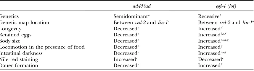
We sequenced exons and intron–exon boundaries of theegl-4gene inad450sdmutants and identified a single G to A transition at position 1084 of the EGL4A cDNA (Figure 1A). This mutation is predicted to change glycine 362 in the C-terminal cGMP-binding domain into an arginine. This glycine is conserved in all cGMP-dependent protein kinases, in the regulatory subunits of dependent protein kinases, and in the cAMP-binding domain of the E. coli CAP as shown by align-ment of the cyclic nucleotide binding domains of these TABLE 1
Genetic and phenotypic evidence suggesting thatad450sdis a gain-of-functionegl-4allele
ad450sd egl-4 (lof)
Genetics Semidominanta Recessiveb
Genetic map location Betweenced-2andlin-1a Betweenced-2andlin-1b
Longevity Decreasedc Increasedd
Retained eggs Decreasede Increasedb,e,f
Body size Decreasede Increasedd,e,f,g
Locomotion in the presence of food Decreasede Increasedg
Intestinal darkness Decreasede Increasedd,e,f
Nile red staining Increasede Decreasede
Dauer formation Decreasede Increasedf
lof, loss of function. a
Avery(1993). b
Trentet al.(1983). c
Lakowskiand Hekimi(1998). d
Hiroseet al.(2003). eThis work.
fDaniels
et al.(2000). gFujiwara
three protein types (Figure 1B). The absolute conser-vation of glycine 362 suggests that the G362R mutation would have major consequences to enzyme function (seediscussion).
To confirm that the G362R mutation causes the
ad450sddominant phenotypes on body size and track-ing behavior (additional phenotypic details are below), we performed a geneticcis–transtest (Hengartneret al.
1992; Levinand Horvitz1993; Leeet al.1997), using
the following logic. If the G362R mutation is responsible for thead450sddominant phenotypes, then a null muta-tion ofegl-4on the same chromosome asad450sd, thecis
configuration, should occlude the dominant pheno-types ofad450sd. The sameegl-4null mutation, however, should not occlude the effect of thead450sddominant mutation when present on the opposite chromosome, in thetransconfiguration. In contrast, ifegl-4 and the gene mutated to cause the ad450sd phenotypes were separate but linked genes, then the effect of an egl-4
null mutation in thecisandtransconfiguration would be the same.
Following chemical mutagenesis, we searched for loss-of-function mutations inegl-4on the same chromo-some as the G362R mutation by screening for suppres-sors of the small and pale phenotypes ofad450sd(see
materials and methods). We identified four
loss-of-function egl-4 alleles at a rate of 1/937 mutagenized haploid genomes, a rate that is consistent with the isolation of loss-of-function mutants (Brenner 1974).
These four mutants formed an allelic series with respect to the severity of body size and tracking abnormality withegl-4(cs80) being the most severe (Table 2). The body size and tracking phenotypes of cs80 were not significantly different from those of the previously characterized egl-4 null allele n479 (Daniels et al.
2000) (Table 2), suggesting thatcs80too is a nullegl-4
allele. We sequenced theegl-4 exons and intron–exon boundaries in cs80 mutants and identified a C / T
transition that is predicted to result in a mutation of a glutamine to a premature stop codon at amino acid 442 of the EGL-4A protein. Although there are several alternative splice forms of EGL-4 (Stansberry et al.
2001; Fujiwaraet al. 2002; Hiroseet al. 2003), Q442
is in exon eight, the same exon in which the G362R mutation is located, and therefore the G362R muta-tion could not be expressed by alternative splicing. Q442STOP is predicted to truncate the protein before the kinase domain (Figure 1A) and would therefore have no kinase activity.
The cs80 egl-4 null mutation completely suppressed the dominant effect of ad450sd on body length and on locomotion when present in the cis configuration but not when present in thetransconfiguration (com-pare rows 3 and 5 in Table 3). We conclude that the Figure 1.—A conserved glycine in PKG is mutated inad450sdmutants. (A) Do-main structure of EGL-4A with the location ofad450sd and cs80, the two muta-tions discussed in the text, marked. (B) Alignment of the amino acid surround-ing the mutated glycine. Included in the alignment are the protein sequence of EGL-4A from C. elegans (Stansberry et al. 2001), dg2 from D. melanogaster (Kalderon and Rubin 1989), type I cGMP-depen-dent protein kinase from humans (Orstaviket al.1997), type II cGMP-dependent protein kinase from humans (Orstavik et al.1996), the catalytic subnit of cAMP-dependent protein kinase from humans and fromC. elegans(Grosset al.1990), and the catabolite activator protein (CAP) fromE. coli(Aibaet al.1982). The amino acids that form theb3 andb4b-sheets, defined by the crystal structure (Weberand Steitz1987), are underlined below the sequence of CAP.
TABLE 2
Body length and tracking phenotypes ofegl-4loss-of-function alleles generated on anad450sdchromosome
egl-4allele
Body length in micrometers: mean6SD (N)
Tracking:a mean6SD (N)
1 1029683 (5) 112625 (5)
ad450sd 873645 (15) 1366 (6) ad450sd cs78 1122653 (20) 105638 (7) ad450sd cs79 1119662 (21) 151650 (7) ad450sd cs81 1096664 (11) 143630 (7) ad450sd cs80b 1222666 (24) 164669 (7)
N479 1237643 (15) 188628 (6)
aIn this and subsequent tables, tracking refers to the num-ber of 0.5-cm squares entered by the worm in a 17-hr period (seematerials and methods).
b
phenotypes ofad450sdmutants are caused by the G362R mutation inegl-4.
Althoughegl-4(ad450cs80)in thetransconfiguration did not significantly suppress either the small body size or the reduced locomotion phenotype of ad450sd/1, there was a trend for partial suppression for both phe-notypes (compare rows 5 and 6 to row 2 in Table 3). The trend for partial suppression of ad450sd dominant phenotype by theegl-4null allele in the trans configu-ration is consistent with the interpretation thatad450sd
behaves as a genetic hypermorph. That is, it causes an increase in normal egl-4 gene function (Park and
Horvitz1986). To provide additional evidence to
sup-port this interpretation, we tested the effect of theegl-4
null mutantn479intranstoad450sdon male body size. We chose to perform this analysis in males rather than in hermaphrodites because length measurements in males showed lower variance and therefore would be more likely to detect a small effect on size (seematerials and methods). Indeed, we found a small but statistically
significant suppression of the dominant small body size effect ofad450sdby theegl-4null allelen479(Table 4). We conclude thatad450sdis a hypermorphic allele ofegl-4.
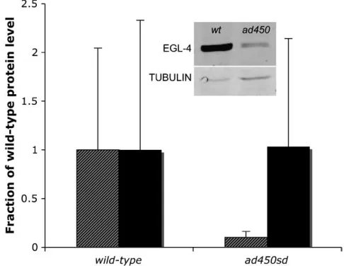
EGL-4 protein level is reduced inad450sd mutants:
One potential explanation for the gain-of-function phe-notype caused by the G362R mutation is an increase in EGL-4 protein abundance. To test for this possibility, we performed quantitative Western blot analysis of EGL-4 in the total protein pool isolated from wild-type and
ad450sd mutant worms. Surprisingly, we observed de-creased rather than inde-creased EGL-4 protein level in
ad450sdmutants (Figure 2). We therefore eliminate one potential explanation for the gain-of-function pheno-type. We consider explanations for this reduction of protein level as well as other models to explain the gain-of-function phenotype in thediscussion.
Phenotypic analysis ofegl-4(ad450sd):ad450sdwas
pre-viously noted to have decreased longevity (Lakowskiand
Hekimi1998), a phenotype opposite to that ofegl-4
loss-of-function mutants, which have increased longevity (Hiroseet al.2003). The decreased longevity ofad450sd
is particularly striking in light of the fact that virtually all other mutants identified as feeding defective have an increased longevity (Lakowski and Hekimi1998). We
found that in contrast to egl-4loss-of-function mutants, which have increased retention of eggs in the uterus (3561 eggs,N¼14) as reported previously (Daniels
et al.2000),egl-4(ad450sd)mutants had a reduced number of retained eggs (861 eggs,N¼16) in comparison to wild-type worms (13 6 1 eggs, N ¼ 13). We analyzed TABLE 3
cis–transtest demonstrating thatad450sdis allelic toegl-4(null)
Relevant genotypea
Body length in micrometers: mean6SD (N)
Tracking: mean6SD (N)
1/1 1029683 (5) 112625 (5)
ad450sd/1 935628 (6) 25611 (5) ad450 cs80/1 1025643 (5)b 110621 (7)b ad450sd/ad450sd 873645 (15) 1366 (6) ad450sd/ad450cs80 951644 (13)c 43616 (8)c ad450sd/n479 ND 36612 (7)c
ND, not done; SD, standard deviation;N, number tested. aIn the first three rows, the worm was heterozygous for nT1[qIs51].
bSignificantly different from
ad450sd/1,P,0.01. cNot significantly different fromad450sd/1,P.0.1.
TABLE 4
Male body length measurements show thatad450sdbehaves as a genetic hypermorph
Genotype Body length in micrometers: mean6SD
n479/n479 965672
1/n479 924646
1/1 891649
ad450sd/n479 856621a,b
ad450sd/1 788641c
ad450sd/ad450sd 721628
For each genotype, shown are the average length and stan-dard deviation (SD) of 8–16 males within 24 hr after reaching the adult stage.
a
Significantly different fromad450sd/1atP,0.001. b
Significantly different from1/1atP,0.01. c
Significantly different fromad450sd/ad450sdatP,0.001.
several additional phenotypes, previously noted to be ab-normal inegl-4 loss-of-function mutants. These pheno-types are summarized in Table 1 and are detailed in the text below.
Body size: Body size ofad450sdmutants is decreased whereas body size of egl-4 (lof) mutants is increased (Tables 2–4). Althoughad450sdmutants show reduced pharyngeal pumping rates, i.e., feeding, when unper-turbed (Avery1993), the small size ofad450sdmutants
is unlikely to be explained solely by caloric restriction. Animals that are doubly mutant forad450sdand foreat-2, a gene required for the normal fast rate of pharyngeal pumping, are significantly smaller than either single mutant despite no significant change in the feeding rate of the double mutants compared to the eat-2 single mutants (data not shown).eat-2(ad1113); ad450sd dou-ble mutants were significantly smaller at 627656mm (N¼6) thaneat-2(ad1116)(848663mm,N¼12) and
ad450sd (851 6 55 mm, N ¼8) single mutants (P ,
0.0001). Previous studies using loss-of-function mutants suggested that egl-4 affects body size by negatively regulating the activity of the TGF-bliganddbl-1(Hirose
et al.2003). Therefore, there appear to be at least two signaling pathways for controlling body size, one that involves TGF-bsignaling and the other that is controlled by caloric intake but whose signaling components have yet to be identified.
Locomotion: ad450sd mutants form fewer tracks on a bacterial lawn, whereas egl-4 loss-of-function mutants form more tracks than wild-type worms, as noted pre-viously (Fujiwaraet al.2002). To demonstrate the
re-duced locomotion behavior of ad450sd mutants, we measured the tracks formed by single animals left unperturbed for several hours (see materials and methods). Whereas wild-type animals move substantial
distances from the point of origin and therefore make tracks that cover close to one-third of the agar surface,
ad450sd animals stay close to the center of the agar surface, where they were placed (Tables 2 and 3).
This reduced tracking behavior is not explained by an inability of the animals to move well. Mechanical stimu-lation of the animals’ tails resulted in brisk forward loco-motion with a mean number of anterior body bends in 20 sec (15.261.4,N¼6) not significantly different from that of wild-type animals (16.361.5, N¼5,P.0.1). Furthermore, introducing a mutation ineat-2to reduce feeding rates and restrict caloric intake of ad450sd
resulted in tracks that were not significantly different from those formed by wild-type animals or by theeat-2
single mutant.eat-2(ad1113); egl-4(ad450sd)double mu-tants entered 83638 0.5-cm squares whereas wild-type animals andeat-2single mutants entered 686 29 and 87627 squares, respectively, in 17 hr. Finally,ad450sd
males appear as active as wild-type males and have normal mating efficiencies (data not shown). Therefore, we con-clude thatad450sd mutants have reduced locomotion only in the absence of sufficient motivation to move.
Reduced tracking despite normal ability to move was previously described for mutants with impaired sensory function (Fujiwaraet al.2002). Many of these mutants
have structurally defective sensory cilia as shown by defective uptake of lipophilic dyes (Starichet al.1995).
In contrast to these chemosensory mutants, sensory cilia stain normally in ad450sd mutants (data not shown). Furthermore, chemotaxis to the volatile odorant diace-tyl is normal inad450sd(data not shown). The reduced tracking behavior of ad450sdmutants is therefore not the result of grossly abnormal chemosensation.
The reduced locomotion of ad450sdmutants is also not explained by signaling changes in the TGF-b path-way that partially mediate the dauer formation and intestinal darkness phenotypes ofad450sdmutants (see below).daf-7(e1372); egl-4(ad450sd)anddaf-8(e1393); egl-4(ad45sd)double mutants formed the same number of tracks asad450sdsingle mutants (Table 5). Therefore, the signaling pathway that mediates reduced locomo-tion byegl-4remains unknown.
Intestinal darkness and fat storage: Intestinal darkness under light microscopy appears decreased in ad450sd
mutants while it appears increased in egl-4 recessive mutants, as reported previously (Daniels et al. 2000;
Hiroseet al.2003). We were able to quantify these
dif-ferences using digital video imaging methods (Figure 2). In addition toegl-4loss-of-function mutants, other mu-tants with the dark intestine phenotype include those with mutations in genes that mediate TGF-bsignaling. These include the TGF-b ligand daf-7, the TGF-b re-ceptordaf-1, and the TGF-bintracellular signaling com-ponents daf-8 and daf-14 (Patterson and Padgett
2000). To determine if the pale intestine phenotype of
ad450sd mutants requires TGF-b signaling, we con-structed strains that were doubly mutant for ad450sd
and each of these TGF-b signaling mutants. The in-testinal darkness of each of these double mutants was not significantly different from that of the respective TGF-b signaling single mutant (Figure 2). Therefore,
egl-4acts upstream or in parallel to TGF-bsignaling to promote a pale intestine.
TheC. elegansintestine has been shown previously to be a storage depot for macromolecules, including fat (Ashrafiet al.2003; McKayet al.2003). To test whether
or not a difference in fat storage could partially explain the difference in intestinal darkness of egl-4 loss- and gain-of-function mutants, we examined Nile red stain-ing in these mutants. Nile red is a vital fluorescent dye that binds to fatty acids and increased Nile red fluorescence has been shown to correlate with increased intestinal fat storage inC. elegans(Ashrafiet al.2003).
promotes intestinal fat storage, particularly in the anterior intestine.
Dauer formation: In addition to the dark intestine phenotype, egl-4 loss-of-function mutants share a de-regulated dauer formation phenotype with TGF-b pathway mutants. When grown in conditions of higher temperatures, crowding, and reduced food,C. elegans
development can proceed through an alternative long-lived third larval stage, called the dauer larva (Riddle
and Albert 1997). At least three signaling pathways
have been identified that control the dauer formation decision, including a cGMP sensory transduction path-way, a TGF-bsignaling pathway, and an insulin-signaling pathway (Riddleand Albert1997).
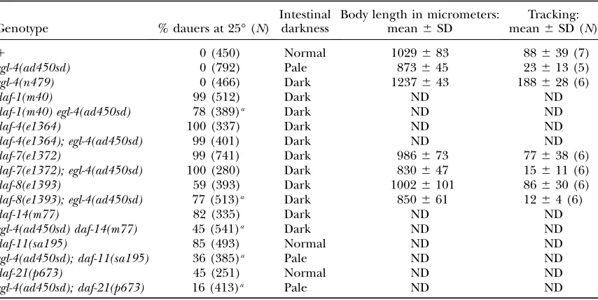
egl-4 loss-of-function mutants have previously been noted to be hypersensitive to dauer pheromone and to have a high rate of dauer formation at 27°, a tempera-ture in which wild-type worms only rarely form dauers (Daniels et al. 2000). Genetic epistasis experiments
suggested that theegl-4loss-of-function Daf-c phenotype is explained by the action of the gene in the TGF-b signaling pathway. Consistent with this placement, we found thatad450sdpartially suppresses the Daf-c phe-notype ofdaf-11anddaf-21and does not suppress the Daf-c phenotypes ofdaf-7 and daf-4 (Table 5). Unlike mutants with defective cilia structure, which completely suppress dauer formation ofdaf-11anddaf-21mutants (Vowelsand Thomas1992), the suppression byad450sd
is partial, a result expected for a gene that functions in parallel todaf-11signaling (Thomaset al.1993).
Although these results taken alone would support a simple single action ofegl-4in the TGF-bbranch of the
dauer formation pathway upstream ofdaf-7, the effect of
egl-4(ad450sd)on the Daf-c phenotype of mutants in the other TGF-b signaling genes suggests a more compli-cated model. We noted a partial suppression of the Daf-c phenotype ofdaf-1anddaf-14mutants and a small but significant enhancement of the daf-8 mutants’ Daf-c phenotype (Table 5). The partial suppression of daf-1
and daf-14 suggests that egl-4 acts at least partially in parallel to TGF-b signaling in regulating the dauer formation decision. Also supporting a site of action that is parallel to TGF-b signaling is the observation by Daniels et al. (2000) that egl-4 loss-of-function
muta-tions enhance the dauer constitutive phenotypes of daf-7anddaf-14mutants. Our observed slight enhancement of daf-8 suggests that egl-4may have dauer-promoting activity in addition to dauer-inhibiting activity, as has been described for other genes that, likeegl-4(Fujiwara
et al.2002), are expressed in multiple sensory neurons (Vowelsand Thomas1992; Coburnet al.1998).
Suppressors of egl-4(ad450sd): Genetic suppressor
screens of gain-of-function mutants are a powerful way to identify components of signaling pathways in C. elegans (Huang and Sternberg 1995). In the same
screen used to identifyegl-4loss-of-function mutations on the same chromosome asad450sd, we also identified three mutants unlinked to egl-4, cs82, cs83, and cs84, which suppress some or all of the ad450sd mutant phenotypes. As expected on the basis of our double-mutant analysis with Daf-c double-mutants described above, we found one new allele of daf-8,cs82, which suppresses the pale intestine phenotype of egl-4(ad450sd). Our assignment ofcs82as adaf-8allele is based on a similar TABLE 5
Genetic interaction betweenegl-4(ad450sd)and dauer constitutive mutants
Genotype % dauers at 25°(N)
Intestinal darkness
Body length in micrometers: mean6SD
Tracking: mean6SD (N)
1 0 (450) Normal 1029683 88639 (7)
egl-4(ad450sd) 0 (792) Pale 873645 23613 (5)
egl-4(n479) 0 (466) Dark 1237643 188628 (6)
daf-1(m40) 99 (512) Dark ND ND
daf-1(m40) egl-4(ad450sd) 78 (389)a Dark ND ND
daf-4(e1364) 100 (337) Dark ND ND
daf-4(e1364); egl-4(ad450sd) 99 (401) Dark ND ND
daf-7(e1372) 99 (741) Dark 986673 77638 (6)
daf-7(e1372); egl-4(ad450sd) 100 (280) Dark 830647 15611 (6)
daf-8(e1393) 59 (393) Dark 10026101 86630 (6)
daf-8(e1393); egl-4(ad450sd) 77 (513)a Dark 850661 1264 (6)
daf-14(m77) 82 (335) Dark ND ND
egl-4(ad450sd) daf-14(m77) 45 (541)a Dark ND ND
daf-11(sa195) 85 (493) Normal ND ND
egl-4(ad450sd); daf-11(sa195) 36 (385)a Pale ND ND
daf-21(p673) 45 (251) Normal ND ND
egl-4(ad450sd); daf-21(p673) 16 (413)a Pale ND ND
Body length measurements are the average of 5–20 worms. Tracking data foregl-4(n479)are the same as those shown in Table 2. ND, not done.
Daf-c and social feeding phenotype, a consistent map position, a failure to complement daf-8 for the Daf-c phenotype, and molecular sequencing data that identi-fied a two-nucleotide deletion in the second exon of the DAF-8 gene R05D11.1 (Riddleand Albert1997).
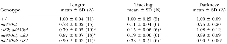
cs83 and cs84 partially suppress the small body, re-duced locomotion, and pale intestine phenotypes of egl-4(ad450sd)(Table 6). Therefore, suppressor screens of
ad450sdpromise to be a useful tool in understanding PKG signaling.
DISCUSSION
We have characterized a novel gain-of-function mu-tation that increases normal activity of the C. elegans
cGMP-dependent protein kinase geneegl-4. We use this
mutation to demonstrate a signaling role for egl-4 in several physiological processes, including the control of body size, intestinal storage of fat, dauer formation, and locomotion in the presence of food. While for many of these phenotypes a role for egl-4 has been previously described on the basis of analysis of mutants with re-duced gene function, analysis ofad450sd, a mutant with increased gene function, allows one to conclude that
egl-4 is not simply permissive for these physiological processes but rather plays an instructive signaling role.
Structure and function of cGMP-dependent protein
kinases:Glycine 362 is conserved in all PKG enzymes as
well as in the regulatory subunits of PKA. On the basis of amino acid alignment with CAP, whose tertiary structure is known (McKayand Steitz1981), a
By analogy to CAP, the structure of the cGMP-binding domain would consist of eight hydrogen-bonded b-strands that form a compactb-roll structure (Weber
and Steitz1987). cGMP binds in a pocket formed by
theseb-strands and onea-helix (Weberet al.1989). At
the vertex of this pocket sits glycine 362 (glycine 45 in the CAP structure), as a connector between strandb3 and strandb4 (Figure 1B). This glycine would not be expected to bind cyclic nucleotide monophosphate directly, but must be critical for maintaining the over-all tertiary structure of the cyclic nucleotide-binding domain.
We do not yet know the biochemical or cell biologi-cal consequences of the G362R mutation and to our knowledge the effect of this particular mutation has never been testedin vitro. The possibility that the gain-of-function phenotype of the G362R mutation is caused by increased steady-state levels of the protein is ex-cluded by our protein level analysis (Figure 2). We in fact observed the opposite result,i.e., of reduced EGL-4 protein level in thead450sdmutants in comparison to that in wild-type worms. The reduction in protein level might be accounted for by reduced protein folding efficiency of the EGL-4 G362R protein, by reduced stability of the folded protein, or by a negative feedback regulation of EGL-4 protein levels byegl-4gene activity. Such a negative feedback may occur, for example, if autophosphorylation of the EGL-4 protein marks it for degradation. Autophosphorylation is known to occur in the case of mammalian PKG (Aitkenet al.1984).
Another explanation for the gain-of-function pheno-type caused by the G362R mutation is that this mutation causes increased kinase activity of the enzyme. This could occur, for example, by causing enzyme activa-tion during basal condiactiva-tions, in the absence of cGMP elevation. Autoinhibition of PKG enzymatic activity is thought to occur via interaction between the catalytic domain and an inhibitory domain localized near the N
terminus of the protein (Yuasaet al.2000a). The change
from glycine, a small nonpolar amino acid, to arginine, a large charged amino acid, may disrupt the integrity of theb-roll structure and lead to easier access for cGMP binding and therefore faster association kinetics. Alter-natively, this mutation may cause such a drastic change to the tertiary structure of the cyclic nucleotide-binding domain with the result that the N-terminal inhibitory domain can no longer effectively inhibit the kinase do-main. In the absence of autoinhibition, the enzyme would be active even in the absence of cGMP. Unlike a constitutively active mutant that lacks the whole N-terminal portion of the protein and contains only the catalytic domain (Browninget al.2001), the G362R
PKG mutant should retain substrate specificity by virtue its normal N terminus.
A final possible explanation of this gain-of-function activity is a change in the subcellular distribution of the enzyme. PKG has been shown previously to undergo nuclear translocation in some cell types (Gudi et al.
1997; L’Etoileand Bargmann2000) and to localize to
other discrete cellular compartments in others (Wyatt
et al.1991; Pryzwanskyet al.1995; Surkset al.1999).
One possibility is that nuclear translocation or another subcellular targeting event is altered in G362R mutants. Distinguishing among these possibilities will require future biochemical and cell biological experiments. Regardless of the mechanism, the genetic evidence we describe here provides compelling evidence that the effect of G362R is to increase normal PKG gene activity. Since G362R is a mutation that would not be expected
a priorito necessarily cause increased gene activity, our finding underscores the importance of an unbiased ge-netic approach for the identification of novel mutants.
The use of the ad450sd mutation to identify
down-stream targets of PKG:On the basis of analysis ofegl-4
loss-of-function and gain-of-function mutant pheno-types, one can conclude that PKG signaling controls TABLE 6
Phenotypic analysis ofad450sdsuppressors
Genotype
Length: mean6SD (N)
Tracking: mean6SD (N)
Darkness: mean6SD (N)
1/1 1.0060.04 (11) 1.0060.25 (5) 1.0060.09
ad450sd 0.7860.02 (15) 0.1160.04 (6) 0.7560.20 cs82; ad450sd 0.7960.05 (19)a 0.1560.06 (6)a 1.0860.12 ad450sd; cs83 0.8760.07 (13)b 0.1960.06 (6)c 0.8960.09d ad450sd; cs84 0.9060.02 (11)e 0.3360.21 (6)f 0.9060.06d
Measurements shown are relative to wild-type measurements. Two-tailedt-tests were performed to determine statistical significance.
a
Not significantly different fromad450sd,P.0.1. b
Different fromad450sd,P¼0.001. c
Different fromad450sd,P¼0.02. d
Different fromad450sd,P,0.01. e
Different fromad450sd,P,0.000001. fDifferent from
multiple physiological processes. The signaling pathway that mediates many of these phenotypes appears dis-tinct (Figure 4). egl-4 promotes reduced longevity via an insulin signaling pathway (Hiroseet al.2003),
pro-motes nondauer development and a pale intestine par-tially via a TGF-bsignaling pathway (Trentet al.1983;
Daniels et al. 2000) (this work), promotes a smaller
body size through a different TGF-bsignaling pathway (Fujiwaraet al.2002; Hiroseet al.2003), and promotes
sensory adaptation through a cGMP-gated cation chan-nel (L’Etoile and Bargmann 2000). The signaling
pathway through which egl-4 promotes reduced loco-motion on food is not yet known (Figure 4).
Previous approaches for the identification of signal-ing elements downstream of PKG in mammalian cells have made use of yeast two-hybrid screens andin vitro
binding and phosphorylation methods (Buttet al.1994;
Voet al. 1998; Surks et al.1999; Yuasaet al.2000b,c).
Verification of the relevance of these targets to mam-malian physiology requiresin vivoinactivation experi-ments, which can be costly and difficult to interpret if gene knockouts have pleiotropic effects.
This G362R gain-of-function mutant offers the op-portunity to identify downstream signaling targets of
egl-4. Indeed, we have demonstrated that extragenic suppressors can be isolated that suppress some or all of the ad450sd phenotypes. Future molecular character-ization of the genes affected in these and other sup-pressors may shed light on PKG signaling in an unbiased hypothesis-independent fashion.
Similarity between the ad450sd phenotype and
lethargus behavior: The phenotype that led to the
iso-lation of ad450sd mutants is the reduced pharyngeal pumping in the presence of abundant food. This mu-tant was subsequently noted to stop moving when left unperturbed. These two behaviors, pumping cessation and reduced locomotion despite unimpaired ability to move and pump, are reminiscent of behaviors normally
exhibited by worms during lethargus behavior. Leth-argus is a period that occurs before each of the four molts that separate the larval stages and fourth larval stage and the adult stage (Singh and Sulston1978).
Little is known about the regulation of behavior during lethargus. In current work we are exploring whether or notegl-4plays a role in the control of lethargus behavior.
We are grateful to Yasumi Ohshima for the gift of the EGL-4 antibody. We thank Young You and Julia George-Raizen for comments on this manuscript. Some nematode strains used in this work were provided by the Caenorhabditis Genetics Center, which is funded by the National Institutes of Health (NIH) National Center for Research Resources. We acknowledge financial support from the NIH grants K08 NS048914 (D.M.R.), R01 HL60287 (A.I.P.), and R01 GM58540 (M.S.) and from the National Alliance for Research on Schizophrenia and Depression (D.M.R.).
LITERATURE CITED
Aiba, H., S. Fujimotoand N. Ozaki, 1982 Molecular cloning and
nucleotide sequencing of the gene for E. coli cAMP receptor
pro-tein. Nucleic Acids Res.10:1345–1361.
Ailion, M., and J. H. Thomas, 2000 Dauer formation induced by
high temperatures inCaenorhabditis elegans.Genetics156:1047–
1067.
Aitken, A., B. A. Hemmingsand F. Hoffman, 1984 Identification of
the residues on cyclic GMP-dependent protein kinase that are autophosphorylated in the presence of cyclic AMP and cyclic
GMP. Biochim. Biophys. Acta790:219–225.
Ashrafi, K., F. Y. Chang, J. L. Watts, A. G. Fraser, R. S. Kamath
et al., 2003 Genome-wide RNAi analysis of Caenorhabditis
ele-gans fat regulatory genes. Nature421:220–221.
Avery, L., 1993 The genetics of feeding in Caenorhabditis elegans.
Genetics133:897–917.
Bradford, M. M., 1976 A rapid and sensitive method for the
quan-titation of microgram quantities of protein utilizing the principle
of protein-dye binding. Anal. Biochem.72:248–254.
Brenner, S., 1974 The genetics ofCaenorhabditis elegans.Genetics
77:71–94.
Browning, D. D., M. McShane, C. Martyand R. D. Ye, 2001
Func-tional analysis of type 1 alpha cGMP-dependent protein kinase using green fluorescent fusion proteins. J. Biol. Chem.276:13039– 13048.
Butt, E., K. Abel, M. Krieger, D. Palm, V. Hoppeet al., 1994
cAMP-and cGMP-dependent protein kinase phosphorylation sites of the focal adhesion vasodilator-stimulated phosphoprotein (VASP) in vitro and in intact human platelets. J. Biol. Chem.
269:14509–14517.
Coburn, C. M., I. Mori, Y. Ohshimaand C. I. Bargmann, 1998 A
cyclic nucleotide-gated channel inhibits sensory axon outgrowth in larval and adult Caenorhabditis elegans: a distinct pathway
for maintenance of sensory axon structure. Development125:
249–258.
Daniels, S. A., M. Ailion, J. H. Thomasand P. Sengupta, 2000 egl-4
acts through a transforming growth factor-b/SMAD pathway in
Caenorhabditis elegansto regulate multiple neuronal circuits in
re-sponse to sensory cues. Genetics156:123–141.
Fujiwara, M., P. Senguptaand S. L. McIntire, 2002 Regulation of
body size and behavioral state of C. elegans by sensory perception
and the EGL-4 cGMP-dependent protein kinase. Neuron 36:
1091–1102.
Gross, R. E., S. Bagchi, X. Luand C. S. Rubin, 1990 Cloning,
char-acterization, and expression of the gene for the catalytic subunit of cAMP-dependent protein kinase in Caenorhabditis elegans. Identification of highly conserved and unique isoforms
gener-ated by alternative splicing. J. Biol. Chem.265:6896–6907.
Gudi, T., H. M. Lohmannand R. B. Pilz, 1997 Regulation of gene
expression by cyclic GMP dependent protein kinase requires nu-clear translocation of the kinase: identification of a nunu-clear local-ization signal. Mol. Cell. Biol.17:5244–5254.
Figure4.—Distinct signaling pathways mediate egl-4 phe-notypes.egl-4promotes a reduced longevity by negatively reg-ulating the activity of the insulin signaling transcription factor daf-16(Hiroseet al.2003), promotes a reduced body size by negatively regulating the activity of thedbl-1TGF-b(Fujiwara et al.2002; Hiroseet al.2003), promotes a pale intestine and nondauer development by positively regulating thedaf-7
Hengartner, M. O., R. E. Ellisand H. R. Horvitz, 1992
Caen-orhabditis elegans gene ced-9 protects cells from programmed
cell death. Nature356:494–499.
Hirose, T., Y. Nakano, Y. Nagamatsu, T. Misumi, H. Ohta et al.,
2003 Cyclic GMP-dependent protein kinase EGL-4 controls body
size and lifespan in C. elegans. Development130:1089–1099.
Hodgkin, J., 1983 Male phenotypes and male mating in
Caenorhab-ditis elegans.Genetics103:43–64.
Huang, L. S., and P. W. Sternberg, 1995 Genetic dissection of
developmental pathways. Methods Cell Biol.48:97–122.
Kalderon, D., and G. M. Rubin, 1989 cGMP-dependent protein
kinase genes in Drosophila. J. Biol. Chem.164:10738–10748.
Lakowski, B., and S. Hekimi, 1998 The genetics of caloric
restric-tion in Caenorhabditis elegans. Proc. Natl. Acad. Sci. USA95:
13091–13096.
Lee, R. Y., L. Lobel, M. Hengartner, H. R. Horvitzand L. Avery,
1997 Mutations in the alpha1 subunit of an L-type
voltage-activated Ca21channel cause myotonia in Caenorhabditis
ele-gans. EMBO J.16:6066–6076.
L’Etoile, N. D., and C. I. Bargmann, 2000 Olfaction and odor
dis-crimination are mediated by the C. elegans guanylyl cyclase ODR-1.
Neuron25:575–586.
L’Etoile, N. D., C. M. Coburn, J. Eastham, A. Kistler, G. Gallegos
et al., 2002 The cyclic GMP-dependent protein kinase EGL-4
regulates olfactory adaptation in C. elegans. Neuron36:1079–
1089.
Levin, J. Z., and H. R. Horvitz, 1993 Three new classes of
muta-tions in theCaenorhabditis elegansmuscle gene sup-9. Genetics
135:53–70.
McKay, D. B., and T. A. Steitz, 1981 Structure of catabolite gene
activator protein at 2.9 A˚ resolution suggests binding to
left-handed B-DNA. Nature290:744–749.
McKay, R. M., J. P. McKay, L. Averyand J. M. Graff, 2003 C.
ele-gans: a model for exploring the genetics of fat storage. Dev. Cell
4:131–142.
Orstavik, S., R. Solberg, K. Tasken, M. Nordahl, M. R. Altherr
et al., 1996 Molecular cloning, cDNA structure, and chromo-somal localization of the human type II cGMP-dependent
pro-tein kinase. Biochem. Biophys. Res. Commun.220:759.
Orstavik, S., V. Natarajan, K. Tasken, T. Jahnsen and M.
Sandberg, 1997 Characterization of the human gene encoding
the type I alpha and type I beta cGMP-dependent protein kinase
(PRKG1). Genomics42:311–318.
Osborne, K. A., A. Robichon, E. Burgess, S. Butland, R. A. Shaw
et al., 1997 Natural behavior polymorphism due to a
cGMP-dependent protein kinase of Drosophila. Science277:834–836.
Park, E. C., and H. R. Horvitz, 1986 Mutations with dominant
ef-fects on the behavior and morphology of the nematode
Caeno-rhabditis elegans.Genetics113:821–852.
Patterson, G. I., and R. W. Padgett, 2000 TGF-brelated pathways:
roles inCaenorhabditis elegansdevelopment. Trends Genet. 16:
27–33.
Pfeifer, A., P. Ruth, W. Dostmann, M. Sausbier, P. Klattet al.,
1999 Structure and function of cGMP-dependent protein
kinases. Rev. Physiol. Biochem. Pharmacol.135:105–149.
Pryzwansky, K. B., S. Kidao, T. A. Wyatt, W. Reed and T. M.
Lincoln, 1995 Localization of cyclic GMP-dependent protein
kinase in human mononuclear phagocytes. J. Leukocyte Biol.
57:670–678.
Raizen, D. M., R. Y. Leeand L. Avery, 1995 Interacting genes
re-quired for pharyngeal excitation by motor neuron MC in
Caeno-rhabditis elegans.Genetics141:1365–1382.
Riddle, D. L., 1977 A genetic pathway for dauer larvae formation in
C. elegans. Stadler Genet. Symp.9:101–120.
Riddle, D. L., and P. S. Albert, 1997 Genetic and environmental
regulation of dauer larva development, pp. 739–768 inC. elegans
II, edited by D. L. Riddle, T. Blumenthal, B. J. Meyerand J. R.
Priess. Cold Spring Harbor Laboratory Press, Plainview, NY.
Riddle, D. L., M. M. Swansonand P. S. Albert, 1981 Interacting
genes in nematode dauer larvae formation. Nature290:668–
671.
Rybalkin, S. D., C. Yan, K. E. Bornfeldtand J. A. Beavo, 2003 Cyclic
GMP phosphodiesterases and regulation of smooth muscle func-tion. Circ. Res.93:280–291.
Siegfried, K. R., A. R. Kidd, M. A. Chesneyand J. Kimble, 2004 The
sys-1 and sys-3 genes cooperate with Wnt signaling to establish the proximal-distal axis of theCaenorhabditis elegansgonad. Genetics
166:171–186.
Singh, R. N., and J. E. Sulston, 1978 Some observations on
moult-ing in Caenorhabditis elegans. Nematologica24:63–71.
Stansberry, J., E. J. Baude, M. K. Taylor, P. J. Chen, S. W. Jinet al.,
2001 A cGMP-dependent protein kinase is implicated in
wild-type motility in C. elegans. J. Neurochem.76:1177–1187.
Starich, T. A., R. K. Herman, C. K. Kari, W. H. Yeh, W. S. Schackwitz
et al., 1995 Mutations affecting the chemosensory neurons of
Caenorhabditis elegans.Genetics139:171–188.
Surks, H. K., N. Mochizuki, Y. Kasai, S. P. Georgescu, K. M. Tang
et al., 1999 Regulation of myosin phosphatase by a specific in-teraction with cGMP- dependent protein kinase Ialpha. Science
286:1583–1587.
Thomas, J. H., D. A. Birnbyand J. J. Vowels, 1993 Evidence for
par-allel processing of sensory information controlling dauer
forma-tion inCaenorhabditis elegans.Genetics134:1105–1117.
Trent, C., N. Tsuingand H. R. Horvitz, 1983 Egg-laying defective
mutants of the nematode Caenorhabditis elegans. Genetics104:
619–647.
Vo, N. K., J. M. Gettemyand V. M. Coghlan, 1998 Identification of
cGMP-dependent protein kinase anchoring proteins (GKAPs).
Biochem. Biophys. Res. Commun.246:831–835.
Vowels, J. J., and J. H. Thomas, 1992 Genetic analysis of
chemosen-sory control of dauer formation inCaenorhabditis elegans.Genetics
130:105–123.
Vowels, J. J., and J. H. Thomas, 1994 Multiple chemosensory defects
in daf-11 and daf-21 mutants of Caenorhabditis elegans.Genetics
138:303–316.
Weber, I. T., and T. A. Steitz, 1987 Structure of a complex of
catab-olite gene activator protein and cyclic AMP refined at 2.5 A˚ res-olution. J. Mol. Biol.198:311–326.
Weber, I. T., J. B. Shabband J. D. Corbin, 1989 Predicted structures
of the cGMP binding domains of the cGMP-dependent protein kinases: a key alanine/threonine difference in evolutionary
diver-gence of cAMP and cGMP binding sites. Biochemistry28:6122–
6127.
Wedel, B., and D. Garbers, 2001 The guanylyl cyclase family at Y2K.
Annu. Rev. Physiol.63:215–233.
Wyatt, T. A., T. M. Lincolnand K. B. Pryzwansky, 1991 Vimentin
is transiently co-localized with and phosphorylated by cyclic GMP-dependent protein kinase in formyl-peptide-stimulated
neutrophils. J. Biol. Chem.266:21274–21280.
Yuasa, K., H. Michibata, K. Omoriand N. Yanaka, 2000a
Iden-tification of a conserved residue responsible for the autoinhibi-tion of cGMP-dependent protein kinase Ialpha and beta. FEBS
Lett.46:175–188.
Yuasa, K., K. Omoriand N. Yanaka, 2000b Binding and
phosphor-ylation of a novel male germ cell-specific cGMP-dependent pro-tein kinase-anchoring propro-tein by cGMP-dependent propro-tein
kinase Ialpha. J. Biol. Chem.275:4897–4905.
Yuasa, K., K. Omori and N. Yanaka, 2000c Specific domain of
cGMP-dependent protein kinase Ibeta but not Ialpha functions
as a transcriptional activator in yeast. IUBMB Life49:17–22.