ISSN(Online) : 2319-8753 ISSN (Print) : 2347-6710
International Journal of Innovative Research in Science,
Engineering and Technology
(An ISO 3297: 2007 Certified Organization)
Vol. 5, Issue 3, March 2016
CLAHE Enhancement Based Baby Vein
Detection Using Live Camera
R.Aravind Raj1, K.Hariharan2, K.Chandra Prasanth3, S.Santhosh4
UG Students, Department of Electronics and Communication Engineering, JNN Institute of Engineering, Chennai,
India123
Assistant Professor, Department of Electronics and Communication Engineering, JNN Institute of Engineering,
Chennai, India4
ABSTRACT: In any surgery, the first important phase is an insertion of an intravenous (IV) catheter. The major problem face by the physicians is difficulty in accessing vein for IV drug delivery or taking blood samples for test. In case of children, adults, critical care patients etc. It is very difficult task. Unnecessary puncturing of veins occurs due to poor visibility. Wrong puncturing may leads many problems such as bruises or permanently damage the vein. To overcome from these problems there is a need of vein detection system. The paper presents an approach for clahe enhancement based Vein detection System. In the proposed work contrast intensity is used to identify the veins appears on the back of the hands and live camera(WEBCAM) is used to capture the images. The preprocessing is done to enhance the vein image using MATLAB 2013b version. The two key characteristics of this device discussed in the paper are portability and low cost.
KEYWORDS: Biometric, vein recognition, Adaptive Histogram Equalization.
I. INTRODUCTION
There are lots of challenges to be found in the design and implementation. Very few devices on infrared radiation have been implemented. In any clinic, hospital or pathology laboratory it is commonly observe that the veins of patients are difficult to detect by the physicians properly, unnecessary of puncturing may occurs which leads to the problem such as swelling, bleeding or may permanently damage the veins. To overcome these problems there is a device like Acuuvein Vein Viewer comes up but the problem is cost and portability factor [1]. The vein pattern is not observable under the visible light. The vein pattern can be detected with help of infrared sensors. There are two different techniques are discussed in paper [3] about the imaging techniques such as far infrared imaging technique (FIR) and near infrared
imaging technique (NIR). FAR works within the range of 8-14μm to capture the large veins on the back of the hand,
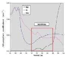
but it sensitive to the ambient condition and does not provide stable image quality.NIR imaging works within the range of 700-1000nm and provide good quality images. There is a medical spectral window from 700 to 900nm in which the light is penetrates into biological tissues up to 3mm of dept, and it allows to non-invasive investigation. These non invasive techniques also help in the treatment of varicose veins, deep vein thrombosis and vascular ailments [4].
1.1 NEEDS FOR VEIN DETECTION
ISSN(Online) : 2319-8753 ISSN (Print) : 2347-6710
International Journal of Innovative Research in Science,
Engineering and Technology
(An ISO 3297: 2007 Certified Organization)
Vol. 5, Issue 3, March 2016
Deep Vein Thrombosis and Varicose Veins, bruises appear on the skin, therefore for the treatment of these diseases, detection of veins is highly essential. Accidents involving first or second degree of bums cause the scarring of the skin. Here appearance of the skin becomes deterred causing the skin to appear whiter or in certain cases darker. The determinations of veins become tough in such cases as well.
d is given to the person intravenously. Blood donation, kidney dialysis also need perfect vein detection.
and infants may be especially difficult and having to puncture them several times with a needle is very frightful and agonizing for the child.
numerous blood tests or medicinal injections and an efficient means of puncture would reduce excessive bruise and enhance the patients overall comfort level [1].
In this paper an approach is presented for vein detection system using Infrared radiation and Smartphone. In the proposed work there are IR LED Ring is used for the illumination and Smartphone is used for capturing the vein image. The captured images are preprocessed to enhance the image adaptive histogram equalization technique is used. Image segmentation is performed to identify the veins. The rest of the paper is organized is as follows. Near Infrared imaging is introduced in section 2. An overview of proposed system is introduced in section 3. Image acquisition and the pre-processing steps are given in section 4. The Experimental results are presented in section 5. Finally the concluding remarks are given in section 6.
II. EXISTING SYSTEM
2.1NEAR INFRARED IMAGING
The special attributes of Near-Infrared imaging which makes it suitable for vein detection are:
3mm of depth, [4].
ISSN(Online) : 2319-8753 ISSN (Print) : 2347-6710
International Journal of Innovative Research in Science,
Engineering and Technology
(An ISO 3297: 2007 Certified Organization)
Vol. 5, Issue 3, March 2016
Figure 1 Optical windows for detecting vein patterns [9]
2.2 OVERVIEW OF THE SYSTEM
Our acquisition system (Fig. 2) is composed of a Smartphone camera with NIR ring of LEDs is used for illuminating the desired body part with infrared light. In this proposed system the Smartphone camera having a resolution of 6MP with 480*640 dimensions is used. The IR LED ring used for illumination is having the wavelength of 850nm. There are 36 IR LED’s are mounted on the ring. First the image is captured using mobile camera and then it is transferred to the laptop via USB cable for further pre-processing. Fig 2 shows the device setup used for vein detection.
Figure 2 Hardware system and IR LED ring used for Illumination.
III. PROPOSED SYSTEM
ISSN(Online) : 2319-8753 ISSN (Print) : 2347-6710
International Journal of Innovative Research in Science,
Engineering and Technology
(An ISO 3297: 2007 Certified Organization)
Vol. 5, Issue 3, March 2016
3.1 ALGORITHM
3.2 IMAGE ACQUISTION
Fig 4 shows the example of captured vein images using proposed system. The software used for pre-processing is MATLAB 2016b version.
Figure 4 Example of vein image captured by proposed system
3.3 RGB TO GRAY SCALE CONVERSION
First of all the color image is converted into gray scale image to make image into black and white image. It converts the true color image RGB to the grayscale intensity image by eliminating the hue and saturation information while retaining the luminance. Rgb2gray converts RGB values to grayscale values by forming a weighted sum of the R, G, and B components: 0.2989 * R + 0.5870 * G + 0.1140 * B (1)
3.4 SMOOTHING AND NOISE REMOVAL
The median filter is a simple edge-preserving smoothing filter. It may be applied prior to segmentation in order to reduce the amount of noise in a stack of 2D images. The filter works by sorting pixels covered by NxN mask according to their grey value. The centre pixel is then replaced by the median of these pixels, i.e., the middle entry of the sorted list.
3.5 CONTRAST ENHANCEMENT
ISSN(Online) : 2319-8753 ISSN (Print) : 2347-6710
International Journal of Innovative Research in Science,
Engineering and Technology
(An ISO 3297: 2007 Certified Organization)
Vol. 5, Issue 3, March 2016
applying CLAHE to the image. sorting pixels covered by NxN mask according to their grey value. The centre pixel is then replaced by the median of these pixels, i.e., the middle entry of the sorted list.
3.6 SEGMENTATION
Segmentation is the process of thresholding the image. By setting a proper threshold the pixel value above the threshold is set to 1 and below the threshold the pixel will be 0. In this system the threshold is set to 90 by experimentally. Figure 5 shows the image after segmentation.
IV. CONCLUSION
Our main goal of obtaining a portable efficient vein imaging system at very low costs accomplished. The proposed system is implemented using available resources therefore the cost of the system is reduced and it is portable. As per the tables provided we have done a pilot study for subjects who include wide range of sex, skin color and age. We have only implemented the acquisition and segmentation of the image which allows for future expansion.
REFERENCES
[1]Manam Mansoor., et al., “Real Time Low Cost Infrared Vein Imaging System.”, In IEEE International conference on Signal Processing, Image Processing and Pattern Recognition [ICSIPR], Coimbatore, pp. 117 – 121. Feb. 2013.
[2]Ajay Kumar, et al., “Personal Authentication Using Hand Vein Triangulation and Knuckle Shape.”, IEEE Transaction on image processing. vol.18, no. 9, pp 2127 – 2136, September 2009.
[3]A. Yuksel., et al., “Hand Vein Biometry Based on Geometry and Appearance Method.”, IET computer vision ., vol. 5, no. 6 pp. 398-406, 2011. [4] Chakravorty, Tanushri, et al. "Low-cost subcutaneous vein detection system using ARM9 based single board computer."
Electronics Computer Technology (ICECT), 2011 3rd International Conference on. Vol. 2. IEEE, 2011.
[5]Deepak Prasanna.R, et al., “Enhancement of vein patterns in hand image for biometric and biomedical application using various image enhancement techniques” International conference on Modeling optimization and Computing, pp. Procedia Engineering 38,elsevier1174 – 1185, 2012.
[6]Jae Hee Song et al., “Vein Visualization using a Smart Phone with Multispectral Wiener Estimation for Point-care-Application.” IEEE Journal of Biomedical and Health Informatics, pp.1-7. 2014.
[7] Ashwini J. Kolar, et al., “Experimental Prototype of Non-Invasive Low Cost Subcutaneous Vein Detection Device”, In IEEE International conference on Signal and Image Processing [ICSIP], pp. 170-179. 2013.
[8]Verma, Dipti, and Sipi Dubey. "Processing and Enhancement of Palm Vein Image in Vein Pattern Recognition System." (2015).
[9] Crisan, Septimiu, Joan Gavril Tarnovan, and Titus Eduard Crisan. "A low cost vein detection system using near infrared radiation." Sensors Applications Symposium, 2007. SAS'07. IEEE. IEEE, 2007.